Note: Descriptions are shown in the official language in which they were submitted.
CA 02212617 1997-08-08
WO 96/25125 PCT/US96l02048
ENDOPROSTHESIS STENT/GRAFT DEPLOYMENT SYSTEM
FIELD OF THE INVENTION
The present invention relates generally to a
deployment system for an implantable endoprosthesis. More
particularly, the present invention relates to a
deployment system for transcutaneous insertion of an
implantable tubularprosthesis supported by a stmt.
BACKGROUND OF THE INVENTION
The implantation of synthetic tubular prostheses to
replace or buttress damaged or diseased vascular vessels
or other luminal passageways within the human body is well
known. Synthetic tubular prostheses include grafts as
well as endoprosthetic devices.
Tubular prostheses such as grafts are most commonly
implanted by surgical techniques. Typically, a surgeon
sutures the graft in place within the blood vessel or
other body passageway to be repaired in an open surgical
technique. Intraluminal implantation is also a common
technique for implanting tubular prostheses. This
procedure typically involves percutaneous insertion of an
endoprosthesis by way of a delivery catheter. This
procedure permits delivery and implementation without the
need for major surgical intervention and the risks
inherent therewith. Thus, intraluminal implantation of
various prosthetic devices via delivery catheters is
becoming increasingly common.
With respect to grafts and other prostheses which may
traditionally be surgically implanted, means other than
suturing must be used to secure these prostheses in place
within the body passageway in order to effectively permit
intraluminal implantation. It is known to employ stems
iri combination with grafts and various other prostheses in
CA 02212617 1997-08-08
WO 96/25125 PC'T/US96/02048
2
order to support and secure such a device in place within
the body passageway after implantation. Stents are
typically radially expandable and/or contractible support
members which are positioned within a graft or other
tubular prosthesis. In common usage, after a prosthesis
has been properly positioned, the stmt is expanded to
anchor the prosthesis within the body passageway.
Since a stent must be expanded to support the
prosthesis within the body passageway for implantation,
the delivery system used to transport the stmt to the
location of implantation must be capable of maintaining
the stmt in a compressed state during delivery and
implantation until such time a stent deployment is
necessitated. Attempts have been made to improve delivery
systems for compressed stents. Several disclosures relate
to such systems.
U.S. Patent No. 4,950,227 discloses a catheter
delivery system for a stmt wherein the stent is
positioned about a balloon-type catheter and held in
position by a sleeve fixing the end of the stmt. As the
balloon is inflated, the stmt is expanded, causing the
sleeve to slide off of the stent and release the stent.
U.S. Patent No. 5,108,416 discloses a catheter
delivery system for a stent wherein the stent is
positioned about a balloon type catheter and held in
position by end caps. As the balloon is inflated, the end
caps move away from the stmt and release the stent.
U.S. Patent Nos. 5,158,548 and 5,242,399 disclose a
stmt delivery system wherein a scent is positioned about
a balloon-type catheter and held in position by an outer
sheath. A guidewire attached to the outer sheath is moved
forward, thereby moving the sheath forward to expose and
release the stent.
CA 02212617 1997-08-08
WO 96/25125 PC'TlUS96102048
3
Accordingly, aneed exists for an effective system
for catheter delivery and deployment of a stent-supported
implantable tubular prosthesis.
SUMMARY OF THE INVENTION
It is an object of the present invention to provide a
deployment system for an implantable endoprosthesis that
is to be delivered transcutaneously.
It is a further object of the present invention to
provide an improved deployment system which provides for --
the implantation of a radially expandable endoprosthesis.
It is a still further object of the invention to
provide a method for=endoprosthetic deployment of an
implantable endoprosthesis.
In the attainment of these and other objects, the
present invention provides a deployment system which
includes a flexible,: elongated, tubular, delivery catheter
with at least one inner lumen, and an opening extending
through the catheter-to the inner lumen. Positioned over
the catheter is an implantable endoprosthesis capable of
radial expansion and having a proximal and a distal end
extent. A support assembly removably maintains the
endoprosthesis in a compressed state, and is located at a
position adjacent the catheter opening. The support
assembly includes an arm which extends through the
catheter opening into the inner lumen. A release
mechanism is insertable through the inner lumen and
includes a distal tip engageable with the arm of the
support assembly. The release mechanism is manipulatable
within the ii~ner lumen so as to remove the support
assembly from the endoprosthesis, permitting radial
expansion of the endoprosthesis for implantation.
CA 02212617 1997-08-08
WO 96/25125 PCT/US96/02048
4
The endoprosthesis is preferably a graft having an
expandable stmt for support, more particularly, a pair of
expandable stems with one at the proximal end extent and
one at the distal end extent. The support assembly is '
preferably a boot. The release mechanism may include a
release rod, and may further include a clip at the distal
tip which is capable-of attachment to the arm.
The support assembly may include a distal support
member, for maintaining the distal end extent of the
endoprosthesis in a compressed state. A proximal support
member anchored to the catheter may further be included
for maintaining the proximal end extent of the
endoprosthesis in a compressed state. The distal end
extent and proximal end extent of the endoprosthesis may
be separate members. In this embodiment, the distal
support member maintains the distal endoprosthesis member
and the proximal support member maintains the proximal
endoprosthesis member.
The deployment system may further include a guidewire
insertable through the inner lumen ofthe catheter. The
catheter may further include a proximal portion and a
distal portion. The proximal portion and distal portion of
the catheter may be separate portions connected by a
connector portion, with the opening through the catheter
located at the connector portion.
The catheter may have a.first and a second inner
lumen, with the opening extending through to the first
inner lumen, the release mechanism insertable through the
first lumen, and the guidewire insertable through the
second lumen.
In its method aspect, the present invention includes
providing a deployment system including: a flexible
elongated tubular delivery catheter having a proximal
CA 02212617 1997-08-08
WO 96/25125 PCT/US96/02048
portion, a distal portion, at least one-inner lumen, and
an opening through the catheter to the inner lumen; an
implantable endoprosthesis capable of radial expansion
positioned over the catheter and having a proximal end
5 extent and a distal end extent; a retractable
endoprosthesis support assembly for removably maintaining
the endoprosthesis in a compressed state located at a
position adjacent the catheter opening and having an arm
extending through the catheter opening into the lumen; and
a release mechanism having a distal tip, insertible
through the inner lumen, engageable with the arm, and
capable of manipulation of the support assembly within the
inner lumen. The deployment system is intraluminally
inserted and positioned at an area of implantation. The
release mechanism is then engaged with the arm of the
support assembly, and the release mechanism is
manipulated within the inner lumen. This manipulation
removes the support assembly from the endoprosthesis and
permits radial expansion of the endoprosthesis. The
deployment system is then removed from the area of
implantation.
BRIEF DESCRIPTION OF THE DRAWINGS
Figure 1 is a perspective view of a preferred
embodiment of the endoprosthetic deployment system of the
present invention.
Figure 2 is a side view of a delivery catheter
assembly used in accordance with the present invention.
Figure 3 is an enlarged cross-sectional view of the
catheter assembly of Figure 2 taken along line A-A
thereof .
Figure 4 is an enlarged cross-sectional view a
component of the catheter of Figure 2 taken along line C-C
thereof .
CA 02212617 1997-08-08
WD 96/25125 PC'T/US96/02048
6
Figure 5 is an enlarged cross-sectional view of the
catheter assembly of Figure 2 taken along line B-B
thereof .
Figure 6 is an enlarged cross-sectional view of a
further embodiment of the catheter assembly of Figure 2
taken along line A-A.
Figure 7 is an enlarged cross-sectional view of the
alternative embodiment of Figure 6 taken along line C-C of
Figure 2.
Figure 8 is a side view of an outer sheath used in
accordance with the present invention.
Figure 9 is a side view of a release mechanism used
in accordance with the present invention.
Figure 10 is a perspective view of an endoprosthesis
including a graft and stmt of the type used with the
delivery system of the present invention.
Figure 11 is a perspective view of a proximal support
member used in accordance with one embodiment of the
present invention.
Figure 12 is a perspective view of a distal support
member used in accordance with the present invention.
DETPILED DESCRIPTION OF THE INVENTION
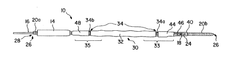
A deployment system 10 for transcutaneous insertion
of an implantable endoprosthesis as contemplated by the
present inventiori is shown in Figure 1. Deployment system
10 includes a flexible elongated delivery catheter
assembly 20. Catheter assembly 20 may be constructed of
any generally flexible biocompatible.material that is
known in the art.
CA 02212617 1997-08-08
WO 96/25125 PCT/US96102048
7
In preferred embodiments, catheter assembly 20
includes a proximal portion 20a and a distal portion 20b,
as shown in Figure 2_ For the purposes of the present
invention, "distal" is used to describe a general location
of delivery system 10 that is first inserted into the
body, while "proximal" is used to describe a general
location of delivery system 10 that is opposite the distal
portion. Proximal portion 20a and distal portion 20b are
typically discrete portions, separated by discontinuity
21. In order to couple proximal portion 20a and distal
portion 20b, a connector portion is employed, shown in
Figures 1 and 2 as connector 40. Connector 40 is
typically an elongated member having tapered ends for
insertion within said proximal portion 20a and distal
portion 20b at discontinuity 21, thereby coupling the two
portions. Connector 40 may be constructed of a
biocompatible material well known in the art. When
coupled together, proximal portion 20a, connector 40 and
distal portion 20b effectively form a single catheter
assembly 20.
Proximal portion 20a includes first inner lumen 26a
and second inner lumen 28a extending therethrough, shown
in Figure 3. Distal- portion 20b includes first inner
lumen 26b extending therethrough, shown in Figure 5.
Connector 40 includes a first inner lumen 26c that
communicates with first inner lumen 26a of proximal
portion 20a, and further communicates with first inner
lumen 26b of distal portion 20b. Connector 40 also
includes a second inner lumen 28c that communicates with
. second inner lumen 2.8a of proximal portion 20a. With the
'proximal portion 20a, connector 40 and distal portion 20b
coupled together; the communicating first inner lumens
26a, 26c and 26b effectively form a single first inner
lumen 26 extending through catheter assembly 20, and
communicating second inner lumens 28a and 28c form a
single second inner lumen 28 extending through catheter
CA 02212617 1997-08-08
WO 96/25125 PCT/US96/02048
8
assembly 20. First -inner lumen 26 may accommodate a
guidewire (not shown) therethrough. As is well known in
the art, guidewires may be commonly used in catheter
delivery systems to locate and guide a delivery catheter
through the vascular system.
An opening 24 exists through the wall of connector
40, extending through to second inner lumen 28c, as
depicted in Figure 4. While the preferred embodiment of
the invention includes connector 40, the present invention
contemplates catheter assembly 20 as a single member
catheter without connector 40, with such an embodiment
including opening 24 through the wall of the single member
catheter and extending through to inner lumen 28.
As can be seen in Figure 1, an implantable
endoprothesis 30 capable of radial expansion is positioned
over catheter assembly 20. Endoprosthesis 30 includes a
proximal end extent 35 and a distal end extent 33.
Endoprosthesis 30, further shown in Figure 10, may be any
type of implantable prosthetic device known in the art.
The present invention contemplates endoprosthesis 30
existing as a straight, tapered, stepped, bifurcated, or
any other type of endoprosthesis useful in implantation
procedures.
In preferred applications, endoprosthesis 30 includes
a vascular graft 32, which may be constructed of braided,
knitted, or woven synthetic yarns such as polyester, or
may be formed of an extruded plastic such as expanded
polytetraflouroethylene (PTFE). Graft 32 is designed for
percutaneous implantation within a diseased or damaged
blood vessel or other like vessel to provide replacement
or reinforcement of the damaged vessel_ In this regard,
graft 32 is f-olded or compressed to facilitate
intraluminal delivery. Such compression or folding is
well known in the art, and the present invention
CA 02212617 1997-08-08
WO 96/25125 PCT/US96/02048
9
contemplates the graft existing in any compressed o~-
folded shape which would permit radial expansion.
Graft 32 may be a self-supporting-type graft known in
the art, or may be supported by other means. For example,
graft 32 may be supported by an expandable stent 34,
further depicted in Figure 10. Stent 34 may be any
conventional stmt constructed of any material known in
the art, such as stainless steel or other metals or
alloys, polymeric materials, or composites of polymers
and metal. Stent 34 is self-expandable in a radial
direction between a compressed diameter and a larger
expanded diameter. Stent 34 may further contain stent
barbs (not shown) extending therefrom, which are commonly
used in stmt applications for aiding in positioning and
anchoring of endoprostheses.
In preferred form, proximal end extent 35 and distal
end extent 33 of endoprosthesis 30 support separate
discrete stent members. This embodiment is particularly
useful when endoprosthesis 30 includes a graft and stent
combination. In such an embodiment, stmt 34 may include
two spaced-apart members, proximal stmt member 34b and
distal stmt member 34a, as shown in Figure 10. In such
an embodiment, distal stent member 34a supports and
anchors distal end extent 33 of graft 32 to the
implantation area, while proximal stmt member 34b
supports and anchors proximal end extent 35 of graft 32 to
the implantation area.
In order to captively retain endoprosthesis 30 in a
compressed state prior to and during implantation, the
present invention employs a removable endoprosthesis
support assembly for removably maintaining endoprosthesis
30 in its compressed state. The support assembly~may
include a stmt boot which maintains endoprosthesis 30 in
a compressed state.
CA 02212617 1997-08-08
WO 96!25125 PCT/US96/02048
In preferred embodiments, the support assembly exists
as two separate members. Referring to Figures 1, 11 and
12, the support assembly is shown as a distal support
member 44 and a longitudinally spaced proximal support
5 member 48. Spaced apart support members 44 and 48 are
particularly useful where endoprosthesis 30 is a
stent/graft combination as shown herein. Distal support
member 44 surrounds and maintains distal end 33 in a
compressed state, while proximal support member 48
10 surrounds and maintains proximal end extent 35 in a
compressed state. Proximal support member 48 is anchored
to catheter assembly 20 along proximal portion 20a.
Adhesive or other fastening techniques may be employed.
Distal support member 44 is located at a position
adjacent opening 24 of connector 40 of catheter assembly
20. Distal support member 44 includes an arm 46, which
extends through opening 24 of connector 40 to second inner
lumen 28c.
A release mechanism 16 having a distal tip 13 is
insertable through second inner lumen 28 of catheter 20.
Release mechanism 16 is retractably movable within
catheter 20. Manipulation of the release mechanism
contemplates any activity which would result in the
removal of the support assembly. For instance, the
release mechanism may be manipulated by movement within
catheter 20 in an axial or longitudinal direction thereby
engaging the support assembly, or the release mechanism
may be manipulated by other means known in the art.
- Release mechanism 16, shown in Figure 9, may include
a release rod, having a clip 18 at distal tip 13. Clip 18
is engagable with and capable of attachment to arm 46 of
distal support member 44, which extends through opening 24
of connector 40. This attachment permits the removal of
distal support member 44 from distal end 33 when release
CA 02212617 1997-08-08
WO 96/25125 PCT/US96/02048
11
mechanism 16 is retractably moved by moving the release
rod in a longitudinal direction toward the distal end of
the deployment system 10.
S As release mechanism 16 need only reach arm 46
extending through connector 40 at opening 24, it is not
necessary for second inner lumen 28 to extend past
connector 40 through. distal portion 20b, although such a
design is within the contemplation of the present
invention.
In preferred applications, the deployment system of
the present invention is utilized with minimally invasive
transcutaneous insertion of an implantable endoprosthesis.
More preferably, the deployment system of the present
invention is utilized with percutaneous insertion of a
stent supported vascular graft. However, it may be
appreciated that the deployment system of the present
invention may be utilized with any endoprosthetic
implantation procedure, including transcutaneous
implantation, percutaneous implantation, cut down
procedures, and the like.
Having described the components of the present
invention, use of the deployment system 16 may now be
described. In this preferred application, a needle (not
shown) is inserted intraluminally into a blood vessel (not
shown). A guidewire (not shown) is then inserted through
the needle and.advanced through the blood vessel to the
area of implantation: The deployment system 10 is then
inserted into the blood vessel and guided over the
guidewire inserted through inner lumen 26 to a position at
the area of implantation.
After reaching the area of implantation, 'release
mechanism 16 can be inserted through second inner lumen
28. Release mechanism 16 may also have been inserted into
CA 02212617 1997-08-08
WO 96/25125 PCT/US96/02048
12
second inner lumen 28 prior to inserting deployment system
intraluminally. Clip 18 of release mechanism 16
engages distal support member 44 at arm 46, which extends
through opening 24 at connector 40. After engaging at arm
5 46, release mechanism 16 is manipulated by moving release
mechanism 16 within second inner lumen 28, in a
longitudinal direction toward the distal end of deployment
system 10. As clip 18 and arm 46 are engaged, movement of
release mechanism 16 causes movement ofdistal support
10 member 44, thereby removing distal support member 44 from
its position maintaining distal stmt member 34a in
compressed state. With distal support member 44 removed,
distal stent member 34a radially expands, and attaches
distal end extent 33 of graft 32 to the inner wall of the
vascular surface.
After distal stent member 34a fully expands, proximal
support member 48, being anchored to proximal portion 20a
of catheter assembly 20, still maintains proximal stent
member 34b in a compressed state. Deployment system 10,
including catheter assembly 20, is then removed from the
area of implantation. As proximal support member 48 is
anchored to proximal portion 20b, this removal causes
proximal support member 48 to be removed from proximal
stent member 34b. This removal permits radial expansion
of proximal stmt member 34b, thereby anchoring proximal
end extent 35 of graft 32 to the vascular wall. With both
ends of the vascular graft 32 now anchored, the graft is
implanted, and deployment system 10 can be completely
removed from the blood vessel.
The present invention further contemplates catheter
assembly 20 existing as a single lumen catheter, with the
guidewire and release mechanism 16 insertable through the
single lumen. The present invention also contemplates
catheter 20 assembly existing as a multiple lumen
catheter, such as a triple lumen catheter as depicted in
CA 02212617 1997-08-08
WO 96/25125 PCTlUS96/02048
13
the cross-sectional view of Figures 6 and 7. In this
alternate embodiment, proximal portion 20a of catheter
assembly 20 has inner lumen 26a, second inner lumen 28a,
and third inner lumen 29a. Also included in this
embodiment is a second opening 27 through catheter 20
assembly at connector 40, extending through to third inner
lumen 29c. Second opening 27 is at a point adjacent
opening 24, but not connected with opening 24. In this
embodiment, inner lumen 26 accepts a guidewire, second
inner lumen 28 accepts release mechanism 16, and third
inner lumen 29 can accept a second release mechanism (not
shown). The second release mechanism can be identical to
release mechanism 16. The second release mechanism is
engaged with a second arm 47 which may be included with
distal support member 44, as depicted in Figure 12.
Second arm 47 extends through catheter assembly 20 at
opening 27 in a similar manner as arm 46 through opening
24. In this embodiment, release mechanism 16 engages arm
46 while second release mechanism engages second arm 47,
thereby permitting removal of distal support member 44
when both the first and second release mechanisms are
moved.
In yet another embodiment of the invention, catheter
assembly 20 exists as a three-lumen catheter similar to
that described above, with the third lumen capable of
accommodating a second release mechanism engaged with the
proximal support member. In this embodiment, a second
opening exists through catheter assembly 20 adjacent the
proximal support member.. The proximal support member
includes an arm which extends through this second opening
of the catheter assembly in'a similar manner as arm 46
extends through opening 24 of the preferred embodiment.
In this alternate embodiment, the second release mechanism
engages the arm of the proximal support member and
manipulation of the second release mechanism causes the
proximal support member to be removed from its position
CA 02212617 2001-11-21
-14-
maintaining the proximal and extent of the endoprosthesis.
Al.ternativel:y, the third lumen may be used for other
purposes, such as dye injection, drug infusion, and other
5 known uses for c<~theter lumens. Additionally, whether a
single .lumen or mul.ti-lumen catheter assembly, the shape
of the .lumen is not. significant to the invention, so long
as the ahape does not preclude the lumen from performing
the fun~~tion intended.
The present invention also contemplates a retractable
outer sheath 14, shown in Figure 8, disposed over catheter
assembly 20 and ~~ndoprosthesis 30, further maintaining
endoprosthesis 30 in a compressed state and in position.
Outer sheath 14 may be constructed of any flexible,
biocompatible material known in the art. Other sheath 14
is retractable after transcutaneous insertion, to permit
exposure of endoprosthesis 30 to the surface of
implantation. Outer sheath 14 is shown in Figure 1 in its
retracted state.
While the invention has been described herein in terms
of certain preferred embodiments, those skilled in
the art will rec:~gnize that various modifications can be
made without dep;~rt:ing from the scope of the present
invention.