Note: Descriptions are shown in the official language in which they were submitted.
96P7523 CA 02212707 1997-os-os
1
METHOD AND SYSTEM FOR DYNAMICALLY ESTABLISHING
FIELD SIZE COINCIDENCE
BACKGROUND OF THE INVENTION
The invention relates generally to a method to improve the
setup of a treatment field for radiation treatment, and relates more
particularly to establishing a field-size coincidence between a setup-mode
light field, a treatment-mode radiation field, and a display that is used to
facilitate the setup of the treatment field.
DESCRIPTION OF THE RELATED ART
Radiation-emitting devices are generally known and used, for
instance, as radiation therapy devices for the treatment of patients. A
radiation therapy device usually comprises a gantry which can be swiveled
around a horizontal axis of rotation in the course of a therapeutic
treatment. A linear accelerator is located in the gantry for generating a
high-energy radiation beam for therapy. This high energy radiation beam
can be an electron radiation or photon (X-ray) beam. During treatment,
this radiation beam is trained on a zone of a patient lying in the isocenter
of the gantry rotation.
To control the radiation emitted toward an object, a beam-
shielding device such as a plate arrangement and/or collimator is usually
provided in the trajectory of the radiation beam between the radiation
source and the object. This beam-shielding device defines a field on the
object to which a prescribed amount of radiation is to be delivered.
The radiation delivered to an object may be analyzed into
primary and scattered components. The primary radiation is made up of
the initial or original photons emitted from the radiation source, and the
scattered radiation is the result of the photons scattered by the plate
3o arrangement. The beam's radiation output in free space increases
because of the increased plate-collimator scatter, which is added to the
primary beam. In other words, a point in the field is subject not only to
96P7523 CA 02212707 1997-os-os
2
direct radiation (which is the primary component), but also to radiation
that is scattered from the plate arrangement. The ratio of the radiation
output in air with the scatterer to the radiation output without the
scatterer for a reference field (for instance 10 x 10 cm) is commonly called
the "output factor" or the "collimator scatter factor." The concept and
definition of the output factor are well understood in the art.
Thus, due to these scattered photons, the dose rate applied
to the surface of the object changes dependent on the size of the opening
in the plate arrangement, that is, on the field size. This means that the
radiation emitted to the same spot, for instance in the center of the
radiation beam onto the object, changes according to the size of the
opening in the plate arrangement. When the plate arrangement shows
only a small opening, then the accumulated dose at the same spot is less
than the accumulated dose at the same spot when the opening is big.
The field size of a radiation therapy device is important, since
it determines the region of the patient that will be exposed to the
radiation. In the setup mode of operation of the device, a source of
visible light may be activated to project a light field onto the patient from
the treatment head. The light field facilitates the adjustment of beam
2o parameters and the proper positioning of the patient relative to the
treatment head.
The field size is adjusted by varying an aperture through a
collimator in the treatment head. The aperture is defined by settings of X-
axis collimator jaws and Y-axis collimator jaws. The jaws are blocks of
25 radiation-attenuating material that determine the field size by limiting
the
angular spread of the beam. By convention, the X-axis jaws are located
below the Y-axis jaws.
Ideally, the field size of an X-ray radiation beam is a duplicate
of the field size of the light that is used in the setup for the patient.
30 However, there are factors that make it difficult to achieve the radiation
field size-to-light-field size coincidence. The characteristics (light
intensity, spot size, and position) of the visible light beam and the X-ray
96P7523 CA 02212707 1997-os-os
3
beam are significantly different. Moreover, different X-ray energies have
different scattering components, another phenomenon that renders field
size coincidence difficult. The penumbra of the two field edges will be
dissimilar. As a consequence, if the collimator jaws are adjusted during a
setup procedure so as to illuminate only the area to be treated, the field
size of the X-ray beam may be significantly different.
In order to increase the coincidence between the light field
size and the radiation field size, trimmers may be attached to the edges of
the jaws. The trimmers are formed of a material that is transparent to the
1 o X-ray radiation but that blocks the visible light. For example, the
trimmers
may be formed of aluminum. Generally, the light field is greater than the
X-ray field, so that trimmers of the appropriate width will at least
decrease the difference. The trimmers may be X-ray transparent blades
that project slightly (e.g., 4.3 mm) beyond the faces of collimator jaws to
~ 5 trim the light field. However, the phenomena that create the
non-coincidence are partially dependent upon energy levels. Trimmers
that are suitable when the radiation system is set to provide radiation at a
relatively low energy level (e.g., 6 MV) will be less effective in establish-
ing coincidence if the system is reset to provide radiation at a higher
2o energy level (e.g., 23 MV). Optionally, the width of the trimmers may be
selected to achieve field size-to-field size coincidence at the center of the
range of energy levels that can be generated by the system, but this
requires a user to concede to non-optimum conditions at the high and low
ends of the energy capabilities of the system.
25 What is needed is a method and system that dynamically
establish coincidence between field sizes of a light field in a setup mode
and a radiation field in an operation mode of a radiation system, regardless
of radiation energy levels.
3o SUMMARY OF THE INVENTION
Field size coincidence for a radiation system is provided by
automatically adjusting field-defining structure each time that the system
CA 02212707 2004-09-23
2D365-3739
' 4
is switched between a light-emitting mode and a radiation-
emitting mode. In one embodiment, the field-defining
structure is a jaw assembly of a collimator of an X-ray
system in which the light-emitting mode is used in a setup
procedure for applying radiation to a preselected area. The
automatic adjustment of the field-defining structure is
implemented to compensate for a dimensional difference
between a light field that is defined in the light-emitting
mode and a radiation field that is defined in the radiation-
emitting mode. The required compensation is dependent upon
at least one variable, such as the dimensions of the area
that is to be radiated and the energy level of the
radiation. Therefore, the preferred embodiment includes
determining and storing data that is indicative of desired
increments of compensation at various settings of the field-
defining structure and at various energy levels of
radiation.
In accordance with one aspect of this invention,
there is provided a method of establishing coincidence
between a dimension of a light field used in a setup mode
and a corresponding dimension of a radiation field used in
an operation mode of a radiation system having field-
defining structure that determines said dimensions, said
method comprising steps of: determining a dimensional
difference between said light field and said radiation field
at each of a plurality of settings of said field-defining
structure; and when switching between said setup mode and
said operation mode of said radiation system, automatically
adjusting said field-defining structure to compensate for
said dimensional difference.
In accordance with another aspect of this
invention, there is provided a method of establishing
coincidence of a light field and a radiation field emitted
CA 02212707 2004-09-23
2365-3739
' 4a
from an X-ray collimator comprising steps of: (a) adjusting
jaws of said collimator to provide a first setting of said
jaws; (b) separately directing X-ray radiation and visible
light through said jaws; (c) storing data indicative of a
difference between sizes of a radiation field and a light
field formed when respectively directing said X-ray
radiation and said visible light through said jaws;
(d) changing settings of said jaws a plurality of times and
repeating steps (b) and (c) for each setting, thereby
forming a first set of data indicative of differences of
said sizes; and (e) based upon generating a radiation field
having a desired field size and, based upon said stored
data, automatically adjusting said jaws to at least
partially offset said difference between said sizes of said
radiation and light fields.
In accordance with a further aspect of this
invention, there is provided a system to establish
coincidence of a light field and a radiation field emitted
from an X-ray collimator comprising: a first source of a
beam of X-ray radiation; a second source of visible light
aligned to direct a beam of light along an axis that is
generally coaxial with said beam of said X-ray radiation;
movable jaws positioned along said axes of said beams of
light and X-ray radiation to define a light field and a
radiation field, respectively; control means, connected to
said first and second sources, for selectively switching
between activating said second source when said system is in
a setup mode and activating said first source when said
system is in an operation mode; memory means for storing a
table of data indicative of differences between sizes of
said light field and said radiation field at a plurality of
settings of said movable jaws; and automated means,
responsive to said control means, for varying said movable
CA 02212707 2004-09-23
20365-3739
' 4b
jaws by an increment determined by said table of data when
said control means switches between said setup mode and said
operation mode.
BRIEF DESCRIPTION OF THE DRAWINGS
Fig. 1 is a perspective view of a radiation system
for providing automated field size coincidence in accordance
with the invention.
Fig. 2 is a block diagram of the radiation system
of Fig . 1 .
Fig. 3 is a schematical representation of field-
defining structure for the collimator of Fig. 2.
Fig. 4 illustrates one embodiment of a process
flow for implementing automated field size coincidence in
accordance with the invention.
Fig. 5 is a table of field sizes that are
calculated and stored during implementation of the process
of Fig. 4 .
DETAILED DESCRIPTION
With reference to Fig. 1, a radiation system 10
for medical applications is shown as including a gantry
which can be swiveled around a horizontal axis of rotation
14 in the course of a therapeutic treatment. A treatment
head 16 of the gantry directs a radiation beam along axis 18
96P7523 CA 02212707 1997-os-os
toward a patient 20. The radiation beam is generated by a linear
accelerator within the gantry. The radiation beam may be electron
radiation or photon radiation, i.e., X-ray radiation. The radiation beam is
trained on a treatment zone 22 of the patient.
The treatment parameters of a particular therapeutic session
are defined when the radiation system 10 is in a setup mode. The
treatment zone 22 is properly positioned relative to the gantry 12 by
rotating the gantry about the horizontal axis 14 and by moving a treat-
ment table 24 on which tha patient 20 rests. After the treatment zone
has been properly positioned, beam parameters are set. Preferably, the
radiation system 10 allows a selection of energy levels, such as X-ray
energy levels of 6 MV, 15 MV and 23 MV. The dimensions of the radia-
tion field should match the dimensions of the treatment zone 22, so that
only that region of the patient that is to be treated will be exposed to
~ 5 radiation. As will be explained more fully below, the dimensions of the
radiation field are determined by field-defining structure within the treat-
ment head 16. During the setup stage, the beam that is projected along
axis 18 is a beam of visible light that allows a user to non-intrusively
adjust the aim and the dimensions of the beam that is projected along the
axis 18. A projection lamp may be activated during the setup mode of
the system to provide the desired light field at the treatment zone 22.
When the system is switched to the operation mode, the visible light is
replaced with the radiation beam.
A concern in the use of the visible light beam to set up the
dimensions of the subsequently used radiation beam is that the effects of
scattering and diffraction are partially dependent upon frequency and
energy level. Consequently, there is often a difference in the field size of
the light beam during the setup mode and the field size of the radiation
beam during the operation mode of the radiation system 10. Typically,
3o the field size of the light field is greater than the field size of the
radiation
field. Trimmers may be used to reduce the light field size without affect-
ing the radiation field size, but unless the trimmers are changed with
96P7523 CA 02212707 1997-os-os
6
almost each change in the desired treatment beam parameters, a substan-
tially exact coincidence between the two field sizes is not possible.
Therefore, the invention provides a dynamic adjustment to compensate for
the dimensional differences between the field sizes.
Referring now to Fig. 2, a conventional linear accelerator
("linac") 26 may be used to generate the electron beam that is emitted
from the radiation system 10 of Fig. 1. The energy level of the electron
beam is determined by a controller 28 that activates an electron gun of
the linac. The electrons from the electron gun are accelerated along a
1 o waveguide using known energy-transfer techniques.
The electron beam from the waveguide of the linac 26 enters
a conventional guide magnet 30, which bends the electron beam by
approximately 270°. The electron beam then exits through a window
that is transparent to the beam, but preserves the vacuum condition
within the linac.
Along the axis 32 of the electron beam is a scattering foil or
a target 34. If the element 34 is a scattering foil, the electrons are spread
to form a conical beam. On the other hand, if the element 34 is a target,
the radiation beam is an X-ray beam.
2p A collimator 36 is positioned along the radiation beam path.
The collimator functions to limit the angular spread of the radiation beam.
For example, blocks of radiation-attenuating material may be used to
define a radiation field that passes through the collimator to a receptor 38.
The receptor may be the patient, or may be structure that is used to
calibrate the radiation system during the calibration process that will be
described below.
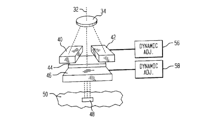
In one embodiment, the collimator 36 includes Y-axis jaws
and X-axis jaws. In Fig. 3, Y-axis jaws are represented by first and
second blocks 40 and 42 of radiation-attenuating material. Below the
3o Y-axis jaws are the third and fourth blocks 44 and 46 that form the X-axis
jaws. The spacing between the first and second blocks defines one
dimension of the target zone 48 on a body 50, while the spacing between
96P7523 CA 02212707 1997-os-os
7
the third and fourth blocks defines the perpendicular dimension. As used
with regard to the invention, "field-defining structure" refers to devices
such as those shown in Fig. 3 for determining the dimensions of the
target zone 48.
SOFTWARE SOLUTION TO COINCIDENCE PROBLEM
In the setup stage of the radiation system, the linac 26 is
deactivated and a light source 52 is energized. The light source directs
visible light to an optical element, such as a beam splitter, for redirecting
t o the light into the collimator 36. The light source may be a 150 W quartz
halogen lamp, but this is not critical. The optical element 54 should be
transparent to radiation from the linac 26. When the light has passed
through the collimator, the beam can be used to properly position the
jaws of the collimator. For example, if the target zone 48 on the body 50
~ 5 of Fig. 3 is a tattooed area on a patient, the blocks 40-46 may be
adjusted until the light field that is emitted from the collimator coincides
with the area of the target zone. The radiation system can then be
switched to an operation mode in which the radiation beam takes the
place of the light beam. However, unlike prior art radiation systems,
2o dynamic adjusters 56 and 58 automatically vary the settings of the X-axis
jaws and the Y-axis jaws in order to compensate for any inherent
dissimilarities between field sizes of the light beam and the radiation
beam. Adjustment of X-axis and Y-axis jaws could also take place when
the light is turned on.
25 The dynamic adjusters 56 and 58 are controlled by the
controller 28. In the preferred embodiment, the increments of adjustment
are calculated according to tables stored at element 60 in Fig. 2. Thus,
the "trimming" of the light field is enabled by computer software. For a
given energy level of X-ray radiation and for a desired field dimension of
30 10 cm, it may be known from data stored at component 60 that for the
appropriate setting of the jaws for the light field there will be a difference
of 0.2 cm when the radiation beam is activated during the operation
96P7523 CA 02212707 1997-os-os
8
mode. The dynamic adjusters will automatically vary the blocks 40-46 to
compensate for the difference.
The structure for forming the dynamic adjusters 56 and 58 is
not critical to the invention. Any device that can be electronically con-
trolled to manipulate the settings of the blocks 40-46 may be utilized.
- Fig. 4 illustrates one embodiment of a process for establish-
ing coincidence between field sizes of a light field and a radiation field for
the system 10 of Fig. 1. In step 62, the collimator field size is calibrated
for the radiation output. Calibration of a radiation system for X-ray output
is well known in the art, and any of the known techniques may be utilized
in executing step 62. For example, a tank of water may be used to
simulate a human body or other object and a probe may be used to
measure radiation through the water. With the water surface at a
900 mm target-surface distance (TSD) and the probe at isocenter, the
~ 5 field size may be measured for a particular setting of the jaws. Conven-
tionally, the field size measurement with regard to the fifty percent
maximum dose value of the radiation. This measurement is stored at
element 60 of Fig. 2. The procedure is repeated for a number of settings
of the jaws, and each measurement is recorded. Preferably, the energy
2o level of the radiation and the TSD are constant throughout the calibration
step, since these two factors affect scattering and diffraction of the
beam. However, as will be explained more fully below, the process steps
preferably are carried out with regard to more than one energy level and/or
with regard to more than one TSD.
25 In step 64, the measurements of field sizes acquired during
step 62 are coordinated with values on a display that is employed by a
user of the radiation system. A display monitor 66 is shown in Fig. 2.
The monitor will include designations of dimensions. In the exemplary
embodiment of Fig. 4, the indications are manipulable, so that the display
3o values can be coordinated with the measured field sizes at each of the
various settings of the jaws. In step 68, the data from steps 62 and 64
are recorded.
96P7523 CA 02212707 1997-os-os
9
The light 52 of Fig. 2 is then turned "on," as shown in step
70.of Fig. 4. For the same jaw settings that were used in the calibration
of the radiation output at step 62, the light field sizes are measured in
step 72. This may be done using conventional techniques. For example,
the tank of water used in step 62 may be replaced with graph paper or
- with a film pack. The portion of the graph paper or film that is illuminated
by the light beam is measured for each setting. Next, the dimensional
difference between the light field and the radiation field is determined for
each jaw setting. The delta values are recorded at step 74. In one
embodiment, a delta value is merely a difference between the measured
light field size from step 72 and the display monitor value for the
particular setting of the jaws. Because the phenomena of scattering and
diffraction will have different effects upon the X-ray beam and light beam,
the delta values will at least partially be indications of the different
~ 5 effects. Following step 74, a table may be formed from the data stored
at component 60 of Fig. 2. Such a table 76 is shown in Fig. 5.
Within step 70, the first row indicates the various settings of
the jaws that affect the field size of concern. That is, the first row is an
indication of the setting 78 of the field-defining structure that can be
2o dynamically adjusted. The second row 80 shows the measured field sizes
for the X-ray field at an energy level of 6 MV. The values of rows 78 and
80 are identical, since the radiation output was calibrated at step 62.
In the third row 82, the display values of the monitor 66
have been recorded. Since the X-ray field sizes and the display values
25 were coordinated at step 64 of Fig. 4, the values of row 82 are identical
to the values of row 80. With the X-ray output turned "off" and the light
52 turned "on," the light field sizes were measured at step 72 and
recorded.in the fourth row 84 of table 76. The final row 86 records the
delta values that are the dimensional differences between the X-ray field
3o size of row 80 and the light field size of row 84.
In step 88 of Fig. 4, the steps for generating the table 76
may be repeated for alternative energy levels of the radiation system.
96P7523 CA 02212707 1997-os-os
Since the radiation field sizes may vary depending upon the energy level
of fihe radiation beam, there are potentially different delta values for the
different energies. By generating separate tables that are implemented
based upon the setting of energy levels, the dynamic compensation
5 process is not a process that requires significant compromise, as would be
- the case if aluminum micro-trimmers were used to achieve field size
coincidence.
The dynamic adjusters 56 and 58 are enabled at step 90 of
the process of Fig. 4. Then, in the operation of the radiation system 10
of Figs 1-3, a user selects a setting for the field-defining structure (i.e.,
the blocks 40-46) in the conventional manner of forming a light field that
corresponds to the target zone of the patient 20. This target zone may be
delineated by a tattoo on the patient 20, but this is not critical. The light
field is provided by activating a light source 52 to pass light through the
~ 5 collimator 36. The adjustment of the field-defining structure is
accomplished during the setup mode of the radiation system.
Once the blocks 40-46 have been properly set with respect
to the dimensions of the light field, the system may be switched to the
operation mode. The controller 28 may provide the switching capability.
2o As the switching function is executed, the dynamic adjusters 56 and 58
are varied by an increment defined by the appropriate delta value in row
86 of Fig. 5. For example, if the light field that was established during
the setup mode had a field size that spaced the first block 40 away from
the second block 42 by a distance of 4.5 cm, the dynamic adjustment will
25 be an incremental increase of 0.5 cm. This spaces the blocks apart by a
distance of 5 cm. The incremental adjustment is performed in software,
so that the user is not required to provide further adjustments. Based
upon the data of table 76, the controller 28 is able to interpolate and/or
extrapolate the information in order to provide the appropriate
3o compensation for a setting that is not contained within stored memory.
While the preferred embodiment of Fig. 4 uses the X-ray
output to calibrate the system in steps 62 and 64, there may be some
96PT523 CA 02212707 1997-os-os
11
applications in which the light field is used in the calibration process. The
invention may be used in applications outside of the medical environment.
OPTICAL SOLUTIONS TO THE COINCIDENCE PROBLEM
While less desirable than the use of computer software to
provide field size coincidence, there are optical solutions. One such
solution is to provide a curved mirror to compensate for the dimensional
error between the light field size and the radiation field size. It has been
determined that an asymmetrically shaped mirror provides the better
results. Specifically, a parabolic mirror is preferred. The equation for the
mirror surface and the positioning of the mirror relative to a target area
has the form:
f (x? = Ax2 + Bx + C
wherein the coefficient "A" describes the curvature of the mirror, "B" is a
~ 5 tilt term, and "C" is an offset term that by convention determines a
vertical position. When the term "A" is 0, the mirror is flat, while a
negative value describes a convex surface.
It has been determined that the mirror should be asym-
metrical, because the intersection of the light with the mirror for a given
2o field size is not equally spaced from the collimator axis when comparing
opposite jaws. Therefore, one side of the mirror has a greater curvature.
An acceptable shape of a mirror 92 is shown in Fig. 6, but the curvature
is exaggerated for purposes of illustration. Actual curvature is more likely
to be a subtle curvature of approximately 0.002 to 0.003 inches per inch.
25 One difficulty with this mirror solution is that while it works
well with positive field sizes, the use of the curved mirror increases the
error if one of the collimator jaws defines a negative field size, i.e., both
of the blocks of the jaw are on the same side of a midline. Another
problem is that the mirror does not rotate with the collimator, so that this
30 approach to correcting the light penumbra only works with the collimator
at 0 degrees. When the collimator is at 90 degrees, there is no correc-
tion. It may be feasible to provide a three-dimensional shape that would
96PT523 CA 02212707 1997-os-os
12
be adequate, but this may not be cost efficient.
Another possible solution is to provide a more compact light
source and a second mirror, with the second mirror being placed in the
head area of the radiation system. By adding an elliptical reflector behind
a conventional light source that produces approximately 20 lux at
isocenter, the illuminance may increase to 100 lux. This increase in
illuminance reduces the perceived light field penumbra without an increase
in the cost of the light source. However, the uniformity at the edges of
the light field is obtained at the cost of creating a dark spot at the beam
axis, since the lamp blocks some of the light reflected by the elliptical
reflector. It may be possible to use frosted glass or some other method to
diffuse the light, but the diffusion reduces the efficiency.
Alternate light sources provide some benefits. For example,
an arc lamp may be used in place of the conventional tungsten-filament,
~ 5 halogen-filled, quartz lamp. The arc lamp provides a significantly greater
illuminance. When focused on a small aperture, no field size compensa-
tion was required. However, the arc lamp is an expensive alternative and
because of size and safety reasons, the best location for the lamp and
power supply would be in the machine stationary structure, so that light
2o guidance (e.g., a fiber optic bundle) may be required.
Another alternative light source is a laser. The laser provides
the advantage of producing a small and well-collimated beam. The beam
could be focused by a converging lens through a very small aperture to
produce little or no penumbra. To achieve a 40 lux, 50 cm diameter field,
25 however, would require 8 lumens of luminous power. At the wavelength
of a typical HeNe laser, one watt of power is approximately equal to 250
lumens. Consequently, a very powerful and perhaps prohibitively
expensive 32 mW laser would be required.
As previously noted, fiber optics may be utilized. Because
3o there is limited space within the head area of the radiation device, the
application of fiber optics is attractive. The challenge with this approach
is keeping the efficiency sufficiently high to provide at least equal illumina-
96PT523 CA 02212707 1997-os-os
13
tion that at least equals that of the existing system.
A final approach is to provide a light source on the target
slide of the radiation device. The advantage of this approach is that the
use of a mirror would not be required and the alignment procedure would
be simplified. The space that the mirror occupies on conventional radia-
tion devices could be used for shielding material, automated wedges, or
the like. To implement this approach, the light source would occupy a
new position on the target slide, and the target slide would move between
a patient setup position and an actual treatment position. The
1 o conventional thickness of the target slide presents difficulties in
placing
the light source directly on the slide, so that there may be a need to
increase the structural integrity of the target slide or to provide a remote
light source that is optically coupled to a fiber optic bundle.