Note: Descriptions are shown in the official language in which they were submitted.
CA 022l276X 1997-08-08 --
Attorney Docket No. HOOV10lPCT
APPARATUS AND h~-~O~ FOR
MORSELATING AND REMOVING TISSUE FROM A PATIENT
INVENTOR: MICHAEL D. HOOVEN
The present invention relates generally to apparatus
and methods for removing tissue from the body of a human
patient. More particularly, the present invention
concerns novel apparatus and methods for morselating and
removing body tissue through a relatively small incision
in the patient.
BACRGROUND OF THE INVENTION
So-called m;n;m~lly invasive surgery has become
increasingly popular in a variety of surgical procedures.
~;n;mally invasive surgery typically involves introducing
surgical devices into a patient through small access
incisions, in contrast to obtaining full and open access
to the surgical site through large incisions.
Briefly, ;n;~lly invasive surgery is typically
carried out through one or more relatively small
incisions, which are usually between approximately 1/2 and
l1/2 inches in length, and through which an entry tube or
trocar is placed. Optical and medical instruments are
inserted through the trocar(s) to allow the physician to
view the surgical area and to target the organ or tissue
that is the subject of the surgery, and then to carry out
the desired surgical procedure. Because of the relatively
small diameter of the trocar(s), however, withdrawal of
the target tissue therethrough can be difficult,
particularly if the tissue is dense or muscular, such as
a kidney, uterus or uterine myoma.
One previously accepted technique for removing such
tissue required manually cutting the tissue into smaller
pieces within the body cavity, which pieces were then
- CA 02212768 1997-08-08 ~
removed through the trocar by graspers. This procedure,
however, in addition to being very tedious and time
consuming, also suffers from a number of other possible
drawbacks, including possible excessive bleeding, possible
accidental cutting of other tissue and possible
contamination of the abdominal cavity with target tissue.
A morselation device is described in U.S. Patent No.
5,290,303. That device uses an inner rotating tube and an
outer stationary sheath, the inner tube extends beyond the
sheath and has a tapered end for severing tissue. A
further outer tube or shield may also be used over the
sheath. This device may be used with a tissue bag, such
as shown in U.S. Patent No. 5,037,379. The tissue to be
morselated may be placed in the bag and the device
inserted into the bag to carry out the morselation within
the pouch.
One of the concerns with the device shown in the '303
patent is possible puncture of the pouch by the rotating
tube, with accompanying potential contamination of the
anatomical space, as well the time consuming insertion and
placement of a new pouch within the body cavity. Although
the bag disclosed in the '379 patent has two layers,
including a puncture resistant inner layer, the above-
mentioned concern is still present. In addition, the
multi-layer construction with a higher strength inner
container may make folding and insertion o~ the pouch into
the abdominal cavity more cumbersome.
More recently, in U.S. Patent No. 5,304,124, an
apparatus and method were disclosed for removing a uterine
myoma. In that method, a tube is inserted through the
trocar and into the myoma. A wire loop, which may
energized by radio frequency energy, is located at the
distal end of the tube to cauterize the tissue as the tube
is inserted into the myoma, resulting in a core of tissue
being located within the tube. A separate morselator is
then inserted into the tube to cut up the tissue (such as
-- CA 02212768 1997-08-08
by rotary blades, laser, or a rotary whip), and the
morselated tissue is then evacuated. Although such a
procedure may be an advance over a purely manual
procedure, this procedure is still relatively complicated,
requiring separate steps and apparatus to core and to
morselate the tissue.
In addition, the procedure described in the '124
patent, when utilized with radio frequency (~'RF") energy,
uses a separate grounding or return electrode or antenna
in contact with the skin of the patient, for example, that
the patient lies on. As is well known in the art, such an
application of RF energy has certain shortcomings. It
re~uires the energy to travel between the electrodes, a
relatively long distance through the body, with possible
adverse effect on other body tissue. It also may result
in accidental injury to non-target tissue, for example, if
the active electrode is inadvertently brought into contact
with non-target tissue. Also, this patent discloses a
relatively complex mechanism to sever the tissue core from
the myoma.
Accordingly, it is a general object of the present
invention to provide apparatus and methods for removing
target tissue through a trocar, which apparatus and method
are more simplified and/or easier to use than the
apparatus and method described above, and reduces the risk
of accidental injury to non-target tissue.
GEN~RAL SUMMARY OF THE lNv~NlION
As set forth in the appended claims, the present
invention is generally embodied in apparatus and methods
for morselating and/or removing target tissue from the
body cavity of a patient, such as through the relatively
small incision(s) that are typically used in m;n;r~lly
invasive surgical procedures.
More particularly, the present invention is generally
embodied in a morselator, a tissue container for
cont~; n; ng resected tissue to be morselated, and their
- CA 02212768 1997-08-08
methods of use. In general, the morselator of the present
invention may comprise an elongated shaft having an inner
tube and an outer tube extending between proximal and
distal end portions. At least one of the tubes is
rotatable and an electrode surface is carried by the
rotatable tube(s) in proximity to the distal end thereof.
The foregoing apparatus may be used ~or removing
tissue from within a body cavity of a patient by inserting
the distal end through an incision in the patient,
energizing and rotating the electrode and advancing the
electrode into the resected tissue in order to morselate
it. The morselated tissue is then removed through the
lumen of the inner tube.
Preferably a second electrode of opposite polarity is
used with the first mentioned electrode, with one of the
terminals being a RF energy active electrode and the other
being a RF energy return electrode, to morselate tissue
therebetween. The additional electrode may be located, in
one embodiment, at the distal end of the shaft or, when
the morselator is used to morselate tissue within a
resected tissue container, the additional electrode may be
defined within the container, such as by a conductive
inner surface of the container or by having the additional
electrode otherwise disposed within the container.
In accordance with ~urther aspects of the present
invention, the combination of a resected tissue container
and a morselator may be provided for morselating resected
tissue within the body cavity of a patient. In such a
combination, the tissue container is insertable through an
incision into a body cavity of a patient. The container
defines an interior chamber for cont~; n; ng the resected
tissue. The morselator has a proximal end portion and a
distal end portion. One electrode is carried on the
distal end portion and is operable to assist in the
morselation upon insertion through an incision and into
the resected tissue container. A second electrode o~
CA 02212768 1997-08-08
opposite polarity also is disposed in the container. In
this combination, the resected tissue container contains
the resected tissue and helps protect surrounding tissue
from inadvertent or undesirable contact with the
electrodes or RF energy associated therewith.
In accordance with another aspect of the present
invention, a tissue container is provided for containing
resected tissue during morselation. The tissue container
is comprised of a flexible wall which defines an inner
chamber adapted to be received within a body cavity of a
patient. The wall comprises a non-conductive outer
surface and a conductive inner surface, which inner
surface also may serve as an electrode of opposite
polarity when only one type of electrode (e.g., active or
return) is carried on the morselator.
The above is only a summary o~ the present invention
in certain of its more general aspects. Accordingly, for
a more complete understanding of these and other features
and advantages of the present invention, reference should
be made to the following detailed description.
- CA 02212768 1997-08-08
BRIEF DESCRIPTION OF THE DRAWINGS
The present invention is set forth in greater detail
in the following description of the attached drawings, of
which:
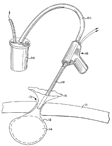
Figure 1 is an overall perspective representation of
the present invention, including morselator extending into
a tissue container in a small incision in the body cavity
of a patient.
Figure 2 is an overall perspective representation of
apparatus embodying the present invention and that may be
used in practicing the method of the present invention,
including a tissue container, hand-held morselator, and
specimen collection jar.
Figure 2a is a cross-sectional view of the tissue
container of Figure 2.
Figure 3 is a perspective view of the morselator
shown in Figures 1 and 2 and embodying the present
invention.
Figure 4 is a cross-sectional view of the apparatus
of Figure 3.
Figure 5 is a cross-sectional view of a multiple-tube
elongated shaft portion of the apparatus of Figures 3 and
4.
Figure 5a is a perspective view of an inner tube of
the multiple-tube shaft shown in Figure 5.
Figure 5b is a perspective view of the distal end of
the multiple-tube elongated shaft shown in Figure 5
Figures 6a - 6g are perspective views of different
configurations of the distal end of the multiple-tube
elongated shaft shown in Figure 5.
Figure 7a is a cross-sectional view of an elongated
shaft for removable attachment as part of a morselator.
Figure 7b is a cross-sectional view of the shaft of
Figure 7a taken along lines 7b-7b of Figure 7a.
Figure 8 is a perspective view of snap collar
employed in the apparatus of Figures 7a-7b.
- CA 02212768 1997-08-08
Figure 9 is a perspective view of a main collar
employed in the apparatus of Figures 7a-7b.
Figure 10 is a perspective view of an outer tube
guide employed in the apparatus of Figures 7a-7b.
-- CA 02212768 1997-08-08
DETAILED DESCRIPTION OF THE DRAWINGS
The present invention is particularly useful in
morselating and removing tissue from a body cavity of a
patient through relatively small incisions, such as those
employed in so-called m; n;m~l invasive surgery.
An initial description of the method of use may aid
in understanding the apparatus of the present invention.
In ~; n; ~1 ly invasive surgery, one or more relatively
small incisions 10 are normally made in the patient. As
is well known in the art, particularly in abdominal
surgery, these incisions are typically made by first
inflating the abdominal cavity to raise the skin away from
the underlying organs. The skin is then grasped, and a
trocar, which may have a puncture tip, is inserted through
the skin and peritoneal membrane 11 and into the abdominal
cavity forming a relatively small access incision or
opening through the skin. Surgical instruments, optical
fiber devices, light sources and the like may then be
inserted through the trocars to carry out the desired
surgical procedure on whatever target tissue is involved.
Although described generally in terms of abdominal
surgery, the method and apparatus of the present invention
are not limited to a specific type or location of surgery.
The method of the present invention is typically
carried out after the surgery, such as that described
above, has been carried out, and the target tissue has
been resected. The target or resected tissue may be any
organ, tumor, growth, or other tissue, although it is
contemplated that the present invention is particularly
useful for tissue that is especially dense or muscular,
such as a uterus or kidney, and unsuited for simple
withdrawal through a trocar, or for tissue that may be
infectious or malignant. Similarly, the present invention
is not limited to any particular technique or apparatus
for the resection of the target tissue.
Referring to Figures 1 and 2, in accordance with one
aspect of the present invention, at least a portion of a
- CA 02212768 1997-08-08
tissue container such as a flexible bag or pouch 12, is
inserted through the incision 10 and into the patient's
body cavity. It is anticipated that in most procedures
the entire bag or pouch 12 will initially be inserted into
the body cavity by rolling or folding it and inserting it
through the trocar located in the incision. The resected
tissue 14 is then placed into the bag or pouch. The bag
or pouch may be located entirely within body cavity or the
lip or marginal edge 15 of the bag or pouch may then
pulled through the incision, for example after removal of
the trocar, to allow the bag or pouch to be held or
gripped by the surgeon during the morselating.
Morselator 16 of the present invention is then
inserted through incision 10 into the bag or pouch 12.
When the bag or pouch has been inserted into the body
cavity, and the lip 15 of the bag or pouch is withdrawn or
pulled up through the small incision 10 and externalized,
the distal end 17 of elongated shaft 18 of the morselator
16 is inserted through the lip or opening 15 of the bag or
pouch 12 and through the incision to the resected tissue
14. A rotating electrode on the distal end o~ the sha~t
morselates the resected tissue by coring it or slicing it
while it remains within the portion of the bag or pouch 12
that is within the body cavity. As used herein,
morselating means cutting, coring, slicing, chopping or
any other way of sub-dividing tissue into smaller pieces.
The morselated tissue is then removed, such as by suction
through the shaft.
Turning now to the illustrated morselator employed in
and embodying the present invention, Figure. 3 is a
perspective view of the preferred morselator 16 shown in
Figures 1 and 2. For the purposes of this description and
the claims, the morselator 16 may also be referred to
generically as an ~electrosurgical device~.
As shown in Figure 3, the morselator 16 includes, in
addition to the elongated shaft 18, a hand piece 20 at the
proximal end 21 of the elongated shaft for gripping by the
~ =
- CA 02212768 1997-08-08
surgeon and for mounting the elongated shaft 18 and the
controls for the morselator. Referring first to the
elongated shaft, which is also shown in Figures 5-5b, the
shaft is made up of a stationary outer tube 22 and an
inner tube 24 rotatably received within the outer tube.
The elongated shaft may be permanently attached to the
hand piece, with the entire morselator being disposable or
reusable. Alternatively, as discussed later, the
elongated shaft may be disposable and removably attachable
to a reusable hand piece.
As shown in Figures 4 and 5, the inner tube extends
from the distal end 17 of the elongated shaft 18, through
nose 26 of hand piece 20 and terminates in a proximal
suction connection fitting 28 on the hand piece. The
outer tube 22 extends from the distal end 17 of the shaft
18 to a proximal tube collar 30, which is fixedly attached
to the nose of the hand piece.
The inner tube 24 has an inner tube collar 32
attached at the pro~; ma 1 end portion of the inner tube.
The inner tube collar is received within and pre~erably
keyed or otherwise attached in rotationally locked
engagement with an inner body sleeve 34 mounted in the
hand piece 20. A keyed arrangement between the inner tube
collar and the inner body sleeve permits easy removal and
disposal of the inner tube and reuse of the hand piece.
Alternatively, the inner tube collar may be permanently
attached or bonded to the inner body sleeve 34, and the
entire morselator may be disposable or resterilizable.
Although not necessarily preferred, the inner tube 24
may be axially movable relative to the outer tube 22, and
spring loaded, such as by a compressed spring (not shown)
between inner tube collar and the nose 26, to bias the
inner tube to a position where the distal end of the inner
tube extends slightly beyond the distal end of the outer
tube. As a result, when an axial force is exerted on the
distal end of the inner tube, such as when it contacts the
tissue container, the inner tube is forced back into the
CA 02212768 1997-08-08
outer tube. This feature may prevent unnecessary damage
to the electrodes at the distal end of the inner tube and
to the tissue container.
The inner and outer tubes are preferably made of a
substantially electrically non-conductive material, such
as a fiber glass-epoxy composite or a polymer.
Alternatively, the walls of the inner and outer tubes may
have a metal core for strength and be coated with a
substantially non-conductive or insulating material. The
diameter and thickness of the inner and outer tubes may be
selected depending on the desired procedure and/or target
tissue involved. For morselating dense or muscular
tissue, fiberglass epoxy inner and outer tube walls of
approximately .007 inches thick have been found suitable.
Because of their thinness, the tubes are shown simply as
lines in Figures 4 and 5.
For morselating resected tissue, a pair of electrode
surfaces 36 of opposite polarity are preferably provided
at the distal end 17 of the shaft 18. In the illustrated
embodiment, the electrode surfaces are provided at the
distal end o~ thin conductive metal strips 38 that extend
along and are bonded to the outer surface of inner tube
24. The distal ends of these strips, which provide the
electrode surfaces, terminate in proximity to the distal
ends of the inner and outer tubes, although they may
extend slightly beyond the distal end of the inner and
outer tubes, be recessed slightly between the inner and
outer tubes, or terminate at the distal end and still
function satisfactorily.
The conductive strips 38, such as aluminum or
stainless steel strips or foil, may be placed on the tube
wall by, for example, bonding the strips to the tube or
manufacturing the tube in such a way that the strips 38
are located inside the wall of the tube, and the strips
exposed at the distal end. The outer tube 22, which may
be made completely of insulating material or have an
interior surface of non-conductive material, insulates the
CA 02212768 1997-08-08
electrical conductors on the inner tube from surrounding
tissue, and the insulating material on the inner wall of
the inner tube insulates the conductors from any tissue
within the inner tube. (~Insulating~ and "non-conducting~
are used interchangeably in this description.)
The present invention, howe~er, is not limited to the
use of conductive strips to transmit electrical energy
from the proximal to the distal end of the elongated
shaft. Any type of conductor or conductive material can
be used. Thin and relatively wide conductive strips are
preferred, however, for delivering high power RF energy to
the electrode surfaces because they allow a m~x;ml7~ amount
of current to be carried along the length of the inner
tube without unduly increasing the spacing between and/or
wall thickness of the tubes. Thin conductors are also
ideal for RF energy because the high frequency current
travels primarily on the surface of a conductor, so a thin
conductor with a large surface area, such as the thin
strips 38 in Figure 5a, offers what is believed to be the
best possible combination of current carrying capability
and m; n; ~1 tube wall spacing and/or wall thickness.
Further, the conductive strips conduct RF energy much more
efficiently than a wire of the same cross-section since a
thin strip conductor has a much larger surface area than
a wire of the same cross-section.
Referring to the hand piece 20 of the morselator 16,
shown in Fig. 3 and in vertical cross-section in Figure 4,
the hand piece 20 includes a lower handle portion 40 for
the surgeon to grip and an upper body portion 42 to mount
the elongated shaft 18. The hand piece also includes the
controls for the morselator. The body portion 42 of the
morselator includes a motor 44, which is preferably a
lower power-consumption high torque motor such as the type
commonly used in electric screwdrivers, for rotating the
inner tube 24 of the elongated shaft 18. In order to
rotate the inner tube, the motor 44 is connected to a
drive gear 48 that mates with a driven gear 46 which is
- CA 02212768 1997-08-08
connected to the inner body sleeve 34. As explained
above, the inner body sleeve 34 i8 rotationally locked to
the inner tube 24 through collar 32. Accordingly, when
the motor 44 is engaged, it causes the inner body sleeve
34 and inner tube 24 to rotate. In the preferred
embodiment, only the inner tube rotates. It is within the
scope of this invention, however, for the outer tube to
rotate, and the inner tube to remain stationary or for
both tubes to rotate.
The handle portion 40 of the hand piece 40 of the
morselator 16 not only includes a grip 50 for the surgeon
to hold while using the morselator, but also may include
a battery pack 52 and a switch or trigger 54 to turn the
morselator on and off. The battery pack 52 may be
rechargeable for a reusable handle or a one-time use
battery for a disposable handle. To turn the morselator
on and off, the battery pack 52 is electrically coupled to
the motor 44 via the switch 54, which is preferably in the
form of a trigger.
The switch 54 also serves to the couple RF contacts
56 to tube contacts 58 on the inner body sleeve 34. Tube
contacts 58 are in the form of spaced-apart rings. Each
ring is in electrical contact through the inner body
sleeve, with one of the conductor strips on the inner
tube. Brushes or sliding contacts 60 are in direct
contact with the rings. One of the brushes is directly
coupled to one of the RF contacts. The other brush is
coupled to the other RF contact through a circuit
controlled by switch 54.
Therefore, when the switch 54 is turned on or the
trigger is depressed, it completes the circuit from the
battery pack 54 to the motor 44 and activates the motor
44, which causes the gear assembly to turn, and, the inner
body sleeve 34 and the inner tube 24 to rotate.
Additionally, when the switch is turned on or the trigger
depressed, it completes the circuit between at least one
of the RF contacts 56 and the tube contacts 58, allowing
- CA 022l2768 l997-08-08
14
RF electrical energy to flow from the RF contacts 56 to
the conductors (see e.g. Figure 5a) located on the surface
of the inner tube 24, and to the electrode surfaces 36 at
the distal end of the morselator 16.
Preferably, morselated tissue is removed through the
morselator 16 via an inner tube lumen 62 extending between
the distal 17 and proximal 21 ends of the morselator. A
suction source or apparatus (not shown) may be used to
suction the tissue and remove it from the body cavity.
More specifically, the suction apparatus is coupled to
connection fitting or tube 28, which communicates with
proximal end 21 of the inner tube 24 of the morselator 16,
and is used to suction the morselated tissue therethrough.
The suction apparatus may include a suction tube 64
coupled to a specimen collection canister 66, which is
then coupled to the suction source. The suction may be
manually controlled by the surgeon, or the morselator 16
also could include a suction control (not shown), such as
a rocker switch or similar mechanism, to allow the suction
control to operate at the same time or in timed relation
with the motor and/or RF energy activation. Alternately,
other devices such as a grasping instrument or a myoma
screw could be used to pull the morselated tissue up
through the inner tube of the morselator while the tissue
is being morselated.
The morselator of the present invention preferably
uses an RF energy source of the type well known in the
~ield. As commonly known in the field, such a radio
frequency energy source may provide high voltage
electrical current at a frequency between 100 KHz and 1
MHZ. The energy source can use either an RF monopolar or
a bipolar RF energy source. In the present invention,
however, because of the small surface area (the end edge
of strip 38, for example) of the electrodes it is
particularly desirable to use RF monopolar power source,
with one of the electrodes connected to the monopolar
~-- CA 02212768 1997-08-08
terminal of a typical RF energy power supply and the other
electrode attached to the return.
For morselating tissue there are various
configurations of electrodes, as shown in Figures 5b and
6a - 6g, that can be used in the present invention. It is
understood that these are merely examples of different
configurations and that other configurations,
modifications, and variations would be readily apparent to
one skilled in the art and are intended to be included
within this application. In each configuration shown in
Figures 5b and 6a - 6g, at least one electrode is active
and can be electrically coupled to a source of electrical
energy, such as for example a RF monopolar current source,
via one of the RF contacts 56 and a conductor such as
conductive strip 38. One or more of the other electrodes
is electrically coupled to a ground/return circuit, such
as a monopolar ground return, via the other RF contact 56.
Alternatively, bipolar energy could be used.
Figures 6a - 6d show the distal end 17 of the
elongated shaft 18 wherein, both electrodes are located
between the inner tube 24 and the outer tube 22 and can be
carried by the inner tube 24. Figure 6a shows two arcuate
or half-circle type electrodes 68, 69. These electrodes
may be, for example, wires or strips. One electrode would
be electrically coupled to ground/return, and the other
would be electrically coupled to a monopolar active
current. The embodiment shown in Figure 6b utilizes a
different configuration with wire electrodes 70, 71 on the
outside of the inner tube 24. These electrodes may be in
the shape of wire loops. The wires extend slightly beyond
the distal end of the inner tube. As a result, the inner
tube 24 may be recessed slightly in relation to the outer
tube 22 with the ends of the wires 70, 71 just flush with
the end of the outer tube 22. As in the embodiment in
Figure 6a, one electrode would be electrically coupled to
ground/return, and one would be electrically coupled to a
monopolar active current. In the embodiment in Figure 6c,
- CA 022l2768 l997-08-08
16
solid metal portions or strips 72, 73 are used instead of
wires. These metal portions may be located on opposite
sides of the inner tube and extend beyond the distal end
of the shaft, and may also be electrically coupled to
ground/return and a monopolar active current,
respectively. In the embodiment shown in Figure 6d, there
are four electrodes 74, 75, 76, 77 wherein two electrodes
80, 82 would be electrically coupled to ground/return, and
two electrodes 81, 83 electrically coupled to a monopolar
active current.
In the embodiment shown in Figures 6e and 6f, a
different configuration is shown where one electrode is
located between the inner tube 24 and the outer tube 22
while the end surface and/or outer surface of the outer
tube 22 functions as the other electrode. Specifically,
Figure 6e shows a scoop type wire 75 which could be
electrically coupled to a monopolar active current. The
outer surface of the outer tube 22 is covered with a
conductive material and acts as the other electrode, which
can be electrically coupled to ground/return. In the
eIElbodiment shown in Figure 6f, a single electrode 80,
which may be a wire, strip, or simply the exposed end of
a conductive tube, extends around the distal end of the
inner tube and may be electrically coupled to a monopolar
active current, with the ground/return electrode being the
outer surface of the outer tube 22.
It may be necessary in some instances to have a
portion of the side of the elongated shaft adjacent to the
tissue to be removed. In those circumstances, the tissue
can be shaved using the embodiment shown in Figure 6g. In
this embodiment, the inner and outer tubes 24, 22 are
slotted at a distal side port, and a wire or strip
electrode 82 extends the length of the side port and may
be electrically coupled to a monopolar active current and
function as a cutting edge as the inner tube 24 rotates
inside the outer tube 22 to shave any tissue within t:he
cutout portion of the tubes. Again, the edge surface or
-. CA 02212768 1997-08-08
outer surface of the outer tube 22 is conductive and may
be electrically coupled to ground/return. In all of these
electrode configurations, the outer tube 22 may be covered
with a conductive coating and used to provide a
ground/return path.
Alternatively, the elongated shaft may be a single
rotatable tube with a pair of electrodes of opposite
polarity at the distal end. For example, a solid metal
tube could be employed with an electrode of selected
polarity carried at the distal end and a conductor
extending along the length of the tube between the
proximal end and the electrode at the distal end portions.
Of course, to avoid short circuiting, the conductor would
need to be insulated from the metal tube. In this
alternative, the distal end of the metal tube itself could
act as the other electrode of opposite polarity, and the
tube body would function as the conductor between the
proximal and distal end portions. In a single tube
arrangement, various other combinations of conductive and
non-conductive materials also could be used, such as a
tube wall of non-conductive material with a pair of
electrodes at the distal end and a pair of conductors
extending along the tube body. Also, the wall of the tube
could have three layers with non-conductive inner and
outer surfaces and a metal core therebetween with the
distal end of the metal core forming the other electrode
and the core itself being a conductor.
Turning now to a more detailed description of the
tissue container that may be used in accordance with the
present invention, the container may be of various shapes,
sizes or materials without departing from the present
invention. As noted above, however, the tissue container
is preferably in the form of a flexible bag or pouch 12.
The bag or pouch 12 generally has side wall 84, bottom
wall 86, and marginal edge or lip 15 defining the opening
into the bag or pouch. The walls of the bag or pouch are
preferably sufficiently flexible to allow the bag or pouch
CA 02212768 1997-08-08
to be flattened or folded or rolled for insertion through
a trocar or a relatively small incision into the body
cavity.
In use in the preferred embodiment, the tissue is
morselated by the rotating action of the inner tube and
electrode and by pushing the distal end of the shaft
against the bottom of the tissue container to sever the
morselated tissue from the remainder of the resected
tissue. Accordingly, the tissue container should be
mechanically strong enough to withstand the force and
abrasion caused by a morselator, as well as the local
heating caused by electrodes. For this reason, it is
preferred that the walls of the vessel be made as thick as
possible, yet not so thick that the vessel cannot be
folded or rolled up to fit through a trocar.
To insulate and protect surrounding tissue, the walls
of the pouch or bag should be of a substantially non-
conductive or electrically insulating material. High
thermal resistance silicone rubber or fabric reinforced
silicone rubber in the range of 0.010 - 0.015 inches
thickness has the desired characteristics of flexibility,
strength and non-conductivity, and is the preferred
material. It is expected, however, that other materials
may also be suitable and the present invention, in its
broader respects, is not limited to a particular material
for the tissue container.
As described briefly above, the preferred morselator
may have both active and ground electrodes located on the
shaft. Alternatively, only one electrode may be located
on the shaft and the cooperating electrode be in the form
of conductive surface on the inside of the tissue
container. For the latter application, the tissue
container preferably has a non-conductive outer surface,
such as silicone, and a conductive inner surface such as
a flexible metallic film or foil, such as aluminum or
stainless steel foil. With proper attachment, the
CA 02212768 1997-08-08
conductive inner surface can act as the ground/return
electrode for RF energy application.
Alternatively, the tissue container may contain a
separate conductor or an electrode there within, such as
a wire or strip electrode of opposite polarity, to assist
in morselating the resected tissue. The separate
electrode could also be electrically coupled to active or
ground/return and operate in conjunction with the
electrode on the morselator for creating the needed
cutting action.
As described briefly above, the inner and outer tubes
24 and 22 forming the elongated shaft may be removably
attachable to the hand piece for disposability. Figures
7a-7b and 8-10 depict aspects of a snap-lock arrangement
for removable inner and outer tubes. In Figure 7a, outer
tube 22 is attached or bonded to an outer tube collar 88.
One end of outer tube collar 88 is received within outer
tube guide 90, which is bonded to main collar 92, which is
bonded to snap collar 94. Inner tube collar 96 is bonded
to the inner tube 24 and held in spaced relationship from
the outer tube collar by outer tube spacer 98 and from the
snap collar by inner tube spacer 100. This construction
allows the inner tube to rotate relative to the outer tube
and both tubes to be snapped into a receiving nose collar
that interfits with spring arms 102 of the snap collar.
In such an embodiment, the inner tube could be
rotationally locked to inner tube sleeve by a keyed
fitting, such as a mating spline and groove or the like.
Electrical contact with the contacts on the tube sleeve
could be achieved by slidable spring contacts. Also,
outer tube spacer 98 and/or inner tube spacer 100 could be
in the form of a spring to allow relative axial movement
between the inner and outer tubes.
The features and method of the present invention have
been described in connection with the accompanying
drawings for the purposes of illustration and not
limitation. It is intended that this application include
- CA 02212768 1997-08-08
those modifications, variations and additions that would
be readily apparent to one of ordinary skill upon reading
this description. Accordingly, for ascertaining the scope
of the present invention, reference must be made to the
appended claims.