Note: Descriptions are shown in the official language in which they were submitted.
,~' CA 02213091 1997-08-14
96P7526
ALIGNMENT SYSTEM AND METHOD
FOR INTRA-OPERATIVE RADIATION THERAPY
BACKGROUND OF THE INVENTION
The invention relates generally to aligning elements for
applying radiation to a patient and more particularly to systems and
methods for properly aligning a source of radiation with an applicator for
intra-operative radiation therapy.
DESCRIPTION OF THE RELATED ART
Radiation-emitting devices are generally known and used,
for instance, as radiation therapy devices for the treatment of patients.
A radiation therapy device typically includes a gantry which can be
swiveled about a horizontal axis of rotation in the course of a therapeutic
session. A linear accelerator is located in the gantry for generating a
high-energy radiation beam. The high-radiation beam can be electron
radiation or photon (X-ray) radiation. During treatment, the radiation
beam is trained on a treatment site of a patient Iying in the isocenter of
the gantry rotation. Typically, the patient is supported on a rotatable
table. The combination of movements of the gantry and the table
permits movement of the patient about mutually perpendicular X, Y and
Z axes. These rotations are sometimes referred to by the terms "tilt,"
"roll" and "yaw," respectively.
Prior to the application of radiation, a treatment setup
process is followed. This process includes setting beam parameters
such as radiation energy, field size, exposure times, dose and distance.
Moreover, the process includes aligning the gantry, a collimator and the
patient. The radiation beam is directed at diseased material, but with a
goal of minimizing any adverse effect upon adjacent healthy tissue.
For intra-operative treatments, the alignment process also
includes aligning an applicator relative to the patient and the source of
radiation. Intra-operative treatment typically includes forming an incision
through which an electron beam is directed to a treatment site. The
applicator is both mechanically and electrically isolated from the source,
i.e. the gantry. Mechanical independence is desirable, since the mass of
the gantry operates against the ability to manipulate the radiation beam
to enter a relatively small operative incision without significant risk to the
patient. The applicator is fixed relative to the patient, typically by
~ CA 02213091 1997-08-14
-2-
attachment to the table. The applicator provides beam collimation close
to the patient by establishing a radiation field-defining aperture. Thus,
the mechanical isolation reliably limits exposure to the desired treatment
site.
Electrical isolation is a factor, since any leakage currents
from the gantry to the patient place the patient at risk. U.S. Pat. No.
4,638,814 to Spanswick, which is assigned to the assignee of the
present invention, asserts that a patient cannot be subjected to ground
leakage currents which exceed five micro amperes because blood and
0 body fluids are good electrolytes and because any electrical devices that
are in contact with the patient may be disturbed. Spanswick describes a
method of aligning an electron applicator with an electron beam source.
A number of laser units project beams of light toward a support ring of
the electron applicator. The beams are arranged in a mutual orientation,
such as four iaser units arranged at 90~ intervals. Each of the four laser
units includes a beam splitter, so that eight beams are formed. The eight
beams form four beam pairs, with the two beams of a pair overlapping at
a predetermined point from the electron beam source. Consequently,
when the support ring is along the plane through the points of intersec-
tion, the eight beams form only four areas of illumination. The electron
applicator is attached to the operating table, so that the operating table is
moved until there are only the four illuminated regions. In addition to
aligning the electron applicator and the electron beam source, the use of
the intersecting beams determines the spacing between the applicator
and the source.
While the system described in Spanswick provided an
improvement over the prior art, further improvements are available.
Since the positioning of the electron applicator based upon overlapping
beams is performed visually, the process is subject to human error.
Moreover, the patent points out that the beams must be "exceedingly
sharp" in order to achieve precise positioning. As a result, the accuracy
of the method depends upon the selection of the sources of the light
beams. Another concern relates to the ability to change the spacing
between the electron applicator and the electron beam source. This
spacing will partially determine the intensity of the electron beam at the
treatment site of the patient. If the intersection of beams is to be used
to determine the spacing between the electron applicator and the elec-
tron beam source, the light beam axes must be adjusted from session to
~ CA 02213091 1997-08-14
.
session when the electron beam intensities vary among sessions. This
increases the setup time for equipment which is in demand.
What is needed is a system and method for accurately and
efficiently positioning a beam applicator without requiring the beam
5 applicator to be connected to a source of the beam.
SUMMARY OF THE INVENTION
A system for applying radiation therapy includes a radiation
source that emits a radiation beam into an applicator that is spaced apart
0 from and mechanically independent of the radiation source. An array of
targets is affixed to the applicator and at least one imaging device is
affixed to the radiation source to form image data representative of the
targets. The image data is processed to determine the positions of the
targets. In one embodiment, the determination of the target positions is
used to automatically adjust either the applicator positioning or the
radiation source positioning until the target positions match predefined
coordinates. Preferably, the target positioning is determined in three
dimensions.
A method of applying the therapeutic radiation includes
20 attaching the applicator so that it has an orientation that is substantially
fixed relative to a patient. The applicator is imaged by the imaging
devices that are affixed to the radiation source. Based upon the image
data, the system determirses whether a desired source-to-applicator
alignment has been achieved. The relative positioning of the radiation
25 source and the applicator is adjusted until the desired source-to-applicator
alignment is achieved. A radiation beam is then directed into the appli-
cator for applying localized radiation to a treatment site. In the preferred
embodiment, the method is used for intra-operative radiation therapy.
30 BRIEF DESCRIPTION OF THE DRAWINGS
Fig. 1 is a schematical view of a system for applying
localized radiation for intra-operative radiation therapy in accordance with
the prior art.
Fig. 2 is a schematical view of a system of applying
35 localized radiation in accordance with the invention.
Fig. 3 is a top view of a radiation applicator having targets
in accordance with the invention.
CA 02213091 1997-08-14
.
Fig. 4 is a process flow view of a method for utilizing the
system of Fig. 2.
Fig. 5 is a front view of a display screen for the applicator of
Fig. 3.
DETAILED DESCRIPTION
With reference to Fig. 1, a patient 10 is shown as resting on
a table 12 under a gantry 14 of a radiation therapy machine. A radiation
beam is directed from a collimator 16 of the gantry toward the patient.
10 The radiation beam is generated by a linear accelerator within the gantry
and is emitted from the collimator. The radiation beam may be electron
radiation or photon radiation, i.e. X-ray radiation. The gantry is known in
the art.
Typically, the collimator 16 determines the final beam
15 geometry. The beam is directed at a treatment site, such as diseased
brain tissue of the patient 10. The table 12 and the gantry 14 are
maneuvered to provide the desired alignment of the patient 10 to the
radiation beam, and the beam is then generated. However, there are
circumstances in which it is undesirable to use the collimator 16 as the
20 component for final direction of the radiation beam at the patient. For
example, within an intra-operative treatment an incision is formed for
passage of an electron beam to a treatment site. An electron beam
tends to expand more ~uickly than an X-ray beam, so that there is
greater concern that hea!thy tissue will be exposed. To reduce the risk, a
25 radiation applicator 18 is utilized. The radiation applicator is spaced apart from the collimator 16 and may have an output end inserted into the
incision of the patient 10. The radiation applicator is formed of a
material that is opaque to the electron beam, but includes a passageway
to the treatment site. The radiation applicator localizes the therapy to
30 the desired treatment site.
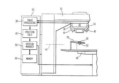
Referring now to Figs. 2 and 3, a radiation applicator 20 in
accordance with the preferred embodiment of the invention is shown as
including four targets 22, 24, 26 and 28. The targets may be recesses
within the surface of the applicator, but preferably are separate members
35 that are formed of a material that facilitates imaging of the targets. As
will be explained more fully below, the targets are imaged in order to
calculate the spacing and the alignment of the radiation applicator relative
to a collimator 30 of the gantry 32 shown in Fig. 2.
CA 02213091 1997-08-14
.
While not critical, the targets 22, 24, 26 and 28 are
preferably fabricated in the manner described in U.S. Pat. No. 5,446,548
to Gerig et al., which is assigned to the assignee of the present inven-
tion. The Gerig et al. patent describes a patient positioning and monitor-
5 ing system that can be utilized in combination with the invention to bedescribed below.
The targets 22, 24, 26 and 28 preferably include retroreflec-
tive material. The arrangement of the targets on the surface of the
applicator 20 is not critical. The targets are imaged by a pair of cameras
0 34 and 36. The Gameras may be charge coupled device (CCD) cameras,
but other imaging devices may be utilized. The image signals from the
cameras 34 and 36 are input to an image processing circuit 38. The
image processing circuit cooperates with a position calculation circuit 40
to determine position data for the radiation applicator 20. The image and
15 position processing may include a visual-based coordinate measurement
(VCM) system to determine target positioning in three-dimensional space.
In the preferred embodimentf the VCM system is a software package
which can be integrated with commercially available solid-state cameras,
image acquisition and processing boards, and computer hardware. The
20 VCM system combines principles of stereo vision, photogrammetry and
knowledge-based techniques to provide precise coordinate and dimension
measurement of objects. The two cameras 34 and 36 and the three-
dimensional image and position processing of circuits 38 and 40 are
calibrated such that the frame of reference is coincident with the system,
25 with an isocenter defined as 0,0,0. The coordinate system is defined
such that the X axis lies on a horizontal plane perpendicular to a gantry
axis 42 of rotation and passes through the system isocenter, the Y axis
is parallel to the gantry axis of rotation and passes through the isocenter,
and the Z axis is mutually perpendicular to the other two axes and
30 defines patient height.
Light sources 44 and 46 may be used to enhance per-
formance of the target imaging. In the preferred embodiment, the light
sources provide infrared radiation, and each of the cameras 34 and 36
includes an infrared filter. The infrared radiation enables the system to
35 more reliably distinguish light reflected from the targets 22-28, as
opposed to background radiation that may be present in the therapy room
under ambient light conditions. The light sources may be infrared lasers,
with the infrared radiation being spread by lenses, not shown. The use
CA 02213091 1997-08-14
-
of laser light sources provides the advantage that the spectral bandwidth
of the radiation is narrow, providing a further reduction in background
interference. Equipping the cameras 34 and 36 with infrared filters
reduces the susceptibility of the cameras to background radiation.
The radiation applicator 20 of Figs. 2 and 3 is shown as
being attached to a displaceable table 48 by an L-shaped support device
50. The mechanism for suspending the radiation applicator is not critical.
In fact, the applicator may be fixed to the patient, rather than to the table
48. For example, headgear may be fitted to the patient to attach the
radiation applicator to the patient.
The radiation applicator 20 is shown as having a truncated
cone-shaped beam outlet end 52. The configuration of the inlet and
outlet ends of the applicator will depend upon the gantry 32 and the
treatment plan of the patient. In the view of Fig. 3, the sloping interior
surface 54 is shown as terminating in a circular outlet 56. However,
other geometries are contemplated.
The determination of the positions of the targets 22-28 by
the image and position processing circuitry 38 and 40 is input to a
session manager 58. Based upon inputted data and/or stored data in
memory 6C), the session manager controls the variable components of
the system. !n the preferred embodiment, the session managing is
completely automated. However, manual adjustments may be required.
The session manager 58 may therefore include an operator console and
input devices, such as a keyboard.
The session manager 58 compares the positions of the
targets 22-28 to preselected coordinates. If the positions of the targets
are different than the desired positions, either or both of the gantry 32
and the table 48 are manipulated to reposition the targets. The session
manager is housed within a stationary portion 62 of the system that
supports the rotatable portion of the gantry 32. The rotatable portion
rotates about the gantry axis 42. The table 48 accommodates reposi-
tioning along the X axis and the Z axis. Preferably, the circuitry within
the stationary portion 62 of the system utilizes a servo approach, so that
periodic image captures via the cameras 34 and 36 are utilized to estab-
lish the desired target coordinates. Since the table 48 supports the
patient, repositioning the radiation applicator 20 relative to the gantry 32
also repositions the patient. As a consequence, manipulation of the
CA 02213091 1997-08-14
gantry 32 or the table 48 does not affect the position of the applicator
20 relative to the patient.
The operation of the system of Fig. 2 is described with
reference to Figs. 2-4. In step 64, the alignment of the applicator 20 to
the patient is established. In one embodiment, the applicator-support
device 50 is attached to the table 48. While not shown, the device 50
preferably includes an adjustment mechanism. For example, the device
may include slide mechanisms that permit vertical and horizontal reposi-
tioning of the applicator 20. In another embodiment, the applicator 20 is
0 supported directly by the patient.
The applicator is secured to provide the desired angular
alignment relative to a treatment site of the patient. This reduces the
risk that healthy tissue will be unnecessarily exposed to radiation. The
alignment of the applicator also includes setting the distance between the
treatment site and the beam outlet end 52 of the applicator 20.
At step 66, the cameras 34 and 36 of Fig. 2 acquire an
image of the targets 22-28. Each camera detects the reflected radiation
from the targets. As previously noted, the preferred embodiment
includes infrared lasers 44 and 46 and infrared filters in order to reduce
the effects of background radiation on the image processing at circuit 38.
At least two cameras 34 and 36 are employed in order to
permit position calculation 68 in three dimensions. Stereo vision tech-
niques of a video-based coordinate measurement system are executed
within the position calculation circuit 40 to determine coordinates within
a coordinate system defined such that the X axis lies in a horizontal plane
perpendicular to the gantry axis 42, the Y axis is parallel to the gantry
axis, and the Z axis is perpendicular to the other two axes and defines
patient height. Each of the three axes of the coordinate system passes
through the isocenter of the radiation system.
In step 70, a determination is made as to whether the
calculated coordinates of the targets 22-28 match desired coordinates.
The position data related to the desired coordinates may be stored in
memory 60 of Fig. 2. The determination of whether a correlation exists
preferably takes place in software. However, referring briefly to Fig. 5,
the determination may be made by an operator using a display 72 that
shows both the desired positions 74, 76, 78 and 80 of the targets and
the actual positions 82, 84, 86 and 88. If the desired positions and the
actual positions are aligned, the applicator 20 is properly aligned with the
-
. CA 02213091 1997-08-14
-8-
gantry 32. Consequently, the treatment site of the patient is properly
aligned with the radiation beam that will be emitted from the gantry. In
such case, the source of radiation can be activated, as shown at step 90
in Fig. 4. If at step 70 no correlation is determined between the coordi-
5 nates calculated in step 68 and the desired target coordinates, the
~ gantry-to applicator alignment is adjusted at step 92. The realignment
may be executed in alternative manners. The stationary portion 62 of
the gantry 32 may rotate the displaceable portion about gantry axis 42.
Alternatively, the table may be manipulated to correct for tilt and roll.
10 The collimator 30 of the gantry 32 is also adjustable, as is well known in
the art. Of course, the gantry-to-applicator alignment may be a combina-
tion of these adjustments.
Following the realignment at step 92, the process returns to
step 66 in order to acquire an updated image for calculation of updated
1~ position data in step 68. Preferably, the steps 66, 68, 70 and 92 utilize
servo techniques to automatically and efficiently obtain the desired
gantry-to-applicator alignment. When the alignment is achieved, the
radiation therapy is initiated at step 90. The arrangement of targets
22-28 is not critical. Preferably, there are three or four targets, but
20 performance may be enhanced in some applications by providing a
different number. As previously noted, the targets may be merely
recessed or raised areas of the applicator servo, but retroreflective
targets enhance the image processing by reducing the effect of
background radiation. Fluorescent and phosphorescent materials may
25 also be utilized with the appropriate camera filters to enhance selectivity
of reception.
In another embodiment, the targets 22-28 are fixed within
the sloping interior surface 54 of the applicator 20 of Fig. 3. This allows
the targets to be at different distances from the collimator 30 of Fig. 2,
30 even when the applicator is in the desired position relative to the
collimator. The variations in distance facilitate distinguishing actual
positions of targets from desired target positions.