Note: Descriptions are shown in the official language in which they were submitted.
CA 02214168 1999-11-19
ELECTROLYTICALLY DEPLOYABLE BRAIDED VASO-OCCLUSION
DEVICE
Field of the Invention
This invention is a br;~ided tubular device used in the occlusion of various
lumen or cavities in the body. In particular, it may be used to form an
endovascular occlusion. Most desirably, it is braided of a majority of super-
elastic
alloy ribbons and therefore is both inherently non-stretching and most
difficult to
permanently deform.. It may be deployed using an electrolytically severable
joint.
A radio-frequency modulated. current may optionally be applied to the device
after
its placement in the body. The elongated device is insulated along its length
to
optimize its occlusive activity without harm to the body.
Background of the Invention
A wide variel:y of medical procedures are facilitated by occluding such
body lumens and cavities as the arteries, veins, vascular aneurysms, various
vascular malformations (e.g., AVM's), fallopian tubes, vas deferens, ureters,
and
the like. For instance, an extravascular approach to treatment of aneurysms
involves surgically e:Kposing or stereotaxically reaching an aneurysm with a
probe.
The wall of the aneurysm is then perforated from the outside and various
techniques are used to occlude the interior in order to prevent it from
rebleeding.
The techniques used to occlude the aneurysm include electrothrombosis,
adhesive
embolization, hog hair emboLization, and ferromagnetic thrombosis. These
procedures are discu:;sed in U.S. Patent No. 5,122,136 to Guglielmi et al.,
the
entirety of which is incorporavted by reference.
A still further approach is the least invasive and is additionally described
in
Guglielmi et al. It is the endovascular approach. In this approach, the
interior of
the aneurysm is entered by use of a catheter such as those shown in Engelson
(Catheter Guidewire), U.S. Pa.tent No. 4,884,575 and also in Engelson
(Catheter for
Guidewire Tracking)., U.S. Patent No. 4,739,768. These procedures utilize
CA 02214168 1999-11-19
endovascular guidewires and catheters, introduced quite remotely, to access
the
aneurysm. Specifically by the use of catheters having very flexible distal
regions
and guidewires which are ste;erable to the region of the aneurysm, embolic
devices
which may be delivered through the catheter are an alternative to the
extravascular
and extra-intravascu.lar approaches.
The endovascular approach typically includes two major steps. The first
step involves the introduction of the catheter to the aneurysm site using
devices
such as shown in the; Engelson patents. The second step often involves filling
the
aneurysm in some fashion or another. For instance, a balloon may be introduced
into the aneurysm from the distal portion of the catheter where it is
inflated,
detached, and left to occlude the aneurysm. In this way, the parent artery is
preserved. Balloons are becoming less in favor because of the difficulty in
introducing the balloon into t:he aneurysm sac, the possibility of an aneurysm
rupture due to overinflation of the balloon within the aneurysm, and the risk
associated with the traction produced when detaching the balloon.
A highly desirable occlusive device which may be introduced to a selected
body site using endowascular placement procedures, is found in U.S. Patent No.
4,994,069, to Ritchart et al. 'Chere is described a device -- typically a
platinum/tungsten alloy coil having a very small diameter -- which may be
introduced to the selected site; through a catheter such as those described in
Engelson above. These coils are often made of wire having a diameter of 2-6
mils.
The coil diameter may be 10-30 mils. These soft, flexible coils may be of any
length desirable and appropriate for the site to be occluded. For instance,
the coils
may be used to fill a berry aneurysm. Within a short period of time after the
filling
of the aneurysm with the embolic device, a thrombus forms in the aneurysm and
is
shortly thereafter complemented with a collagenous material which
significantly
lessens the potential for aneurysm rupture.
Coils such as found in Ritchart et al. may be delivered to the vasculature
site in a variety of ways including, e.g., mechanically detaching them from
the
delivery device as is shown in U.S. Patent No. 5,250,071, to Palermo or by
CA 02214168 1999-11-19
electrolytic detachment as is shown in Guglielmi et al. (U.S. Patent No.
5,122,136)
as was discussed above.
Guglielmi et al. shows an embolism-forming device and procedure for
using that device. Specifically, Guglielmi et al. fills a vascular cavity such
as an
aneurysm with an embolic dcwice such as a platinum coil which coil has been
endovascularly delivered. The coil is then severed from its insertion tool by
the
application of a small electric current. Desirably, the insertion device
involves a
guidewire which is attached ;~t its distal end to an embolic device by an
electrolytic,
sacrificial joint. Guglielmi et al. suggests that when the embolic device is a
platinum coil, the platinum coil may be 1-50 cm. or longer as is necessary.
Proximal of the embolic coil is a guidewire, often stainless steel in
construction.
The guidewire is used to push the platinum embolic coil, obviously with great
gentleness, into the vascular site to be occluded. The patent shows a variety
of
ways of linking the embolic coil to the pusher guidewire. For instance, the
guidewire is tapered at its distal end and the distal tip of the guidewire is
soldered
into the proximal end of the embolic coil. Additionally, a stainless steel
coil is
wrapped coaxially about the distal tapered portion of the guidewire to provide
column strength to the guidewire. This coaxial stainless steel wire is joined
both to
the guidewire and to the embolic coil. Insulation may be used to cover a
portion of
the strength-providing stainless steel coil. This arrangement provides for two
regions which must be electrolytically severed before the embolic coil is
severed
from the guidewire.
A further variiation of the Guglielmi detachable coil is one in which the
distal tip of the stainless steel guidewire is not soldered to the proximal
end of the
embolic device. A simple conical stainless steel wire is included from the
stainless
steel guidewire to the; embolic; coil.
A further variation found in Guglielmi et al. includes a thin, threadlike
extension between the guidewire core and the proximal end of the embolic coil.
In
this way, the guidewiire does not extend to the embolic coil, but instead
relies upon
a separately introduced extension.
3
CA 02214168 1999-11-19
A continuation-in-part application to the Guglielmi et al patent discussed
above, U.S. Pat. No. 5,354,295, "IMPROVEMENTS IN AN ENDOVASCULAR
ELECTROLYTICA,LLY DETACHABLE WIRE AND TIP FOR THE
FORMATION OF ~f HROIVI:BUS IN ARTERIES, VEINS, ANEURYSMS,
VASCULAR MALIFORMATIONS AND ARTERIOVENOUS FISTULAS"
issued October 1 l, 1994, describes the use of mechanically detachable embolic
devices as well as those which are electrolytically detachable. The embolic
devices
may be augmented with attached filaments.
Dr. Taki et al. describe a detachable coil Taki et al. describe a detachable
coil having a copper link between the core wire and the coil, described in
Treatment of a Spontaneous Carotid Cavernous Fistula Using an
Electrodetachable
Microcoil, American Journal! ofNeuroradiology, Vol. 14 (1993).
None of these devices utilize an electrolytically detachable braid element
which comprises a majority of super-elastic alloy ribbons, a radio-opaque
marker
element, and an outer insulative layer.
SUMMARY OF THE INVENTION
Accordingly, the present invention provides an occluding device for
placement in the human body comprising an elongated braided body member
comprising woven ribbons, at least a majority of which comprise super-elastic
alloys, and further having a proximal end and a distal end and a lumen between
said proximal and distal ends, and electrolytically detachable joint attached
to said
body member proxinnal end capable of conducting an electrical current through
said joint to said body member, and wherein said body member is substantially
covered with a polynneric insulative materials.
As noted above, this invention is a device used in forming an occlusion at a
selected site typicall~J within the human body. In general, the device
comprises a
braided elongated body having a proximal end and a distal end. The braided
elongate tubular body is made using a majority of super-elastic alloy ribbons
and
some type of a radio-opaque marker. The body length between those ends has a
longitudinal axis and typically a lumen running within the elongated body. The
4
CA 02214168 1999-11-19
elongated body is typically tubular although it need not be. An
electrolytically
detachable joint is often found at the proximal end of the elongated body
member.
Central to this invention is the presence of an insulating layer over the
exterior of
the body member. T he proximal portion and the connective joint are
electrically
conductive. Because' of the use of the braided structure and the super-elastic
alloy,
the device retains its shape and does not deform to any appreciable extent
during a
deployment procedure.
The inventive device is typically used in conjunction with a DC source for
dissolution of the joint and miay also be used with an AC source, or a
modulated
RF source in such a way that it either produces an occlusion in the chosen
body site
or constricts the lumen into vrhich it is placed. In the latter instance, the
device is
often left at the selected site but in some occasions may be removed if such
is
desired by the attending physician.
BRIEF DESCRIPTION OF THE DRAWINGS
Figure 1 shows a side view of a typical device made according to this
invention.
Figure 2 shows a side view partial cutaway of the electrolytic joint made
according to the invewtion.
Figures 3 and 4 schematically depict the method for deploying the vaso-
occlusive device.
DESCRIPTION OF THE INVENTION
Figure 1 provides a side view of a generic representation of the inventive
device (100). In this view, three important portions of the device may be
seen: the
braided occlusive device (10f.), the pusher element (104) and the connective
joint
( 106).
As has been discussed above, this invention may be used in conjunction
with the procedure discussed in the Guglielmi patents. In the earlier
described
Guglielmi procedures, a DC current is sent through an insulated wire or pusher
connected to the vaso-occlusive device. The current is held at a level
sufficient to
s
CA 02214168 1999-11-19
cause a specially designed joint located just proximal of the vaso-occlusive
coil
itself to erode thereby allowing the connective wire to be withdrawn. Once the
connective wire is withdrawn, the coil forms an embolus at the desired site in
the
vasculature. Such a site might be, for instance, within an aneurysm. This
invention may be used in that variation of the Guglielmi procedure.
Optional to this inverution is the radio-frequency variation of the Guglielmi
procedure. In essence, the latter variation desirably involves the imposition
of a
radio-frequency signal into the device for the specific purpose of causing a
spasm
in the blood vessel (or other lumen or cavity) and thereby causing a collapse
of the
vessel wall onto the coil. It is this formation of a region of collapse that
distinguishes the later Guglielmi procedure from the earlier method.
The invention described herein may be used in either procedure. We have
observed that when using the radio-frequency version of the method, that if at
least
the distal end of the device is left unprotected (that is to say
"uninsulated") then the
distal end has a tendency to erode or even to perforate the vessel wall.
The braided section ( l~ 02) comprises a braided tubular structure made up of
a plurality of interwoven ribbon or fibrous members, a majority of which
comprise
one or more super-elastic and (preferably) ternary alloys of nickel, titanium,
and
optionally at least about 1.5°/<. (wt) of one or more alloying members
selected from
the group consisting of vanadium, chromium, manganese, iron, and cobalt. The
braided structure ma;y contain a minority of fibrous members of radio-opaque
materials, polymeric materials, other metals or alloys, and highly conductive
materials. Highly conductive materials are considered to be those having a
specific
resistance less than about 100 ohms per foot, preferably less than 50 ohms per
foot,
and most preferably Mess than about 10 ohms per foot. Once the braid is woven,
it
preferably is heat treated to "set" the woven structure in its tubular form.
The braid
structure of this invention is particularly desirable because of its
consistency of size
(e.g., diameter) and physical properties (e.g., flexibility). Because of the
suppleness
of the smaller sizes of the component braid, it is especially useful as a vaso-
occlusive device since it does not cause substantial damage to the intima. Yet
the
material and structure: provides significant ability to maintain a desired
position in
CA 02214168 1999-11-19
the selected vascular site through the gentle pressure against the vascular
wall. The
device may be used as electromagnetic shielding during various diagnostic
procedures.
Figure 2 shows one variation of the metallic braid ( 102) made up of a
number of metallic ribbons (108). A majority of the metallic ribbons (108) in
braid
(102) are super-elastic alloys. In this variation, there is a significant
amount of
spacing between adjacent turns of the braid ribbons.
A technical basis for ;super-elastic alloys is found in the class of
titanium/nickel materials known as nitinol -- alloys discovered by the U.S.
Navy
Ordnance Laboratory. These materials are discussed at length in U.S. Patent
Nos.
3,174,851 to Buehler et al., 3,351,463 to Rozner et al., and 3,753,700 to
Harrison et
al. Alloys especiall~~ suitable; for use in the inventive device are those
which also
contain at least 1.5°/. (wt) and up to about 8% (wt) or more, of one or
more
alloying members selected from the group consisting of vanadium, chromium,
manganese, iron, and cobalt.
When using such super-elastic alloys, an additional step may be desirable to
preserve the shape of the braid. For instance, with a Cr-containing Ni/Ti
super-
elastic alloy which has been rolled into a 1 x 4 mil ribbon and formed into a
16-
member braid, some heat treatment is desirable. The braid may be placed onto a
mandrel, usually metallic, of an appropriate size. The braid is then heated to
a
temperature of 650°-750°F for a few minutes, possibly (but not
necessarily)
annealing the constituent ribbon. After heat treatment, the braid retains its
shape
and the alloy retains its super-elastic properties.
In the event tlhat a coil-like shape such as is shown in Figure 1 is desired,
the mandrel may have the coil-like shape shown there as well. Alternatively,
the
braid with (or without) its heat treatment mandrel may be woven onto a second
mandrel for a secondary heat treatment step to provide the coil-like shape
shown in
the Figure 1. Other shapes are obviously desirable as well.
Metallic ribbons (108;1 that are suitable for use in this invention are
desirably between 0. 25 mil and 3.5 mil in thickness and 2.5 mil and 12.0 mil
in
width. The term "ribbon" is intended to include elongated shapes, the cross-
section
CA 02214168 1999-11-19
of which are not square or round and may typically be rectangular, oval or
semi-
oval. They should have an aspect ratio of at least 0.5 (thickness/width).
The braid shown in the Figures may contain a minor number of ribbons
(108) which are non-super-elastic materials. Although metallic ribbons may be
preferred as the ancillary materials because of their strength-to-weight
ratios,
fibrous materials (both synthetic and natural) may also be used. Preferred,
because
of their radio-opacity, are radio-opaque metals and alloys, e.g., gold,
platinum,
palladium, rhodium, rhenium, tungsten, their alloys and mixtures, etc. A
platinum
alloy with a few percent of tungsten is preferred partially because of its
radio-
opacity. In certain applications, where cost, strength, and ready availability
are
criteria, stainless stef;ls (SS3(14, SS306, SS308, SS316, SS318, etc.) and
tungsten
alloys may comprise the ribbons.
Suitable non-metallic ribbons include high performance materials such as
those made of polyanamids (e.g., KEVLAR) and carbon fibers.
The braids of this invention may be made using commercially available
tubular braiders. The term "braid" is meant to include tubular constructions
in
which the fibrous materials making up the construction are woven in an in-and-
out
fashion as they cross to form a tubular member defining a single passageway.
The
braids may be made up of a suitable number of ribbons, typically six or more.
Ease of production on a commercial braider typically results in braids having
eight
or sixteen ribbons.
The braided structures shown in Figures 1 and 2 have a nominal pitch angle
of 45°. Clearly the invention is not so limited. Other braid angles
from 20° to 60°
are also suitable. An important variation of this invention is the ability to
vary
controllably the pitch angle o:Fthe braid either at the time the braid is
woven or at
the time the braid is assembled into another device.
Although the braid (102) shown in the Figures has a single size of ribbon,
the braid need not be so limitf:d; multiple sizes of ribbon may be used as
desired.
The major limitations are simply the size, e.g., diameter, of the overall
braid as
finally constructed and the desired added stiffness to be added to the braid
structure.
CA 02214168 1999-11-19
The braids typically useful in this invention comprise an even number of
ribbons: one half of the ribbons wound one way, i.e., clockwise, and the
remainder
are wound the other way. A typical braid will be of four to 16 ribbons. The
braid
may have a single piach, an angle of a constituent ribbon measured against the
axis
of the braid, or it many have a pitch which varies along the axis of the
braid.
The braid structure ( 102) shown in Figure 1 has a relatively constant
diameter. Although the heat treatment step noted above in conjunction with the
specified alloys results in a tubular structure having a shape corresponding
to the
particular mandrel cl'nosen for the heat treating step, the shape of the
mandrel and
hence the shape of the tubular structure may have a varying, e.g., an
increasing or
decreasing diameter.
The braid structure (102) may be rough to the touch if not covered or
further processed. Procedures such as rolling, sanding, or grinding may be
used to
smooth the surface of the braid structure if so desired. Removal of any
produced
particulates is, of course, desirable.
The spacing between the adjacent ribbons (108) may be minimal. That is to
say that each ribbon (108) is adjacent the next. This tight structure is
typically
stiffer than more loo;~ely woven braids.
Another variation of the depicted braid is a structure in which the
filamentary member:~ are not a single weave as is shown in the Figures above.
Instead, the filamental-y members weave around the tubular structure in a band
of
(for instance) four to five filaments much in the same way that the single
ribbon is
woven around the Figure 1 arid 2 devices. This variation is nominated a
"multiple
member braid structure."
The axial len;;th of thc; device as deployed will usually fall in the range of
0.10 to 100 cm. If u:>ed with a radio-frequency version the length is
typically 0.25
to 0.75 cm., more preferably about 0.5 cm. If used in other procedures, the
length
is more usually 2.0 to 40 cm. Depending upon usage, the braid may well have 10-
75 pics per centimetesr, preferably 10-40 pics per centimeter. For most
neurovascular indications, they preferable device diameter is 0.006 to 0.018
inches.
Each of the dimensions is provided only as a guideline and is not critical to
the
9
CA 02214168 1999-11-19
invention. However', only dimensions suitable for use in occluding sites
within the
human body are included in t:he scope of this invention.
Figure 2 shows an exterior covering (110) of an insulative polymer placed
directly upon the braid ribbons (108). In general, by "insulative" is meant
that the
S insulator has a resistance of 500 kilohms/cm or greater. The insulation
typically is
a polymer such as polyethylene, polypropylene, polyurethane, polyethylene
terephthalate, polyvinylchloriide, polytetrafluoroethylene or the like and may
be
applied by a number of procedures, depending in large part on the composition
of
the polymer. They rnay be applied by shrink-wrapping the insulators onto the
device in the form oi.-" tubing. The device may be dipped in molten polymer.
The
insulation may be sprayed on in the form of a suspension or latex. Each of
these
procedures and polymers has benefits and detriments, e.g., added stiffness or
complicated adjuvant process. steps.
One very desirable thermoplastic insulator is generically known as
parylene. There are a variety of polymers (e.g., polyxylylene) based on para-
xylylene. These polymers are typically placed onto a substrate by vapor phase
polymerization of the monorr~er. Parylene N coatings are produced by
vaporization
of a di(P-xylylene) dimer, pyrolization, and condensation of the vapor to
produce a
polymer that is maintained at a comparatively lower temperature. In addition
to
parylene-N, parylene~-C is derived from di(monochloro-P-xylylene) and parylene-
D
is derived from di(dichloro-P-xylylene). There are a variety of known ways to
apply parylene to substrates. Their use in surgical devices has been shown,
for
instance, in U.S. Patf;nt No. 5,380,320 (to J.R. Morris), in U.S. Patent No.
5,174,295 (to Christian et al.), in U.S. Patent No. 5,067,491 (to Taylor et
al.) and
the like. A coating of less than about 0.001 " is highly desirable, preferably
less
than about 0.00075", e.g., about 0.0002". A parylene coating has the benefits
of
being very thin and very flexible. Because it may be applied in a vapor-phase
process, the masking of the electrolytically erodible region (112) is easily
accomplished during coating of the insulated regions.
io
CA 02214168 1999-11-19
Figure 2 also shows a. plug or tip (114) which may also be a polymeric
material such as various thermoplastics or epoxides and a pusher facing ( 118)
which is also formed of an insulative material.
Figure 2 also shows tlhe essential details of the electrolytic joint (112).
The
detachable embolic braided device (102) is insulated from the surrounding
blood
(or other ionic fluid) and consequently when a current is applied to the core
wire
( 116), the current flows into t:he surrounding ionic medium through the
electrolytic
joint (112) back to the power source (not shown) while dissolving the joint
(112).
The areas just adjacent the electrolytic joint (112) are insulated, perhaps
much in
the same way and wiith the same material that the braid structure is covered.
The
length of the exposed electrolytic dissolution area (112) is quite short. For
instance, it may be as short as 0.010 inches, and typically is no longer than
0.150
inches in length.
As noted above, it is useful to add a measure of radio-opacity to the braided
vaso-occlusive device. An alternative to using radio-opaque materials as braid
ribbons is shown in 1~ figure 2. A radio-opaque member ( 120) is included in
the
lumen within the braid member and passes from one end of the braid member to
the other. The radio-opaque member ( 120) may be a ribbon or wire or the like
and
is preferably joined to the core wire (116) as shown.
Additional details on the construction of effective electrolytic joints may be
found in U.S. Pat. No. 5,423,;829, to Pham et al, and in U.S. Ser. No
08/367,061
and its continuation I)8/485,5~02, to Gia et al, the entirety of which are
incorporated
by reference.
Placement of the device ( 100) in the body may be achieved by the methods
described in a variety of patents, e.g., U.S. Patent No. 4,994,069, to
Ritchart et al.
In this approach, a chosen vascular site, such as an aneurysm, is entered by
use of a
catheter such as those shown in Engelson (Catheter Guidewire), U.S. Patent No.
4,884,575 and also in Engelson (Catheter for Guidewire Tracking), U.S. Patent
No.
4,739,768. These patents describe procedures using guidewires and catheters
which allow access to the site from remote portions of the body. Specifically,
by
the use of catheters having very flexible distal regions and guidewires which
are
n
CA 02214168 1999-11-19
steerable to the region of the aneurysm, embolic devices may be delivered
through
the catheter to the remote vascular site. The guidewires described in these
patents
typically have a soft distal tip which may be bent or "formed" by the
physician
using the device to allow the guidewire to be used to select a path at a
junction
between vessels.
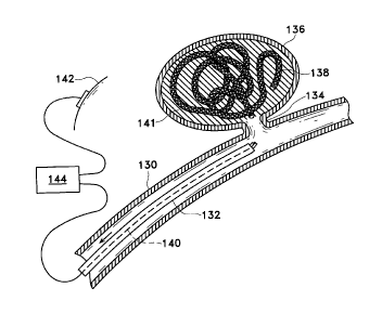
Figure 3 shov~rs the placement of the inventive devices shown above
within a vessel (130) with the tip of catheter (132) placed near neck (134) of
aneurysm (136). Braided vaso-occlusive device (138) is fed into aneurysm (136)
at
least until sacrificial link ( 112) is exposed beyond the distal tip of the
catheter
(132). A positive electric current of approximately 0.01-2 mini-amps at 0.1-6
volts
is applied to core wire ( 140) to form a thrombus ( 141 ) within aneurysm (
136). The
negative pole (142) of power supply (144) is typically placed in electrical
contact
with the skin. It is also desirable that the current be allowed to return
through a
conductor placed in the wall of the catheter (or the guide catheter used in
conjunction with the catheter).
As the thrombus ( 141 ) is formed and the aneurysm ( 136) occluded,
vaso-occlusive device (138) is detached from core wire (140) by electrolytic
disintegration of sacrificial li~rik (112).
After sacrifici;~l link (112) is completely dissolved by electrolytic
action, typically within 5 seconds to 5 minutes, the core wire ( 140) and
catheter
(132), are removed from the vessel (130), leaving aneurysm (136) occluded as
shown in Figure 4.
The process is typically practiced under fluoroscopic control with
local anesthesia. A transfemoral catheter is utilized to treat a cerebral
aneurysm
and is usually introduced at the groin. When the core wire and pertinent
portions
of the supporting coils at the distal tip of the guidewire are adequately
coated with
insulating coverings, only the exposed portion at the sacrificial link (112)
is
affected by the electrolysis.
Procedures for using this invention in non-vascular systems of the
body are carried out i.n a similar fashion. The chosen site must be accessible
and
12
CA 02214168 1999-11-19
the site must provide; a local medium of sufficient ionic nature to allow
electrolysis
of the sacrificial joint to take place.
The illustrated embodiments have been used only for the purposes of clarity
and should not be taken as limiting the invention as defined by the following
claims.
13