Note: Descriptions are shown in the official language in which they were submitted.
CA 022l4449 l997-09-02
W O 96/27331 PCTrUS96/02433
Needle Driving A,uparal-Js and Methods of Suturing Tissue
BACKGROUND OF THE INVENTION
Field of the Invention
The present invention pertains to suturing of bodily or anatomical tissue and,
more particularly, to methods and ap~aralus for suturing tissue during endoscopic and
open surgical procedures.
Discussion of the Prior Art
Suturing of bodily tissue is a time consuming part of most surgical procedures
including both open surgery and enrioscopic or minimally invasive surgery. By "open"
surgery is meant surgery wherein the surgeon gains access to the surgical site via a
relatively large incision, and by "encJoscopic" surgery is meant surgery wherein the
surgeon gains ~ccess to the surgical site via one or more portals through which
O enrloscopes are introduced to view the surgical site and through which various
instruments are introduced to the surgical site. There are many common endoscopic
surgical ,~,r~cedures, including a. U " oscopy, lapa, oscopy (pelviscopy), gastroentroscopy
CA 02214449 1997-09-02
W O96/27331 PCTrUS96/02433
and lary"yol)rc" ,~:hoscG~y, for example. In the past, suturing was accomplished with
the use of a sharp metal suture needle attached to the end of a length of suture
material. Depending on the size of the suture needle and the type of surgery being
performed, the suture needle was either grasped manually or with a for~;eps and
ç~sed to penel,ale and pass through anatomical tissue thereby pulling the suture
material through the tissue. Once the suture material was pulled through the tissue,
the surgeon tied a knot in the suture material and adjusted the tension on the suture
material to acco" ,r"odate the particular tissue being sutured and to control
approki",ation, occl~ ~sion, attac~", lenl or other conditions of the tissue. However, the
pr~cess of tissue penel, ~lion and kl ,ulliny of the suture " "3lerial can be time consuming
and tediQus work, particularly when performed in connection with microsurgery and
e., ' ss ,~i~ surgery, and can unduly prolong the duration of surgery and thererore the
period in which the patient is under anesthesia. Nevertheless, enr~oscopic surgery is
prefel led over open surgery due to the greatly re:luced trauma and wound healing time
for the patient and due to concomila"l cost savings ~ssoci~ted with shorter hospital
stays and pe~ rul ming surgery in non~ ,ospilal or out-patient surgery sites. Accordingly,
there has been much effort spent to develop techniques for facililaling the suturing
.)c~...ally ~Jel ru..-,ed by use of suture needle and a length of suture ..,alerial. Alternative
techniques proposed have incl~ ed electrical coagulation, mechanical devices such
as clips, clamps and staples, and lasers; however, no aller-,~live technique has yet
been well acc~,Led by su,gec"s to produce the results obtained by suturing and tying.
Thus, there is a great need for suturing techniques useful in en-JoscG~ic surgery that
permit surgeons to suture a,-dlol"ical tissue using suturing needles and lengths of
suture material in a time erri--~ ", co, ~sisle, ll and precise Illafll .er.
CA 02214449 1997-09-02
W O 96/27331 PCTrUS96102433
SUMMARY OF THE INVENTION
Accordingly, it is a primary object of the present invention to overcome the
above-mentioned disadva. ~layes of the prior art and to improve methods and apparatus
for suturing anatomical tissue.
Another object of the present invention is to permit suturing of thick tissue by
extending a suture needle from a tissue penetrating tip of a hollow needle guide.
An additional object of the present invention is to expand the range of motions
by which a surgeon can drive a needle through anatomical tissue to form a suture.
The pr~senl invention has a further object in preventing e. Ildn91e'l 1 lenl of a length
of suture material allached to a suture needle when the needle is inserted in an
a..al~,...ical cavity.
Some of the adva- ~layes of the p- esenl invention are that suturing of analomical
tissue can be ac~nlplished in a time efficient, consistent and precise manner, that
anatomical tissue of varying thickness can be sutured, that suturing can be
ac~mplished using -clandal d suture needles and filamentous suture materials without
the need of having to manually grasp the nee~'ss thereby reducing the risk of inr~cli. .9
medical per~o",)el, that the size of needles used for suturing thick tissue can be
r~ Iced lhereby reducing the space needed to suture and facilitating insertion of the
needles through portals as part of an endoscopic procedure, and that familiar wrist and
arm n.olio. .s can be utilized alone or in combination with simple hand motions to drive
the suture needles through a. ,alo,oical tissue.
The present invention is generally characterized in an apparatus for suturing
anatomical tissue including a hollow needie guide having a distal end, a needle
CA 02214449 1997-09-02
W O96/27331 PCTrUS96/02433
movably ~I;spose~i within the needle guide and having proximal and distal ends suture
",ale,ial co""e-ted with the needle and manually articulable needle receiving means
coupled with the needle guide for guiding the distal end of the needle as the proximal
end of the needle is advanced distally through the needle guide and capturing the
needle when the pro~i",al end of the needle emerges from the distal end of the needle
guide.
Another aspect of the present invention is generally characterized in an
apparatus for suturing anatomical tissue including a hollow needle guide having a
sharp tissue penelrali.,g distal end a needle disposed within the needle guide and
having proximal and distal ends the needle being movable between a retracted
position where a distal end of the needle is pro~i"lally spaced from the distal end of the
needle guide and an exle"ded position where the distal end of the needle protrudes
distally from the distal end of the needle guide and suture material connected with the
needle.
A further aspect of the present invention is generally cl,aracle,i~ed in an
apparatus for suturing anatomical tissue including a rorce,t,s having opposed jaws
defining a hollow needle guide with a distal end, a needle disposed within the needle
guide and having ~.,,u,.i,,,al and distal ends the needle being movable between a
r~tr~3~ted position where the proximal end of the needle is disposed within the needle
guide and an exle"ded posilio" where the proximal end of the needle is disposed
e,~le" ,ally of the needle guide and suture material Col "~e~;led with the needle.
Yet another aspect of the present invention is generally c;l,araclerized in a
method of suturing a"alG",ical tissue including the steps of penetrating through the
CA 02214449 1997-09-02
O96/27331 PCTrUS96/02433
tissue with a distal end of a hollow needle guide and extending a needle carrying
suturing material from the distal end of the hollow needle guide.
Still a further aspect of the present invention is generally characlerized in a
n~tllocl of suturing anatGi"ical tissue including the steps of positioning a distal end of
a hollow needle guide ~urokin,ale the a,)ato",ical tissue to be sutured extending a
needle carrying suture material from the distal end of the hollow needle guide
pe"el,d(ing through the tissue with the needle guiding the needle as it is extended
from the distal end of the hollow needle guide and capturing the needle when a
proximal end of the needle emerges from the distal end of the hollow needle guide.
An additional aspect of the present invention is generally characterized in a
method of suturing a"alo"~ical tissue including the steps of pel,alrdling part way
through the a"alo" ~ical tissue with a distal end of a hollow needle guide and extending
a needle carrying suture material from the distal end of the hollow needle guide to
complete penelrdliorl of the andlor"ical tissue.
Other objects and adva~ ~lages of the present invention will become ap~are,)l
from the following description of the prefel,~d e",bodiments taken in conjunction with
the acc~r"~dnying dr. ~;ngs wherein like parts in each of the several figures are
identified by the same referer,ce numerals.
BRIEF DESCRIPTION OF THE DRAWINGS
Fig. 1 is a side view of a suturing apparatus according to the present invention.
Fig. 2 is a side view partly in section of the suturing ap~aralus shown in Fig.
1.
CA 02214449 1997-09-02
W O96/27331 PCTrUS96/02433
Fig. 3 is an exploded fragmentary view of the needle guide and inner member
of the suturing apparatus of Fig. 1.
Fig. 4 is an enlarged fragmentary view of the needle receiving asse" ,bly of the
suturing apparatus of Fig. 1.
Fig. 5 is a pe, :,pe~,ti~e view of the distal end of the suturing apparatus shown in
Fig. 1.
Fig. 6 is a side view, partly in section, illusl, alir)g a suture needle being loaded
into the suturing apparatus of Fig. 1.
Figs. 7 -10 are side views, partly in section, illustrating use of the suturing
a~ a(dlus of Fig. 1.
Fig. 9A is a side view of a modified needle guide accordi"g to the present
invention.
Figs. 11 - 13 are side views, partly in section, illustrating another use of the
suturing a~ aralus of Fig. 1.
Figs. 14 -16 are side views, partly in section, illustrating yet another use of the
suturing apparatus of Fig. 1.
Fig. 17 is a fray",e,)ta~ side view, partly in section, illusl, ali"y a modification of
the suturing apparal.Js of the present invention.
Figs. 18 and 19 are fragmentary side views of a modified needle receiving
asse" ,bly for the suturing ap~,aral,Js of the present invention.
Figs. 20 - 23, 25 and 26 are side views of alternate needle guide assemblies for
use with the suturing apparalus of the present invention.
Fig. 24 is a frontal view of the needle guide assembly shown in Fig. 23.
CA 02214449 1997-09-02
W O96/27331 PCTrUS96/02433
Figs. 27 - 30 are fragmentary side views of alternate distal end configurations
for the needle guide asse-nblies of the present invention.
Figs. 31 - 34 are rrag,oe"lary side views of alle" ,dle needle receiving
assel"blies for use with the suturing apparatus of the present invention.
Fig. 35 is a r. ag",el ,lary side view, partly in section, illustrating a modification of
an inner member of the suturing apparatus of the present invention.
Figs. 36 - 40 are side views illusl(ali"g all~r"ale pusher rods for mounting at the
distal end of the inner member of Fig. 34.
DESCRIPTION OF THE PREFERRED EMBODIMENTS
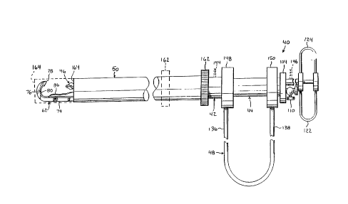
A suturing apparatus 40 according to the present invention, as shown in Figs.
1 and 2, includes an outer member 42, an inner member 44 telescG,.~ically fitted within
the outer member, a needle receiving forceps assembly 46 disposed within the inner
member, and a handle assembly 48 connected between proximal ends of the outer
member 42 and the inner member 44. Also shown is a protective sleeve 50
telescopically fitted over the outer member 42 for reasons detailed below.
As best seen in Figs. 2 and 3, outer member 42 includes a tubular body portion
52 and a needle guide asse",bly 54 at the distal end of the tubular body portion.
Tubular body portion 52 and needle guide assembly 54 can be of integral, one-piece
construction but are prererably configured in a manner to be detachably coupled.
Needle guide assembly 54 includes a cylindrical base 56 having proximal and distal
ends 58, 60 and a generally J-shaped needle guide 62 extending from the distal end
of the cylindrical base. The proximal end 58 of the cylindrical base 56 is externally
-
CA 02214449 1997-09-02
W O96/27331 PCTrUS96/02433
threaded as indicated at 68 and the distal end 70 of the tubular body portion 52
includes an inle"~ally threaded lumen 72 to matingly receive the needle guide
assembly. Needle guide 62 has a proximal shank portion 74 extending from a lateral
distal edge of the cylindrical base 56 and a distal curved portion 76 terminating at a
sharp, tissue penel,dlir)g tip 78. Shank portion 74 and curved portion 76 of the needle
guide are hollow and a continuous slot 80 is formed along an inner surface of the
needle guide with a size to permit p~sage of conventional suture material through the
slot.
Referring further to Fig. 2, a standard curved suture needle 82 with a tissue
~n~l,aling distal end 84 is disposed within the hollow curved portion 76 of the needle
guide 62. A length of fila"~enlous suture material 86 extends from a proximal end 88
of the needle and p~sses through slot 80 into the tubular body portion 52 of the outer
, . ,~" Iber 42. Suture r"dle, ial 86 can be of finite length, in which case the proximal end
of the suture ,.,alerial can be disposed within the tubular body pollio" of the outer
me" IL,er, or the suture " ,dle, ial 86 can be fed continuously from a spool mounted at or
near a ~,oxi,.,al end of the inner member.
Inner member 44 includes an elo"gale tubular shaft 90 having proxi",al and
distal ends 92, 94 and a solid cylindrical pusher rod 96 extending from a lateral edge
of the distal end of the shaft. Pusher rod 96 is prere, dbly made of a flexible or semirigid
,.,alerial that can be ~lerili~ed, such as teflon, nylon or a spring steel, and is configured
to fit telescopically within needle guide 62 so that when the tubular shaft 90 is fitted
within the tubular body ~,~,llio" 52 of the outer member 42 and advanced distally
relative to the outer me"~l,er, the pusher rod 96 will slide through the sl,aiyl,l and
curved portions 74 and 76 of the needle guide. A cylindrical wall or partition 98
CA 02214449 1997-09-02
W O96/27331 PCTrUS96/02433
extends longitudinally along an inner surface 100 of inner member 44 diametrically
opposed from pusher rod 96 to form a passage 102 for the needle receiving assembly
46. The ~ i" ,al end 92 of inner member M is closed by a cap 104 having an opening
U ,e~i n communicating with r~ss~ge 102 and anoll ,er opening 106 communicating with
the central lumen 108 d~ri, led by the tubular shaft 90. A stopcock valve 110 regulates
p~ss~ge of fluids and instruments through the second opening 106 and thus the central
lumen 108. A round flange 112 is distally spaced from the cap 104.
N~e~'Q receiving asse",l,ly 46 includes a tubular member 114 having proximal
and distal ends 116 and 118, a central member 120 telescopically fitted within the
tubular member 114, and a pair of U-shaped handles 122 and 124 connecting proximal
ends of the central and tubular members. As best shown in Fig. 4, central member 120
has a forceps 126 at a distal end made of a material having sufficient elastic memory
to be spring urged into a normally open configuration as shown. The forceps 126
shown include jaws 128 and 130 with opposed grasping surfaces 131 and 133 and
op~sed concave pcllio.ls 132 and 134 proximally spaced from the grasping surfaces.
Concave portions 132 and 134 cooperate to define a suhst~ntially circular aperture
when the jaws of the fc,r~ s are closed together; and, when the forceps is
pr~,~,. ialely positioned relative to the needle guide, the aperture serves to guide the
distal end of the needle as a proximal end of the needle is advanced distally through
the needle guide and also r, i~1io~ Ially e~ ~yages the needle when the prokimal end of the
needle has emerged from the distal end of the needle guide so that the needle iseffectively captured or immobilized. Jaws 128 and 130 can be opened to release the
captured suture needle and can also be used to grasp suture material, anatomicaltissue and other objects using grasping surfaces 131 and 133. Handles 122 and 124
CA 02214449 1997-09-02
W O 96127331 PCTrUS96/02433
normally urge proximal ends of the central and tubular members apart so that the distal
end of the tubular member will slide over the jaws causing the jaws to close together
as shown in Fig. 2.
Referring again to Fig. 2, handle assembly 48 is generally U-shaped and
includes a pair of handle " ,e,nbe, s 136 and 138 coupled with the ~roxi" ,al ends of the
outer and inner members 42 and 44, respectively. Handle assembly 48 can be
bifurcated centrally at 140, as shown in phantom in Fig. 2, and provided with a leaf
spring 142 to urge the handle members 136 and 138 apart. AlLel"ali-lely the U-shaped
handle assembly can be of one-piece spring material construction.
Outer member 42 and10r needle receiving asse" Ibly 46 can each carry electrical
con"ectors as shown in p hal llom at 144 and 146 in Figs. 1 and 2, so that the needle
guide 62 and/or needle grasping forceps 126 can be utilized as conductive elements
in a conve, llional r, Idl 11 ,er to ~, ru" " unipolar or bipolar electrosurgical procedures. For
example outer ,nel,lber 42 and needle receiving assembly 46 can both carry an
electrical con"e~;tor and be pr~pe, Iy ir-sl ~ tr-l SO that the tip of the needle guide 62 and
for~eps 126 serve as spaced ele.;t,udes to form a bipolar electrosurgical instrument.
The suturing appdr~ s can be provided with parts assembled as shown in Figs.
1 and 2 or in a ~l:s~cselnbled state wherein parts of the suturing apparal-ls are provided
s~parately and asse",bled by the user. If provided in a ~iis~ssembled state assembly
of the parts ~isa~ssecl so far involves choosing an appropriate needle guide assembly
based upon the type of analo",ical tissue to be sutured and the suture needle to be
used and attaching the needle guide assembly to the distal end 70 of the tubular body
po, lion 52 of the outer member 42 as shown in Fig. 3 for needle guide assembly 54.
With a needle guide asselllbly threaded onto the tubular body portion 52 of outer
CA 02214449 1997-09-02
WO 96/27331 PCT/US96/02433
" ,~" ll~r 42 inner member 44 can be inserted into outer " ,e" ,ber 42 and rotated to align
pusher rod 96 at the distal end of the inner member with the straight shank portion 74
of needle guide 62. Inner member 44 is then advanced distally relative to outer
",ember 42 until pusher rod 96 is received within shank portion 74 of needle guide 62
at which point inner and outer memL,ers 44 and 42 are restrained from rotating relative
to one another.
Handle assembly 48 can be coupled with outer member 42 and inner member
44 as shown in Fig.2 by atlbCI)irly handle member 136 to the proximal end 64 of outer
member 42 and handle member 138 to the proximal end 94 of inner member 44.
Handle members 136 and 138 include cylincllical collars 148 and 150 respectively that
are slipped over flanges 66 and 112 at the proximal ends of the outer and inner
members. As mentioned previously handle members 136 and 138 are biased apart
for exd",~le by a leaf spring 142 so that ,~rrJxi",al ends of the outer and inner members
42 and 44 will r..s"nally be biased apart as shown in Fig. 2. As a result pusher rod 96
is normally held in a rel,a~ ted position near the proximal end of the needle guide 62.
If not already installed within inner member 44 needle receiving assembly 46
can be inse, led into p~-~sage 102 fo""ed by cylindrical wall 98 inside the inner member
and advanced distally until rorceps 126 is disposed distally of the cylindrical wall. As
r,e"lioned previously rorce~s 126 is normally closed; and in order to facilitate passage
of the rorceps through p~-cs~ge 102 concave portions 132 and 134 of the forceps can
be configured to extend radially outward no further than tubular member 114. For
pu",oses of clarity handles 122 and 124 are shown in the plane of the drawing of Fig.
2; however as can be seen in Fig. 5 in use forceps 126 is preferably rotated 90~ from
tl .e position shown in Fig. 2 so that the o,~eni"~ defined between concave portions 132
11
CA 022l4449 l997-09-02
W O96/27331 PCTrUS96/02433
and 134 of the rorceps is aligned with the path of a needle emerging from the distal end
of the needie guide.
Suture needle 82 can be loaded into the curved portion 76 of needle guide 62
by inse, lirlg the proxi-,~al end 88 of the needle into the distal end of the curve needle
guide portion and guiding the suture " ,~le, ial 86 along slot 80 so that it trails behind the
needle. An optional loading device, shown in Fig. 6 at 152, can be used to load the
needle into the needle guide 62 without fear of cGI)la~lil lg the sharp tip 78 of the needle
guide. The loading device 152 resembles a funnel with a hollow conical mouth portion
154 tape, i~ ~y to a curved throat portion 156 configured to fit within or around the needle
guide. In use, the throat portion 156 of the loading device is fitted against the open
distal end of needle guide 62 and needle 82 is dropped into the mouth portion and
received within the needle guide, after which the loading device is removed.
Needle 82 co"ro""s s~ ~hst~ntially to the curvature at the distal end of needle
guide 62 and is thus prevented from migrating into the straight shank portion 74 of the
needle guide by geometrical constraint. In the loaded or reL,dcled position, shown in
Fig. 2, the sharp, tissue penel, ali"g tip 84 of the needle is proximally spaced from the
sharp tip 78 of the needle guide 62 and the proximal end 88 of the needle is distally
.sp~ced from the distal end of the pusher rod 96.
Referring still to Fig. 2, it can be seen that, in use, handle assembly 48 can be
grasped by the user and manipulated by use of various wrist and arm motions to
.osilion the suturing apparalus 40 at a surgical site without inducing substantial relative
movement between outer and inner members 42 and 44 of the apparatus. Accordingly,
one melhod of suturing a"ato" ,ical tissue with the suturing apparatus, as shown in Figs.
7 - 10, involves utilizing the needle guide 62 as a needle to penetrate through
12
CA 022l4449 l997-09-02
WO 96/27331 PCT/US96/02433
~nalo,nical tissue T with the suture needle 82 in the ,el.a~;ted position. More
s~ilically the sharp tip 78 of needle guide 62 is used to penetrate into the analo"-ical
tissue by a~ ~r~,- idle marip~ ion of the suturing a,~a, dl~ls and is passed through the
tissue until the tip er"er~~es from the tissue at a location spaced from the point of initial
penetration as shown in Fig. 7. Turning again to Fig. 2, it can be seen that needle
receiving assembly 46 can be advanced distally relative to inner member 44 and
rotated a,c~,ro~rialely to position forceps 126 at a location suitable for receiving the
curved suture needle 82 when the needle protrudes from needle guide 62.
Rere. . i. ,9 still to Fig. 2, it will also be seen that needle 82 can be driven through
needle guide 62 by 5~ ee~ing handle members 136 and 138 together which in turn
o~uses inner member 44 to be advanced distally relative to outer member 42 and
pusher rod 96 to slide within needle guide 62 into abutment with the proximal end 88
of needle 82. Pusher rod 96 is flexible and can thus confor", to the shape of needle
guide 62 as the rod is advanced. Continued s~ue~ing of handle members 136 and
138 toward one another c~ ~ses pusher rod 96 to drive needle 82 in a distal direction
through needle guide 62, as shown in Fig. 8, until the tip 84 of needle 82 emerges from
the distal end of the needle guide and is received by the aperture formed between jaws
of the forceps 126. Suture ,.,alerial 86 is pulled through the tissue T along slot 80 in
response to distal movement of needle 82. The shape and size of the aperture are
such that when the proximal end 88 of needle 82 emerges from needle guide 62, the
needle is captured bet~voon jaws of the rc;,r~;eps and frictionally engaged so that handle
pressure can be r~cluce~i. allowing pusher rod 96 to recede from the curved portion 76
of needle guide 62 to its original position within the straight shank portion 74. Suture
r)~terial 86 is attached to the proximal end 88 of needle 82 and thus remains threaded
13
CA 02214449 1997-09-02
W O96/27331 PCTrUS96/02433
through the analor"ical tissue T when the needle guide is removed from the tissue as
shown in Fig. 9. If necess~ry needle receiving assembly 46 can be manually moved
p~Cil I Idlly relative to inner rr,er"ber 44 to facilitate removal of the needle guide from the
tissue by increasing the spacing between the distal end of the needle guide and the
ror~,us.
For purposes of illustration a spherical knotting element 158 is shown
sc~ ,e"~alically in Fig. 9 dlLached to the proximal end of the suture material to serve as
a knot for joining opposile ends of the suture material into a loop; however any type
of suture material or knotting technique can be used. A forceps 160 is used to grasp
the knotting element 158 in such a way that the distal end of the suture material is
received within an engaging portion of the knotting element and the k-,olling element
is moved toward tissue T while the suturing apparatus 40 is retracted to adjust the
tension of the suture material. Aller"dlively, the needle guide 62 could be formed of
laterally o~,osecl jaws 161 163 of a forceps 165, as shown in Fig. 9A such that
grasping and manipulation of the knotting element can be performed without the need
for a separate forceps. The modified needle guide is prererably coupled with any
suitable handle structure such as handle assembly 48 such that the jaws can be
normally biased to a closed condition to form a tissue penetrating tip 167 and opened
manually when it is desired to grasp an object such as knotting element 158. When the
suture ",ale,ial is a~"~ro~ riately tensioned the knotting element can be closed around
the suture material to form a closed loop suturing the tissue as shown in Fig. 10.
Various types of h lollin~J elements that can be used with the suturing apparal.Js of the
present invention are described in copending patent applications Serial No.
14
CA 022l4449 l997-09-02
W O96/27331 PCTrUS96/02433
08/366285 filed December29 1994and Serial No. 08/ filed January25 1995
the disclosures of which are inco, ~.u, aled herein by reference.
Anothemllell ,od of suturing a, ,dlum;cal tissue according to the present invention
is illustrated in Figs.11 - 13 wherein only the suture needle is utilized in penetrating the
tissue. The suturing a,u~,aralus 40 is manipulated to position anatomical tissue T
between the distal tip 78 of needle guide 62 and forceps 126. Needle guide tip 78 is
then posilio"ed pr~"~i")dle the tissue as shown in Fig. 11 to precisely locate the point
of entry for suture needle 82. Pusher rod 96 is advanced in the manner previously
cles~iL,ed driving needle 82 and suture material 86 through needle guide 62 and into
the tissue as illustrated in Fig. 12. When the distal end of the needle penetrates
through the tissue the needle is received by forceps 126 and held therein during the
tying procedure as shown in Fig. 13.
Figs. 14 - 16 illustrate yet another method of suturing ar,aloi"ical tissue
according to the pr~:senl invention wherein the needle guide 62 is utilized as a needle
to pe"el, dle a"alun,i-~' tissue part way through the thickness of the tissue and suture
needle 82 is extended to complete penetration through the tissue. It will be
appr~c~ therefore that use of the suturing apparatus 40 in such a manner permits
suturing of andlol,, ~l tissue thicker than that no" ~ ~ally able to be sutured by use of the
suture needle alone. The method involves penet, aling into anatomical tissue T using
the tip 78 of needle guide 62 and positioning the curved portion 76 of the needle guide
within the tissue as shown in Fig. 14 such that a portion of the a,);atcii"ical tissue is
~osilioned between tip 78 of needle guide 62 and forceps 126; Pusher rod 96 is then
advanced distally by s~ ~ee~irl9 handle me" ,ber~ 136 and 138 tc yetl ,er to extend suture
needle 82 from the tip of the needle guide and through the tissue positioned between
CA 02214449 1997-09-02
W O96/27331 PCTrUS96/02433
the tip and rorceps as illustrated in Fig. 15. Needle 82 is received by forceps 126 and
held therein while needle guide 62 is removed from the tissue leaving suture material
86 in place within the tissue to form a suture using any conventional knotting or tying
technique.
The suturing aupar~ s can be used to suture anatomical tissue in open or
e"doscopic procedures. In the case of encloscopic procedures the suturing apparalus
will be inserted into an a"alor"ical cavity through a portal formed in the wall of the
cavity; and it will be appreci ~lecJ that the sharp needle guide of the suturing apparatus
can snag structures within the portal or the andlu".icdl cavity. Referring to Fig.1 it will
be seen that protective sleeve 50 can be used to cover the needle guide 62 of the
suturing auu~ldlLls 40 when the apparatus is inserted through a portal or any time it is
desired that the needle guide not be exposed. Protective sleeve 50 is telescopically
fitted over outer member 42 and includes a collar 162 at a proximal end for being
grasped by the user. Collar 162 can be grasped and moved distally as shown by
phantom line in Fig. 1 to position the distal end 164 of protective sleeve 50 around
needle guide 62 so as to protecl the needle guide during insertion. Once positioned
within the anaLo",.--' cavity sleeve 50 can be retracted or moved proximally to expose
the needle guide prior to suturing tissue.
Fig. 17 illustrates a modification of the suturing apparatus according to the
present invention wherein the modified suturing apparal.ls 166 includes an inner
",e",ber 168 similar to inner member 44 but with a c~lil ldl iCdl housing 170 at a proximal
end and a needle receiving assembly 172 coupled with the housing. Tubular member
174 of the needle receiving assembly is similar to tubular member 114 and in addition
includes a flange 176 near a proximal end of the tubular member. Flange 176 is
16
CA 02214449 1997-09-02
WO 96t27331 PCT/US96/02433
disposed within housing 170 and is biased toward a proximal end wall 178 of the
housing by a helical coil spring 180 disposed around tubular member 174 and held in
co""~r~ssion between a distal end wall 182 of housing 170 and flange 176. Suturing
apparal.ls 166 also differs in that the central member 184 of the needle receiving
asse,nbly is hollow and a valve 186 is mounted at a proximal end of the central member
to control p~ssage of fluids and instruments through the central member.
Referring still to Fig. 17 it can be seen that in use, inner member 168 can be
moved distally relative to an outer member by o~,eralion of handle assembly 188. As
deswibed previously in cc ""e~ion with suturing apparatus 40 distal movement of the
inner ",emberwill cause a pusher rod at the end of the inner member to slide through
a needle guide into abutting relation with a suture needle disposed within the needle
guide. Housing 170 is also moved distally relative to the outer member causing
~.ru~imal end wall 178 of the housing to bear aya;l lsl flange 176 of tubular member 114
driving the needle receiving asser"bly 172 forward in a distal direction at the same time
the needle is driven from the needle guide by the pusher rod. As a result the suture
needle can be extended and received by the needle receiving assembly in response
to a single hand motion such that the number of hand ",oliGns required to suture are
reduced and the suturing ~roced-Jre is simplified.
Figs.18 and 19 illustrate a modified needle receiving assembly 190 for use with
the suturing a~,ar~lus of the present invention wherein the distal end 192 of the
modified needle receiving assembly has a predetermined shape and is made of a
.,alerial having an elastic memory such that the needle receiving assembly normally
assumes the prede~e",~ined shape when in a relaxed state. For example central
..~r,lber 194 of needle receiving assembly 190 can have a predetern-ined shape with
CA 02214449 1997-09-02
W O96/27331 PCT~US96/02433
a bend 196 and tubular member 198 can be made of a flexible material to co, ~rOl l l l to
the shape of the central member. In the position shown in Fig. 18, needle receiving
assembly 190 is prevented from bending because the normally bent portion of the
asse,nbly is tel~sc~icallyfitted within a relatively rigid straight section of the cylindrical
wall 98. When needle receiving assembly 190 is advanced distally relative to the
cylindrical wall 98, as shown in Fig. 19, the assembly assumes the predetermined
shape, which can for example be chosen to facilitate positioning of the forceps 199 to
receive a particular type of suture needle. Needle receiving assembly 190 can have
any precleter~"ined shape useful for suturing tissue, including bent or curved
configurations, for example.
Referring now to Figs. 20 - 25, various needle guide assemblies for mounting
on the distal end of the outer member 42 are shown. In accordance with the present
invention, these "~odiried needle guide assemblies share a common base structure 56
so that they can be readily i"lercl)a"ged depending on the specific surgical application
for the suturing apparatus of the present invention. The needle guide assembly
illustrated in Fig. 20 at 200 is similar to needle guide assembly 54 but with a hollow
needle guide 202 having a continuous outer surface without a slot. Fig. 21 shows a
needle guide assembly 204 having a generally L-shaped needle guide 206 with a
straight p,u,(i",al portion 208 extending from a distal edge of the base 56 to a bend 210
joining the shank with a sl,ai~l ,t distal portion 212 oriented s~ ~ ,st~nlially perpendicular
to a longitudinal axis of the needle guide. Proximal and distal portions 208 and 212 of
the needle guide are hollow and are shown with a slot 214 extending continuously
along an inside surface of the needle guide. Bend 210 of the needle guide is shown
as being acutely angled but can also be curved as shown in pllal)lGIll in Fig. 21.
18
CA 022l4449 l997-09-02
WO 96/27331 PCTnUS96/02433
Another needle guide asse~-~biy is illustrated in Fig. 22 wherein the needle guide
asse",bly 216 includes a needle guide 218 having a straight proximal portion 220
extending from base 56 along a central longitudinal axis of the base, for example from
a support extending diametrically across the open distal end of the base, and an
arcuate distal portion 222 extending distally from the straight proximal portion of the
needle guide in a plane conlai"i"g the central longitudinal axis of the needle guide. A
slot 224 exteu ds continuously along the length of the needle guide 218. Needle guide
assembly 226, as shown in Figs. 23 and 24, includes a needle guide 228 having a
straight proximal portion 230 similar to that shown in Fig. 22 and an arcuate distal
portion 232 forming multiple coils of inc~easi"g diameter in a plane perpendicular to the
longitudinal axis of the needle guide. The needle guide assembly 234 shown in Fig.
25 is similar to needle guide assembly 226 but with an arcuate portion 236 of the
needle guide 238 coiling around the straight proximal portion 240 in a proximal
direction. Fig. 26 shows another modification of the needle guide assembly wherein
the modified needle guide assembly 242is similar to needle guide assembly 226 but
with the arcuate portion 244 forming coils that protrude distally from the straight portion
246 like a corkscrew.
The needle guides shown and desc.iL,ed herein can have various distal end
configurations, as illuslrale-J in Figs. 27 - 31, depending on the operational
requirements of the suturing apparatus and the tissue to be sutured. For example, a
needle guide distal end 248 having a sharp tissue penet~alir,g tip 250 and a small notch
252 formed near the tip is shown with a slot 254 in Fig. 27 and without a slot in Fig. 28.
Another needle guide distal end 256 including a blunt tissue penetrating tip 258 and
a notch 260 formed near the tip is shown with a slot 262 in Fig. 29 and without a slot
19
=:
CA 022l4449 l997-09-02
W O96/27331 PCT~US96/02433
in Fig. 30. Notches 252 and 260 are generaliy V-shaped to receive and frictionally
el ,yaye suture male, ial drawn into the notches so that the suture material can be held
in a fixed position relative to the needle guide during knotting and tying procedures.
Various needle receiving assemblies that can be used with the suturing
appa(alus of the prese,)l invention to guide and capture suture needles extended from
a needle guide are shown in Fig. 31 - 33. The needle receiving assembly 264 shown
in Fig. 31 includes a tubular member 266 like tubular member 114 and a central
member 268 with an eyelet 270 at a distal end. Eyelet 270 can be protruded distally
from tubular member 266 by operation of a handle mechanism (such as handles 122
and 124) to receive a suture needle but is biased to a retracted posilio" so that when
the handles are released, the needle is captured between the eyelet and a distal end
of the tubular member. Fig. 32 illustrates yet another needle receiving assembly 272
having a tubular member 274 and a central member 276 with a hollow cylindrical
s~lllenl 278 mounted transversely at the distal end of the central member for receiving
and frictionally engaging a suture needle. Yet another needle receiving assembly is
shown in Fig. 33 at 280 wherein the tubular member 282 of the needle receiving
asse"~bly is closed at a distal end to form a forceps jaw 284 and an aperture 286 is
rolll-ed in a sidewall of the tubular ",e",ber at a posilion pr~xi",ally spaced from forceps
jaw 284 to permit pA-csaye of a suture needle into the tubular member. Central member
288 of needle receiving assembly 280 is movable within the tubular member and is
biased in a distal direction so that the distal end 290 of the central member functions
as an o,u~,osed rorceps jaw to capture the suture needle passing through aperture 286
The needle receiving asse",bly 292 shown in Fig. 34 is similar to needle receiving
assembly 280; however the tubular member 294 carries an L-shaped outer jaw 296
CA 02214449 1997-09-02
W O96127331 PCTrUS96/02433
having longitudinal and transverse legs 298 and 300 and the central member 302
carries an L-shaped inner jaw 304 having a longitudinal leg 306 that slides within the
longitudinal leg of the outer jaw and a transverse leg 308 that can be moved toward the
transverse leg of the outer jaw to ,ue,rurl" the functions of a forceps such as, for
example, capturing suture needles, holding suture material or applying knotting
~lemenls.
Refer.ing now to Figs. 3~ - 40, various pusher rods for mounting on the distal
end of the inner member 44 are shown. In accorda-lce with the present invention,
these modified pusher rods can be formed as an integral part of the inner member or
can be configured to be detachably coupled with the inner member so as to be readily
inlercl ,a"ged depending on the operational requirements of the suturing apparatus of
the ,~r~:senl invention. The pusher rod illustrated in Fig. 35 at 310 is similar to pusher
rod 96 but with ll--eads 312 at a proki-.,al end 314 for being received by a threaded
r~t~le 316 formed at the distal end of the inner member. Fig. 36 shows a pusher
rod 318 which is similar to pusher rod 310 but with a rounded distal end 320. Another
n~diri~ pusher rod, shown in Fig. 37 at 322, includes a hollow tube 324 with a flat or
rounded distal end 326 and a II.readed proximal end 328 for being received by a
U ~-eaded bore 330 formed at the distal end of the inner member in communication with
the oentral lumen. The hollow pusher rod 322 can thus be used to pass fluids and/or
suture male. ial through the needle guide. Fig. 38 illustrates another hollow pusher rod
332 having a threaded proximal end 334, a rounded or flat distal end 336 and a slot
338 formed longitudinally between proximal and distal ends of the pusher rod, for
example to permit lateral passage of suture material into and out of the pusher rod.
The mc,diried pusher rod shown in Fig. 39 at 340 can be hollow or solid and includes
21
CA 02214449 1997-09-02
W 096/27331 PCT~US96tO2433
a proximal end 342, a distal end 344 and a notch 346 formed at the distal end for
holding suture material. Yet another modified pusher rod 348 includes a forceps 350
for grasping the proximal end of a suture needle so that the needle can be retracted
pl UAil "ally into the needle guide from an extended position, for example after the suture
,..ale,ial has been yld~Jed. Forceps 350 includes a tube 352 and an elo,)yale member
354 rlisposed within the tube. The elongate member 354 has a pair of opposed jaws
356 and 358 at a distal end that are elastically biased apart. A handle assembly (not
shown) can be connected between the tube 352 and the elongate member 354 to
normally bias the tube over the jaws 356 and 358 so that the jaws are closed to form
a needle driving distal end and can be opened to receive the proximal end of the suture
needle.
From the above, it will be appreci~lecl that the suturing apparatus of the present
invention can be used to suture anatomical tissue of varying thickness by positioning
a hollow needle guide ~5 -cenl to, within or through the tissue to be sutured and
extending a suture needle carrying suture material from the distal end of the needle
guide. Any type of suture needle can be utilized with the suturing apparatus including,
for example, slrai!Jl,l or curved and blunt or sharp needles in hollow or solid
configurations with or without slots. The suture material can be attached to the suture
n~dle at ~Jroxi",al, distal or intermediate portions of the needle or advanced through
the suture needle if the needle is hollow. Also, the suture needles can have two or
more lengths of suture ".alerial attached. In addition, the suturing apparatus can be
used to apply bic~l~sorbable suture needle devices such as those desc, ibed in U.S.
ralenl~ 4,932,962, 4,981,149 and 5,074,874 to Yoon et al; and 5,053,047, 5,222,976
and 5,330,503 to Yoon; the ~I;sclos~ ~res of which are inco, ~oraled herein by reference.
22
CA 02214449 1997-09-02
W O96/27331 PCTrUS96/02433
The needle guide can have any configuration for carrying a suture needle, including
straight or curved configurations with blunt or sharp tissue penetrating tips. By "hollow"
is meant defining a p~ssa.3e or lumen between open ends; and, in addition to being
hollow, the needle guides of the ,c"-~se, ll invention can also have slots formed part way
or along the entire length of the needle guides in communication with the lumens to
permit p~-ss~e of suture material from spaces within the suturing ap,c,aralus to points
of allac;l""enl with suturing needles disposed within the needle guides. Needle
receiving assemblies mounted by the suturing apparatus can be moved axially, bent
transversely and, in the case of ~or~;eps, opened or closed to grasp the suture needle,
suture material or a knotting element, such as the knotting element shown
schen,alically in Figs. 7-10 and desc~ ibe-l in my pe"dil ,9 patent appli~tions Serial Nos.
08/~66,285 and 08/_, filed Decer.,ber 29, 1994 and January 25, 1995, respectively.
Moreover, axial movement of the needle receiving assemblies can be indexed for a
particular suture needle curvature.
While electrical conne~lo, s have been shown and described as forming contacts
with the outer member and/or needle receiving assembly of the suturing apparatus, it
will be appreciated that any number of conventional electrical connectors can be
,~osilio"ed at various lo~lio"s and cor"-e~ed with various components of the suturing
a~,,uardlus for performing unipolar or bipolar electrosurgical procedures. Also, inner
surfaces of the outer member, inner member or any other member defining a passage
or lumen through the suturing appar~ s can be electrically insulated to permit insertion
of electrosurgical instruments through the passage as a backup.
The U-shaped handles shown and described herein for sliding the inner member
within the outer member are exemplary of the types of conventional handle
23
CA 02214449 1997-09-02
W O96/27331 PCTrUS96/02433
" ,e~;l ,a, li3, 1 15 51 lit~hl~! for use with the suturing apparalus of the present invention; and,
accordingly, the handles can have any configuration for producing relative movement
between components of the suturing apparatus including, for example, scissors-type
handles having ~ ossed legs with a pivot, handles having a fixed leg or grip connected
with one cor, IpGI lel 11 and a pivoted leg connected with the other component, and pistol
grips having movable ll igger:j. Moreover, the handles can have any orientation relative
to the longitudinal axis of the suturing apparatus including, for example, substantially
transverse orientations whereby the handles extend transversely from a longitudinal
axis of the suturing apparatus, substantially longitudinal orientations whereby the
handles extend longitudinally from a proximal end of the apparatus or rotatable
configurations whereby the handles can be moved between transverse and longitudinal
orientations as desired.
The co" "~nenls of the suturing appa~ ~lus of the present invention can be made
of any suitable, I,.edical grade materials to permit sterilization for re-use or for single
patient use. The cc r"po, lenls can be made of multiple parts of various configurations
and ,..alerials to reduce cost. The various p~ss~ges and lumens formed through the
apparatus can have various valves, stop cocks and seals to control the flow of fluid and
instruments U .er~U " uugh, such as the valve 1 10 shown in Fig. 1 or the valve shown in
Fig. 17at186.
It will also be appreciated that the suturing apparatus of the present invention
can be used to apply single or multiple stitches in open or endoscopic surgical
procedures. Furthermore, the central channel of the suturing apparatus can be used
for i, . iyalion or aspiration and can serve as a space for holding the suture material or
as a portal for the introduction of medical instruments. The features of the various
24
CA 02214449 1997-09-02
W Og6/27331 PCTAUS96/02433
embodiments described above can be combined in any manner desired dependent
upon the oper~lional requirements of the ~r~ced.lre to be pe~ ro~",ed and the complexity
of the suturing a~,par~lus.
Inasmuch as the present invention is subject to many variations, modifications
and changes in detail, it is intended that all subject matter disclJssed above or shown
in the accc "~pa,)ying drawings be interlJI eted as illustrative only and not be taken in a
li-l-ili- ~y sense.