Note: Descriptions are shown in the official language in which they were submitted.
CA 02214516 2000-09-OS
STERILIZABLE ENDOSCOPE WITH SEPARABLE
AUXILIARY ASSEMBLY
Technical Field
This application is a continuation-in-part
application of my U.S. Patent No. 5,489,256 and
entitled "Sterilizable Endoscope With Separable
Disposable Tube Assembly".
This invention relates to an endoscope that is
shielded by a sterile end cap with an attached sterile
drape wherein a sterile separable channel section
removably attaches to the end cap. The end cap, drape
and channel section are disposable after use and the
same endoscope may be used for subsequent operative
procedures without resterilization.
Backaround Art
In recent years the popularity of endoscopic
surgery has proliferated. This has occurred because
of the advances in technology which allow smaller and
smaller endoscopes to be used, thereby permitting
operative procedures to be undertaken in a less
invasive manner than was previously possible. Thus,
the patient suffers less trauma, recuperates much more
rapidly and experiences less pain and discomfort than
with more conventional surgical procedures.
Because of the sophisticated optics and electro-
optics contained in modern endoscopes, they generally
are very expensive. In order for this expense to be
justified, they must be reused with a large number of
patients.
CA 02214516 1997-09-03
WO 96127322 PCTlUS96/03028
2
Of course, reuse requires that the endoscope must
be sterilized or at least disinfected after use with
each patient prior to use with the next patient. One
protocol for sterilization involves immersing the
endoscope in a disinfectant solution for a
predetermined period of time. It is also important to
flush the channels inside the endoscope which carry
gases, fluids, or which receive operative instruments.
When using the disinfectant, sometimes the endoscope
is not placed in the disinfecting solution for a
sufficient length of time nor are the channels flushed
out completely because of the urgency to get the
endoscope back into service as soon as possible. Over
time, the disinfectant solution may lose some of its
strength, thereby limiting its effectiveness. Another
protocol is to heat sterilize the endoscope by placing
it in an autoclave. However, the optics and
electronics of many endoscopes will not permit them to
be subjected to heat sterilization.
Because of these shortcomings, studies have shown
that transmission of infectious diseases from one
patient to another has occurred in many instances. By
way of example, transmission of salmonella typhi has
been reported. In addition, pseudomonas aeruginosa
has been linked to endoscopy. Also, an outbreak of
serratia marcescens has been associated with the use
of a bronchoscope. Furthermore, Hepatitis B has been
transmitted by endoscopes when the endoscopes were
processed in an inappropriate manner between patients.
Finally, with respect to endoscope use on patients
with acquired immune deficiency syndrome (AIDS), it
has been found that the sterilizing procedures have
not always removed contamination of the human
immunodeficiency virus (HIV). This list is not
exhaustive by any means.
CA 02214516 1997-09-03
WO 96/Z73Z2 PCT/I1S96/03028
3
A high-level of disinfection failures among
gastrointestinal endoscopes have been noted, as well
as failures in bronchoscopes, laryngoscopes and other
. devices. This may be due to the fact that they are
long and narrow and have internal channels that are
difficult to sterilize.
Another shortcoming of the prior art is that when
a tube assembly is attached to the distal end of an
endoscope not having integrally formed internal
passageways, the resulting cross-sectional shape was
greatly increased because the tube assembly was not
structurally designed to mate with the endoscope. A
larger cross-sectional shape results in increased
trauma to patients undergoing surgery.
From the foregoing, it is apparent that
endoscopes are needed that can be more easily and
effectively sterilized, and that maintain a small size
to accommodate minimal invasive surgery.
Disclosure of the Invention
In accordance with the present invention, an
endoscope with a separable auxiliary assembly in one
configuration is provided. The separable auxiliary
assembly includes a sterile disposable separable
channel section, and a sterile disposable end cap with
an attached sterile drape. The endoscope is of the
type that includes an elongated capsule, of the size
of a medicinal capsule, and a substantially
cylindrical housing with a transparent window at the
distal end thereof for containing the endoscope
' 30 optics. An image sensor such as a CCD is mounted
adjacent the window within the capsule. An image
transmitting cable or conduit with multiple conductors
each has a distal end connected to the image sensor
circuit board and a proximal end connected through a
CA 02214516 1997-09-03
WO 96!27322 PC~'lITS96103028
4
series of cables to a video control unit. From the
video control unit, signals are transmitted to a video
monitor which displays the image in black and white or
color. A plurality of light transmitting fibers are
disposed within the capsule, each having a distal end
adjacent to the window within the capsule, the fibers
extending proximally from the capsule for transmitting
light to a site under investigation from a remote
light source.
An end cap is provided that is configured to
house the distal end of the capsule and shield it from
the sterile operating environment. The proximal end
of the end cap provides a means of attachment for the
sterile drape that encloses the remaining portion of
the capsule and the trailing endoscope cables.
The separable channel section is removably
attached to the end cap in a fixed relationship and
has at least one longitudinal channel extending
internally therethrough for transmitting fluids, gas
or for receiving an operative instrument. A flexible
tube is connected to the proximal end of the channel
for supplying fluid, gas or the operative instrument
from a remote location. The separable channel section
and end cap with attached drape are disposable after
use on a patient. Thus, the endoscope is available
for reuse with another separable auxiliary assembly on
the next patient without having to resterilize or
disinfect the endoscope.
In operation, the capsule portion of the
endoscope is inserted within the end cap and the drape
is extended over the capsule to completely shield the
endoscope and its trailing cables from the sterile
operating area. The separable channel section may
then be attached to the end cap.
CA 02214516 1997-09-03
WO 96/27322 PCT/US96/03028
More particularly, the separable channel section
is configured to attach to the end cap to minimize the
cross-sectional size thereof. The separable channel
section includes a longitudinal guide or key which may
5 be snap fit or slidably engageable with a longitudinal
guideway or keyway formed on the exterior surface of
the end cap. Conveniently, the separable channel
section is configured such that when the end cap and
the separable section are attached, they form a
circular, oblong or substantially oval cross-sectional
shape.
Other means of attachment may be used to attach
the separable channel section to the end cap. For
example, a magnetic attachment or the use of an
elastic band may be used. Those schooled in the art
may envision other methods of holding the separable
channel section in the releasable yet fixed
relationship to the end cap.
The same inventive concept of using a separable
channel section can be used with a steerable
endoscope. The separable channel section may include
a guide wire channel which receives a guide wire
extending from the endoscope. Thus, if the separable
channel section and end cap are constructed of a
flexible material, the guide wire extending through
the guide wire channel can be used to steer the distal
end of the endoscope capsule housed within the end
cap.
In operation, the endoscope capsule housed by
the separable auxiliary assembly is inserted into a
bodily passageway to the desired operative site. The
site is investigated through the image produced by the
' endoscope and the necessary operative procedures are
carried out through channels within the separable
channel section. The endoscope and separable
CA 02214516 1997-09-03
WO 96127322 PCTY~S96I03028
6
auxiliary assembly are then removed from the bodily
passageway whereupon the endoscope capsule may then be
separated from the auxiliary assembly. Since the
endoscope is completely shielded from the operative
site, the same endoscope may be used in a subsequent
surgical procedure by simply utilizing a new separable
auxiliary assembly. Thus, the need to sterilize the
endoscope is eliminated which greatly improves
surgical procedures both in terms of the cost and
labor associated with sterilization of such endoscopic
devices.
In order to enhance the viewing field of the
operative sight, a detachable viewing head may be
attached to the distal end of the endoscope prior to
placing the end cap over the endoscope. Detachable
viewing heads offer a wide variety of optical lens
combinations which enable a surgeon to view the
operative site at the desired angle or magnification.
With this described apparatus, a method has also
been provided for utilizing a separable auxiliary
assembly on a conventional endoscope. The method
includes placing a sterile end cap with attached drape
over a conventional endoscope and extending the drape
over the exposed length of the endoscope and its
trailing cables. The separable channel section is
then releasably attached to the distal end of the end
cap in a fixed position. The operative site is
investigated and the necessary operative procedures
are performed. After use, the endoscope is removed
from inside the extended drape and end cap, and the
separable channel section, end cap and drape may be
thrown away. A detachable viewing head may be used in
conjunction with the aforementioned method enabling
precise and selectable viewing of the operative area.
CA 02214516 1997-09-03
WO 96127322 PC."T/US96103028
7
Brief Description of the Drawincrs
Figure 1 is a fragmentary perspective view of the
sterilizable endoscope with separable auxiliary
assembly of this invention, with the drape extended;
Figure 2 is a fragmentary exploded perspective
view of the invention of Figure l, with the drape in a
rolled position prior to use;
Figure 3 is a partial longitudinal section taken
along line 3-3 of Figure 1 illustrating the interior
relationship of the elements making up the invention
when the invention is assembled for use;
Figure 4 is a fragmentary exploded perspective
view of another embodiment of the invention that
illustrates a detachable viewing head that is
engageable with the distal end of the endoscope, with -
the drape rolled up;
Figure 5 is a fragmentary perspective view of the
invention of Figure 4 with the drape extended and
having phantom lines illustrating the relationship
between the end cap, detachable viewing head and
endoscope;
Figure 6 is a partial longitudinal section taken
along line 6-6 of Figure 5 illustrating the interior
relationship of the elements making up the invention
when the invention is assembled for use;
Figure 7 is a perspective view of the invention
of Figure 1 connected through a control member to
suitable gas supplying and instrument supplying
channels, and connected to a video monitor;
Figure 8 is a fragmentary exploded perspective
view of a third embodiment of this invention wherein a
viewing head enables side viewing and the separable
channel section attaches to the end cap resulting in a
substantially cylindrical cross-section;
CA 02214516 1997-09-03
WO 96127322 PGTlIlS96/03028
8
Figure 9 is a fragmentary perspective view of the
invention in Figure 8;
Figure 10 is a partial longitudinal section taken
along line 10-l0 of Figure 9 illustrating the interior
relationship of the elements when assembled for use;
Figure 11 is a fragmentary exploded perspective
view of a fourth embodiment of this invention, with
the drape rolled up, illustrating a separable channel
section that includes a flat mating surface having a
magnet for attachment to a ferrous surface on the end
cap;
Figure 12 is a fragmentary perspective view of
the invention in Figure 11 with the drape extended;
Figure 13 is a fragmentary exploded perspective
view of a fifth embodiment of this invention
illustrating a separable channel section attachable to
the end cap by a retaining band;
Figure 14 is a fragmentary perspective view of
the invention in Figure 13; and
Figure 15 is a fragmentary perspective view of a
sixth embodiment of this invention illustrating a
separable channel section like that shown in Figure 8
but wherein the endoscope is an end viewing device.
Best Mode For Carrying Out the Invention
In accordance with one embodiment of this
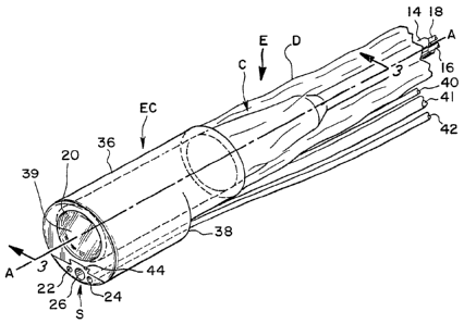
invention, an endoscope E is provided, as best seen in
Figures 1-3, which includes a capsule portion C that
is inserted within a protective end cap EC. A rolled-
up drape D is attached to a proximal end of the end
cap EC. Releasably attached to the end cap EC is a
disposable or throwaway separable channel section S.
The end cap EC, drape D and separable channel section
S comprise the separable auxiliary assembly of this
invention. The end cap EC, attached drape D and
CA 02214516 1997-09-03
WO 96127322 PGT/OS96/03025
9
separable channel section S are packaged prior to use
as completely sterile items. As shown in Figure 2,
the endoscope E includes the distal portion or capsule
C which has a window 10 for sealing the interior of
the capsule C from the environment. Typically, one or
more optical fibers 20 terminate within the capsule C
at the window 10. Disposed within the interior of
capsule C is an imaging chip such as a CCD (not shown)
and associated electrical leads (not shown) which
connect to transmission wires 18 within conduit 14.
In operation, the optical fibers 20 provide light to
the operative site and the imaging chip receives a
visual image of the operating site which is then
transmitted via the electrical leads through wires 18.
As shown in Figure 1, the proximal end of capsule C is
connected to the conduit 14 which extends proximally
away from the capsule C and which houses wires 18 and
bundled optical fiber cable 16 that branches out
within capsule C into the peripherally arranged fibers
20. Although one embodiment of an endoscope is
illustrated herein, it will be understood that any
endoscope with a generally cylindrical shape may be
used in conjunction with the separable auxiliary
assembly of this invention.
The separable channel section S may include a
pair of fluid or gas channels 22 and 24 and an
operative channel 26. In communication with channels
22, 24 and 26 are corresponding tubes 40-42,
respectively, which allow fluids, gas or a surgical
instrument to be passed therethrough so that the
- fluids, gas or operative instrument may be applied to
the operative site. Figures 1 and 2 illustrate a pair
of fluid or gas channels and a single operative
channel, however, it will be understood by those
skilled in the art that any number or combination of
CA 02214516 1997-09-03
WO 96/27322 PCTlUS96103028
channels may be used with the separable channel
section S in order to transmit the needed operative
fluid, gas or surgical instruments to the operative
site. Conveniently, the channel section S includes a
5 pair of flats 28 which are located on opposing sides
of a key 30. Accordingly, end cap EC includes a pair
of corresponding flats 32 with a keyway 34 positioned
therebetween. Thus, in order to attach the separable
channel section S to the end cap EC, the channel
10 section S is simply snapped into place, or
alternatively, key 30 is slid along keyway 34 until
the distal end of the separable channel section S is
flush with the distal end of the endoscope EC. When
the end cap EC and separable channel section S are
joined, a substantially circular, oblong, or oval
cross-sectional shape is achieved depending upon the
particular end cap and separable channel section
embodiment.
Although key 30 is shown as being formed on the
separable channel section S, it is to be understood
that key 30 could be formed on the end cap EC.
Accordingly, keyway 34 would be formed on separable
channel section S to complement the protruding key 30
located on the end cap EC.
Central axis A-A is shown extending
longitudinally through the invention such that when
the separable section S and end cap EC are attached,
the resulting cross-sectional area can be defined from
the axis. Specifically, axis A-A passes through the
approximate geometric center of the resulting cross-
sectional area.
As further illustrated in Figure 2, the end cap
EC includes an outer end cap wall 36 having a proximal
.end which attaches to the sterile drape D which may be
rolled as shown prior to use. As best seen in Figure
CA 02214516 1997-09-03
WO 96!27322 PGT/US96103028
11
3, the end cap EC also includes an inner wall 46
wherein the outer wall 36 and inner wall 46 capture
the distal end of the drape D, thus holding it in a
fixed relationship thereto. The end cap EC also
includes a window 39 attached to the distal end of the
end cap EC which enables light emanating from optical
fibers 20 to be conveyed to the operative site. In
order to compensate for tubes 40-42 which are
positioned exteriorly of capsule outer surface 48, the
end cap EC is configured to include a protrusion 38
which conveniently allows the tubes 40-42 to extend
proximally away from the end cap EC and to be kept in
parallel relationship with the proximally extending
conduit 14.
In the event that endoscope E is of the type that
includes a steering mechanism, guide wire channel 44
is formed integrally with separable channel section S
along one flat 28 enabling some articulation of the
very distal tip of the capsule C. Additionally, one
or more guide wire channels (not shown) may be formed
on the inner wall 46 of end cap EC so that the distal
end of the endoscope may be articulated in a more
precise direction. The end cap EC and separable
channel section S may be made of a flexible material
to enhance omni-directional articulation of the distal
end of the endoscope.
Figure 3 illustrates the interior relationship of
the elements of the invention when the endoscope is
placed in use. As shown, the drape D is unrolled to
extend over the capsule C and the trailing conduit 14.
' The endoscope E includes a plurality of optical fibers
20 which terminate at the window 10 as shown in Figure
2. As is commonly known in the art, endoscope E may
include one or more endoscope lenses, such as lenses
CA 02214516 1997-09-03
W~ 96!27322 PCTlITS96103028
12
52 and 53, in order to provide the desired image to
the CCD located within endoscope E.
As illustrated in Figures 4 and 5, this invention
may include an alternative embodiment wherein a
viewing head VH is attachable to the distal end of the
endoscope E. A detachable viewing head VH of the type
shown in Figure 4 can be used to further enhance
options to the surgeon in viewing the operative site.
Each viewing head may contain its own set of lenses or
optics, such as lenses 54 and 55 as shown in Figure 6,
in order that the desired type of image is transmitted
to the CCD. The viewing head VH further includes a
proximal window 56 and a distal window 57 enabling
light to be transmitted from the light fibers 20 and
through the viewing head VH to the operative site.
According to one method, the viewing head VH may be
attached to the endoscope E by simply utilizing
internal threads 58 on the viewing head and
corresponding external thread 60 on the endoscope E.
It will be understood by those skilled in the art that
a number of other ways may be used to attach the
viewing head to the endoscope, such as by a ball
detent, compression fitting or other appropriate
means.
Figure 6 illustrates the internal relationship of
the elements of the embodiment of Figure 4, wherein
the viewing head VH includes the plurality of viewing
head lenses 54 and 55 in order that a surgeon may
obtain the desired image of an operative site.
Figure 7 illustrates the invention of Figure 1 as
it is used with equipment that provides operative
fluid, gas, instruments and a viewing monitor. This
figure shows the endoscope and separable auxiliary
assembly greatly enlarged in comparison to the
equipment in order to illustrate structural detail of~
CA 02214516 1997-09-03
WO 9127322 PCT/US9G103028
13
the endoscope and separable auxiliary assembly.
> Specifically, endoscope E' may be of the type having
an elongated barrel 70. The proximal end of drape D
. may be attached to the barrel 70 by means of a
retainer band 62. A miniaturized video camera 71
attaches to the endoscope E'. Running into one end of
the camera 71 may be a light source cable 72 which in
turn connects by fitting 74 to a source of light 76.
Camera 71 may include a plurality of controls 80. An
image conduit 82 extending from the camera 71 connects
with a video control unit 84 which in turn connects to
a video monitor 86 via cable 88. Thus, an image
received on the CCD (not shown) is transmitted through
electrical leads (not shown) to transmission wires 18,
to the camera 80, through image conduit 82 to the
video control unit 84, and then to the video monitor
86 via the cable 88. Camera 71 may also be of the
type that includes a steering knob 90 which is
connected to a plurality of steering wires (not shown)
which extend through barrel 70 and communicate with
guide wire channel 44 of the separable channel section
S and guide wire channels (not shown) formed on the
inner wall 46 of end cap EC. Tubes 40-42 parallel the
barrel 70 and are attached to the camera 71 by means
of a tube housing 92. As shown in Figure 7, tubes 40-
42 may communicate with sources such as vacuum, fluid,
or instruments. Accordingly, vacuum hose 94
communicates with vacuum pump 96. Fluid hose 98 is
attached to a fluid pump 100. Instrument tube 102
receives forceps 104 which travel through tube 102,
tube 41 and channel 26.
Figure 8 illustrates yet another embodiment of
this invention wherein the viewing head VH1 allows side
viewing of the operative site. Accordingly, end cap
EC1 also includes side viewing window 128 to match
CA 02214516 1997-09-03
~V~ 96127322 PC2YIJS96/03028
14
viewing head window 138 of viewing head VHl so that
side viewing may be achieved. Additionally, separable
channel section S1 may be modified such that it has a
crescent shape exterior 112, resulting in a
substantially oval or oblong cross-sectional shape
when the end cap EC1 is attached to the separable
channel section S1. As with the previous embodiments,
the separable channel section S1 may include a
plurality of channels 106 to transmit fluid, gas or to
provide a channel for receiving an operative
instrument. Communicating with the plurality of
channels 106 are corresponding plurality of tubes 114
which extend proximally away from the separable
channel section S1 and which communicate with the
respective sources of fluid, gas or operative
instruments. In this embodiment, the drape D' is
accordion folded as opposed to roll folded as
illustrated in the previous embodiments. Depending
upon how the end cap is packaged prior to use, either
a roll fold or accordion fold may be advantageous.
The end cap EC1 includes an outer end cap wall 126 and,
as previously mentioned, a viewing window 128 enabling
side viewing of the operative site. A keyway 130 is
formed along the outer end cap wall 126 and is
engageable with a key 132 of the separable channel
section Sl. Thus, in order to engage the separable
channel section S1 with the end cap EC1 one simply may
snap the elements together, or alternatively, key 132
may be slid along keyway 130. As with the previous
embodiment, viewing head VH1 may also include internal
threads 134 to engage with external threads 60 of
capsule C. As best seen in Figure 10, viewing head VH1
includes a proximal viewing head window 136, a side
viewing window 138 and viewing head lenses 144 and 145
in order that the desired type of image may be
CA 02214516 1997-09-03
WO 96!27322 PCTIUS96/03028
transmitted to the CCD located within the capsule C.
A prism 142 adjacent lenses 144 and 145 causes
reflected light to be bent at a 90° angle so that side
viewing can be achieved. As with the previous
5 embodiments, the end cap EC1 may be attached to the
drape D' by capturing the end of the drape between
outer end cap wall 126 and inner end cap wall 140.
In yet another embodiment of the invention, as
best seen in Figures 11 and 12, a separable channel
10 section SZ may be modified to include a magnet 156
which is engageable with a ferrous engaging surface
154 of end cap ECz. The separable channel section S2
of this embodiment may also include a guide wire
channel 158 in the event a steerable endoscope is
15 used. A plurality of channels 160 communicate with
corresponding tubes 166 in order to allow transmission
of fluid, gas or operative instruments to the
operative site. In operation, the separable channel
section S2 is simply pressed against the ferris
engaging surface 154 wherein the magnetic attraction
between the members allows the separable channel
section S2 to be releasably held against the end cap
ECM .
Although the magnet 156 is shown as attached to
the separable channel section S2, it is well within the
knowledge of those skilled in the art to place the
magnet 156 on the end cap ECZ in order to engage with a
ferrous engaging surface 154 locatable on the
separable channel section S2.
Figure 13 is yet another embodiment of the
disclosed invention wherein a separable channel
section S3 is attached to an end cap EC3 by means of a
retainer band 174 that fits in retainer slot 172. The
retainer slot 172 extends circumferentially around the
end cap EC3 and corresponds with a retainer slot 173
CA 02214516 1997-09-03
R'O 96/27322 PCTYUS96103028
16
formed in circumferential relationship around the
separable channel section S3. As with the other
embodiments, a plurality of channels 178 are provided
that communicate with plurality of tubes 180 for
transmission of fluid, gas or an operative instrument
to the operative site. As shown in Figure 14, placing
the retainer band 174 in retainer slots 172 and 173
results in attachment of the separable channel section
S3 to the end cap EC3 such that a substantially
circular or oval cross-section shape is achieved.
Accordingly, the separable channel section S3 includes
a second flat mating surface 182 that engages with a
first flat mating surface 176 of the end cap EC3.
In yet another embodiment, as shown in Figure 15,
separable channel section S4, which is similar to that
shown in Figure 8, may also be configured so that the
endoscope is an end viewing device. Crescent shape
exterior 184 and outer end cap wall 36 form a smooth
and contiguous cross-sectional shape when the
separable channel section S4 is attached to the end cap
E C4 .
From the foregoing, the advantages of this
invention are readily apparent. An endoscope with a
separable auxiliary assembly has been provided having
a disposable sterile separable channel section and a
disposable end cap with attached drape so that after
use, the end cap, drape and separable channel section
can be separated from the endoscope and thrown away.
Since the endoscope is completely shielded from the
operative or surgical site, it is ready for subsequent
use in another operative procedure without the need
for disinfecting or sterilization. The use of a
viewing head attached to the distal end of the
endoscope enable a surgeon to obtain a multitude of
desired viewing possibilities. Because of the novel
CA 02214516 1997-09-03
WO 96/27322 PCTlUS96/03028
17
arrangement of the end cap and separable channel
section, the endoscope when introduced into a body
cavity maintains a small size which reduces trauma for
the patient. With the use of guide wires from a
steerable endoscope, the distal end of the endoscope
is steerable and may be precisely positioned during an
operative procedure.
This invention has been described in detail with
reference to particular embodiments thereof, but it
will be understood that various other modifications
can be effected within the spirit and scope of this
invention.