Note: Descriptions are shown in the official language in which they were submitted.
CA 0221~049 1997-09-09
W096/28088 PCT~S96103092
--1--
TITLE
VENOUS PUMP EFFICIENCY TEST SYSTEM AND METHOD
OF T~E lNV~llON
This invention relates to a system and method for
testing the efficiency of venous valves, and more particu-
larly to a noninvasive device for testing the function of the
venous pump in the leg muscles to identify incompetent venous
valves.
The human venous system of the lower limb consists
essentially of the superficial venous system and the deep
venous system with perforating veins connecting the two
systems. The superficial system includes the great saphenous
vein and the small saphenous vein. The deep venous system
includes the anterior and posterior tibial veins which unite
to form the popliteal vein which in turn becomes the femoral
vein when joined by the small saphenous vein. The venous
systems contain a plurality of valves for directing blood
flow to the heart.
Venous valves are usually bicuspid valves, with
each cusp forming a sack or reservoir for blood which, under
pressure, forces the free edges of the cusps together to
prevent retrograde flow of the blood and allow only antegrade
flow to the deep veins and heart. When an incompetent valve
attempts to close in response to a pressure gradient across
the valve, the cusps do not seal properly and retrograde flow
of blood occurs. Venous insufficiency is a chronic disease
in which incompetence of venous valves is thought to be an
important factor in the pathophysiology.
Chronic venous insufficiency is a problem caused by
hydrodynamic forces acting on the lowest part of the body,
the legs, ankles and feet. As the veins dilate due to
- increased pressure, the valves in the veins become less able
to withstand the weight of the blood above them. The weight
of the blood causes the veins to dilate further and the
valves in the veins to fail. Localized incompetence of a
venous valve allows re~lux of blood from the deep venous
system to the superficial venous system. Such incompetence
=~
CA 022l~049 l997-09-09
W096/28088 PCT~S96/03092
--2--
is traditionally thought to arise at the saphenofemoral
junction, but may also start at the perforating veins.
Patients who develop or have chronic venous
insuf~iciency of the lower extremities frequently develop
complications of this disease. These mani~estations range
from skin discoloration to pain~ul varicose veins, to
disabling skin ulcerations. Frequently, these patients also
develop blood clots in their legs which can travel to their
lungs, resulting in death from pulmonary embolism.
Patients who develop these complications do so over
time, with increasingly severe damage to the veins and valves
within the veins. Certain surgical procedures can help
correct or mitigate the progression o~ the disease. However,
correct diagnosis requires evaluation o~ the veins and valves
at segmental levels of the leg, with therapy directed
specifically to the type and level o~ disease present. Vein
incompetence at the ankle or cal~ requires treatment
different from venous incompetence which has affected the
veins in the upper leg. Diagnosis requires specific
morphological in~ormation. The international scheme for
categorization of venous disease requires this morphological
information. Further, researchers must compare patients by
classi~ying them morphologically in order to be able to
compare results o~ treatments.
The current "gold standard" for morphology o~
venous disease is the descending venogram. This study
requires that patients have an intravenous catheter placed in
their groin and have multiple injections o~ radiographic
contrast material injected while having multiple x-rays taken
o~ the legs. The patient is held in various positions and
tilted to allow the contrast material to ~low into the veins.
The descending venogram test has many limitations.
For example, the contrast agent has inherent medical risks o~
allergic or anaphylactic reactions. Also, the test requires
that needles and canulas be placed into the patient at
multiple sites for dye injection. In addition, it is a
- static test and does not provide in~ormation about dynamic
blood flow in the veins. Furthermore, the descending
CA 022l~049 l997-09-09
W096/28088 PCT/U~'''03092
--3--
venogram may give data which is artifactual, because the dye
may cause venoconstriction or dilation and change the
valw lar function.
Another test to determine venous incompetency is
the duplex ultrasound. This test gives good information
regarding locating the point(s) of incompetence and
determining vein size. If color-flow is added, then duplex
ultrasound can differentiate veins from arteries. In
addition, the test can be used to localize perforating
vessels. The primary limitation of duplex ultrasound is that
it is extremely operator dependent, making it difficult to
reproduce results from one institution to the next.
Additionally, duplex ultrasound is a test which can only
provide information about the existence and patency of
vessels, and is much less useful in providing a functional
assessment of the venous system.
The directional h~n~held Doppler can provide
information about flow direction in a given vessel. With
proper usage, information about valvular competence and
venous flow through the superficial and parts of the deep
system can be obtained. However, results from the
directional handheld Doppler is highly dependent upon the
technician performing the test. The angle of the Doppler
probe in two axes, the thoroughness of the test and the
consistency of the provocative tests (usually valsalva and
manual leg compression) all are possible sources of
variability. Also, the handheld Doppler does not provide
information regarding quantification of flow, size o~ the
vessels, or direction of flow. Similarly, the handheld
Doppler does not indicate competence of perforating veins,
especially in the calf where the distance between the skin
surface and the vessel is greater than the rest of the leg.
Moreover, the usefulness of the handheld Doppler is limited
by the presence of large varicose veins and cannot be used
effectively on patients who have had recanalization with
unpredictable anatomy.
CA 0221~i049 1997~09~09
WO 96/28088 PCT/u~r~ ~0;~092
.
--4--
Air impedance plethysmography is a test wherein an
air bladder is placed about the lower leg and inflated only
to sufficient pressure to make contact with the skin
throughout the surface of the bladder. Plethysmography works
by measuring the change in volume o~ the lower leg with
exercise and as the leg is moved from a horizontal to a
vertical dependent position. The rate of filling for normal
venous systems has been empirically determined. More rapid
filling in moving to a dependent position is a sign of
possible venous valve incompetence. Additionally, the change
in volume with tip-toe exercise can be measured and provides
data about the output of blood resulting from the calf muscle
pump. This can be combined with a tourniquet placed around
the leg just tightly enough to obstruct flow through the
superficial veins. Some practitioners believe that the deep
vein function can be isolated in this manner; however, such a
belief is not completely accepted by the medical community.
While air plethysmography is less dependent on the
operator's skill than the other tests previously described,
it is limited to measuring venous function in the lower leg.
Air plethysmography is also incapable of further delineating
or localizing the point(s) of incompetence, since perforating
vein incompetence results in the same changes in venous
filling rates as deep or superficial vein incompetence.
Hence, while being a fairly reproducible test of lower leg
calf pumping function, air plethysmography does not provide
anatomical or morphological information which is needed for
patient disease classification and treatment.
Photoplethysmography is identical to the tip-toe
air plethysmography described previously, except that the
transducer is a LED/photodetector pair which is mounted on
the skin, instead of an air bladder. The amount of blood in
the skin changes with exercise and results in a tracing
similar to that resulting from the air plethysmograph. The
disadvantages o~ photoplethysmography are the same as those
described herein for air plethysmography.
Testing methods employing devices having mercury in
silastic and pneumoplethysmography were predecessors to
CA 0221~049 1997-09-09
WO 96/28088 PCI'/u~ 92
--5--
impedance plethysmography and photoplethysmography tests
discussed above. The impedance plethysmography test included
a tourniquet placed on the thigh and another on the calf
which was attached via a pressure transducer to an amplified
5 strip chart recorder. The leg would be elevated so as to
drain the blood. The cuff on the thigh would be inflated to
just above venous pressure. The leg then would be lowered
and the patient would stand. The tourniquet would be quickly
released and the venous filling time would be measured, as
described for air and photoplethysmography. The criteria for
normalcy was determined empirically by testing normal legs.
A more rapid filling time would indicate venous re~lux.
Although relatively reproducible, impedance plethy-
smography has the same limitations as air and photoplethy-
smography, but required more coaching from the technician andwas, therefore, more technician dependent. Blood clots in
the vein can produce false negatives in plethysmography tests
by reducing the change in volume, since the fixed volume of
the clot prevents additional blood from entering the vessel.
Plethysmography tests also require that the patient is able
to stand.
There is a great need for a diagnostic system which
would provide ~unctional assessment about the ability of the
veins to conduct blood back to the heart. Such a system
would provide a morphologic assessment about the level at
which the veins are failing. Similarly, there is need for a
diagnostic system that differentiates deep from superficial
and perforating vein function. Moreover, a diagnostic system
is needed that provides quantitative assessment for
comparison over time and after an intervention. Preferably
the ideal system also would be only minimally dependent on
technician skill, so as to be reproducible and comparable
from day-to-day and institution-to-institution. The system
and method of the present invention provides these features
while solving many of the deficiencies found in the prior art
test systems.
CA 022l~049 l997-09-09
W096/28088 PCT~S96/03092
--6--
SUMMARY OF T~E lN V~N'l lON
Brie~ly, and in general terms, the present
invention provides a system and method ~or testing the
e~iciency o~ the venous pump in leg muscles and ~or
identi~ying incompetent venous valves. The system includes a
test station at which a patient is connected to a pneumatic
legging having a plurality o~ air in~latable bladders. The
bladders are connected to a mani~old, which is connected to a
system controller. The controller restricts the blood ~low
in the patientls leg by sequentially in~lating the bladders
in the legging. The controller receives ~emoral blood ~low
measurements from a Doppler ~low transducer.
The test per~ormed by the system and method o~ the
present invention is non-invasive, while providing both
morphological and ~unctional data. The test results are not
operator dependent, and, there~ore, are highly reproducible.
Similarly, the testing can be per~ormed without patient
e~ort being a determining factor. Moreover, the results are
autostandardized so that there is no dependency on empirical
population-based data. Consequently, pre-intervention and
post-intervention studies can be compared in the same patient
and results can be compared ~rom di~erent medical centers.
Thus, results ~rom new therapies can be correlated and
evaluated against other existing modalities. In addition,
the system design allows it to potentially predict results
a~ter a success~ul venous valve repair procedure. Also, the
~ormat o~ the output ~rom the controller is easily con~igured
to be understood by the re~erring physician and vascular
surgeon.
The present test system comprises a legging or
stocking having in~latable bladders, a mani~old and a system
controller. The mani~old includes valves in ~luid communi-
cation with in~lation tubes connected to the in~latable
bladders o~ the legging. A pressurized air source is
provided which is in fluid communication with the mani~old
and the in~lation tubes. The controller sequences the valves
in the mani~old to in~late and de~late the bladders o~ the
legging. The test system ~urther includes a blood ~low
CA 0221~049 1997-09-09
W O 96/28088 PC~rrUS96/03092
--7--
sensor interfaced to the controller, e.g., a Doppler flow
transducer. The controller uses a microprocessor for
coordinating the flow sensor measurements with the bladder
inflation sequence. The patient is positioned on a
reclinable test station, upon which the controller and
manifold may be mounted.
The test system's Doppler blood flow transducer is
configured with a first pair of crystals and a second pair of
crystals directed at 180 degrees in opposite direction to the
first pair of crystals. The sensor is configured with a
housing which contains a rotatable disk, upon which the two
pairs of crystals are disposed. A strap or similar mechanism
is used to secure the housing proximate to a location on a
leg of a patient corresponding to a vein or artery for which
blood flow is to be measured. The controller causes a pulsed
sound signal to be emitted and received through the crystal
pairs for measuring blood flow in the patient's vein or
artery, such that the sound signals are centered within the
blood vessel. The sensor can also measure the distance
between the walls of the blood vessel.
The method in accordance with the present invention
determines the location of an incompetent venous valve in a
leg of a patient. The method uses a legging having bladders
or a similar device to sequentially restrict the blood flow
along the veins in a patient's leg. The total or maximum
output flow from the leg is measured and the blood flow is
subsequently measured after the bladders are sequentially
inflated above a first location on the leg of the patient.
Reduced output flow from the maximum flow indicates reflux
and identifies the location of an incompetent valve. Further
steps of the method include restricting the blood flow at a
higher locations on the leg of the patient and measuring the
~ output blood flow through the femoral vein of the patient.
These and other features and advantages of the
present invention will become apparent from the following
more detailed description, when taken in conjunction with the
accompanying drawings which illustrate, by way of example,
the principles of the invention.
CA 0221~049 1997-09-09
W096/28088 PCT~S96103092
--8--
BRIEF ~ESCRIPTION OF THE DRAWINGS
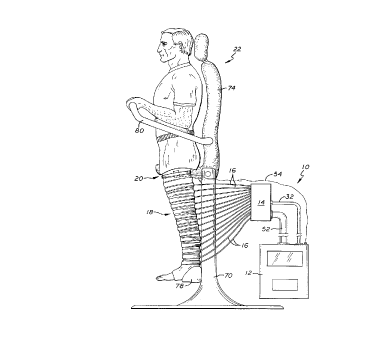
FIGURE 1 i~ a side plan view o~ the test system o~
the present invention showing a patient prepared ~or testing
disposed within a pneumatic legging attached to a mani~old
and system controller.
FIG. 2 is a side plan view o~ the test system of
the present invention and partial schematic showing the
system controller, manifold, inflation tubes, legging and
doppler ~low transducer and their interconnections.
FIG. 3 is a ~ront perspective view o~ the Doppler
flow transducer, housing and sensor lead.
FIG. 4 is a side plan view showing the testing
station, including the stand in a vertical position and the
~rame, seat and backrest rotated to a horizontal position.
FIG. 5 is a rear perspective view of the legging of
FIG. 2, showing a closure system.
FIG. 5A is a partial rear perspective view o~ the
enclosure system o~ FIG. 5 showing a strap and ~astener.
DESCRIPTION OF THE ~K~r~ED EMBODIMENTS
As shown in the exemplary drawings, the invention
is embodied in a diagnostic system 10 having a controller 12
connected to a manifold 14 by a plurality of inflation tubes
16 which are ~urther connected to an in~latable legging 18.
A Doppler ~low transducer 20 strapped to the upper thigh o~ a
patient provides measurements o~ blood ~low in the ~emoral
vein to the controller. As shown in FIG. 1, the patient is
positioned on a test station 22 so that he or she may be
moved to positions where the ~oot is elevated equal to the
level o~ the heart.
CA 0221~049 1997-09-09
WO 96/28088 PCT/US96/03092
_g_
It is possible to mimic valvular competence with
dynamic tourniquet obstruction. Externally compressing a
vein and preventing it from filling simulates closing a
venous valve at the point of the tourniquet. Furthermore,
the muscle pump function can be mimicked by external
compression of the leg by a pneumatically inflated series of
tourniquets. The narrower each tourniquet, the higher is the
resolution of the device.
In accordance with the present invention, multiple
pneumatically activated tourniquets may be placed along the
lower and upper leg to empty the blood in the veins of the
leg. Emptying of veins in this way is similar to the use of
an Esmark bandage, whereby an arterial tourniquet is placed
on the high thigh and an elastic bandage is wrapped around
the leg from foot to thigh, upon which the arterial
tourniquet is inflated to keep the leg vessels empty of
blood. The deep veins can be isolated from the superficial
veins by controlling the pressure of inflation in the
bladders at each level.
By measuring venous outflow through the only normal
exit vessel, the common femoral vein, and by taking measure-
ments with the leg level with the heart and dependent (but
without muscular activation) one can account for changes in
arterial inflow due to position change. In addition, the
amount of blood outflow can be obtained, as a proportion of
maximum flow. By measuring the changes in outflow with
compression and "competent valve simulation," one can
determine morphologically where the valvular incompetence
exists. Because of the resolution between tourniquets, it is
possible to demonstrate perforator incompetence separate from
longitudinal vein incompetence. Finally, with the tip-toe
exercise, a measurement of calf pump output efficiency can be
obtained.
As shown in FIG. 2, the test system 10 includes a
long-leg tubular legging 18 having a series of inflatable
bands or bladders 30. The legging has a longitudinal slit
105 in the back so that the legging may be wrapped around the
patient's leg. Each bladder or cuff is substantially
CA 0221~049 1997-09-09
W O 96/28088 PC~rrUS96/03092
--10--
cylindrical in shape, having first and second ends which abut
at the back of the legging. The semicircular or "C-shaped'
bladders are spaced approximately equally apart along the
legging, wrapping around the patient leg.
Forming cuffs 30 with closed ends in the back of
the legging 18, the bladders have a relatively small diameter
at the ankle, progressing to the largest diameter cuff at the
upper thigh. The legging is preferably made of a material
such as coated cotton or vinyl; whereas, the bladders are
made of a ~luid tight material capable of maintaining the
liquid or gas used for inflation, such as rubber or vinyl.
The legging may be disposable, such that the body is
constructed from molded or a sheet of heat sealed plastic.
Similarly, the bladders may be formed as an integral part of
the legging body.
Each bladder 30 may be connected to an inflation
tube 16 which is attached to a coupling or manifold 14 in
fluid communication with a pressurized fluid source 34.
Alternatively, the bladders may be connected directly to the
coupling. The in~1ation tubes are made from material
suitable to contain the fluid at inflation pressures, such as
rubber or nylon. The pressurized fluid is provided to the
manifold through a similarly constructed conduit 32.
Pressurized air, oxygen, carbon dioxide, or other
suitable gas is provided to the manifold 14 from a source 34
located in the controller 12. Alternatively, the gas source
may be external to the controller. Similarly, a hydraulic
system, rather than a pneumatic system, may be utilized to
inflate the bladders, for example, with water or oil. The
materials of construction disclosed herein are by way of
example only, and are not intended to limit the scope of the
invention.
As shown in FIGS. 5 and 5a, a closure system 90 may
be used with the legging 18 to configure the test device ~or
different size patient legs. A plurality of first tapered
tabs 92 are secured to or manufactured as part o~ the legging
and extend from one outside edge to the center o~ the back o~
the legging. A plurality of second tapered tabs 94 are
CA 0221~049 1997-09-09
W O 96/28088 PC~rrUS96/03092
--11--
similarly secured to or manufactured as part of the opposite
side of the legging and extend to the center of the back of
the legging proximate to the end of the corresponding first
tapered tab. The plurality of sets of first and second tabs
are spaced approximately equally apart from the ankle to the
upper thigh.
In the closure system 90, a strap 96 is fixed at
its first end 97 to one side of each first tapered tab 92 and
is configured to be slidably disposed within a slot 98 in the
corresponding second tapered tab 94. The strap's second end
99 is removably secured to the first tapered tab by a
suitable fastener affixed to either the first end of the
strap or directly to the first tapered tab, for example, by a
hook and loop connector (VELCRO~), snap or buckle. The strap
is used to tighten the first and second tapered tabs together
to adapt the legging to the diameter of the patient's leg.
The legging is secured sufficiently tight around the leg to
effect a tourniquet when the cuffs 30 are inflated; however,
the closed legging should not be unduly tight around the leg
when the cuffs are deflated.
Referring to FIG. 2, the coupling manifold 14
houses multiple electrically or pneumatically activated
valves 40. The manifold is preferably constructed from a
plastic, such as polycarbonate, and may be mounted on the
test station 22. The manifold valves can be selectively
opened to pass air to each connected bladder 16, opened to
vent air from the bladder or left closed to maintain bladder
pressure. Pressure monitoring is accomplished through a
pressure transducer (not shown) in the air conduit 32 or at
the source 34. Sequencing of the valves is initiated by a
control circuit 50 disposed within the controller 12. The
control circuit is connected to the valves by standard
electronic cabling or pneumatic tubing shielded within
conduit 52.
As shown in FIG. 2, the controller 12 includes a
microprocessor or similar control circuit 50 which operates
the manifold valves 40. The sequence of inflation and
deflation of the bladders 30 is controlled by sequentially
CA 022l~049 l997-09-09
W 096/28088 PC~r~US96103092
-12-
opening and closing the mani~old valves. The sequencing may
be accomplished by software in the microprocessor or by an
equivalent electronic or mechanical device. In addition, the
control circuit communicates with the Doppler flow sensor 20
via a sensor lead 54 to coordinate the valve sequencing with
the blood flow measurements. The pressure of the in~lation
fluid is monitored by a sensor (not shown) in the conduit 32.
The pressure signals are transduced, amplified and recorded
by the controller.
The data gathered by the controller 12 and the
location of any incompetent valves or similarly computed
in~ormation are sent to a printer via a standard communica-
tion port (not shown). The interpretation o~ the study is
also programmed with user configurable text phrases and
output patterns, ~or example, using a ~orty character LCD
display. Output displays may be available to provide
pressure and blood flow data and to provide system error
messages. Similarly, touch keys may be configured to
inter~ace with the controller to initiate and/or terminate
the testing and to allow input of test parameters and patient
specific data.
As shown in FIG. 3, the Doppler transducer 20
comprises two pairs oE crystals 60 mounted on a sur~ace 62,
such as a rotatable disk, disposed within a housing 64. Each
crystal pair is directed 180 degrees ~rom the other pair and
secured at about a thirty to forty-five degree angle of
incidence to the contact sur~ace o~ the leg (sixty to forty-
~ive degrees ~rom the mounting sur~ace). The transducer is
connected to a pulse Doppler generator (not shown) located
within the controller 12. The pulse Doppler generator causes
the crystal to emit and detect sound signals which may be
interpreted ~or a Doppler ~requency shi~t by the control
circuit 50. The disk is rotated by a microprocessor-
controlled motor, rotor or other device (not shown) also
located within the transducer housing. The housing is
secured to the patient thigh by a strap or similar mechanism.
When the Doppler transducer 20 is in operation, the
disk 62 iS rotated while the legging bladders 30 positioned
CA 022l~049 l997-09-09
W096/28088 PCT~S96/03092
-13-
at the patient's leg are sequentially inflated. Thus, the
sound signal frequency shift produced by the distally
directed crystals is equal to the signal magnitude produced
by the proximally directed crystals. The signals from the
crystal pairs, however, will be opposite in direction, such
that one signal is increasing and the other signal is
decreasing. This rotation verification assures that the
crystals are pointed along the long axis of the vein. An
inability to accomplish this results in control circuit 50
generating an error message at the display 56 and the
technician is prompted to reposition the device.
The pulse timing sequence is then changed by the
control circuit 50 so that the center of the vessel is
located, and so that the size of the vessel is calculated by
the controller 12. Determining these parameters allows the
control circuit to make uniform measurements of the fluid
velocity and to make uniform calculations of the fluid flow.
The control circuit calculates the fluid flow at the center
of the vessel. Assuming a round vessel, the volume of blood
flow in a given period of time is calculated as the integral
over time of the velocity of the blood times the cross-
sectional area of the blood vessel.
As noted, the Doppler ~low transducer 20 is config-
ured with two paired transmitter/receiver units 60. One
crystal of each pair is configured to transmit a pulsed sound
signal, while the other crystal of each pair is configured to
receive the re~lection of the pulsed signal. The
transmitting crystals are driven by a frequency generator
within the controller 12 which causes the transmitter
crystals to intermittently send a sound signal. The
frequency of any sound signal reflected back is detected by
the receiving crystal in the pair.
- The ~requency shi~t (Doppler) is interpreted by the
control circuit 50. If the sound signal is reflected from
moving blood cells, then the frequency of the signal is
changed upward by blood moving toward the receiving crystal
and downward by blood moving away from the crystal. By
configuring two pairs of crystals 180 degrees offset from
,
CA 0221~049 1997-09-09
W096/28088 PCT~S96/03092
-14-
each other, the magnitude of the flow represented by the
Doppler shift will be equal from each pair, but will be
opposite in direction, so long as the crystal pairs are
sensing along the same long axis of the vessel. By turning
the sensor's rotatable disk 62 until the Doppler shift is
equal, but opposite, one can effectively set the crystals
parallel to the blood flow in the vessel.
To obtain reproducible blood flow measurements, the
sensor 20 must consistently measure from the same place
within the blood vessel. The most accurate place to measure
average flow is in the center of the vessel's cross section.
By using a pulsed Doppler transmitter and sensor combination,
one can not only place the field of measurement into the
center of the vessel, but can also obtain relatively
accurate measurements of the flow in that vessel. The flow
measurement requires a calculation of the integral of flow
over time multiplied by the cross-section of the vessel.
Because flow is not constant but instead is pulsatile, a
calculational integration is necessary for accurate
measurement.
The method of accomplishing this "centering"
requires determining the location of the back wall and front
wall of the vessel. The vessel diameter is calculated from
the wall locations and the control circuit 50 sets the timing
of the send/receive crystal pair to the midpoint of the
vessel diameter. For instance, the control circuit initiates
the transmitting crystal to send or emit a short sound signal
and uses the receive crystal to detect the reflected signal
at some interval of time. By changing that interval, the
flow measured changes. As the interval is lengthened, the
flow measured changes until a point when it reaches zero.
When the interval is long enough, the sound travels past the
back wall and is no longer reflected. The distance to the
back wall is calculated as a function of the first interval
of time when the signal is not reflected and the speed of
sound through the tissue in the leg.
The time interval between when the first crystal
sends its signal and when the control circuit 50 detects the
CA 022l~049 l997-09-09
W096/28088 PCT~S96/03092
-15-
re~lected signal in the second crystal is then shortened
incrementally until no Doppler shi~t is calculated (i.e.,
zero flow). The ~irst time interval where no shi~t is
detected is used to calculate the distance to the ~ront wall
o~ the vessel. The di~erence in distance ~rom the ~ront to
the back wall divided by two is the measured midpoint of the
vessel. The control circuit then sets the send/receive
interval to the computed time in which a signal will travel
~rom the ~irst crystal to the vessel's midpoint and back to
the second crystal o~ the same pair. The control circuit
"listens" to the vessel's center ~or the remainder o~ the
test.
Because the system o~ the present invention creates
~low by compressing the veins with pneumatic cu~s, the
impulse and blood ~low measured by the sensors during this
"set up" or calibration phase can be relatively consistent.
The timing o~ the cu~ compression and transmitted sound can
be empirically set to maximize the ~low and there~ore the
accuracy o~ the centering and axial localization phases o~
the calibration.
As shown in FIG. 4, the testing station 22 is
con~igured with a central stand 70, a bicycle-type seat 72
and backrest 74 secured to a reclinable ~rame 76 for
positioning the patient. This assembly can be pivoted
backward so that the seat backrest is horizontal. In
addition, there is a cup-like device 78 a~fixed to the bottom
o~ the station ~rame. The cup accommodates the patient's
heel, and there is one heel cup on each side o~ the frame. A
handrail 80 is secured to the backrest or ~rame so the
patient may balance himsel~ or hersel~ when the ~rame is
rotated to the horizontal position. The controller 12
electronic and mechanical components and the mani~old 14 may
be part o~ or mounted to the test station stand or ~rame.
The patient is seated on the seat 72 of the test
station 22 with his or her back against the rest 74 and heels
in the cups 78. The in~latable legging 18 is placed on the
patient's leg and closed by a zipper or closure system 90 as
described hereto~ore. The mani~old 14 is connected to the
CA 022l~049 l997-09-09
W096/28088 PCT~S9G~J0~2
-16-
controller 12 via main air conduit 32 and electrical conduit
52. The Doppler sensor 20 is attached to the patient over
the common ~emoral vein proximal to the entry point o~ the
greater saphenous vein. The patient is tilted back on the
station ~rame 76 so that his or her legs are at the same
level above the ~loor as the patient's heart.
Each patient leg is tested and studied separately.
First, the control circuit 50 calibrates the Doppler
transducer 20 to assure that the transducer is properly
centered over the ~emoral vein. The bladders or cu~s 30 in
the legging 18 are in~lated ~rom the ankle or ~oot to the
upper thigh in sequence so as to cause restrictions which
milk the blood out o~ the leg. The cu~s are in~lated
su~iciently to compress the veins, but not compress the
arteries. The pressure in the cu~fs caused by the sequence
o~ restrictions is high enough to force blood out o~ the deep
and super~icial veins. The volume o~ venous output blood
~low through the leg (~emoral vein) is calculated by the
control circuit and is termed the Maximal Venous Output
(MVO).
The cu~s 30 are de~lated by the controller 12 and
the patient is tilted so that the legs are dependent. The
patient is instructed not to move his or her legs. The
control circuit 50 again centers the Doppler transducer 20.
The cu~s are then in~lated by the controller sequentially
~rom ~oot to thigh and the venous output blood volume is
again calculated by the control circuit. This volume o~
venous output blood ~low through the leg (~emoral vein) is
termed the Maximal Dependent Output (MDO).
The patient is le~t in this position with legs
dependent. The controller 12 then sequences in~lation and
de~lation o~ the cu~s 30 to test venous valve competence and
pump output e~iciency at each level o~ the leg. To begin
the procedure ~or identi~ying the location of an incompetent
valve, the controller in~lates the lowest or ~irst cu~
(closest to the ankle). This pushes blood to the level o~
the next cu~ above the ~irst cu~, i.e. the second cu~,
which r~m~i n.~ de~lated. The controller then releases or
CA 022l~049 l997-09-09
Wos6/28088 PCT~S96/03092
-17-
deflates the first cuff. If the venous valve at the location
of the first cuff is competent, then the blood pushed above
the first valve will remain above the valve. If that valve,
however, is incompetent, then some or all the blood pushed
above the first valve will reflux below the valve.
The control circuit 50 inflates the second cuff and
the remaining upper cuffs to the upper thigh are inflated in
sequence to milk the blood out of the leg and past the
diagnostic flow sensor 20 at the common femoral vein. The
controller 12 uses the signal from the flow sensor placed
proximate the upper thigh proximate the femoral vein to
calculate the total volume of blood which flows out of the
leg after the first valve was released. Any difference in
flow between this measurement and the MDO is the amount of
reflux at the first cuff level.
If the valves proximate the first cuff are
competent, then the blood moved by the compression by the
first cuff prior to its deflation will remain at that level
for the other cuffs to push out. Thus, the measured volume
of blood flow for a test at a competent valve will
approximate the MDO. If the valves are incompetent, the
blood will regurgitate back down and the amount of blood
pumped out will be less than the MDO by the amount which
refluxes through the incompetent valves.
The test is repeated, except that this time the
first and second cuffs are inflated to move the blood in the
leg past the second cuff. The first and second cuffs are
deflated to permit blood to drain through any incompetent
valves proximate the second cuff. The third cuff (next above
the second cuff) and all subsequently higher cuffs are then
inflated to move the blood past the flow sensor. The total
blood flow is again calculated for this valve sequence. This
- venous output will indicate any reflux at the level of the
second cuff when compared to the MDO. Such valve sequencing
is repeated until the top most cuff is tested.
The evaluation of each leg is then repeated, except
that instead of compressing to the point of deep venous com-
pression, the cuffs are initially compressed fully, then
CA 022l~049 l997-09-09
W096/28088 PCT/U~5~'~3092
-18-
deflated to superficial vein compression only. At this com-
pression, differences in flow represents incompetence of the
deep veins, whereas previous measurements were for total vein
reflux. Location of deep venous valves as well as
perforators which are incompetent can be determined in this
way. Additionally, the incompetence of the saphenofemoral
junction can be ascertained in this manner.
Another evaluation can be accomplished by having
the patient perform a tip-toe exercise while measuring venous
output as described heretofore. The measured blood flow
volume divided by the MDO is the Dependent Pump Efficiency
(DPE). A similar evaluation can be obtained with the patient
supine, such that the measured venous output divided by the
MVO represents the Maximum Pump Efficiency (MPE). The
difference between the DPE and MPE is the amount of decrease
in efficiency due to the longitudinal valvular incompetence,
since the incompetence of the longitudinal vein is decreased
to near zero by eliminating the effect of gravity.
By appropriately timing the cuff inflation at
selected levels, and combining this with tip-toe exercise,
the test system is capable of predicting the improvement
which one might get by re-establishing valvular competence at
any given level, or at multiple levels in combination. The
timing sequence would be determined empirically by changing
the time between calf muscle contraction and inflation until
the maximum pump output was obtained M~ m~ 1 Compensated Pump
Output, MCPO.
Additionally, an evaluation may be performed which
simulates the efficiency of the venous pump likely to be
gained by valve repair. During the procedure, the patient is
allowed to stand with the Doppler flow sensor in place over
the femoral vein. The patient is asked to do a tip-toe
exercise and the total volume of output blood flow is
calculated. This is then compared with the MDO. Addition-
ally, the effect of valve repair can be assessed by doing asimilar exercise as above, but instead of allowing the blood
to flow as it naturally would, the cuff at the level of
proposed valve repair would be inflated at the appropriate
CA 0221~049 1997-09-09
W O 96/28088 PC~rrUS96/03092
--19--
tlme after tip-toe to maximize venous outflow. The increase
of venous blood volume over baseline would be the approximate
improvement which a venous repair could expect to achieve.
Thus, the test system method of the present
invention meets the need for a diagnostic system which
provides analysis of the ability of the veins to conduct
blood to the heart. Also, the system provides a morphologic
assessment as to the level at which the veins are failing.
In addition, the system differentiates deep from superficial
and perforating vein function. Moreover, the present system
provides quantitative assessment for comparison over time and
after an intervention, being only m;n;m~l ly dependent on
technician skill, so as to be reproducible and comparable
from day-to-day and institution-to-institution.
While several particular forms of the invention
have been illustrated and described, it will be apparent that
various modifications can be made without departing from the
spirit and scope of the invention. Accordingly, it is not
intended that the invention be limited, except as by the
appended claims.