Note: Descriptions are shown in the official language in which they were submitted.
CA 02215060 1997-09-10
WO 97/26830 PCT/US97J00I06
ULTRASHORT PULSE HIGH REPETITION RATE LASER SYSTEM
FOR BIOLOGICAL TISSUE PROCESSING
t
Field of the Invention
The present invention is directed to the field of ultrashort pulse duration
Iaser systems
suitable for material and biological tissue processing and in particular to a
material removal
apparatus and method in which ultrashort pulse Iaser systems are operable at
pulse repetition
rates in excess of at least 10 Hertz so as to efficiently remove substantial
material volumes while
substantially eliminating collateral damage.
AcknowIed~ment ofU.S. Government Support
This invention was made with U.S. Government support under Contract No. DE-
FG03
9IER61227, awarded by the U.S. Department of Energy, Grant No. N0014-91-C-
0134, awarded
by the O~ ce of Naval Research, and Grant No. RRO I I 92, awarded by the
National Institute of
Health. The United States Government has certain rights in this invention.
Baclc~round of the Invention
Laser interaction with organic and inorganic targets has been investigated for
the past
thirty five years for applications as diverse as material processing and
surgical tissue ablation.
One significant challenge to laser tissue processing is the need to maximize
ablation efficiency
while, at the same time, minimizing collateral damage to adjacent material.
Recent years have brought increased interest in the use of lasers as a
therapeutic and
preventive tool in various dental applications such as removal of carious
lesions (removal of
tooth decay), surgical treatment of oral malignancies and periodontal
diseases, and preparation
and sterilization of root canals. In spite of these advances, Iasers remain
limited in their ability
to remove sound (hard as opposed to soft) tooth structure since the lasers
currently in use for
dental procedures generate unacceptable heat levels which cause collateral
damage to the tooth
surface and in the tooth pulp. Early procedures for removal of hard dental
substances involved
optical drilling using CO2, ruby and Nd:YAG (Neodymium doped Yttrium Aluminum
Garnet)
lasers requiring high radiant exposure and resulting in considerable damage to
surrounding
tissue. As a consequence, it was generally concluded in the mid 1970s that
lasers would not
become a common drilling tool unless a new method was found to reduce
collateral damage.
Optical dental drilling with Er:YAG (Erbium doped YAG) lasers yielded
encouraging
results in the early 1990s, and has shown capabilities to perform as an
efficient drill with out
. generating excessive damage to surrounding tissue. The success of Er:YAG
systems, operating
in the nanosecond to microsecond pulse duration regime, in minimizing thermal
damage has also
_1_
CA 02215060 1997-09-10
WO 97/26830 PCT/US97/00106
1
been observed in other areas of application in medicine, and can be attributed
to the high
absorption coefficient of biological tissues at the particular wavelengths
characteristic of the
system (2900 nm), when used in combination with nanosecond to microsecond
pulse durations. x
Neev et al., Dental Ablation With Three Infrared Lasers, Lasers in Surgery ahd
Medicine,
Vol. 17, 1995, discloses three laser systems adapted to hard tissue
processing, such as dentin and ,
enamel removal in dental applications. The laser systems disclosed (Er:YSGG,
Ho:YSGG, and
Q-switched Nd:YAG) all operate in the near IR region of the electromagnetic
spectrum and are
pulsed in two different regimes: about 250 microsecond pulse durations for the
Er:YSGG and
. Ho:YSGG lasers, and about 15 nanosecond pulse durations for the Er:YAG
system.
While the disclosed removal rate is in the range of approximately tens of
micrometers per
pulse, the disclosed laser systems exhibit classical spectrum selectivity
(wavelength dependent
absorption) and effect high removal rates by operating at pulse energies in
excess of 20 to 30
millijoules per pulse. Enhancing material removal by increasing laser power
is, however,
accompanied by increased photothermal and photomechanical effects which causes
collateral
damage in adjacent material. In addition, increasing power leads to plasma
decoupling of the
beam, e.g., incident laser energy is wasted in heating the ambient in front of
the target.
High intensity pulses additionally cause very loud acoustic snaps, when the
laser pulse
interacts with tissue. These snaps or pops include a large high frequency
component which is
very objectionable to a user or, in the case of a medical application, to a
patient. In addition to
the psychological impact of such noise, these high frequency snaps are able to
cause hearing loss
in clinicians when repeated over a period of time.
U.S. Patent No. 5,342,198, to Vassiliadis, et al., discloses an Er:YAG IR
laser system
suitable for the removal of dentin in dental applications. The laser produces
a pulsed output
having a beam with a pulse duration in the range of several tens of
picoseconds to abort several
milliseconds. Although disclosed as being efficient in the removal of dentin
and dental enamel,
the mechanism by which material removal is effected is not understood.
Significantly, however,
the only laser systems disclosed as suitable for the process are those which
operate at
wavelengths (1.5 to 3.5 microns) that have proven to be generally effective
for enamel
interaction. Thus, the absorption characteristics of the material target are
of primary concern to
the removal rate. In addition, high energy levels are required to remove
enamel and dentin,
leading to the problem of thermal damage and acoustic noise.
Additional possibilities for the application of lasers to the field of
dentistry in particular,
and to hard tissue ablation in general, have been proposed by the use of
excimer lasers that emit
high intensity pulses of ultraviolet (UV} light, typically with pulse
durations in the approximately
1 to 100 nanosecond range. Both the short wavelengths and nanosecond range
pulse durations
used by excimer lasers contribute to defining a different regime of laser-
tissue interaction. Short
-2-
CA 02215060 1997-09-10
WO 97!26830 PCT/US97l00106
1
wavelength ultraviolet photons are energetic enough to directly break chemical
bonds in organic
molecules. As a consequence, UV excimer lasers can often vaporize a material
target with
minimal thermal energy transfer to adjacent tissue. The resultant gas (the
vaporization product)
is ejected away from the target surface, leaving the target relatively free
from melt, recast, or
other evidence of thermal damage.
Another important characteristic of UV excimer lasers is that materials which
are
transparent to light in the visible or near infra-red portions of the
electromagnetic spectrum often
begin to exhibit strong absorption in the UV region of the spectrum. It is
well established that
the stronger a material's absorption at a particular wavelength, the shallower
the penetration
achieved by a laser pulse having that wavelength. Thus, in many types of
materials, a UV pulse
typically only penetrates to a depth in the range of from about 1 to about 4
micrometers. By
simply counting pulses, great precision can be achieved in defining removal
depths_ In addition,
organic tissue is strongly absorbent in the UV wavelengths (193 nm for ArF,
for example)
therefore~allowing the laser-tissue interaction region to be controlled with
great precision.
Notwithstanding the relatively damage free material removal characteristics of
UV
excimer lasers, these systems suffer from several disadvantages which limit
their applicability
to biological tissue processing. The reports of damage free tissue removal
result from
evaluations performed on single pulses, or on pulses with a very low
repetition rate (typically
about 1 to 5 Hertz}. Because of the low volumett~ic removal per pulse of
excimer systems
(material removed per unit time is poor), efficient material removal can only
be accomplished
by high pulse repetition rates. However, when the pulse repetition rate
exceeds about 3 to 5
Hertz, considerable thermal and mechanical collateral damage is observed.
While UV photons
are sufficiently energetic to directly break chemical bonds, they are also
sufficiently energetic
to promote mutagenic effects in tissue irradiated at LJV wavelengths, raising
concerns about the
long term safety and health of a system operator. The scattered Light produced
by excimer lasers
also presents a significant threat to the clinician and/or the patient. Even
low intensity scattered
radiation, with wavelengths below 300 nanometers, is able to interact with the
ambient
environment to produce atomic oxygen and other free radicals. These can, in
turn, react with the
lens and cornea of the eye, producing cataracts, and produce burns on the skin
equivalent to sun
burns. As a consequence, excimer laser systems have been found to be most
suitable for
inorganic material processing applications, such as thin coating patterning or
dielectric or
semiconductor material etching.
~35 In addition, the operational parameters of excimer laser systems are such
that material
removal remains a wavelength and beam energy dependent process (although
weakly dependent
~ on wavelength). Even when pulsed in the tens of nanoseconds pulse duration
regime, excimer
lasers are configured to deliver energy in the range of from about 10 to about
1000 miIlijoules
-3-
CA 02215060 1997-09-10
WO 97/26830 PCT/US97/00106
1 - -_
per pulse. At the higher energies, excimer lasers suffer from the same
problems caused by
plasma decoupling and pulse to poise interaction as 1R lasers. Additionally,
as pulse energy
increases, so too does the intensity of the associated acoustic snap.
Summary of the Invention
A
There is, therefore, provided in the practice of this invention a fast,
e~cient, and collateral
damage free apparatus and method for selective removal of material through
material-laser
interaction between biological tissue and a pulsed laser operating in the
femtosecond to
picosecond pulse duration regime at high pulse repetition rates.
The process of the present invention results in material removal rates which
meet or
exceed the removal rates of mechanical drilling systems while far exceeding
the accuracy and
precision of low removal rate laser systems.
In one embodiment of practice of the present invention, the process for
selective
biological tissue removal processing comprises providing a pulsed laser
operated so as to
produce a pulsed output beam which includes individual pulses each having a
pulse duration in
the range of from about 1 femtosecond to about 100 picoseconds. The pulsed
beam is directed
onto a target material, such as biological tissue, from which removal is
desired.
Each pulse interacts with a thin layer portion of said biological tissue sa as
to form a
plasma which decays in the time period between pulses such that the impacted
tissue portion is
removed by ablation. The plasma formation step is repeated at a pulse
repetition rate greater
than or equal to I O pulses per second until a sufficient depth of tissue has
been removed with
substantially no transfer of thermal or mechanical energy into the remaining
tissue and
substantially no collateral damage thereto.
According to one aspect of the invention, the plasma is formed by mufti-photon
absorption
and/or collisional ionization of the atoms and molecules comprising the tissue
material. Each
pulse of the pulsed output beam has an energy in the range of from about 0.1
to about 100
millijoules, the pulsed beam having a diameter at the tissue target such that
the tissue experiences
an energy fluence in the range of from 'about 0.1 to about 15 Joules per
square centimeter
depending upon tissue type, laser pulse duration and laser wavelength. When so
operated, the
pulsed beam exhibits a material removal rate in the range of from about 0.1 to
about 2.0
micrometers per pulse, with the removal rate being substantially constant
without regard to
variations in tissue chromophore, tissue hardness or tissue state.
According to an additional aspect of the invention, the method of the present
invention
is practiced by laser systems operating in the 200 to 2000 nanometer region of
the
electromagnetic spectrum.
CA 02215060 1997-09-10
WO 97/26830 PCT/C1S97100106
1
In a more detailed embodiment of the present invention, a chirped-pulse
amplified, solid
state laser is used to provide an about 500 micrometer diameter pulsed beam,
which provides
pulses, having durations in the 0.02 to 100 picosecond region at an adjustable
repetition rate from
d
to 2000 Hertz with pulse energies of about 3 millijoules. The pulsed beam is
used to
selectively ablate undesired material, such as carious lesions, dentin, enamel
and/or soft tissue
in a dental procedure at a removal rate which meets or exceeds the removal
rate of a mechanical
dental drill. The precision and selectivity of material removal by the
apparatus and method of
10 the present invention enables additional delicate surgical procedures,
particularly in cases where
diseased or undesired tissue is interspersed with healthy tissue, or in cases
where the working
area is exceptionally close or exceptionally delicate, such as brain and
spinal surgery, bone
removal in neural surgical applications, and orthopaedic surgery.
20
?5
3~
-5-
CA 02215060 1997-09-10
WO 97/26830 PCT/US97/00106
1
Brief Description of the Drawings
These and other features, aspects, and advantages of the present invention
will be more
fully understood when considered with respect to the following detailed
description, appended
claims, and accompanying drawings, wherein:
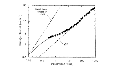
FIG. 1 (a) is a graphical representation of experimentally determined values
of laser ,
damage fluences in Joules per square centimeter, plotted as a function of
pulse duration in
picoseconds, for fused silica showing the monotonically decreasing threshold
and fluence
departure from root tau dependence on pulse duration, when pulse duration is
reduced in
accordance with practice of principles of the invention;
FIG. 1 (b) is a graphical representation of experimentally determined values
of laser
damage fluences in Joules per square centimeter, plotted as a function of
pulse duration in
picoseconds, for fused silica showing the wavelength independence of fluence
departure from
IS root tau scaling;
FIG. 2 is a graphical representation of experimentally determined values of
ablation
thresholds plotted as a function of pulse duration for materials having
various absorption
characteristics, depicting the independence of ablation threshold values on
material
chromophore;
FIG. 3 is a graphical representation of experimentally determined values of
material
removal rates in microns per pulse, plotted as a function of laser fluence, of
ultrashort laser
pulses for exemplary dentin and enamel material, depicting the independence of
removal rates
at a given laser fluence on material properties;
FIG. 4 is a graphical representation of experimentally determined values of
residual pulse
heat, plotted as a function of time, for ultrashort laser pulses (solid
diamonds) as compared to
nanosecond laser pulses (triangles) at a 10 Hertz repetition rate;
FIG. 5 is a graphical representation of experimentally determined values of
residual pulse
heat, plotted as a function of time, for ultrashort laser pulses operating at
a 1000 Hertz repetition
rate;
FIG. 6 is a simplified block level schematic diagram of a chirped pulse
amplified solid-
state laser system suitable for practice of principles of the invention; and
FIG. 7 is a simplified block level schematic diagram of an exemplary dental
drilling
apparatus incorporating the ultrashort pulse duration, high repetition rate
laser system of FIG.
6.
-6-
CA 02215060 1997-09-10
WO 97/26830 PCT/US97/00106
1
Detailed Description of the Preferred Embodiment(sl
The principles of operation of an exemplary laser system, which will be
described in detail
in following sections, will now be developed in connection with certain
mechanisms for hard
tissue removal, for example, the ablation of dentin during removal of carious
lesions. The
description of the operation of the laser system with respect to dental
applications is for
exemplary purposes only and is not intended to limit the application of the
laser of the present
invention. As will be described in greater detail below, the laser system of
the present invention
has application to a wide variety of biological tissue removal processes as
well as exceptional
utility for general material removal and micro-machining. Those having skill
in the art will
immediately recognize the utility and applicability of the laser system's
novel operational regime
to Laser-tissue interactions in the general sense.
Utilizing the laser system which will be described further below, the
inventors have
l 5 identified a laser operational parameter regime which provides hard tissue
interaction
characteristics that are superior to conventional laser systems and provides
material removal
rates, for exemplary dental material, on a par with mechanical drill
technology. Advantageously,
the tissue ablation methods of the laser system of the present invention
provide for efficient
material ablation because the input laser energy per ablated volume of tissue
is small, resulting
in a decrease in the amount of energy required to ablate a given volume of
material achievable
with conventional prior-art lasers employed for cutting, drilling and
sculpting of biological
tissue. The laser system's high ablation efFciency and the short duration of
the stress impulse
results in negligible collateral mechanical damage, while the extremely short
energy deposition
time results in minimal collateral thermal damage.
The ablation threshold and removal rate are only minimally dependent on tissue
type and
condition, thus, ablation (material removal) is generally chromophore
independent as well as
generally insensitive to tissue linear absorption characteristics, tissue
moisture content, material
morphology and micro-architecture, and tissue hardness. Precision of ablation
depth is achieved
by removing only a small volume of material with each pulse, while volumetric
ablation is
controlled by the repetition frequency of the ablation pulses.
Precise spatial control of tissue ablation and removal as well as precise
control of ablation
depth has been determined by the inventors as resulting from an intensity-
dependent multiphoton
initiation and plasma termination process. Formation of a critical density
plasma by both
multiphoton and collisional ionization processes eliminates significant energy
deposition below
3~5 a depth approximately that of the wavelength of the laser Light, when
energy deposition takes
place in less than about 10 picoseconds. This "self termination" insures a
high precision of tissue
removal for each pulse and is primarily responsible for the high ablation
efficiency, defined as
the magnitude of laser energy required to effect removal of a given volume of
tissue or material,
CA 02215060 1997-09-10
WO 97/25830 PCT/LJS97/00106
1
of ultrashort pulses in accord with the invention. Ablation efficiencies have
been demonstrated,
in accordance with the present invention, at approximately 0.1 cubic
millimeters of material
removed per Joule of laser energy, for hard, dielectric materials, e.g., fused
silica, .bone, enamel,
or the like. Conventional nanosecond pulse duration laser systems have
substantially lower
ablation effciencies, in that laser energies must be increased significantly
in order to remove the
same amount of material with substantially the same laser beam size.
An additional advantage of the method of the present invention, is that longer
wavelength,
ultrashort pulse duration laser systems can be utilized in most, if not all,
of the procedures
currently employing lasers which operate in the ultraviolet region. Replacing
ultraviolet lasers
with the longer wavelength ultrashort pulse lasers of the invention would
provide the benefit of
eliminating the risks associated with mutagenic radiation produced by short
wavelength lasers,
and the attendant dangers posed to clinicians and their patients.
The operational characteristics of an ultrashort pulse width, high repetition
rate laser
system, in accordance with practice of principles of the invention will now be
described with
reference to FIGS. 1 (a), 1 (b), 2, and 3 . .
I. Principles of Operation: Ultrashort Pulse Durations
Previously known and used long pulse laser systems, operating in the
nanosecond to
microsecond pulse duration regime, have shown themselves to be generally
inefficient in their
ability to remove substantial amounts of tissue without causing extensive
collateral damage. in
a conventional long pulse laser system (conventional Nd:YAG or Er:YAG TR.
lasers, for
example), much of the optical energy delivered to a material removal target
site has not gone into
disrupting the structural integrity of the target material, but rather is
transferred into the
surrounding tissue as thermal, acoustic or mechanical energy. This energy
propagates through
the surrounding tissue as both mechanical shock waves and heat which manifest
themselves as
undesirable cracks, material charring, discoloration, surface melting and
perceived pain.
Conventionally, for pulses longer than a few tens of picoseconds, the
generally accepted
model of bulk material removal involves the heating of conduction band
electrons by an incident
beam of coherent photons and transfer of this thermal energy to the bulk
material lattice.
Damage occurs by conventional heat deposition resulting in melting, boiling,
and/or fracture of
the material in the region in which removal is desired. Because the
controlling rate for material
removal depends on thermal conduction through the material lattice and the
lattice's
thermodynamic properties (heat capacity, heat of vaporization, heat of fusion,
and the like), the
minimum amount of energy required to effect an observable change in the
material's properties,
termed herein as the threshold damage fluence and defined as the incident
laser energy per unit
_g_
CA 02215060 1997-09-10
WO 97/26830 PCT/ITS97/00106
1
area, is dependent approximately on the square root of the pulse duration (l).
Relatively long
pulse durations have, in the past, been considered necessary in order to
obtain adequate material
removal characteristics. Long pulse durations, however, are often the source
of many of the
undesirable side effects exhibited by conventional nanosecond or longer pulse
laser systems.
Unexpected results are obtained, however, when material removal is performed
with
- lasers having pulse durations Iess than the characteristic electron-lattice
energy transfer time for
a particular tissue or material of interest. For the majority of hard,
biologic materials, this
characteristic energy transfer time is on the order of about 10 to 50
picoseconds. However, when
pulsed laser systems are operated in a parametric regime which includes pulse
durations shorter
than this characteristic transfer time, the physical mechanism of material
removal changes as
depicted in FIGS. 1 (a) and 1 (b).
FIG. 1 (a) is a log-log graph depicting the general behavior of laser induced
damage, the
damage fluence in Joules per square centimeter (J/cm2), as a function of beam
pulse duration (z)
in picoseconds for a laser system operating in the 1053 nanometer wavelength
region. At pulse
durations above about 20 picoseconds, the plot of damage fluence as a function
of pulse width
is seen to follow the classical, diffusion dominated root tau (i"~) scaling
characteristic of electron
kinetic energy transfer to the material lattice structure and diffusion during
the Iaser pulse.
Material damage, in this region, is thermal in nature and characterized by
melting, boiling, and/or
fracture of the material surface. However, below 20 picosecond pulse widths,
the inventors have
determined that the damage fluence departs from the root tau model, and
exhibits a steadily
decreasing threshold associated with a gradual transition from the long-pulse,
thermally
dominated regime to an ablative regime characterized by multiphoton and
collisional ionization,
and plasma formation. Short pulse damage is typically confined to small region
bounded by the
peak of the laser beam's Gaussian irradiance distribution. Thus, damage
(material ablation or
removal) occurs only over an area with sufficient beam intensity to produce
ionization.
As the pulse duration decreases to a time period less than the relaxation
time, i.e., the time
required for electrons to transfer energy to the lattice (approximately 20
picoseconds in the case
ofthe exemplary dentin material), the laser energy is non-linearly absorbed to
produce quasi-free
electrons which, in turn, act as seed-electrons which cause an avalanche or
electron cascade by
collisional ionization in which material conduction band electrons,
oscillating in response to the
Iaser optical field, transfer energy by phonon scattering. Once an electron
acquires kinetic
energy equal to the band gap energy for the material, subsequent impact
ionization promotes an
5 additional valence electron into the conduction band. The resulting
avalanche, similar to that
produced by gases for example, Ieads to an irreversible change in the bulk
material structure.
Initial quasi-free electrons are thought to be produced by multiphoton
absorption or
optical field ionization of individual constituent atoms and molecules or
defect sites of the
_g_
CA 02215060 1997-09-10
WO 97/26830 PCT/LTS97/00106
material. The electron avalanche causes a microplasma to be produced on the
surface of the
material which is allowed to decay by ablation after the end of each
individual pulse. For
sufl ciently short pulses, a critical density microplasma can be produced
directly as a result of
optical field ionization (termed muitiphoton ionization herein), with little
or no collisional
ionization. As can be seen in FIG. 1, the experimentally determined damage
fluence, shown as
a function of pulse duration, approaches the multiphoton ionization Limit (no
collisional
ionization) when pulse durations are reduced so as to be in the range of about
10 to 100
femtoseconds or less. It will be realized by those skilled in the art,
however, that significant
benefits will be derived from laser pulse durations in excess of one
femtosecond, but less than
the characteristic lattice coupling time.
Because the mechanism for energy transfer from the laser to the target
material involves
forming a localized, energetic plasma from the target material rather than
melting and boiling
away the target material, there is little energy transfer into the material
bulk before the material
is removed by ablation. As was described above, damage occurs only in an area
irradiated by
sufficient beam intensity to produce ionization. At the pulse durations in
accord with practice
of the invention, there is insufficient time for lattice coupling and,
therefore, negligible diffusion
induced collateral damage. Additional benefit to reduced collateral damage is
realized due to
the monotonically decreasing ablation threshold as the pulse duration is
reduced. Consequently,
ultrashort pulse width laser systems offer a dramatic reduction in the amount
of collateral
damage caused in a material as a result of laser-material interaction. The
damaged area, when
formed by short (<I0 picoseconds) pulses, is typically several orders of
magnitude smaller than
when formed with long (nanosecond to microsecond) pulses.
FIG. 1 (b) is a log-log graph depicting damage fluence in Joules/cm2, for
fused silica, as
a function of pulse duration in picoseconds for laser beams at two
wavelengths, 1053 manometers
(as also described above) and 526 manometers, as experimentally determined by
the inventors.
As can be seen from the graph of FIG. I (b), the departure. of the damage
fluence, as a
function of pulsewidth, from conventional root tau scaling is independent of
wavelength. At
pulse durations in the sub-picasecond region (approximately 0.3 picoseconds)
the damage
fluence values, for the two wavelengths (which may also be expressed in units
of frequency)
differ by only a factor of two (about 2 Joules/cm2 for the I 053 manometer
case, and about 0.9
Joules/cm2 for the 526 manometer case}.
Referring now to FIG. 2, an additional consequence of using ultrashort Laser
pulses to
process tissue is the relative insensitivity of the ablation threshold and
ablation rate (volume of
material removed per pulse} to the laser wavelength, tissue chromophore, and
the structure,
hydration and oxygenation state of the material. FIG. 2 depicts a graphical
representation of the
ablation threshold as a function of pulse duration for materials having
various light-absorption
-I 0-
CA 02215060 1997-09-10
WO 97/26830 PCT/ITS97/00106
1
properties as experimentally determined by the inventors. Collagen gel
(fibrous protein found
in all multicellular organisms) materials were prepared having properties that
mimic the densities
and atomic numbers of living tissue. Different concentrations of aqueous
cupric chloride were
mixed with the gels to provide materials with a range of linear absorptions.
Ablation
measurements were performed using a chirped-pulse amplif cation laser (to be
described in detail
below) which is able to provide pulses of continuously adJustable duration
from about 0.3 to
about 1000 picoseconds.
I0 As can be seen in FIG. 2, the ablation threshold for clear gels (generally
similar to a
human cornea) in the 1000 picosecond pulse duration range (75 Joules/cm2) is
approximately
1000 times higher than the absorption threshold for black gel (0.074
3oulesl/cmz). However, in
the sub-picosecond pulse duration range, the ablation threshold difference
reduces to less than
an order of magnitude; in particular, the difference is only approximately a
factor of six. As is
I S apparent from the representation of FIG. 2, ablation thresholds for
transparent and opaque
materials converge at the ultrashort pulse durations in accordance with the
invention.
Without wishing to be bound by theory, the inventors postulate that the
absorption
threshold insensitivity results directly from the generation of quasi-free
electrons caused by
multiphoton absorption by the material.
20 , Ablation rates, in microns removed per 350 femtosecond pulse, for both an
exemplary
enamel and an exemplary dentin material, are depicted in FIG. 3, at fluences
of from about 0.5
to about 16 Joules per square centimeter. Far purposes of identification,
dentin is represented
by open square shapes, while enamel is represented by open triangles. A "best
fit" curve, as
determined by the inventors, has been superposed on the individual data
points. As can be seen
25 in the figure, both material types exhibit a clear ablation efficiency
saturation pattern as pulse
energy is increased. From the ablation threshold at about 0.5 3ouies per
square centimeter,
ablation rate increases rapidly to about I micron per pulse at a fluence level
of about 3 Joules per
square centimeter, where ablation for both tissue types stabilizes at about
the same rate. Beyond
this point, only a very small increase in ablation rate occurs with increases
in fluence. Ablation
30 rates of 1.5 microns per pulse are achieved for dentin material at I 6
3oules per square centimeter,
which represents only a 50% increase in ablation rate for a five-fold increase
in fluence level,
as compared to 3 Joules per square centimeter level. The diminished return in
ablation efficiency
is thought to be a natural consequence of the microplasma formation by
ultrashort laser pulses.
As the pulse energy is increased, a denser plasma is generated by the leading
edge of the laser
35 pulse which, in turn, absorbs and reflects subsequent radiation, thus
shielding the surface and
preventing additional energy to be used for deposition.
~ For purposes of comparison, the ablation rates of dentin and enamel when
processed with
a nanosecond pulse at a ffuence of 34 Joules per square centimeter are
depicted in FIG. 3 as filled
-1 I-
CA 02215060 1997-09-10
WO 97126830 PCT/US97/00I06
I
square and triangle shapes. As is shown in the figure, nanosecond pulses
exhibit an ablation rate
of about 4 microns per pulse for dentin (the filled square), and about 1.4
microns per pulse for
enamel (the filled triangle), at the 34 Joules per square centimeter fluence
level. The inventors
have determined that a 3 Joule per square centimeter fluence was well below
the ablation
threshold of either dentin or enamel for nanosecond pulses, which threshold
was determined by ,,
experimentation to be in the range of about 20 Joules per square centimeter.
As is additionally clear from FIG. 3, the ablation rates for the same fluence
level in the
nanosecond regime are very different for dentin and enamel, with dentin
ablation being almost
a factor of four greater. Thus, as clearly indicated by FIG. 3, ultrashort
pulses with femtosecond
to picosecond durations, have substantially greater ablation efficiencies than
nanosecond pulses.
Comparison of the ablation rates for nanosecond pulses and the ultrashort
pulses in accordance
with the invention shows a ten-fold efficiency increase over nanosecond pulses
in enamel (3
J/cm2 verses 26 J/cm2 for an ablation depth of about I micrometer) and a three-
fold effciency
increase i~n dentin (3 J/cm2 verses about 10 J/cm 2 for a 1 micrometer
ablation depth). The
increase in ablation efficiency of the present invention, relative to
conventional microsecond
pulse systems, is even greater than the comparison to nanosecond systems
described above.
Additionally, FIG. 3 shows an almost complete lack of material sensitivity of
ultrashort pulses
in accord with the invention, in particular. when compared to pulses in the
nanosecond regime.
It will be apparent to those skilled in the art that substantially all types
of tissue, whether hard,
soft, opaque or transparent, dry or wet, will be removed at approximately the
same rate with a
given laser fluence.
Thus, it has been demonstrated that in the ultrashort pulse duration regime
(about 10
picoseconds decreasing to about 100 femtoseconds or less} laser interaction
with tissue is
substantially different in mechanism from that of any prior long pulse laser
system. Despite the
many advantages of ultrashort pulse lasers, however, the practical application
of this class of
laser system would normally remain infeasible, because many material removal
procedures
require the removal of large volumes of material in a relatively short period
of time. A single
pulse material removal rate of about 1 to 1.5 microns, at a conventional pulse
rate of about 2 to
10 pulses per second is quite inadequate for these procedures.
II. Principles of Operation: High Repetition Rate
In accordance with practice of principles of the invention, these
disadvantages are
mitigated by the use of ultrashort pulse laser systems which generate pulse
repetition rates in the
range of 100 to over 1000 pulses per second (1 kilohertz). Such high
repetition rates, from about
10 Hertz to about 1000 Hertz, and certain instances up to about 2000 Hertz,
are made practical
-I2-
CA 02215060 1997-09-10
WO 97/26830 PCT/US97/00106
1
only because of the low thermal build-up in the material bulk which is, in
turn, a consequence
of the material removal mechanism characteristic of the ultrashort pulse
durations described
above.
With such high repetition rate systems, high material removal rates (of up to
I millimeter
per second) can be achieved with ultrashort pulse duration systems, while
maintaining their
' minimal collateral damage characteristics. Since the ultrashort duration
pulses cause highly
localized, self terminating, shallow (plasma skin depth) energy depositions,
each pulse removes
only a thin layer of material (typically less than 1 micrometer). Varying the
number of pulses
provides a means of controlling the volume of material. For example, if the
laser system were
contemplated as substituting for a paradigm mechanical dental drill, the
system would be
required to drill dental tissue at a rate approximating the 300 micron per
second removal rate of
the mechanical drill. From the discussion of ultrashort pulse ablation rates,
in connection with
FIG. 3, above, it will be clear that a 300 micron per second removal rate can
be easily achieved
by operating the laser system of the invention at a repetition rate of between
about 200 to 300
pulses per second (200-300 Hertz).
Characteristically, prior nanosecond pulse duration systems are unable to
operate at such
high repetition rates because of the high degree of thermal loading in the
ablation area associated
with these systems and the consequent increase in temperature in the
surrounding material.
Various nanosecond systems, operating at IJV wavelengths, have been described
in the literature
as causing objectionable charring in target material when operated at pulse
repetition rates of
about 20 Hertz: Since typical I S nanosecond UV pulses are able to remove
exemplary dentin
material at a rate of about 4 microns per pulse, the maximum repetitive pulse
removal rate would
be on the order of only about 80 microns per second.
Lasers conventionally used for the removal of hard and/or soft tissue operate
in the
infrared region of the electromagnetic spectrum, have pulse durations in the
range of about i 0
nanoseconds to in excess of 350 microseconds, and exhibit characteristic
removal rates of
exemplary dentin-type material of about 20 to SO microns per pulse. IR lasers
are additionally
known to cause objectional charring of target material, such as exemplary
dentin, when operated
at pulse repetition rates as low as 2 to 3 Hertz. Thus, it will be apparent
that conventional pulsed
IR systems are only capable of effecting material removal at a maximum rate of
about 150
microns per second.
In addition, for successful application of a laser system to, for example,
dental processing,
the temperature increase in the pulp vicinity must remain below 5 degrees C in
order to at least
avoid killing nerves. FIG. 4 is a graphical representation of thermographic
measurements of the
v residual temperature increase, as a function of time, in exemplary dentin
material processed with
a laser having I nanosecond pulse durations and a fluence of 34 Joules/cmz
(the filled triangles),
-13-
CA 02215060 1997-09-10
WO 97/26830 PCT/US97/00106
compared to a laser of the present invention having pulse durations of about
350 femtoseconds
at a fluence of 3 Joules/cm2 (the filled diamonds). Both lasers are operated
at 10 Hertz pulse
repetition rates. As can be seen from FIG. 4, the nanosecond laser system
exhibits an 8 degree
C temperature differential over the femtosecond laser after only about 5
seconds operation. The
residual temperature ofthe nenosecond laser continues to increase at a rate of
about 1 degree per
second. In contrast, the residual temperature of the femtosecond laser remains
substantially at
room ambient after application times in excess of one minute.
I O Turning now to FIG. 5, there is depicted a graphical representation of
residual temperature
as a function of time of a laser operating in accordance with the invention at
a repetition rate of
1000 Hertz. The pulse duration is 600 femtoseconds at a fluence of about 02
Joules/cm2. As
can be seen from FIG. 5, the residual temperature increases only slowly to
about five degrees
over room ambient after 60 seconds application time.
Thus, it will be apparent that a laser operating in accordance with practice
of the invention
is able to comprise a material removal system that results in minimal thermal
loading in the
ablation target area and thus can tolerate pulse repetition rates as high as I
000 Hertz, without the
need for any type of additional target cooling mechanism, for periods of time
substantial enough
to effect volume material removal. It is also apparent that such a system
cannot be realized by
a conventional laser operating in the nanosecond pulse duration regime.
In sum, ultrashort pulse duration lasers operated at high repetition rates
have several
advantages over conventional systems. As pulse energy decreases, the energy
density required
to ablate material also decreases making the material removal system of the
invention eff cient.
Minimal collateral damage occurs because ofthe ablation efficiency of the
ultrashort pulses. The
ablated tissue or other removed material carries away a large fraction of the
energy deposited by
the laser. Indeed, the minimal collateral damage and low energy density of
laser systems in
accordance with the invention, allows pulse repetition rates far in excess of
those achievable with
conventional systems, thereby allowing substantially greater bulk material
removal rates.
III. Construction of the System
A block diagram of an ultrashort pulse width, high repetition rate laser
system, suitable
for practice of principles of the present invention is depicted generally at
10 in FIG. 6. The
exemplary laser system depicted represents a laboratory-model prototype device
developed by
the inventors in order to experimentally determine the characteristics and
properties of ultrashort
pulse duration, high repetition rate systems in accordance with principles of
the invention. As
such, the laser system depicted in FIG. 6 and described below comprises a
degree of complexity
-14-
CA 02215060 1997-09-10
WO 9?/26830 PCTlUS97/00106
I ~ _ . _ .
and control variability suitable for laboratory experimentation, but which far
exceeds that which
is necessary for practice of the invention.
The laser system 10 produces a pulsed output beam having a selectively
variable output
pulse duration from about 30 femtoseconds to over 1 nanosecond at a variable
pulse repetition
rate from about 0. I to about 10 Hertz. Increasing the pulse repetition rate
to the range of from
" about 10 to over 1000 Hertz is readily accomplished by changing the pump
laser for the
regenerative amplifier, both of which are described further below. The energy
per pulse,
obtainable from the laser system 10 is variable from about 10 microjoules to
over 50 millijoules,
deliverable in a beam having a spot size variable from about 10 micrometers to
over 1 centimeter
in diameter. These parameters have been determined by the inventors to be
particularly efficient
in ablating all types of material without regard to their material properties
or absorption
characteristics, and without regard to the optical regime (IR, visible, or LTV
wavelengths) in
which the laser system operates.
Although, as will be described in greater detail below, any type of laser
system, capable
of operating within the parameters described above, can be employed in
practice of the invention,
the laser system 10 preferably comprises a mode-locked oscillator 12 which
operates to provide
pulses having the same or shorter durations than the desired final pulse
duration. The mode-
locked oscillator 12 is pumped by an ArgonIon pump laser 14. Commercially
available
oscillators, providing 100 femtosecond pulses, as well as laboratory built
oscillators, providing
20 femtosecond pulses, have shown themselves suitable for practice of the
invention. Both
oscillator embodiments employ Titanium-doped sapphire as the casing material
and utilize the
well known Kerr effect for mode-locking. The pulses produced by such
oscillators are typically
low in energy, particularly on the order of about I .0 nanojoule.
These low energy pulses are then stretched in time by over about four orders
of magnitude
(a factor of ten thousand) by a pulse stretcher 16. The pulse stretcher 16
suitably comprises a
diffraction grating to disperse the various frequency components of the broad-
bandwidth pulse
produced by the oscillator. By transmitting the various frequency components
along different
paths through an imaging telescope, pulses are lengthened in time by an amount
nL/c, where nL
is the difference in the optical path length between the various frequency
components and c is
the speed of light. When the desired final pulse duration is above about 100
femtoseconds, the
imaging telescope employs, preferably, refractive optics (a lens), when the
desired final pulse
duration is less than about 100 femtoseconds, the imaging telescope employs,
preferably,
reflective optics (parabolic mirrors).
The stretched pulse is then amplified by several orders of magnitude,
preferably to the
miliijoule range, in an amplifier stage. The amplifier stage may comprise any
one of various
types of laser' amplifiers familiar to those skilled in the art, but is.
preferably a regenerative
-15-
CA 02215060 1997-09-10
WO 97/26830 PCT/US97/00106
amplifier, wherein a pulse is able to make multiple passes through a single
amplifier media. The
regenerative amplifier 18 employs Titanium-doped sapphire (Tiaapphire) as the
gain medium.
Because of the short storage time of Tiaapphire, a second, pump laser 20, is
used to pump the
Tiaapphire gain medium. In the illustrated embodiment, this pump laser is a
frequency-doubled,
Q-switched, Neodymium-yttrium-aluminum-garnet (Nd:YAG) laser. The energy
required to
pump the Tiaapphire regenerative amplifier I8 is typically greater than five
times the energy
output ofthe regenerative amplifier. The inventors have determined that less
than 50 millijoules
I O per pulse of 523 nanometer light is required to pump the regenerative
amplifier, however, more
energy can be used to produce an output which is insensitive to small
variations iri the alignment
of the pump beam.
The repetition rate of the system is determined by the repetition rate of the
pump laser 20.
The illustrative system typically operates at l 0 Hertz, although by changing
the repetition rate
of the pump laser operation at repetition rates up to and in excess of 1000
Hertz can be achieved.
Switching of the pulses into and out of the regenerative amplifier 18 is
accomplished with
conventional pulse switching technology based on the Pockels effect for
polarization rotation.
Pulses are switched out of the regenerative amplifier when saturation is
achieved. Switchout
after saturation reduces the pulse energy somewhat, since subsequent passes in
the cavity
experience a loss which is greater than the single-pass gain.
The regenerative amplifier 18 produces pulses up to 10 millijoules in energy.
These
pulses can be sent directly to a pulse compressor or, alternatively, further
amplified. in the
exemplary embodiment, fizrther amplification in a Tiaapphire power amplifier
22 is employed
to increase the pulse energy to about 50 millijoules. However, it will be
understood by those
having skill in the art that the need for fizrther amplification will depend
only on the application
of the system, and will not always be necessary. When used, the power
amplifier 22 is also
pumped by a pump laser 24, preferably a frequency-doubled, Q-switched,
Neodymium-yttrium-
aluminum-garnet (Nd:YAG) Laser.
Following amplification, a pulse is compressed by a variable length pulse
compressor 26,
employing a diffraction grating. In a manner similar to the pulse stretcher
16, pulse compression
occurs by controlling the optical path of the various frequency components of
the laser pulse
through the compressor. Different frequency components are directed along
different paths by
the angular dispersion of the grating. By controlling the dispersive path
length taken by the
various frequency components, a variable duration output pulse is obtained.
The exemplary laser system I O has demonstrated a final pulse duration which
is adjustable
in the range of between about 0.03 and about Z 000 picoseconds. Diffraction
gratings have been
used with groove densities from as low as 1200 lines per millimeter to as high
as I 740 lines per
millimeter. The diffraction gratings are conventional holographic or ruled
metal (preferably gold
-I6-
CA 02215060 1997-09-10
WO 97!26830 . . PCT/US97lOOI06
1
or silver) gratings. The energy of a pulse exiting the grating compressor 26
is reduced by
approximately 30 percent from that of a pulse exiting the amplifier stage
because of the 94
S percent diffraction efficiency of the grating.
The laser pulse is directed to a material target 28, through a hand-piece 30,
by a delivery
system which may comprise an open beam transport system, an articulated arm,
an optical fiber,
or a hollow core optical waveguide. If desired, the delivery system may be
adapted to provide
additional compression of the pulse duration. Since the exemplary laser system
10 is a general
IO research tool, the beam transport and focusing system is an open beam
transport system
comprising conventional relay telescopes, well known to those skilled in the
art.
Hand piece 30 suitably comprises a focusing element 32 which focuses the pulse
onto the
material target 28 with the desired irradiance. Suitable focusing elements may
be comprised of
refractive (lenses) or reflective (mirrors) elements. A typical exemplary
focusing element may
I S consist of a simple large f number (f~ 100) singlet lens for focusing the
beam onto the target area
in a spot size greater than 250 micrometers. Spot size is easily adjusted
either by moving the
target away from best focus, or by the simple expedient of changing the Lens.
It is noteworthy
that for ,large f numbers and for spot sizes greater than approximately 100
micrometers,
chromatic and spherical aberation were determined by the inventors to be
unimportant for
20 material ablation.
The laser system 10 of the present invention is thus able to produce a
continuously tunable
output from approximately 800 to over 1064 nanometers by minor changes in
optics and minor
adjustments to the angles of the gratings in the pulse stretcher 16 and
compressor 26. Operation
at the second harmonic (400 to 532 nanometers) is accomplished by passing the
beam through
25 a thin potassium di-deuterium phosphate (KD*P) crystal after compression.
The KD*P crystal
is cut for type-I phase matching and is typically between 0.5 and 4
millimeters in length.
Although the ultrashort pulse width, high repetition rate laser system has
been described
with reference to the exemplary chirped-pulse amplified solid-state laser
embodied in FIG. 6, it
will be understood by those having skill in the art that many different laser
systems, operating
30 in various portions of the electromagnetic spectrum from the near IR to the
W, and capable of
providing pulses having durations of from about 10 femtoseconds to about 1
nanosecond, at
repetition rates of up to 1000 Hertz, are within the contemplation of the
present invention. What
is required from such systems is that pulse durations be significantly shorter
than the relaxation
time scale for electron energy transfer to the material lattice, resulting in
material removal
35 characterized by plasma creation and subsequent ablation or a plasma
mediated ablation process
with substantially no collateral damage to surrounding material. Turning now
to FIG. 7, there
is depicted a simplified block level schematic diagram of a material removal
apparatus (for
example, a dental drilling system) incorporating an ultrashort pulse duration,
high repetition rate
-17-
CA 02215060 1997-09-10
WO 97/26830 PCT/US97/00106
1
laser system 100 in accordance with the present invention. The material
removal apparatus
further includes an optical delivery system, for directing the laser beam to a
specific area of a
material target. The optical delivery system depends on the design parameters
of the material
removal system and may alternatively comprise a fiber optic cable 101, an
articulated arm 102,
or an open beam delivery system including coated reflectors 103 and lenses I04
to focus the
beam. A handpiece I OS is depicted as attached to the distal end of the
articulated arm I 02, to
allow a dentist or clinician to maneuver the beam into close proximity with a
material removal
I 0 target 107, for example, the surface of a tooth. Handpiece 1 OS may also
be fitted onto the distal
end of the optical cable 1 O 1, to allow the cable to be more easily
manipulated.
A laser controller 106 is connected to the laser system 100, and controls the
activation of
the laser, as well as the pulse repetition rate, in response to control
signals provided by the
operator.
An on-off switch 108 {a foot pedal or, alternatively, a hand switch) is
connected to the
laser controller and provides laser activation signals in response to the
dentist or clinician's
depressing the switch. Likewise, a pulse repetition rate controller I 10 is
also connected to the
laser controller and may be provided as a rheostat control which increases or
decreases the pulse
repetition rate of the laser system in response to the clinicians turning the
knob.
A feedback analyzer 120 and feedback transducer I22 operate in conjunction
with the
laser to allow precise control of ablation end points. Because the ultrashort
pulse duration, high
repetition rate laser system of the present invention removes material based
on the principal of
cold plasma formation, tissue-differentiation diagnostics are performed on the
material target
region based on spectroscopic plasma emission signatures. In this case,
feedback transducer I22
is provided in the form of a spectroscope which further includes a collection
fiber for collecting
emitted light from the plasma generated by the removed tissue. The light is
dispersed and
analyzed by the feedback analyzer 120, preferably an intensified, gated,
optical multichannel
analyzer/spectrograph. Emission peaks characteristic of different tissue
types, e.g., dentin,
enamel, and pulp, and different tissue states, e.g., diseased versus normal,
are compared to
reference data contained within the analyzer 120. When tissue characteristics
change, a feedback
signal is provided by the feedback analyzer 120 to the laser controller which
then ceases laser
delivery in response. .
Alternatively, the feedback transducer 122 may be provided in the form of an
optical
coherence tomography head, suitable for performing crater depth diagnostics on
the material
target. As the laser system is ablating material, the depth of the ablation
crater is monitored
continuously by the optical coherence tomography head. Crater depth data is
provided to the
feedback analyzer I20 which, in turn, may be programmed to issue a feedback
signal to the laser
controller and, thus, stop laser delivery when a predetermined crater depth is
reached.
_18_
CA 02215060 1997-09-10
WO 97/26830 PCT/US97/00106
I
The most common application of the apparatus will involve foot-pedal operation
by a
dentist or clinician, who first determines and sets the pulse repetition rate
and who then starts and
stops laser operation on the basis of a visual examination of the target
tissue and evaluation of
the progress of the procedure. Thus, it can be seen that the apparatus is
suitable for performing
many different dental procedures including the elimination of carious lesions,
removal of stains
on the outer tooth surface, and tooth desensitization. Using the apparatus in
combination with
various feedback devices allows the dentist or clinician to perform various
delicate and di~cult
procedures including the ablation of enamel, dentin, diseased soft gum tissue
as well as diseased
nerve tissue in endodontic procedures without fear of damaging healthy pulp or
nerve tissue.
Although the ultrashort pulse duration high repetition rate laser system of
the present
invention has been described in connection with an exemplary dental drilling
application, it will
be clear to those having skill in the art that the laser system has
operational characteristics that
are suitable for a very wide range of material removal applications. For
example, in the
treatment of ear, nose and throat disorders, volumetric material removal is
required in various
surgical procedures, such as middle ear bone surgery, cholesteatoma, skull and
jaw bone surgery,
selective removal of malignant tissue, and tympanic membrane surgery. Many of
these
procedures require the operating physician to have a very deft touch because
the structural
features of interest are in very close proximity with one another. In
addition, because of the
proximity and delicacy of the structure associated with such procedures, great
care must be taken
to process only the target tissue and avoid damaging anything else.
Thus, it can be seen that the characteristics of the laser system of the
present invention
would be eminently suitable for application in such surgical procedures. In
addition, the laser
system of the present invention is suitable for use in the field of burn
debridement. Skin
resurfacing and burn tissue removal are particular applications to which the
ultrashort pulse, high
repetition rate laser may be applied. The precision of material removal of the
present invention
is derived from the fact that only a thin layer of material is removed per
laser pulse. By
controlling the number ofpulses, a surgeon controls the amount of material
that is removed. The
application of this removal method to burn debridement, in combination with a
tissue-
differentiation diagnostic feedback apparatus would allow very precise
texturing of the skin
surface. By either dithering where the laser beam is directed, by rasterizing,
or by controlling
the laser beam profile, a clinician is able to sculpt into a predefined
texture.
Additional procedures in which the laser system of the present invention is
suitable
include arthroscopic surgery, including partial neniscectomy, synovectomy,
chondroplasty,
cartilage and tendon removal, and micro perforation, resurfacing, and
texturing of cartilage,
tendon and bone material.
-19-
CA 02215060 1997-09-10
WO 97/26830 PCT/US97/00106
1
From the foregoing, it can be seen that the present invention provides an
apparatus and
method for fast, e~cient, precise and damage-free biological tissue removal,
including a pulsed
laser system having pulse durations on the order of from about 1 fs to about
100 ps. The duration
of the laser pulse is such that there is insignificant transfer of energy from
the beam to the target
material lattice in the form of thermal energy. As pulse duration becomes
shorter, multiphoton
andlor collisional ionization produces a cold plasma which ablates from the
target surface in the
time period between pulses. When operating with short pulses, energy
deposition is localized
in a small depth and occurs before significant hydrodynamic motion and thermal
conduction can
take place in the material lattice. While the depth of material removed per
pulse is small, the
minimal thermal and mechanical effects associated with plasma mediated
ablation allow
operation of the laser system at a high pulse repetition rate which, in turn,
achieves high material
removal rates.
Those skilled in the art will appreciate that the foregoing examples and
descriptions of
various preferred embodiments of the present invention are merely illustrative
of the invention
as a whole, and that variations in wave length, pulse duration, pulse
repetition rate, as well as
beam energy density, may be made within the spirit and scope of the invention.
Accordingly,
the present invention is not limited to the specific embodiments described
herein, but rather is
defined by the scope of the appended claims.
30
-20-