Note: Descriptions are shown in the official language in which they were submitted.
' CA 02215069 1997-09-09
- . ....
- 1 -
62655/002.588
Esters of 5-aminolevulinic acid as
s. ..
Photosensitizing Accents in Photochemotherapv
The present invention relates to derivatives of 5-
aminolevulinic acid (ALA) and in particular to esters of
ALA for use as photosensitizing agents in
photochemotherapy or diagnosis.
Photochemotherapy, or photodynamic therapy (PDT) as
it is also known, is a recently up-coming technique for _
' the treatment of various abnormalities or disorders of _
the skin or other epithelial organs or mucosa,
especially cancers or pre-cancerous lesions, as well as
certain non-malignant lesions for example skin
complaints such as psoriasis. Photochemotherapy
involves the application of photosensitizing
(photochemotherapeutic) agents to the affected area of -
the body, followed by exposure to photoactivating light
in order to activate the photosensitizing agents and
convert them into cytotoxic form, whereby the affected
cells are killed or their proliferative potential
diminished.
A range of photosensitizing agents are known,
including notably the psoralens, the porphyrins, the
chlorins and the phthalocyanins. Such drugs become
toxic when exposed to light.
Photosensitizing drugs may exert their effects by a
variety of mechanisms, directly or indirectly. Thus for
example, certain photosensitizers become directly toxic
when activated by light, whereas others act to generate
toxic species, e.g. oxidising agents such as singlet
oxygen or other oxygen-derived free radicals, which are
extremely destructive to cellular material and
biomolecules such as lipids, proteins and nucleic acids.
Psoralens are an example of directly acting
photosensitizers; upon exposure to light they form
adducts and cross-links between the two strands of DNA
AMENDED S~iE~T'
/~tlJicP~~ED SHEET
CA 02215069 1997-09-09
R'O 96/28412 PCT/GB96/00553
- 2 -
molecules, thereby inhibiting DNA synthesis. The
unfortunate risk with this therapy is that unwanted
mutagenic and carcinogenic side effects may occur.
This disadvantage may be avoided by selecting
photosensitizers with an alternative, indirect mode of
action. For example porphyrins, which act indirectly by
generation of toxic oxygen species, have no mutagenic
side effects and represent more favourable candidates
for photochemotherapy. Porphyrins are naturally
occurring precursors in the synthesis of heme. In
particular, heme is produced when iron (Fe3+) is
incorporated in protoporphyrin IX (Pp) by the action of
the enzyme ferrochelatase. Pp is an extremely potent
photosensitizer, whereas heme has no photosensitizing
effect.
One such porphyrin-based drug, Photofrin, has
recently been approved as a photosensitizer in the
therapy of certain cancers. The main disadvantage is
that since it must be administered parenterally,
generally intravenously, cause photosensitization of the
skin which may last for several weeks following i.v.
injection. Photofrin consists of large oligomers of
porphyrin and it does not readily penetrate the skin
when applied topically. Similar problems exist with
other porphyrin-based photosensitizers such as the so-
called "hematoporphyrin derivative" (Hpd) which has also
been reported for use in cancer photochemotherapy (see
for example S. Dougherty. J. Natl. Cancer Ins., 1974,
5~; 1333; Kelly and Snell, J. Urol, 1976, : 150).
Hpd is a complex mixture obtained by treating
haematoporphyrin with acetic and sulphuric acids, after
which the acetylated product is dissolved with alkali.
To overcome these problems, precursors of Pp have
been investigated for photochemotherapeutic potential.
In particular the Pp precursor 5-aminolevulinic acid
(ALA) has been investigated as a photochemotherapeutic
agent for certain skin cancers. ALA, which is formed
CA 02215069 1997-09-09
- 3 -
from succinyl CoA and;glycine in the first step of heme
synthesis, is to a limited extent able to penetrate the
skin and le~.cl to a localised build-up of Pp; since the
action of ferrochelatase (the metallating enzyme) is the
rate limiting step in heme synthesis, an excess of ALA
leads to accumulation of Pp, the photosensitizing agent.
Thus, by applying ALA topically to skin tumours, and
then after several hours exposing the tumours to light,
a beneficial photochemotherapeutic effect may be
obtained (see for example W091/01727). Since the skin
covering basilomas and squamous cell carcinomas is more
readily penetrated by ALA than healthy skin, and since _,
the concentration of ferrochelatase is low in skin
tumours, it has been found that topical application of
ALA leads to a selectively enhanced production of Pp in
tumours.
However, whilst the use of ALA represents a
significant advance in the art, photochemotherapy with
ALA is not always entirely satisfactory. ALA is not
able to penetrate all tumours and other tissues with
sufficient efficacy to enable treatment of a wide range
of tumours or other conditions and ALA also tends to be
unstable in pharmaceutical formulations. A need
therefore exists for improved photochemotherapeutic
agents.
The present invention addresses this need and in
particular aims to provide photochemotherapeutic agents
which are better able to penetrate the tumour or other
abnormality, and which have an enhanced
photochemotherapeutic effect over those described in the
prior art.
In one aspect, the present-invention thus provides
compounds being esters of 5-aminolevulinic acids or
pharmaceutically acceptable salts thereof for use in
photochemotherapy or diagnosis.
In the esters of the invention the 5-amino group
may be substituted or unsubstituted, the latter case
being the ALA esters.
AMEfVDED SHEET
CA 02215069 1997-09-09
- 4 -
More particularly, the compounds for use according
to the invention are esters of 5-aminolevulinic acids
with optionally substituted alkanols, ie. alkyl esters
or substituted alkyl esters.
Database Xfire, entries 3060978, 5347132, 5499790,
5620924, 5633390, 5991317 and 6517740 (Beilstein); Cosmo
Sogo Kenkyusho KK, Patent Abstracts of Japan, Vol 16;
No. 156 (C-0930), 16.4.1992; EP-A-316179 (Tokuyama Soda
KK); GB-A-2058077 (Hudson et al) and DE-A-2411382
(Boehringer Sohn Ingelheim) describe alkyl ester
derivative of 5-aminolevulinic acid, and derivatives and
salts thereof and processes for their preparation.
Alternatively viewed, the invention can therefore
be seen to provide compounds of formula I,
RZN-CH2COCH2-CHZCO-OR1 (I)
(wherein R1 may represent alkyl optionally substituted by
hydroxy, alkoxy, acyloxy, alkoxycarbonyloxy, amino,
aryl, oxo or fluoro groups and optionally interrupted by
oxygen, nitrogen, sulphur or phosphorus atoms; and RZ,
each of which may be the same or different, represents a
hydrogen atom or a group R1) and salts thereof for use in
photochemotherapy or diagnosis.
The substituted alkyl Rl groups may be mono or poly-
substituted. Thus suitable R'- groups include for example
unsubstituted alkyl, alkoxyalkyl, hydroxyalkoxyalkyl,
polyhydroxyalkyl, hydroxy poly alkyleneoxyalkyl and the
like. The term "acyl" as used herein includes both
carboxylate and carbonate groups, thus, acyloxy
substituted alkyl groups include for example
alkylcarbonyloxy alkyl. In such groups any alkylene
moieties preferably have carbon atom contents defined
for alkyl groups below. Preferred aryl groups include
AMENDEp ~~~,.
CA 02215069 1997-09-09
- 5 -
phenyl and monocyclic 5-7 membered heteroaromatics,
especially phenyl and such groups may themselves
optionally be substituted.
Representative substituted alkyl groups R1 include
alkoxymethyl, alkoxyethyl and alkoxypropyl groups or
acyloxymethyl, acyloxyethyl and acyloxypropyl groups eg.
pivaloyloxymethyl.
Preferred compounds for use according to the
invention, include those wherein R1 represents an
unsubstituted alkyl group and/or each R2 represents a
hydrogen atom.
As used herein, the term "alkyl" includes any long
or short chain, straight-chained or branched aliphatic
saturated or unsaturated hydrocarbon group. The
unsaturated alkyl groups may be mono- or polyunsaturated
and include both alkenyl and alkynyl groups. Such
groups may contain up to 40 carbon atoms. However,
alkyl groups containing up to 10 eg. 8, more preferably
up to 6, and especially preferably up to 4 carbon atoms
are preferred.
Particular mention may be made of ALA-methylester,
ALA-ethylester, ALA-propylester, ALA-hexylester, ALA-
heptylester and ALA-octylester and salts thereof, which
represent preferred compounds for use according~to the
invention.
The compounds for use in the invention may be
prepared using standard processes and procedures well-
known in the art for derivatization of multi-functional
compounds, and especially esterification. As known in
the art, such esterification of compounds may involve
protection and deprotection of appropriate groups such
that only the required groups remain active and take
part in the reaction under the conditions of the
esterification. Thus for example the substituents of
substituted alkanols used to prepare the esters may be
protected during esterification. Similarly the NR2a
group on the compound contributing this group to
AMEMDED SHEET
CA 02215069 1997-09-09
- 6 -
compounds of formula I may be protected during the
reaction and deprotected thereafter. Such
protectyon/deprotection procedures are well known in the
art for t'he preparation of derivatives, and in
particular, esters of well known amino-acids, see for
example Mcomie in "Protective Groups in Organic
Chemistry", Plenum, 1973 and T.W. Greene in "Protective
Groups in Organic Chemistry", Wiley-Interscience, 1981.
In a further aspect, the present invention thus
provides a process for preparing the compounds for use
in the invention, comprising forming an ester of the
carboxy group of a 5-aminolevulinic acid. _
The invention can thus be seen to provide a process
for preparing the compounds for use in the invention,
comprising reacting a 5-aminolevulinic acid, or an
esterifiable derivative thereof, with an alkanol or an
ester-forming derivative thereof.
More particularly, this aspect of the invention
provides a process for preparing compounds of formula I,
which process comprises at least one of the following
steps:
(a) reacting a compound of formula II
RzN-CH2COCH2-CH2C0-X ( II )
(wherein X represents a leaving group, for example a
hydroxyl group, a halogen atom or alkoxy group or COX
represents an acid anhydride group and R2 is as
hereinbefore defined)
with a compound of formula III
R1-OH ( I I I )
(wherein R1 is as hereinbefore defined); and
(b) converting a compound of formula I into a
AMENDED ~HE~T
CA 02215069 1997-09-09
- 7 _
pharmaceutically acceptable salt thereof.
The reaction of step (a) may conveniently be
carried out in a solvent or mixture of solvents such as
..Jx'
water, acetone, diethylether, methylformamide,
tetrahydrofuran etc. at temperatures up to the boiling
point of the mixture, preferably at ambient
temperatures.
The conditions of the esterification reactions will
depend of the alcohol used and the conditions may be
chosen such that maximum yield of the ester is obtained.
Since the esterification reactions are reversible
'equilibrium reactions, reaction conditions may be
selected in such a way that maximum yield of the ester
product is obtained. Such conditions may be obtained by
selecting a solvent which is capable of removing the
water formed in a typical esterification reaction by
forming an azeotrope with water. Such solvents are
exemplified by aromatic hydrocarbons or others capable
of forming azeotropes with water, e.g. some chlorinated
hydrocarbons such as chloroform. For the formation of
the lower esters of 5-ALA the equilibrium reaction may
be driven to the ester side by using a large excess of
the alcohol. With other esters the equilibrium may be
driven towards the ester product by using a large excess
of the acid.
Esterification reactions are well-known in the art
for example, as described by Saul Patai in "The
chemistry of the carboxylic acids and esters", (Ch. 11,
p. 505, Interscience 1969) and Houban Weyl, (Methoden
der Organische Chemie, Band E5, "Carbonsauren and
carbonsauren-derivate", p. 504, Georg Thieme Verlag,
1985). The formation of derivatives of amino-acids are
described in Band XI/2 of the same series, (Houben Weyl,
Methoden der Organische Chemie, Band XI/2,
"Stickstoffverbindungen", p. 269, Georg Thieme Verlag,
1958 ) .
The reaction will conveniently be carried out in
-AMEND~p S~iE~i'
CA 02215069 1997-09-09
- g _
the presence of a catalyst, eg. an inorganic or organic
acid or an acid binding agent such as a base.
The compounds used as starting materials are known
y. ..t
from the~literature, and in many cases commercially
available, or may be obtained using methods known er
se. ALA, for example, is available from Sigma or from
Photocure, Oslo, Norway.
As mentioned above, the compounds for use according
to the invention may take the form of pharmaceutically
acceptable salts. Such salts preferably are acid
addition salts with physiologically acceptable organic
w or inorganic acids. Suitable acids include, for
example, hydrochloric, hydrobromic, sulphuric,
phosphoric, acetic, lactic, citric, tartaric, succinic,
malefic, fumaric and ascorbic acids. Procedures for salt
formation are conventional in the art.
As mentioned above, the compounds for use according
to the invention and their salts have valuable
pharmacological properties, namely a photosensitizing
agent which renders them useful as photochemotherapeutic
agents.
Like ALA, the compounds exert their effects by
enhancing production of Pp; upon delivery to the desired
site of action hydrolytic enzymes such as esterases
present in the target cells break down the esters into
the parent ALA, which then enters the haem synthesis
pathway and leads to a build-up of Pp. However, the
compounds for use according to the invention have a
number of advantages over ALA itself. Firstly, the
compounds are better able to penetrate skin and other
tissues as compared with ALA; the penetration is both
deeper and faster. This is an-important advantage,
especially for topical administration. Secondly, the
esters have surprisingly been found to be better
enhancers of Pp production, than ALA; Pp production
levels~following administration of the ALA esters are
higher than with ALA alone. Thirdly, the compounds for
r~MENDED SHEET.
CA 02215069 1997-09-09
_ g _
use in the invention demonstrate improved selectivity
for the target tissue to be treated, namely the Pp
production-enhancing effect is localised to the desired
t
target lesion and does not spread to the surrounding
tissues. This is especially evident with tumours.
Finally, the compounds appear to localise better to the
target tissue upon administration. This is especially
important for systemic application, since it means that
undesirable photosensitization effects, as reported in
the literature for other porphyrin-based
photosensitizers, may be reduced or avoided.
A further aspect of the present invention _
accordingly provides a pharmaceutical composition
comprising a compound as described hereinbefore, or a
pharmaceutically acceptable salt thereof, together with
at least one pharmaceutical carrier or excipient.
In a still further aspect, there is also provided
the use of a compound as described hereinbef-ore, or a
pharmaceutically acceptable salt thereof, for the
preparation of a therapeutic agent for use in
photochemotherapy, and especially for the treatment of-
disorders or abnormalities of external or internal
surfaces of the body which are responsive to
photochemotherapy.
The abnormalities and disorders which may be
treated according to the present invention include any
malignant, pre-malignant and non-malignant abnormalities
or disorders responsive to photochemotherapy eg. tumours
or other growths, skin disorders such as psoriasis or
actinic keratoses, skin abrasions, and other diseases or
infections eg. bacterial, viral or fungal infections,
for example Herpes virus infections. The invention is
particularly suited to the treatment of diseases,
disorders or abnormalities where discrete lesions are
formed to which the compositions may be directly applied
(lesions is used here in a broad sense to include
tumours and the like).
AME~SDED SHEEF
CA 02215069 1997-09-09
- 9a -
The internal and external body surfaces which may
be treated according to the invention include the skin
and ally, other epithelial and serosal surfaces, including
for example mucosa, the linings of organs eg. the
respiratory, gastro-intestinal and genito-urinary
tracts, and glands with ducts which empty onto such
surfaces (e. g. liver, hair follicles with sebaceous
glands, mammary glands, salivary glands and seminal
vesicles). In addition to the skin, such surfaces
include for example the lining of the vagina, the
endometrium and the urothelium. Such surfaces may also
include cavities formed in the body following excision
RMEI~DED S~tE~T
CA 02215069 1997-09-09
WO 96/28412 PCT/GB96/00553
- 10 -
of diseased or cancerous tissue eg. brain cavities
following the excision of tumours such as gliomas.
Exemplary surfaces thus include: (i) skin and -
conjunctiva; (ii) the lining of the mouth, pharynx,
oesophagus, stomach, intestines and intestinal -
appendages, rectum, and anal canal; (iii) the lining of
the nasal passages, nasal sinuses, nasopharynx, trachea,
bronchi, and bronchioles; (iv) the lining of the
ureters, urinary bladder, and urethra; (v) the lining of
the vagina, uterine cervix, and uterus; (vi) the
parietal and visceral pleura; (vii) the lining of the
peritoneal and pelvic cavities, and the surface of the
organs contained within those cavities; (viii) the dura
mater and meninges; (ix) any tumors in solid tissues
that can be made accessible to photoactivating light
e.g. either directly, at time of surgery, or via an
optical fibre inserted through a needl-e.
The compositions of the invention may be formulated
in conventional manner with one or more physiologically
acceptable carriers or excipients, according to
techniques well known in the art. Compositions may be
administered topically, orally or systemically. Topical
compositions are preferred, and include gels, creams,
ointments, sprays, lotions, salves, sticks, soaps,
powders, pessaries, aerosols, drops and any of the other
conventional pharmaceutical forms in the art.
Ointments and creams may, for example, be
formulated with an aqueous or oily base with the
addition of suitable thickening and/or gelling agents.
Lotions may be formulated with an aqueous or oily base
and will, in general, also contain one or more
emulsifying, dispersing, suspending, thickening or
colouring agents. Powders may be formed with the aid of
any suitable powder base. Drops may be formulated with
an aqueous or non-aqueous base also comprising one or
more dispersing, solubilising or suspending agents.
Aerosol sprays are conveniently delivered from
CA 02215069 1997-09-09
- 11 -
pressurised packs, with the use of a suitable
propellant.
Alternatively, the compositions may be provided in
a form ac'~apted for oral or parenteral administration,
for example by intradermal, subcutaneous,
intraperitoneal or intravenous injection. Alternative
pharmaceutical forms thus include plain or coated
tablets, capsules, suspensions and solutions containing
the active component optionally together with one or
more inert conventional carriers and/or diluents, e.g.
with corn starch, lactose, sucrose, microcrystalline
cellulose, magnesium stearate, polyvinylpyrrolidone, _
citric acid, tartaric acid, water, water/ethanol,
water/glycerol, water/sorbitol, water/
polyethyleneglycol, propyleneglycol, stearylalcohol,
carboxymethylcellulose or fatty substances such as hard
fat or suitable mixtures thereof.
The concentration of the compounds as described
hereinbefore in the compositions, depends upon the
nature of the compound, the composition, mode of
administration and the patient and may be varied or
adjusted according to choice. Generally however,
concentration ranges of 1 to 50% (w/w) are suitable.
For therapeutic applications concentration ranges of 10
to 50% have been found to be suitable, eg. 15 to 30%
(w/w) .
Following administration to the surface, the area
treated is exposed to light to achieve the photo-
chemotherapeutic effect. The length of time following
administration, at which the light exposure takes place
will depend on the nature of the composition and the
form of administration. This can generally be in the
order of 0.5 to 48 hours, e.g. 1 to 10 hours.
The irradiation will in general be applied at a
dose level of 40 to 200 Joules/cm2, for example at 100
Joules/cm2.
The wavelength of light used for irradiation may be
selected to achieve a more efficacious photochemo-
NMENDED SHEET
CA 02215069 1997-09-09
WO 96/28412 _ PCT/GB96/00553
- 12 -
therapeutic effect. Conventionally, when porphyries are
used in photochemotherapy they are irradiated with light
at about the absorption maximum of the porphyrin. Thus,
for example in the case of the prior art use of ALA in
photochemotherapy of skin cancer, wavelengths in the
region 350-640 em, preferably 610-635 em were employed.
However, by selecting a broad range of wavelengths for
irradiation, extending beyond the absorption maximum of
the porphyrin, the photosensitizing effect may be
enhanced. Whilst not wishing to be bound by theory,
this is thought to be due to the fact that when Pp, and
other porphyries, are exposed to light having
wavelengths within its absorption spectrum, it is
degraded into various photo-products including in
particular photoprotoporphyrin (PPp). PPp is a chlorin
and has a considerable photo-sensitizing effect; its
absorption spectrum stretches out to longer wavelengths
beyond the wavelengths at which Pp absorbs ie. up to
almost 700 em (Pp absorbs almost no light above 650 em).
Thus in conventional photochemotherapy, the wavelengths
used do not excite PPp and hence do not obtain the
benefit of its additional photosensitizing effect.
Irradiation with wavelengths of light in the range 500-
700 em has been found to be particularly effective. It
is particularly important to include the wavelengths 630
and 690 em.
A further aspect of the invention thus provides a
method of photochemotherapeutic treatment of disorders
or abnormalities of external or internal surfaces of the
body, comprising administering to the affected surfaces,
a composition as hereinbefore defined, and exposing said
surfaces to light, preferably to light in the wavelength
region 300-800 em, for example 500-700 em.
Methods for irradiation of different areas of the ,
body, eg. by lamps or lasers are well known in the art
(see for example Van den Bergh, Chemistry in Britain,
May 1986 p. 430-439).
CA 02215069 1997-09-09
- 13 -
The compounds for use in the invention may be
formulated and/or administered with other
photosensitizing agents, for example ALA or photofrin,
or withkother active components which may enhance the
photochemotherapeutic effect. For example, chelating
agents may beneficially be included in order to enhance
accumulation of Pp; the chelation of iron by the
chelating agents prevents its incorporation into Pp to
form haem by the action of the enzyme ferrochelatase,
thereby leading to a build-up of Pp. The
photosensitizing effect is thus enhanced.
Aminopolycarboxylic acid chelating agents are
particularly suitable for use in this regard, including
any of the chelants described in the literature for
metal detoxification or for the chelation of
paramagnetic metal ions in magnetic resonance imaging
contrast agents. Particular mention may be made of
EDTA, CDTA (cyclohexane diamine tetraacetic acid), DTPA
and DOTA. EDTA is preferred. To achieve the iron-
chelating effect, desferrioxamine and other siderophores
may also be used, e.g. in conjunction with
aminopolycarboxylic acid chelating agents such as.EDTA.
The chelating agent may conveniently be used at a
concentration of 1 to 20% eg. 2 to 10% (w/w).
Additionally, it has been found that surface-
penetration assisting agents and especially
dialkylsuphoxides such as dimethylsulphoxide (DMSO) may
have a beneficial effect in enhancing the
photochemotherapeutic effect. This is described in
detail in our co-pending application No. PCT/GB94/01951,
a copy of the specification of which is appended hereto.
The surface-penetration assisting agent may be any
of the skin-penetration assisting agents described in
the pharmaceutical literature e.g. HPE -101 (available
from Hisamitsu), DMSO and other dialkylsulphoxides, in
particular n-decylmethyl-.sulphoxide (NDMS),
dimethylsulphacetamide, dimethylformamide (DMFA),
AMENDED SHEET'
CA 02215069 1997-09-09
- 14 -
dimethylacetamide, glycols, various pyrrolidone
derivatives (Woodford et al., J. Toxicol. Cut. & Ocular
Toxicology,_1986, 5: 167-177), and Azone~ (Stoughton et
al., Drug Dpv. Ind. Pharm. 1983, 9: 725-744), or
mixtures thereof.
DMSO however has a number of beneficial effects and
is strongly preferred. Thus, in addition to the
surface-penetration assisting effect (DMSO is
particularly effective in enhancing the depth of
penetration of the active agent into the tissue), DMSO
has anti-histamine and anti-inflammatory activities. In _
addition, DMSO has been found to increase the activity
of the enzymes ALA-synthase and ALA-dehydrogenase (the
enzymes which, respectively, form and condense ALA to
porphobilinogen) thereby enhancing the formation of the
active form, Pp.
The surface penetration agent may conveniently be
provided in a concentration range of 2 to 50% (w/w), eg
about 10 % (w/w) .
According to the condition being treated, and the
nature of the composition, the compounds for use in the
invention may be co-administered with such other
optional agents, for example in a single composition or
they may be administered sequentially or separately.'
Indeed, in many cases a particularly beneficial
photochemotherapeutic effect may be obtained by pre-
treatment with the surface-penetration assisting agent
in a separate step, prior to administration of the
compounds for use in the invention. Furthermore, in
some situations a pre-treatment with the surface-
penetration assisting agent, followed by administration
of the photochemotherapeutic agent in conjunction with
the surface-penetration assisting agent may be
beneficial. When a surface-penetration assisting agent
is used in pre-treatment this may be used at high
concentrations, e.g. up to 100% (w/w). If such a pre-
treatment step is employed, the photochemotherapeutic
AIvIENDED SHEET'.
CA 02215069 1997-09-09
- 15 -
agent may subsequently be administered up to several
hours following pre-treatment eg. at an interval of 5-60
minutes following pre-treatment.
Viewed from a further aspect, the invention thus
provides a product comprising a compound as described
hereinbefore or a pharmaceutically acceptable salt
thereof, together with at least one surface-penetration
assisting agent, and optionally one or more chelating
agents as a combined preparation for simultaneous,
separate or sequential use in treating disorders or
abnormalities of external or internal surfaces of the
body which are responsive to photochemotherapy.
Alternatively viewed, this aspect of the invention
also provides a kit for use in photochemotherapy of
disorders or abnormalities of external or internal
surfaces of the body comprising:
a) a first container containing a compound as
described hereinbefore or a pharmaceutically
acceptable salt thereof,
b) a second container containing at least one surface
penetration assisting agent; and optionally
c) one or more chelating agents contained either
within said first container or in a third
container.
Where the surface penetration agent is applied in a
separate pre-treatment step, it may be applied in higher
concentration, for example up to 100% (w/w).
It will be appreciated that the method of therapy
using compounds as described hereinbefore inevitably
involves the fluorescence of the disorder or abnormality
to be treated. Whilst the intensity of this
fluorescence may be used to eliminate abnormal cells,
the localization of the fluorescence may be used to
visualize the size, extent and situation of the
abnormality or disorder. This is made possible through
AMENDED S~fEEf
CA 02215069 1997-09-09
- 16 -
the surprising ability of ALA esters to preferentially
localize to non-normal tissue.
The abnormality or disorder thus identified or
confirmed at the site of investigation may then be
treated through alternative therapeutic techniques e.g.
surgical or chemical treatment, or by the method of
therapy of the invention by continued build up of
fluorescence or through further application of compounds
of the invention at the appropriate site. It will be
appreciated that diagnostic techniques may require lower
levels of fluorescence for visualization than used in
therapeutic treatments. Thus, generally, concentration
ranges of 1 to 50% e.g. 1-50 (w/w) are suitable. Sites,
methods and modes of administration have been considered
before with regard to the therapeutic uses and are
applicable also to diagnostic uses described here. The
compounds for use in the invention may also be used for
in vitro diagnostic techniques, for example for
examination of the cells contained in body fluids. The
higher fluoresence associated with non-normal tissue may
conveniently be indicative of an abnormality or
disorder. This method is highly sensitive and may be
used for early detection of abnormalities or disorders,
for example bladder or lung carcinoma by examination of
the epithelial cells in urine or sputum samples,
respectively. Other useful body fluids which may be
used for diagnosis in addition to urine and sputum
include blood, semen, tears, spinal fluid etc. Tissue
samples or preparations may also be evaluated, for
example biopsy tissue or bone marrow samples. The
present invention thus extends to the use of compounds
of the invention, or salts thereof for diagnosis
according to the aforementioned methods for
photochemotherapy, and products and kits for performing
said diagnosis.
A further aspect of .the invention relates to a
method of in vitro diagnosis, of abnormalities or
AMENDED SHEET.
CA 02215069 1997-09-09
WO 96/28412 PCTIGB96/00553
- 17 -
disorders by assaying a sample of body fluid or tissue
of a patient, said method comprising at least the
following steps:
i) admixing said body fluid or tissue with a
compound as described hereinbefore,
ii) exposing said mixture to light,
iii) ascertaining the level of fluorescence, and
iv) comparing the level of fluorescence to control
levels.
The invention will now be described in more detail
in the following non-limiting Examples, with reference
to the drawings in which:
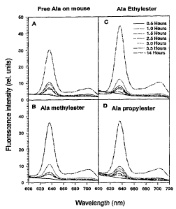
FicTUre 1 shows fluorescence intensity (relative units vs
wavelength (nm)) of PpIX in the normal skin of mice
after topical administration of
(A) free ALA
(B) ALA methylester
(C) ALA ethylester
(D) ALA propylester
after 0.5, 1, 1.5, 2.5, 3, 3.5 -and 14 hours following
administration;
Firnzre 2 shows the distribution of PpIX as measured by
fluorescence intensity (relative units vs wavelength
(nm)) in Brain, dermis, Ear, Liver and muscle 14 hours
after topical administration to the normal skin of mice:
(A) free ALA
(B) ALA methylester
(C) ALA ethylester
(D) ALA propylester;
Figure 3 shows PpIX fluorescence (fluorescence intesity,
relative units vs wavelength (nm)) in the skin of mice
CA 02215069 1997-09-09
R'O 96/28412 PGT/GB96/00553
- 18 -
15 minutes, 1 hour, 4 hours and 10 hours after
intraperitoneal injection of ALA methylester (150
mg/kg) ; '
Figure 4 shows PpIX fluorescence (fluorescence intensity
relative units vs wavelength (nm)) (A) 1.5 hours and (B)
4 hours after topical administration of-ALA methylester
to basal cell carcinoma (BCC) lesions on the skin of
human patients (- tumour; --- normal skin);
Figure 5 shows PpIX fluorescence (fluorescence intensity
relative units vs wavelength (nm)) (A) 1.5 hours and (B)
4 hours after topical administration of ALA ethylester
to basal cell carcinoma (BCC) lesions on the skin of
human patients (- tumour; --- normal skin);
Figure 6 shows PpIX fluorescence (fluorescence intensity
relative units vs wavelength (nm)) (A) 1.5 hours and (B)
4 hours after topical administration of ALA propylester
to basal cell carcinoma (BCC) lesions on the skin of
human patients (- tumour; --- normal skin);
Figure 7 shows PpIX fluorescence (fluorescence intensity
relative units vs wavelength (nm)) (A) 1.5 hours and (B)
4 hours after topical administration of ALA to basal
cell carinoma (BCC) lesions on the skin of human
patients (- tumour; --- normal skin);
Figure 8 shows measurement of PpIX production following
topical application of ALA methylester in human BCC and
surrounding normal skin by CDD microscopy of biopsies
(A) graphical representation showing fluorescence
intensity vs depth (~.~.m) and (B) micrograph;
FiQUre 9 shows measurement of PpIX production following
topical application of ALA in human BCC and surrounding
CA 02215069 1997-09-09
WO 96/28412 PCTlGB96/00553
- 19 -
normal skin by CDD microscopy of biopsies (A) graphical
representation showing fluorescence intensity vs depth
(/.~.m) and (B) micrograph;
F'~g~ure 10 shows PpIX fluorescence (fluorescence
intensity relative units vs wavelength (nm)) 24 hours
following topical administration of ALA methylester to
BCC lesion and to normal skin of human patients.
Ficrure ~.1 shows PpIX fluorescence (fluorescence
intensity relative units vs wavelength (nm)) 24 hours
following topical administration of ALA to BCC lesion
and to normal skin of human patients.
~aure 12 shows measurement of PpIX production 4.5 hours
following topical application of ALA methylester in
human BCC by CDD microscopy of biopsies (A) graphical
representation showing fluorescence intensity vs depth
(gym) and (B) micrograph;
F'ig2re 13 shows measurement of PpIX production 4.5 hours
following topical application of ALA methylester in
human normal skin by CDD microscopy of biopsies (A)
graphical representation showing fluorescence intensity
vs depth (/Cm) and (B) micrograph;
F;cturP 14 shows measurement of PpIX production 24 hours
following topical application of ALA methylesterin
human BCC by CDD microscopy of biopsies (A) graphical
representation showing fluorescence intensity vs depth
(~.cm) and (B) micrograph;
~<zure 15 shows measurement of PpIX production 24 hours
following topical application of ALA methylester in
human normal skin by CDD microscopy of biopsies (A)
graphical representation showing fluorescence intensity
vs depth (gym) and (B) micrograph;
CA 02215069 1997-09-09
WO 96/28412 PCT/GB96/00553
- 20 -
Figure 16 shows measurement of PpIX production 24 hours
following topical application of free ALA in human BCC
by CDD microscopy of biopsies (A) graphical '
representation showing fluorescence intensity vs depth
(um) and (B) micrograph; '
Figure 17 shows measurement of PpIX production 24 hours
following topical application of free ALA in human
normal skin by CDD microscopy of biopsies (A) graphical
representation showing fluorescence intensity vs depth
(~.m) and (B) micrograph;
Figure 18 shows measurement of PpIX production 4.5 hours
following topical application of free ALA and 20o DMSO
in human BCC by CDD microscopy of biopsies (A) graphical
representation showing fluorescence intensity vs depth
(E.r.m) and (B) micrograph;
Figure 19 shows measurement of PpIX production 4.5 hours
following topical application of free ALA and 20o DMSO
in human normal skin by CDD microscopy of biopsies (A)
graphical representation showing fluorescence intensity
vs depth (~.m) and (B) micrograph;
Figure 20 shows a time course (fluorescence intensity
relative units vs time (hours)) of ALA methylester-
induced (PpIX) fluorescence in the mouse skin after
topical application of ALA methylester alone (-~-). AT-'A
methylester plus DMSO (-~-), ALA methylester plus
desferrioxamine (DF) (-~-) or ALA methylester plus DF
and DMSO (-~-). Each point is the mean of measurements
from at least three mice;
Figure 21 shows fluorescence photographs of the mouse
skin taken 1 h after topical application of free ALA
alone (A), ALA methylester (B), ALA ethylester (C) and
ALA propylester (D), showing fluorescence in the
CA 02215069 1997-09-09
WO 96/28412 PCT/GB96/00553
- 21 -
epidermis (Ep), epithelial hair follicles and sebaceous
gland (arrows), but not in the dermis (De). Original
magnification x250.
~cTUre 22 is a graph showing relative tumour volume
against time (days) following treatment of WiDr human
colonic carcinoma transplanted subcutaneously into nude
mice with ALA or ALA methylester plus DF; (-~-) control;
-) DF alone; (-~-) ALA + DF + DMSO; (-~-) ALA
methylester + DF + DMSO.
FicTUre 23 shows PpIX fluoresence ratios between BCC
lesions and surrounding normal skin after topical
application of ALA or its esters.
Example 1
Pr a m 1 m' r hl
To a 500 ml glass reactor containing 200 ml methanol,
was added 1g 5-amino=levulinic acid hydrochloride and 1
drop conc. HCl. The reaction mixture was then stirred
overnight at 60°C. The progress of the esterification
was followed by 1H-NMR. Excess methanol was removed by
distillation, and the product further dried under vacuum
at 30-40°C, giving methyl 5-aminolevulinate
hydrochloride. The structure was confirmed by '-H-NMR in
DMSO-d6 .
1g 5-aminolevulinic acid hydrochloride was added to 200
ml dry ethanol containing 1-2 drops conc. hydrochloric
acid in a 250 ml glass reactor equipped with a stirrer,
reflux condenser and a thermometer. The esterification
was performed at reflux overnight (70-80°C). After the
CA 02215069 1997-09-09
WO 96J28412 PCTlGB96l00553
- 22 -
reaction had gone to completion, the ethanol was removed
under vacuum. Finally, the product was dried under high
vacuum at 30-40°C, giving 0.948 Ethyl 5-aminolevulinate
hydrochloride. Confirmation of the structure was done
by 1H-NMR in DMSO-ds .
Example 3
Preparation of n-prop~rl 5-aminolevulinate hydrochloride
(ALA propylester)
0.5g 5-aminolevulinic acid hydrochloride was dissolved
in 100 ml dry n-propanol containing 1-2 drops of cone.
hydrochloride in a 250 ml glass reactor equipped with a
stirrer, reflux condenser and a thermometer The
reaction mixture was stirred at 70-80°C for approx. 20
hours. After all the 5-aminolevulinic acid was
converted to its n-propylester (followed by 1H-NMR), the
excess propanol was removed, and the product dried under
high vacuum (<1 mBar) at 40-50°C. The reaction gave
0.498 propyl 5-aminolevulinate hydrochloride. The
structure was confirmed by '-H-NMR in DMSO-d6.
Example 4
Preparation of n-hexyl 5-aminolevulinic hydrochloride
(ALA hexvlester)
2 grams of 5-aminolevulinic acid hydrochloride was
dissolved in 25 grams of dry n-hexanol with 5-6 drops of
cone. hydrochloride added in a 50 ml glass reactor
equipped with a reflux condenser and a thermometer. The
reaction mixture was held at 50-60°C for approx. 3 days.
The excess n-hexanol was removed under vacuum and the
product finally dried under high vacuum, giving 2.4
grams of n-hexyl 5-aminolevulinate hydrochloride. The
structure was confirmed by 1H-NMR spectroscopy in DMSO-
ds.
CA 02215069 1997-09-09
WO 96/28412 PCT/GB96/00553
- 23 -
Example 5
f r.
(AT,.A he~t~ylester)
0.5g 5-aminolevulinic acid hydrochloride was added to 30
grams of n-heptanol containing 5 drops of conc.
hydrochloride in a 100 ml glass reactor equipped with a
stirrer, reflux condenser and a thermometer. After all
the 5-aminolevulinic acid had dissolved, the reaction
mixture was stirred at 70-80°C for approx. 48 hours.
After the 5-aminolevulinic acid was converted to its n-
heptyl ester (followed by 1H-NMR), the excess alcohol was
removed, and the product dried under high vacuum (c 1
mBar) at 70°C. The reaction gave 1.5g n-heptyl 5-
aminolevulinate hydrochloride. The structure was
conffirmed by 1H-NMR in DMSO-d6.
Example 6
r n n- r hl
octylester)
1 gram 5-aminolevulinic acid hydrochloride was added to
30 grams of dry n-octanol containing 5-6 drops of conc_
hydrochloride in a 50 ml glass reactor equipped with a
reflux condenser, stirrer and a thermometer. The
reaction mixture was stirred at 65-70°C for approx. 2
days. Excess n-octanol was removed under vacuum and the
product finally dried under high vacuum, giving 1.5
grams of n-octyl 5-aminolevulinate hydrochloride. The
structure was confirmed by 1H-NMR spectroscopy in DMSO-
ds.
20% creams were prepared by admixture of the active
component, ALA, ALA methylester, ALA ethylester, or ALA
CA 02215069 1997-09-09
WO 96/28412 PC'T/GB96/00553
- 24 -
propylester (prepared according to Examples 1 to 3
respectively), with "Urguentum Merck" cream base
(available from Merck) consisting of silicon dioxide,
paraffin liq., vaseline, album, cetostearol.,
polysorbat. 40, glycerol monostearate, Miglyol°812 (a -
mixture of plant fatty acids), polypropyleneglycol., and
purified water.
Corresponding creams were also prepared, additionally
containing 3-20o DMSO.
Example 8
Determination of protoporphyrin IX production in the
skin of mice by CCD microscopy of biopsies:
A commercial oil-in-water cream containing (20o w/w) one
of the chemicals (free ALA, ALA methylester, ALA
ethylester and ALA propylester) (see Example 1) was
t-opically applied to the normal skin of nu/nu nude mice
for 0.5, 1, 3 and 6 hours, then biopsied and evaluated
by means of microscopic fluorescence photometry
incorporating a light-sensitive thermol-electrically
cooled charge coupled device (CCD) camera. The results
show that free ALA and its three ester derivatives are
taken up by the skin tissue, the esterified ALA
derivatives are being deesterified in the skin, and
converted into protoporphyrin IX (PpIX) 0.5 hours after
topical application. The fluorescence intensity of PpIX
in the skin increased with the time of the application
and the maximum amounts of the fluorescence were seen
about 6 hours (the latest time point studied) after the
application in all cases.
CA 02215069 1997-09-09
WO 96/28412 PCT/GB96/00553
- - 25 -
The aim of this study was to investigate the build-up of
esterified ALA ester-induced porphyrins fluorescence in
the normal skin of nude mice ~, vivo after topical or
systemic administration of ALA ester derivatives.
Chemicals. 5-aminolevulinic acid (ALA) methyl-, ethyl-
and propyl-esters (HZN-CH2COOCH2-CH2C00-R; R can be CH3,
CH2-CH2-CH3) were prepared by Norsk Hydro Research Center
(Porsgrunn, Norway) as described in Examples 1 to 3.
Free ALA hydrochloride and desferrioxamine mesylate (DF)
were purchased from Sigma Chemical Company (St. Louis,
Mo, USA). Dimethyl sulphoxide (DMSO) was obtained from
Janssen Chimica (Geel, Belgium). Commercial oil-water
creams (Unguentum Merck, Darmstadt, Germany) containing
200 one of the ALA ester derivatives (w/w), 20% free
AT.A_, 20o ALA methylester plus 5o DF, 20o ALA methylester
plus 20o DMSO, or 20o ALA methylester plus 5o DF and 200
DMSO were freshly prepared prior to use. All creams
were made by the Pharmacy at the Norwegian radium
Hospital. For intraperitoneal injection, ALA and its
methylester were freshly dissolved in saline. All other
chemicals used were of the highest purity commercially
available.
Animals. Female Balb/c nu/nu athymic nude mice were
obtained from the Animal Laboratory at the Norwegian
Radium Hospital and kept under specific-pathogen-free
conditions. At the start of the experiments the mice
were 6-7 weeks old weighing 18-24 g. Three mice were
housed per cage with autoclaved covers in a dark room
during the experiments.
'Treatment r~roced»r-P . One of the creams was painted on
the normal skin at right flank region of each mouse, and
covered by a semi-permeable dressing (3M, St Paul, MN,
CA 02215069 1997-09-09
WO 96/28412 PCT/GB96/00553
- 26 -
USA) for various time intervals (from 0.25 to 24 h)
before fluorescence measurements in situ or being
biopsied for microscopic fluorescence imaging. About
0.2g cream was applied to an approximate 2.25 cm2 area of
the skin. In the case of i.p. injection the mice were
given ALA or its methylester at a dose of 150 mg/kg. At
least three mice were used for each condition.
F r A
perkin Elmer LS-50 fluorescence spectrometer equipped
with a red-sensitive photomultiplier (Hamamatsu R 928)
was used. This instrument has a pulsed Xenon arc light
source and phase sensitive detection, such that
fluorescence can be readily measured. Part of the
excitation beam (set at 408 nm for fluorescence
measurements) was reflected into a 600 ~m core
multimodus optical quartz fiber (No. 3501 393, Dornier
Medizintechnik, GmbH, Germering, Germany) by means of a
mirror for application onto the subject through a hand
held probe. Emission in the region of 550-750 nm was
measured via emission fibres collecting information
through the probe.
Fluorescence microscopy. After the creams were
topically applied to the skin of mice for various times
(as indicated above), the skin was biopsied and the
frozen tissue sections were cut with a cryostat to a
thickness of 8 E.cm. The fluorescence microscopy was
carried out using an Axioplan microscope (Zeiss,
Germany) with a 100 W mercury lamp. The fluorescence
images were recorded by a light-sensitive thermo-
electrically cooled charge coupled device (CCD) camera
(resolution: 385x578 pixels with a dynamic range of 16
bits per pixel)(Astromed CCD 3200, Cambridge, UK) and
hard copies on a video printer (Sony multiscan video
printer UP-930). The filter combination used for
detection of porphyrin fluorescence consisted of 390-440
CA 02215069 1997-09-09
WO 96/28412 PCT/GB96/00553
- 27 -
nm excitation filter, a 460 nm beam splitter and a >600
nm emission filter.
~2Pa"~ r~
PpIX fluorescence was measured in situ by an optical-
fiber based system in the normal skin of nude mice 0.5,
1, 1.5, 2.5, 3, 3'.5 and 14 hours after topical
application of free ALA or one of its ester derivatives
as described above. As shown in Figure 1, the PpIX
fluorescence was already built-up 1 hour after topical
application in the case of all derivatives, while the
fluorescence was seen 1.5 hours after the application of
free ALA. The maximum fluorescence intensity was found
14 hours after the application in all cases, but PpIX
fluorescence induced from ALA esters in the skin was
stronger than that from free ALA. Furthermore, as can
be seen in Figure 2, 14 hours after the application no
fluorescence of ALA-esters-induced PpIX was detected in
other areas of the skin and internal organs including
ear, dermis, muscle, brain and liver. However, in the
case of free ALA, a strong fluorescence was also seen in
the ear as well as in the other areas of the skin.
Thus, after topical application ALA-ester-induced PpIX
was found locally in the skin, whereas free ALA-induced
PpIX distributed not only locally, but also in other
areas of the skin. We suggest that ALA is transported
in the blood and that PpIX is subsequently formed in all
organs containing the enzymes of the heme synthesis
pathway and/or PpIX is formed in the skin and then
transported to other tissues via blood circulation. The
latter situation may lead to skin photosensitivity in
areas where free ALA is not topically applied. In
addition, after intraperitoneal injection of ALA
methylester at a dose of 150 mg/kg, the PpIX
fluorescence in the skin of mice was built-up 15 minutes
after the injection and the peak value was found around
CA 02215069 1997-09-09
WO 96/28412 PCT/GB96/00553
- 28 -
4 hours, and the fluorescence disappeared within 10
hours post the injection (Figure 3). This kinetic
pattern is similar to that of the fluorescence of free
pT~A_-induced porphyrins in the skin following i.p.
injection of the same dose, although the fluorescence ,
decreased faster in the case of the ester than in the
case of the free ALA.
Examxale 10
"rtPasu~-ements of protopornhyrin IX production in human
1 r 'n a B n orm 1 s 'n
by optical-fiber based svstem
The PpIX fluorescence in the BCC lesions and surrounding
normal skin of human patients was measured in situ by
optical-fiber based system after topical application of
20o free ALAand its derivatives for various time
intervals.
Figures 4, 5, 6 and 7 show that, compared to free ALA,
the ALA derivatives-induced PpIX was built up faster,
produced more and localized more selectively in the BCC
lesions (i.e. much less fluorescence in the surrounding
normal skin), particularly for ALA methylester.
Example 11
I r s m n s o
- h BCC d surrou ndin normal skin
~ by
on in uman an q,
pY of b~ ies
oduct ops
c''~-'D microsco~v
In a 78 years old Caucasian male presenting multiple
ulcero-nodular BCCs lesions were exposed to commercial
oil-in-water-creams containing either ALA alone (20%
w/w) or ALA methyl ester (20% w/w) (as described in
Example 7) covered by a semi-permeable dressing for 24
hours. After removal of dressings and cream in vivo
fluorescence was measured at the surface of tumor tissue
CA 02215069 2003-08-13
- 29 -
and adjacent normal skin by means of a spectrofluorometer.
Punch biopsies of the same areas were removed and samples were
immediately immersed in liquid nitrogen. The tissue sections
were cut with a cryostat microtome to a thickness of 8 Vim.
The localization pattern of the porphyrin fluorescence in the
tissue sections was directly observed by means of fluorescence
microscopy. The same frozen sections were subsequently
stained with routine H&E staining for histological
identification. Fluorescence microscopy was carried out with
an Axioplan microscope (Zeiss, Germany). Fluorescence images
and quantitative measurements were performed by a light-
sensitive thermol-electrically cooled charge coupled device
(CCD) camera (Astromed CCD 3200, Cambridge, UK) and an image
processing unit (Astromed/Visilog, PC 486DX2 66 MHz VL). The
main purpose for such quantitative measurements is to
determine the exact penetration of ALA-induced porphyrins from
tissue surface to the bottom layers of cancer lesions. The
results are shown in Figures 8 and 9 in which the fluorescence
intensity is expressed as a function of depth of cancer
lesion.
As shown in Figures 8 and 9, a homogeneous distribution of
PpIX fluorescence is seen from the top to the bottom of the
whole BCC lesions after use of either free ALA or its
methylester. This suggests that ALA methylester is at least
as good as free ALA in terms of penetration and PpIX
production in the BCC lesion. In addition, no PpIX
fluorescence was seen in the surrounding normal skin after
topical application of ALA methylester, indicating that ALA-
methylester-induced PpIX highly selectively took place only in
the BCC lesion.
In vivo fluorescence' after 29 hours showed at least
CA 02215069 2003-08-13
- 30 -
doubled fluorescence intensity for ALA methyl ester
compared to ALA for the selected tumors and also an
increase for corresponding norTnal tissues, however this
only of about SOo. The ratio between tumor and normal
tissue was about 1.2: 1 for ALA and 2: 1 for the ALA
methyl ester. The results are shown in Figures 10 and
11.
At control one week after treatment all treatment fields
presented a central necrotic area corresponding to the
tumor. In the adjacent normal skin exposed to cream and
light irradiation there was observed a marked erythema
for the ALA while for the ALA methyl ester only moderate
erythema was observed.
Example 12
v'
CCD microscopx of biopsies
The present data show the localization patterns and
production of porphyrins (mainly protoporphyrin IX
(PpIX)) after topical application of free ALA and one of
its derivatives (methyl ester) f or 4.5 and 24 hours in
the nodular basal cell carcinomas (BCCs) and surrounding
normal skin of patients. The tests were performed as
described in Example 11.
Each of the following figures show both (B) fluorescence
images of either the bottom layer of BCC lesions or of
the surrounding normal skin. Curves indicating the
fluorescence intensity as a function of depth of the BCC
lesions or of the normal skin are also shown (A) .
Figure 12 shows a homogenous distribution of PpIX
fluorescence generated by ALA methyl ester in the bottom
CA 02215069 2003-08-13
- 31 -
layer of a BCC 4.5 hours after topical application.
There is also some porphyrin fluorescence in surrounding
normal skin (Figure 13). The fluorescence intensity
ratio between BCC and the normal skin is about 2.
Moreover, the absolute amount of the fluorescence
induced by ALA methyl ester is higher than that induced
by free ALA and 20% DMSO after topical application for
4.5 hours, as shown below.
Figures 14 and 15 show a uniform distribution of
porphyrin fluorescence induced by topical application of
ALA methyl ester for 24 hours in the bottom layer of BCC
and surrounding normal skin. The ratio of the
fluorescence in BCC and that in normal skin is also
about 2. Furthermore, the fluorescence intensity of ALA
methyl ester-induced porphyries in the BCC is almost
twice as high as that in BCC after topical application
of free ALA alone for 24 hours, as shown below.
Figures 16 and 17 show a homogenous distribution of free
ALA-induced porphyries in the bottom layer of BCC and
surrounding normal skin 24 hours following topical
application. However, the ratio of the fluorescence
intensity between BCC and normal skin is about 1, which
indicates a low selectivity of this treatment. Moreover
the production of porphyries in BCC is less than that in
the case of ALA methyl ester.
Figures 18 and 19 show a homogenous distribution of ALA-
induced porphyries in the bottom layer of BCC and
surrounding normal skin after topical application of
free ALA and 20% DMSO for 4.5 hours. However, the ratio
of the fluorescence intensity between BCC and normal
skin is only slightly larger than 1, which demonstrates
that the DMSO probably reduces the tumor selectivity of
the porphyries produces. Moreover, also in this case
less porphyries are produced in BCC than in the case of
CA 02215069 1997-09-09
WO 96128412 PGT/GB96/00553
- 32 -
the application of ALA methyl ester.
Example 13 -
nv a 'na a
desfer~-ioxamine (DF) and/or DMSO and fluorescence of .
skin
I. The effect of DF and/or DMSO on the build up of
fluorescence in the normal skin of mice in situ was
ascertained various times after topical administration
of ALA-methylester. Methods were performed as descrir~ed
in Example 9.
Topical application of the cream alone containing only
DMSO did not show any fluorescence in the normal mouse
skin, but there was some fluorescence of PpIX after DF
alone was applied.
DF or DF plus DMSO (a well-known skin penetration
enhancer) significantly enhanced the production of ALA
methylester-induced PpIX.
II. Fluorescence imaging of the skin treated with three
derivatives (performed as described in Example 9) showed
fluorescence of the ester derivative-induced porphyrins
in the epidermis, epithelial hair follicles and
sebaceous glands 1 h after topical application (Figure
21). The fluorescence intensity of the porphyrins
increased with the time after the application.
A large number of patients with basal cell carcinomas
(BCCs) has topically been treated with ALA-based PDT in
our hospital during the past five years and more than
90~ of superficial BCCs have shown a complete
CA 02215069 1997-09-09
WO 96/28412 PCT/GB96/00553
- 33 -
regression. However, nodular BCCs had a low complete
response rate due to a poor ALA retention and,
consequently, a low ALA-induced porphyrin production in
the deep layers of the lesions. In order to improve the
technique, we used ALA ester derivatives instead of free
AT.A_. The present data obtained presented in this
Example and in Example 9 by means of both fluorescence
spectroscopic measurements ~n sltll and fluorescence
microscopy of tissue biopsies, indicate that all three
ester derivatives studied were taken up, de-esterified
and finally converted into porphyrins in the epidermis,
epithelial hair follicles and sebaceous glands of the
nude mice with a higher porphyrin production than that
of free ALA. This is in agreement with the preceding
Examples concerning a study of human nodular basal cell
carcinoma that demonstrate that the fluorescence of the
ALA ester-induced porphyrins was built up faster with a
higher intensity and a more homogenous distribution than
those of free ALA-induced porphyrins in the lesions.
The present study also shows that DF has a significant
effect in enhancing the production of ALA methylester-
derived PpIX in the normal skin of the mice after
topical application.
Interestingly, a strong fluorescence of free ALA-induced
porphyrins was found in regions of the skin outside the
area where the cream was topically applied (Figure 2).
This indicates that after topical application free ALA
is transported in the blood and porphyrins are
subsequently formed in all organs containing the enzymes
of the heme synthesis pathway or porphyrins are
initially formed in the skin or/and liver, then
transported to other tissues via blood circulation.
This may lead to skin photosensitivity in areas where
free AIjA is even not topically applied. However, none
of the ester derivatives studied induced porphyrin
CA 02215069 1997-09-09
R'O 96/28412 PCT/GB961005~3
- 34 -
fluorescence in other parts of the skin.
Examgle 14
Effects of ALA meth3rlester or ALA DF and DMSO PDT on
t~.mor growth in W'Dr human colonic carcinoma
~rar~splanted nude mice
Nude-mice were transplanted with WiDr human colonic
carcinoma cells by subcutaneous injection into the right
flank region. The following creams, formulated as
described in the preceding Examples, were applied
topically to the site of the tumor: 10% DF alone; 20%
ALA + 10% DF + 20% DMSO; or 20% ALA methylester + 10% DF
+ 20% DMSO, followed, 14 hours later by laser light
irradiation (632 nm, 150 mW/cm2 for 15 minutes). A
separate group of animals bearing the same tumor model,
but receiving no topical application of the cream,
served as a control. The responses of the treated
tumors were evaluated as tumor regression/regrowth time.
When the tumors reached avolume 5 times that of the
volume on the day of light irradiation, the mice were
killed. The results are shown in Figure 22. (Bars:
standard error of mean (SEM) based on 3-5 individual
animals in each group). The results show that it took
34 days for tumors treated with ALA methylester + DF +
DMSO to reach a volume five times that of the volume on
the day just before light irradiation, whereas in the
case of free ALA + DF + DMSO it took 24 days for the
treated tumors to grow to 5 times size. Thus, ALA
methylester is more effective than ALA in slowing tumor
regrowth.
CA 02215069 1997-09-09
R'O 96/28412 PCT/GB96/00553
- 35 -
The PpIX fluorescence ratios between BCC lesions and
surrounding normal skin after topical application of AT.A
or its esters (20% for 4 hours), was examined using
methods described in previous examples. The results are
shown in Fig.23 and indicate that all esters can more
selectively induce PpIX in BCC lesions than free ALA,
particularly in the case of ALA-methylester and ALA-
hexylester.