Note: Descriptions are shown in the official language in which they were submitted.
CA 02215392 2005-02-24
INFLATABLE DEVICES FOR SEPARATING LAYERS OF TISSUE
Background of the Invention
The present invention relates to the field of inflatable tissue separation
devices and methods
of using such devices. The apparatus and methods of the present invention may
be used in any
procedure requiring dissection andlor retraction of tissue planes throughout
the body including
inguinal hernia repair, pelvic lymphadenectomy and bladder neck suspension in
the preperitoneal
space; renal, adrenal, aortic and anterior spinal access in the
retroperitoneal space; penile prosthetic
reservoir placement in the anterior abdominal wall; and augmentation
mammaplasty prosthetic
placement. By way of example only, use of such devices and methods for hernia
repair will be
described.
A hernia is the protrusion of part of a body part or structure through a
defect in the wall of a
surrounding structure. Most commonly, a hernia is the protrusion of part of
abdominal contents,
including bowel, through a tear or weakness in the abdominal wall, or through
the inguinal canal into
the scrotum.
An abdominal hernia is repaired by suturing or stapling a mesh patch over the
site of the tear
or weakness. The mesh patch has a rough surface that can irritate the bowel
and cause adhesions. It
is therefore preferred to install the patch properitoneally. It is intended
that the terms properitoneal
and preperitoneal by synonymous. The mesh patch is preferably attached to the
properitoneal fascia
of the abdominal wall, and covered by the peritoneum. To attach the mesh patch
to the properitoneal
fascia, the peritoneum must be dissected from the properitoneal fascia. This
is a difficult process
which involves the risk of puncturing the peritoneum. Moreover, strands of
properitoneal fat
interconnecting the peritoneum and the properitoneal fascia make it difficult
to see the site of the
hernia.
The use of laparoscopic techniques to perform hernia repair is becoming
increasingly
common. In the conventional procedure for carrying out a hernia repair
laparoscopically, an
endoscope and instruments are introduced into the belly through one of more
incisions in the
abdominal wall, and are advanced through the belly to the site of the hernia.
Then, working from
inside the belly, a long incision is made in the peritoneum covering the site
of the hernia. Part of the
peritoneum is dissected from the properitoneal fat layer to provide access to
the fat layer. This is
conventionally done by blunt dissection, such as by sweeping a rigid probe
under the peritoneum. In
this procedure, it is difficult to dissect the peritoneum cleanly since patchy
layers of properitoneal fat
tend to adhere to the peritoneum.
-1-
CA 02215392 2005-02-24
In an alternative known laparoscopic hernia repair procedure, the belly is
insufflated. An
incision is made in the abdominal wall close to the site of the hernia. The
incision is made through
the abdominal wall as far as the properitoneal fat layer. The peritoneum is
then blunt dissected from
the properitoneal fat layer by passing a finger or a rigid probe through the
incision and sweeping the
finger or rigid probe under the peritoneum. After the peritoneum is dissected
from the properitoneal
fat layer, the space between the peritoneum and the properitoneal fat layer is
insufflated to provide a
working space in which to apply the mesh patch to the properitoneal fascia.
During the blunt dissection process, it is easy to puncture through the
peritoneum, which is
quite thin. Additionally, after initial dissection of the properitoneal space,
known surgical procedures
require introduction of various instruments in the space to conduct the
surgery. These instruments can
cause inadvertent puncture of the peritoneum wall after the initial
dissection. A puncture destroys the
ability of the space between the peritoneum and the fascia to hold gas
insufflation; pressurized gas can
travel through a puncture in the peritoneum to allow the fluid to migrate to
the abdominal cavity and
degrade the pressure differential maintaining the properitoneal cavity. Also,
it is difficult to dissect
the peritoneum cleanly since patchy layers of properitoneal fat tend to adhere
to the peritoneum.
Clearing difficult adhesions can sometimes result in a breach of the
peritoneum itself.
United States Patent No. 5,309,896, discloses a laparoscopic hernia repair
technique that
enables a mesh patch to be attached to the properitoneal fascia without
breaching the peritoneum. An
incision is made through the abdominal wall as far as the properitoneal fat
layer. A mufti-chambered
inflatable retraction device is pushed through the incision into contact with
the peritoneum, and is
used to separate the peritoneum from the underlying layers. The main end
chamber of the inflatable
retraction device is then inflated to elongate the inflatable retraction
device towards the site of the
hernia. As it inflates, the inflatable retraction device generally separates
the peritoneum from the
underlying layers. Once the main chamber of the inflatable retraction device
is fully inflated, a
second inflatable chamber is inflated. The second inflatable chamber enables
the inflatable retraction
device to continue to separate the peritoneum from the underlying layers after
the main inflatable
chamber has been deflated.
One or more apertures are then cut in the envelope of the main inflatable
chamber to provide
access to the site of the hernia for instruments passed into the main chamber.
With such an
arrangement, instruments pass through the main chamber situated between the
peritoneum and the
underlying layers. In this way, a patch can be attached to the properitoneal
fascia without breaching
the peritoneum.
-2-
CA 02215392 2005-02-24
Another device for separating tissue layers is disclosed in U.S. Patent No.
5,468,248 (which
corresponds to PCT International App. Publication No. WO 93/09722). This
apparatus includes a
main envelope that defines a main inflatable chamber, and also includes an
introducing device for
introducing the main envelope in a collapsed state between the first layer of
tissue and the second
layer of tissue. The introducing device inflates the main envelope into an
expanded state to separate
the first layer of tissue from the second layer of tissue, and to create a
working space between the first
layer of tissue and the second layer of tissue. Finally, the apparatus
includes an insufflating device for
introducing insufflation gas into the working space between the first layer of
tissue and the second
layer of tissue.
In a method according to PCT International App. Publication No. WO 93/09722,
of
separating a first layer of tissue from a second layer of tissue, a main
envelope and insufflation gas are
provided. The main envelope defines a main inflatable chamber. The main
envelope is introduced in
a collapsed state between the first layer of tissue and the second layer of
tissue. The main envelope is
inflated into an expanded state to separate the first layer of tissue from the
second layer of tissue, and
to create a working space between the first layer of tissue and the second
layer of tissue. Finally,
insufflation gas is introduced into the working space between the first layer
of tissue and the second
layer of tissue.
In a first practical embodiment of an apparatus according to PCT International
App.
Publication No. WO 93/09722, the main envelope and the introducing device
constitute a first
component that separates the first layer of tissue from the second layer of
tissue to create the working
space. The insufflation device constitutes a second component, which
insufflates the working space
to maintain the separation of the first layer of tissue from the second. The
insufflation device is
tubular, has an anchor
-2a-
CA 02215392 1997-09-15
flange slidably mounted on it, and has a toroidal inflat~ble cha-r.ber at its
distal Pra. ThP anchor flange ;
and toroidal inflatable chamber together form a gas-tight seal with the second
layer of tissue.
In a method according to PCT International App. Publication No. WO 93/09722 of
using the two-
component apparatus, the introducing device is used to push the main envelope
in a collapsed state
through an incision through the second layer of tissue to place the main
envelope between the first layer
of tissue and the second layer of tissue. The main envelope is then inflated
to gently separate the first
layer of tissue from the second layer of tissue, and to create a working space
between the two layers of
tissue. An endoscope may be passed through the bore of the introducing device
into the main chamber to
observe the extent of separation of the layers of tissue. The main envelope is
then returned to a
collapsed state, and the main envelope and the introducing device are removed
from the incision.
The insufflating device is inserted into the incision so that its distal end
projects into the working
space between the two layers of tissue. The toroidal inflatable chamber is
inflated into an expanded state.
The anchor flange is slid distally along the insufflating device to compress
the second layer of tissue
between it and the expanded toroidal inflatable chamber, and thus to form a
gas-tight seal. Insufflating
gas is then passed through the insufflating device into the working space to
maintain the separation of the
first layer of tissue from the second. An endoscope may be passed through the
bore of the insufflating
device into the working space to observe within the working space.
In a first embodiment of a one-component apparatus according to PCT
International App.
Publication No. WO 93/09722, the introducing device is also used for returning
the main envelope to a
collapsed state. A single elongated tube provides the introducing device and
the insufflating device. The
main envelope is detachable from the single elongated tube. The single
elongated tube has an anchor
flange slidably mounted on it, and has a toroidal inflatable chamber at its
distal end. The anchor flange
and toroidal inflatable chamber together form a gas-tight seal with the second
layer of tissue.
In a method according to PCT International App. Publication No. WO 93/09722 of
using the first
embodiment of a one-component apparatus to separate a first layer of tissue
from a second layer of
tissue, the elongated tube is used to push the main envelope in a collapsed
state through an incision
through the second layer of tissue to place the main envelope between the
first layer of tissue and the
second layer of tissue. The main envelope is then inflated to gently separate
the first layer of tissue from
the second layer of tissue, and to create a working space between the two
layers of tissue. An endoscope
may be passed through the bore of the single elongated tube into the main
chamber to observe the extent
of separation of the layers of tissue. The main envelope is then returned to a
collapsed state, detached
from the elongated tube, and removed from the working space between the layers
of tissue through the
bore of the elongated tube. The toroidal inflatable chamber at the distal end
of the elongated tube is then
inflated into an expanded state. The anchor flange is slid distally along the
elongated tube to compress
the second layer of tissue between it and the expanded toroidal inflatable
chamber to form a gas-tight
seal. Insufflating gas is passed through the elongated tube into the working
space to maintain the
separation of the first and second tissue layers. An endoscope may be passed
through the bore of the
single elongated tube into the working space to observe within the working
space.
In a second embodiment of a one-component apparatus according to PCT
International App.
Publication No. WO 93/09722, the introducing device is an outer elongated
tube, and the insufflating
device is an inner elongated tube mounted in the bore of the outer elongated
tube. The proximal ends of
the tubes are flexibly coupled together. The main envelope is a cylindrical
piece of elastomeric material.
One end of the main envelope is evened with respect to the other, and is
attached to the distal end of the
outer elongated tube. The other end of the main envelope is attached to the
distal end of the inner
elongated tube. The main inflatable chamber defined by the main envelope is
thus substantially toroidal.
The outer elongated tube has an anchor flange slidably mounted on it. The
anchor flange and the main
inflatable chamber together form a gas-tight seal with the second layer of
tissue.
-3-
sLISSTm slur A~~~ENDED S~Et~'
CA 02215392 1997-09-15
In a method according to PCT International App. Publication Drc. WO 93/09'/22
0~ using the
second embodiment of a one-component apparatus to separate a first layer of
tissue from a second layer
of tissue, the outer elongated tube is used to push the main envelope in a
collapsed state through an
incision through the second layer of tissue to place the main envelope between
the first layer of tissue
and the second layer of tissue. The main envelope is then inflated to gently
separate the first layer of
tissue from the second layer of tissue, and to create working a space between
the layers of tissue. An
endoscope may be passed through the outer elongated tube into the main chamber
to observe the extent
of separation of the layers of tissue. The anchor flange is slid distally
along the introducing device tube
to compress the second layer of tissue between it and the main inflatable
chamber, to form a gas-tight
seal. Insufflating gas is then passed through the bore of the inner elongated
tube and the bore of the
main envelope into the working space to maintain the separation of the first
layer of tissue from the
second. An endoscope may be passed through the bore of the inner elongated
tube and the bore of the
main envelope into the working space to observe within the working space.
In a further method according to PCT International App. Publication No. WO
93/09722, access
through the abdominal wall to repair a hernia is provided. The abdominal wall
includes the peritoneum
and an underlying layer. A main envelope and an insufflation gas are provided.
The main envelope
defines a main inflatable chamber. The main envelope is introduced in a
collapsed state between the
peritoneum and the underlying layer. The main envelope is inflated into an
expanded state to separate
the peritoneum from the underlying layer, and to create a working space
between the peritoneum and the
underlying layer. Insufflation gas is introduced into the working space, and
the hernia is repaired using
an instrument passed into the working space.
In a final method according to PCT International App. Publication No. WO
93/09722, access is
provided through the abdominal wall from near the umbilicus to repair a
hernia. The abdominal wall
includes the peritoneum and an underlying layer. A main envelope and
insufflation gas are provided.
The main envelope defines a main inflatable chamber. An incision is made at
the umbilicus through the
abdominal wall, including the underlying layer, excluding the peritoneum. The
main envelope is
introduced in a collapsed state into the incision to bring the main envelope
into contact with the
peritoneum. The main envelope is inflated into an expanded state to separate a
portion of the peritoneum
from the underlying layer, and to create a space between the portion of the
peritoneum and the
underlying layer. The main envelope is returned to a collapsed state. The main
envelope is advanced in
the direction of the hernia to the boundary of the separated portion of the
peritoneum. The main
envelope is re-inflated into an expanded state to separate an additional
portion of the peritoneum from the
underlying layer, and to enlarge the space. Finally, insufflation gas is
introduced into at least part of the
space.
In a variation, the collapsing, advancing, and re-inflating steps are repeated
with the main envelope
being expanded to a partially expanded state to create a narrow tunnel between
the incision at the
umbilicus and the hernia. At the hernia, the main inflatable chamber is
inflated into a fully expanded
state to create a working space that is later insufflated.
Before being inserted into a patient, the inflatable envelopes and chambers
are deflated and packed
into a sheath. A known method of packing the chamber in the deflated, compact
state is to roll the
chamber inwardly from opposing lateral sides as shown in Figure 18.
PCT International Application Publication No. WO 92/21292 discloses a device
for separating
tissue layers, including a first inflatable chamber at the end of a first tube
and a second inflatable
chamber at the end of a second tube, with the second inflatable chamber
disposed within the interior of
the first inflatable chamber.
Referring to Figure 34, a problem which occurs when mounting a balloon to the
distal end of
delivery device is that the balloon becomes skewed and off center when
inflated. The balloon becomes
skewed and off center since the balloon does not have structural support
during inflation.
-4-
SUBSTITUTE SI~E~f
AI~9E~lD''D ~~iEET.
CA 02215392 1997-09-15
A method of preventing the balloon from ~ecom:ng ske~,rcd and off cente,~ -
lurinL ~rflation ~s to
attach the balloon away from the distal end so that a length of the cannula
extends into the interior of the
balloon as shown in Figure 35. During inflation, the length of cannula inside
the balloon provides
structural support and prevents the balloon from becoming skewed and off
center.
In many known methods of dissecting and retracting tissue layers, dissection
is performed with one
device and retraction is performed with another device. After dissection is
performed, the dissection
device is withdrawn and the retraction device is then introduced into the
patient. A problem which
occurs when changing from the dissecting device to the retracting device is
that the user may end up in
the wrong spacial plane with the retraction device.
Summary of the Invention
The present invention provides a device which performs dissection and
retraction of tissue layers
while at least a part of the device remains in the patient throughout the
dissection and retraction
procedure so that the user does not have to search for the dissected spacial
plane.
In a preferred method, the distal end of the device is moved to a position
between tissue layers in
the patient. A first balloon is then inflated between the tissue layers to
dissect the tissue layers. A
second balloon, which is used to retract the tissue layers, is then inflated
between the tissue layers. The
distal end of the delivery device remains in the patient until the second
balloon has been inflated so that
the tissue layers remain at least partially separated. After retracting the
tissue layers with the second
balloon, the first balloon is then deflated, preferably by puncturing the
balloon to create an opening in the
first balloon. Instruments are then introduced into a working space through
the opening in the first
balloon.
In a preferred embodiment of the device, the second balloon is positioned
within the interior of the
first balloon. The second balloon is also preferably configured to seal the
working space so that the
insufflating fluid is impeded from escaping.
In another aspect of the present invention, a supporting portion is provided
which is movable
between an extended position, in which the supporting portion is positioned
within the interior of the
inflatable balloon, and a retracted position, in which the supporting portion
is positioned outside the
interior of the inflatable balloon. The supporting portion provides support
for the balloon so that the
balloon does not become skewed and off center during inflation.
Other features and advantages of the invention will appear from the following
description in which
the preferred embodiment has been set forth in detail in conjunction with the
accompanying drawings.
Brief Description of the Drawings
Figure 1 is a cross-sectional view of the abdominal wall showing the
peritoneum, the
properitoneal fat layer, the properitoneal fascia, and other tissue layers.
Figures 2A through 2E show a two-component apparatus according to the
invention,
wherein:
Figure 2A shows the separation component of the two-component apparatus
according to the
invention.
Figure 2B shows part of the distal part of the separation component of the two-
component
apparatus according to the invention with the main envelope in its everted
position.
Figure 2C shows part of the distal part of the separation component of the two-
component
apparatus according to the invention with the main envelope in its inverted
position.
Figure 2D shows the insufflation component of the two-component apparatus
according to
the invention with the toroidal inflatable chamber in its collapsed state.
Figure 2E shows the insufflation component of the two-component apparatus
according to
the invention with the toroidal inflatable chamber in its expanded state.
-5-
SUBSTITUTE SI-AEI'
~~~E~VD~D ~t;EEi
CA 02215392 1997-09-15
WO 96/28098 PCT/CTS96/02838
Figures 3A through 3I are longitudinal cross sections of the abdomen
illustrating the method
according to the invention of using a two-component apparatus according to the
invention to separate the
peritoneum from the underlying Iayer, wherein:
Figure 3A shows an incision made through the abdominal wall, including the
properitoneal
fat layer, excluding the peritoneum.
Figure 3B shows the distal part of the separation component of a two-component
apparatus
according to the invention inserted into the incision. The separation
component includes the main
envelope in its collapsed state.
Figure 3C shows the main envelope inflated to its expanded state to separate
the peritoneum
from the underlying layer.
Figure 3D shows the main envelope returned to its collapsed state.
Figure 3E shows the separation component removed from the incision.
Figure 3F shows the distal part of the insufflation component of the two-
component
apparatus according to the invention inserted into the incision.
Figure 3G shows the toroidal inflatable chamber of the insufflation component
inflated to its
expanded state and the anchor flange slid into contact with the skin of the
abdominal wall to
provide a gas-tight seal.
Figure 3H shows the working space between the peritoneum and the underlying
layer
insufflated with a gas passed through the bore of the insufflation component.
Figure 3I shows additional instruments passed through gas-tight trocar sheaths
into the
insufflated working space to repair the hernia by attaching a mesh patch to
the properitoneal fascia.
Figures 4A through 4C show the main embodiment of the first one-component
apparatus
according to the invention, wherein:
Figure 4A shows the main embodiment of the first one-component apparatus
according to
the invention with the main envelope in its expanded state.
Figure 4B shows details of the area marked "A" at the distal end of the tube
assembly in
figure 4A.
Figure 4C shows the distal part of the tube assembly with the toroidal
inflatable chamber in
its expanded state.
Figures SA through SD show the alternative embodiment of the first one-
component
apparatus according to the invention, wherein:
Figure SA shows the alternative embodiment of the first one-component
apparatus according
to the invention with the main envelope in its expanded state.
Figure SB shows the elongated main envelope of the alternative embodiment of
the first one-
component apparatus according to the invention.
Figure SC shows the distal part of the tube assembly of the alternative
embodiment of the
first one-component apparatus according to the invention with the main
envelope in its everted state.
Figure SD shows the distal part of the tube assembly of the alternative
embodiment of the
first one-component apparatus according to the invention with the main
envelope in its inverted state.
Figures 6A through 6H are longitudinal cross sections of the abdomen
illustrating the
method according to the invention of using a first one-component apparatus
according to the invention to
separate the peritoneum from the underlying layer, wherein: .
Figure 6A shows an incision made through the abdominal wall, including the
underlying
layer, excluding the peritoneum.
-6-
CA 02215392 1997-09-15
WO 96/28098 PCT/US96/02838
Figure 6B shows the distal part of the .tube assembly of a one-component
apparatus
according to the invention inserted into the incision. The tube assembly
includes the main envelope in its
collapsed state.
Figure 6C shows the main envelope inflated to its expanded state to separate
the peritoneum
from the underlying layer.
Figure 6D shows the main envelope returned to its fully collapsed state.
Figure 6E shows the apparatus advanced into the incision such that the
envelope of the
toroidal inflatable chamber clears the incision.
Figure 6F shows the toroidal inflatable chamber inflated to its expanded
state.
Figure 6G shows the anchor flange slid into contact with the skin of the
abdominal wall.
The anchor flange together with the expanded toroidal inflatable chamber
provides a gas-tight seal.
Figure 6H shows the space between the peritoneum and the underlying layer
insufflated with
a gas passed through the bore of the apparatus.
Figures 7A and 7B show a second embodiment of a one-component apparatus
according to
the invention, wherein:
Figure 7A shows the second one-component apparatus according to the invention
with the
main envelope in its expanded state.
Figure 7B shows the second one-component apparatus according to the invention
with the
main envelope in its collapsed state.
Figure 8A shows the second one-component apparatus according to the invention
with the
main envelope in its expanded state and an endoscope passed through the bore
of the outer tube into the
main inflatable chamber.
Figure 8B shows the second one-component apparatus according to the invention
with the
main inflatable chamber in its partially expanded state and an endoscope
passed through the bore of the
inner tube and through the bore of the main envelope.
Figures 9A through 9F are longitudinal cross sections of the abdomen
illustrating the method
according to the invention of using a second one-component apparatus according
to the invention to
separate the peritoneum from the underlying layer, wherein:
Figure 9A shows an incision made through the abdominal wall, including the
underlying
layer, excluding the peritoneum.
Figure 9B shows the distal part of the tube assembly of a one-component
apparatus
according to the invention inserted into the incision. The tube assembly
includes the main envelope in its
collapsed state.
Figure 9C shows the main envelope inflated to its expanded state to separate
the peritoneum
from the underlying layer.
Figure 9D shows the main envelope returned to its partially-collapsed state.
Figure 9E shows the anchor flange slid into contact with the skin of the
abdominal wall.
The anchor flange and the partially-collapsed main inflatable chamber together
provide a gas-tight seal.
Figure 9F shows the space between the peritoneum and the underlying layer
insufflated with
a gas passed through the bore of the inner tube of the apparatus.
Figures l0A through l0I illustrate the alternative method according to the
invention of using
any of the apparatus according to the invention to separate the peritoneum
from the underlying layer near
the groin, with the apparatus inserted through an incision near the umbilicus.
Figures l0A through lOH
are longitudinal cross sections of the abdomen, wherein:
Figure IOA shows an incision made through the abdominal wall, including the
underlying
layer, excluding the peritoneum.
Figure lOB shows the distal part of the apparatus according to the invention
inserted into the
incision. The tube assembly includes the main envelope in its collapsed state.
_7_
CA 02215392 1997-09-15
WO 96/28098 PCT/ITS96/02838
Figure lOC shows the main envelope inflated to a partially-expanded state to
separate part of
the peritoneum from the underlying layer.
Figure lOD shows the main envelope returned to its collapsed state.
Figure l0E shows the apparatus advanced in the direction of the groin to bring
the main
envelope to the limit of the separated part of the peritoneum.
Figure lOF shows the main envelope re-inflated to a partially-expanded state
to separate an
additional part of the peritoneum from the underlying layer.
Figure IOG shows the main envelope advanced to close to the site of the hernia
and re-
inflated to its fully inflated state to create a working space.
Figure lOH shows the introducer component advanced through the tunnel into the
working
space, and the toroidal inflatable chamber inflated to form a gas-tight seal
with the entrance of the tunnel.
Figure l0I is a plan view of the abdomen showing the insufflator component in
position
with its distal end in the working space and its toroidal inflatable chamber
forming a gas-tight seal with
the entrance of the tunnel. The figure also shows the lesser extent to which
the peritoneum is detached
in the tunnel compared with in the working space.
Figures I lA through I1C show a retraction device having a first inflatable
chamber for
maintaining separation between two tissue layers, wherein:
Figure I lA shows the first inflatable chamber in a collapsed state and
contained within a
perforated sheath.
Figure 11B and 11C show the first inflatable chamber in an expanded state.
Figures 12A and I2B show a second inflatable chamber for maintaining
separation between
two tissue layers, wherein:
Figure 12A is an end view of the second inflatable chamber for maintaining
separation
between two tissue layers.
Figure 12B is a side view of the second inflatable chamber in the expanded
state.
Figures 13A through 13C show the construction of the first inflatable chamber,
wherein.
Figure 13A shows the orientation of the first and second sheets, baffles and
release agent
before RF welding the baffles and sheets.
Figure 13B shows an exploded cross-sectional view of Figure 13A with the RF
welding
electrodes in position.
Figure 13C shows the baffles attached to the first and second sheets.
Figures 14A and 14B show a third inflatable chamber for maintaining separation
between
two tissue layers, wherein:
Figure 14A is an end view of the third inflatable chamber.
Figure 14B is a side view of the third inflatable chamber.
Figures 15A and ISB show a fourth inflatable chamber for maintaining
separation between
two tissue layers, wherein:
Figure 15A is an end view of the fourth inflatable chamber.
Figure ISB is a side view of the fourth inflatable chamber.
Figures 16A and 16B show a fifth inflatable chamber for maintaining separation
between
tissue layers, wherein:
Figure 16A is an end view of the fifth inflatable chamber.
Figure 16B is a side view of the fifth inflatable chamber.
Figures 17A and 17B show a retraction device having the fourth inflatable
chamber
advanced through a tunnel into a working space and an additional instrument
passing adjacent the fourth
inflatable chamber.
Figure 18 shows a balloon rolled in the known manner with two rolls formed by
rolling the
balloon inward from opposing outer edges;
_g_
CA 02215392 1997-09-15
WO 96/28098 PCT/US96/02838
Figure 19 shows deployment of the balloon of Figure 18 with the top of the
rolls rubbing
against the upper tissue layer;
Figure 20 shows an isometric view of an inflatable balloon;
Figure 21 shows a plan view of the inflatable balloon of Figure 20;
Figure 22 shows a first portion of the balloon of Figure 20 displaced
inwardly;
Figure 23 shows a rolling device grasping an end of the first, inwardly-
displaced portion
between two rods;
Figure 24 shows the rolling device of Figure 23 used for rolling-up the first
inwardly-
displaced portion of the balloon;
Figure 25 shows the rolling device during rolling of the first portion of the
balloon;
Figure 26 shows a cross-sectional view of the balloon of Figure 20 with first
and second
inwardly-displaced portions rolled-up into first and second rolls and an
obturator positioned therebetween;
Figure 27 shows the balloon of Figure 26 during inflation and deployment
between tissue
layers;
I S Figures 28 and 29 show a cross-sectional view of a balloon packed in
accordance with
another preferred method of packing a deflated balloon;
Figures 30 and 31 show a cross-sectional view of a balloon packed in
accordance with
another preferred method of packing a deflated balloon.
Figure 32 shows a balloon having accordion-folds;
Figure 33 shows the balloon of Figure 32 in a compact state;
Figure 34 shows a balloon mounted to a distal end of a delivery device with
the inflated
balloon being skewed and off center;
Figure 35 shows a balloon mounted away from the distal end of a delivery and
inflation
device;
Figure 36 shows a first balloon cannula system having a delivery device, an
insert and
obturator;
Figure 37 is an end view of the delivery device and insert with the insert
having lips which
engage recesses in the delivery device to lock the insert to the delivery
device;
Figure 38 is an end view of the insert showing the opening adapted to receive
an instrument;
Figure 39 shows the first balloon cannula system with the balloon in a
deflated state;
Figure 40 shows the first balloon cannula system with an endoscope inserted
through a
proximal end of the insert;
Figure 41 shows the first balloon cannula system with the tubular insert in an
extended
position so that a supporting portion of an inner cannula extends into the
interior of the balloon during
inflation;
Figure 42 shows the first balloon cannula system with the tubular insert in a
retracted
position so that the supporting portion of the inner cannula is housed within
an outer cannula;
Figure 43 shows a second balloon cannula system having an outer cannula
slidably coupled
to an inner cannula;
Figure 44 shows the second balloon cannula system with a supporting portion of
the inner
cannula being in a retracted position;
Figure 45 is a cross-sectional view of a sleeve and a lock ring used to lock
the outer cannula
to the inner cannula;
-9-
CA 02215392 1997-09-15
R'O 96/28098 PCT/LTS96/02838
portion;
position;
Figure 46 shows a third balloon cannula system with the outer cannula having a
contracting ,
Figure 47 shows the third balloon cannula system with the inner cannula in a
retracted
Figure 48 is a partial cross-sectional view of the distal end of a third one-
component
apparatus for dissecting and retracting tissue layers;
Figure 49 is a partial cross-sectional view of the third one-component
apparatus of Figure 48
with a contracting portion in an extended position:
Figure 50 shows an incision made through the abdominal wall;
Figure 51 shows the distal end of the third one-component apparatus inserted
into the
incision;
Figure 52 shows a first balloon inflated to a partially-expanded state to
separate part of the
peritoneum from the underlying layer;
Figure 53 shows the first balloon returned to its collapsed state;
Figure 54 shows the third one-component apparatus advanced in the direction of
the groin to
bring the first balloon to the limit of the separated part of the peritoneum;
Figure 55 shows the first balloon re-inflated to a partially-expanded state to
separate an
additional part of the peritoneum from the underlying layer;
Figure 56 shows the first balloon advanced to a position close to the site of
the hernia and
re-inflated to its fully inflated state to create a working space;
Figure 57 shows a second balloon inflated within the first balloon;
Figure 58 shows the first balloon deflated and the contracting portion in a
retracted position
so that the first balloon is pulled taught over the distal end;
balloon; and
Figure 59 shows a trocar inserted through the third one-component device to
pierce the first
Figure 60 shows a plan view of the working space with an instrument passing
through the
third one-component device for performing the hernia repair.
Detailed Description of the Invention
A cross-sectional view of the abdominal wall is shown in figure 1. The
abdominal wall includes
the various layers of tissue shown. The peritoneum P is the innermost layer.
Underlying the peritoneum
are several layers of tissue, including the properitoneal fat layer FL and the
properitoneal fascia F. The
properitoneal fascia is the layer to which the mesh patch is preferably
attached in hernia repair. The
properitoneal fat layer separates the peritoneum from the properitoneal
fascia. The properitoneal fat layer
is relatively weak, which enables the peritoneum to be separated relatively
easily from the fascia.
When the peritoneum is separated from the fascia, separation takes place at or
in the properitoneal
fat layer. The properitoneal fat layer can remain attached to the
properitoneal fascia, or can come away
with the peritoneum. Alternatively, part of the properitoneal fat layer can
remain attached to the
peritoneum and part of the fat layer can come away attached to the peritoneum.
Because of the
uncertainty in the point of separation, the Payer which is detached will be
called the peritoneum, and the
layer from which the peritoneum is detached will be called the underlying
layer. Additional layers of
tissue lie between the properitoneal fascia and the skin S.
An inguinal hernia occurs when the contents of the abdominal cavity break
through the abdominal
wall. As described above, a hernia is repaired by attaching a piece of mesh to
the abdominal wall. To
prevent the mesh from causing trauma to the bowel, either through irritation
of the bowel by the rough
surface of the mesh, or by adhesion of the bowel to the mesh, it is preferred
to attach the mesh to the
properitoneal fascia. With the mesh attached to the fascia, the peritoneum
covers the mesh and isolates
the bowel from the mesh.
- 10-
CA 02215392 1997-09-15
WO 96/28098 PCT/US96/02838
Conventional techniques of attaching the mesh patch to the properitoneal
fascia, both laparoscopic
and normal, involve blunt dissecting the peritoneum away from the
properitoneal fascia, working from
inside or outside the belly. The apparatus and methods according to the
invention enable the peritoneum
to be separated from the properitoneal fascia and the mesh patch attached to
the fascia without entering
the belly.
Although the following description will describe the apparatus and methods
according to the
invention with respect to hernia repair, the apparatus and methods are not
restricted to hernia repair. The
apparatus and methods can equally well be used in other procedures in which
one layer of tissue is
separated from another to form a working space between the layers. These
procedures include
thoracoscopy in patients with pleural adhesions; pericardioscopy, or the
introduction of an endoscope into
the pericardial cavity, in patients with pericardial adhesions:
retroperitoneal lymph node dissection. in
which the peritoneum on the distal aspect of the abdominal cavity is separated
from the underlying tissue
which includes lymph nodes; and in separating a blood vessel from surrounding
connective tissue in the
course of, for example, a femoropopliteal arterial bypass graft procedure.
I S I . TWO-COMPONENT APPARATUS AND METHOD OF USING
The two-component form of the apparatus according to the invention is shown in
figures 2A
through 2C. Figure 2A shows a partially cut-away view of the separation
component 1 of the apparatus.
In the separation component, the introducer tube 3 is a rigid tube having a
bore with a circular cross
section that can accommodate an endoscope.
The proximal end of the introducer tube is fitted with a port 5, in the
proximal end 7 of which is
mounted a flapper valve 2. The shutter 6 of the flapper valve is operated by
the button 9. The seat 4 of
the flapper valve additionally forms a gas-tight seal with an endoscope or
other instrument inserted
though the flapper valve into the bore of the introducer tube 3. The port 5 is
also fitted with a valve I I
to which a supply of a suitable inflation fluid can be connected.
The main envelope 12 defines a main inflatable chamber 13. The main envelope
is fitted to the
distal end 15 of the introducer tube 3. The main envelope and main inflatable
chamber are shown in
their collapsed states. The dotted line 12X indicates the extent of the main
envelope when the main
inflatable chamber 13 in its expanded state. It should be noted that although
the main envelope 12 is
illustrated as generally spherical, it can be formed as oblong, "hockey puck"
or disc shaped, kidney bean
shaped or other shapes as are suited for the particular dissection
contemplated.
The main envelope 12 is preferably formed from an elastomeric material, such
as latex, silicone
rubber, or polyurethane. The main envelope can also be formed from a thin,
inelastic material such as
Mylar~, polyethylene, nylon, etc. If an inelastic material is used, it should
be suitably packaged to fit
inside the bore of the introducer tube 3 when in its collapsed state.
The preferred elastomeric main envelope 12 can be simply attached to the
distal end 15 of the
introducer tube 3 by stretching the main envelope over the distal end of the
introducer tube, as shown in
figure 2B. The main envelope is then kept in place by friction resulting from
the tension caused by
stretching. A suitable adhesive, such as an epoxy or cyanoacrylate adhesive,
may additionally or
alternatively be used. Other means of attaching the main envelope to the
inside or the outside of the
introducer tube can be used.
After attachment, the main envelope 12 is inverted into the bore of the
introducer tube, as shown
in figure 2C. Inverting the main envelope into the bore of the introducer tube
makes it easier to use the
introducer tube to pass the main envelope through an incision and place it
adjacent to the peritoneum, as
will be described next.
The first part of a method according to the invention of using the separation
component 1 of a
two-component apparatus according to the invention to separate a first layer
of tissue from a second layer
- 11 -
CA 02215392 1997-09-15
WO 96/28098 PCT/US96/02838
of tissue will next be described. As an illustration, separating the
peritoneum from the properitoneal
fascia in the course of repairing a hernia will be described.
Figures 3A through 3H show a longitudinal cross section of the lower abdomen.
An incision about
12-15 mm. long is made in the abdominal wall AW, and is carried through the
abdominal wall as far as,
and including, the properitoneal fat layer FL. The distal end 15 of the
introducer tube 3 of the separation
component I is then inserted into the incision to bring the distal end into
contact with the peritoneum P.
Additional gentle pressure detaches the part of the peritoneum in the
immediate vicinity of the incision
from the underlying layer, as shown in figure 3B. Figure 3B shows the
peritoneum detached from the
properitoneal fat layer FL. The main envelope cannot be seen in these figures
because it is inverted
within the bore of the introducer tube 3.
A source of a suitable inflation fluid (not shown) is connected to the valve
11. A gas, preferably
air, is the preferred inflation fluid, but other gases, such as carbon
dioxide, can be used. A liquid, such
as saline sotution, can be used, but liquids are less preferable to gases
because they change the optical
properties of any endoscope inserted into the main inflatable chamber 13. The
flow of inflation fluid is
turned on, which ejects the main envelope 12 of the main inflatable chamber 13
from the bore of the
introducer tube 3.
The inflation fluid progressively expands the main envelope 12, and hence the
main inflatable
chamber 13 defined by the main envelope, into an expanded state. The main
envelope expands between
the peritoneum and the properitoneal fascia, and gently and progressively
detaches an increasing area of
the peritoneum from the underlying layer as it expands. When the main envelope
is in its expanded state,
the main inflatable chamber is preferably about 4"-6" (100-150 mm) in
diameter.
Early in the process of expanding the main envelope 12, an endoscope E is
inserted into the
flapper valve 2 in the port 5, as shown in figure 3C. The endoscope E is
passed through the bore of the
introducer tube 3 into the main inflatable chamber 13. Once partially
expanded, the main envelope 12 is
sufficiently transparent for the extent of the detachment of the peritoneum to
be observed through the
endoscope.
When a sufficient area of the peritoneum has been detached, the supply of
inflation fluid is turned
off. The inflation fluid is then vented from the main inflatable chamber, and
the main envelope 12
progressively returns to its collapsed state. The peritoneum remains detached
from the properitoneal
fascia, however, as shown in figure 3D. The separation component I, including
the collapsed main
envelope, is then withdrawn from the incision I (figure 3E).
The insufflation component 21 of the two-component apparatus, shown in figure
2D, will next be
described. The insufflation component 21 comprises an inner tube 35 and an
outer tube 37 mounted
coaxially, with the outer tube covering the inner tube over most of the length
of the inner tube. The
inner tube is similar to the introducer tube 3 (figure 2A), and is a rigid
tube having a bore with a circular
cross section that can accommodate a 10 mm endoscope.
The proximal end of the inner tube 35 is fitted with a port 25, the proximal
end 27 of which has a
flapper valve 32. The shutter 36 of the flapper valve is operated by the
button 29. Additionally, the seat
34 of the flapper valve forms a gas-tight seal with an endoscope (not shown)
or an obturator, such as the
obturator 33, inserted though the flapper valve into the bore of the inner
tube 35. The port 25 is also
fitted with a first valve 31 to which a supply of a suitable insufflation
fluid can be connected.
The distal end 41 of the outer tube 37 stops short of the distal end 39 of the
inner tube 35. The
insufflation component 21 includes a toroidal inflatable chamber 43. The
envelope 45 of the toroidal
inflatable chamber is a cylindrical piece of a thin elastomeric material, such
as latex, silicone rubber, or
polyurethane. The envelope 45 is placed over the distal ends of the inner tube
and the outer tube. The
proximal end 47 of the envelope is attached to the distal end 41 of the outer
tube, and the distal end 49
of the envelope is attached to the distal end 39 of the inner tube 35.
- 12-
CA 02215392 1997-09-15
WO 96/28098 PCT/US96/02838
The bore of the outer tube 37 is spaced from the outer surface of the inner
tube 35. The annular
space 51 between the inner tube and the outer tube inter connects the toroidal
inflatable chamber 43 and
a second valve 53. The second valve 53 is connected to a source of a suitable
inflation fluid (not
shown). Thus, the toroidal inflatable chamber 45 can be inflated using an
inflation fluid passing into the
toroidal inflatable chamber via the second valve 53 and the annular space 51.
The toroidal inflatable
chamber is shown in its collapsed state in figure 2D, and in its expanded
state in figure 2E.
The anchor flange 55 is slidably mounted on the outer tube 37, and can be
locked in a desired
position along the length of the outer tube with a simple over-center action
locking lever (not shown).
As will be described in detail below, the anchor flange and the toroidal
inflatable chamber, in its
expanded condition, enable the insuffl'ator component 21 to form a gas-tight
seal to prevent insufflation
gas passed through the insufflator component from escaping.
The use of the insufflation component 21 in the second part of the method
according to the
invention of using the two-component apparatus according to the invention will
next be described. It is
preferred to use the insufflation component 21 in conjunction with the first
part of the method and the
separation component 1 for dissecting the first and second tissue layers,
however, the second part of the
method and the insufflation component 21 may be used in conjunction with any
other dissection method
or apparatus including manual dissection with an endoscope, graspers,
operating scope or any blunt
instrument which may be used to dissect the tissue layers by sweeping the area
between the layers.
An obturator 33, having a blunt tip 59, is preferably inserted through the
flapper valve 32 in the
port 25 into the bore of the inner tube 35. The tip of the obturator projects
beyond the distal end of the
inner tube to provide the insufflation component 21 with a blunt nose. The
blunt nose enables the distal
end of the insufflation component to be atraumatically inserted into the
properitoneal space through the
incision I. The insufflation component is advanced through the incision until
the proximal end of the
cylindrical envelope 45 is in the properitoneal space, clear of the incision,
as shown in figure 3F.
A suitable source (not shown) of an inflation fluid is attached to the second
valve 53. A gas, such
as air or carbon dioxide, can be used for the inflation fluid; alternatively,
a liquid, such as saline can be
used. Since the volume.of inflation fluid required to inflate the toroidal
inflatable chamber is small,
about 15 ml in the preferred embodiment, the inflation fluid can be forced
into the toroidal inflatable
chamber from a large syringe. Inflation fluid is fed into the toroidal
inflatable chamber 43 to expand the
toroidal inflatable chamber to its expanded condition, as shown in figure 3G.
The anchor flange 55 is then advanced in the direction of the arrow 59 along
the outer tube 37 to
bring the anchor flange into contact with the skin S of the abdominal wall AW.
The insufflation
component 21 is then gripped, and the anchor flange is further advanced
slightly. This forces the
expanded toroidal inflatable chamber 43 into contact with the underlying
layer, and slightly compresses
the abdominal wall, including the underlying layer, but excluding the
peritoneum P, between the toroidal
inflatable chamber and the anchor flange. Once adjusted, the anchor flange is
locked in position on the
outer tube. The expanded toroidal inflatable chamber is held against the
underlying layer, and forms a
gas-tight seal between the insufflation component and the abdominal wall,
including the underlying layer,
excluding the peritoneum.
A suitable source (not shown) of an insufflation gas is attached to the first
valve 31, and
insufflation gas is passed through the bore of the inner tube 35 into the
working WS space between the
peritoneum P and the underlying layer created by separating by the peritoneum
from the underlying layer
using the separation component of the apparatus in the first part of the
method described above. The
pressure of the insufflation gas re-separates the peritoneum from the
underlying layer, as shown in figure
3H, and provides a working space in which repair of the hernia can be carried
out. The obturator is
removed from the bore of the inner tube 35. The bore of the inner tube 35 can
then be used to pass
instruments, such as the endoscope E, into the working space to perform the
repair procedure.
Insufflation pressure is maintained by the flapper valve 32.
-13-
CA 02215392 1997-09-15
WO 96/28098 PCT/US96/02838
As part of the hernia repair procedure, additional gas-fight trocar sheaths
are inserted through the
abdominal wall into the working space WS, shown in figure 3I. An endoscope
(not shown) can be
passed into the working space through the bore of the inner tube 35. or
through one of the additional
trocar sleeves for observation. If the properitoneal fat layer FL remains
attached to the properitoneal
fascia F, it is scraped off the fascia around the site of the hernia so that
the patch can be attached directly
to the fascia.
A patch M, preferably a Dacron~ or Teflon~ mesh, is shown gripped by the
grippers G, and
passed through the trocar sleeve TS2 info the working space. Using the
grippers, the patch is
manipulated to place it in contact with the properitoneal fascia F over the
site of the hernia. The patch is
attached to the properitoneal fascia by staples inserted using the stapler ST
passed through the trocar
sleeve TS1 into the working space. Sutures can alternatively be used to attach
the patch to the
properitoneal fascia.
After the treatment procedure is completed, the first valve 31 is operated to
release the insufflation
gas from the working space. The second valve 53 is operated to release the
inflation fluid from the
toroidal inflatable chamber 43. The envelope 45 of the toroidal inflatable
chamber returns to its
collapsed state, flush with the outer surfaces of the inner tube and the outer
tube. The insufflating
component is then withdrawn from the incision, and the incision is closed
using sutures or clips. The
pressure of the viscera against the peritoneum returns the peritoneum into
contact with the underlying
layer. Over time, the peritoneum reattaches to the underlying layer.
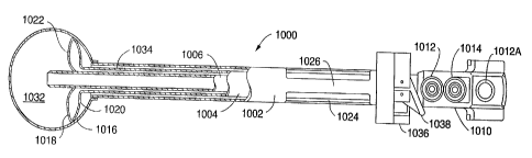
2. FIRST ONE-COMPONENT APPARATUS
(a) Main Embodiment
The separation component can be dispensed with, and the insufflation component
can be modified
to provide the first embodiment of a one component apparatus according to the
invention. The first one-
component apparatus is shown in figure 4A. The first one-component apparatus
121 is similar to the
insufflation component just described. Like components will use the same
reference numbers with 100
added. The first one component apparatus comprises a tube assembly 160,
including an inner tube 135
coaxially mounted inside an outer tube 137. The outer tube covers the inner
tube over most of the length
of the inner tube. The inner tube is a rigid tube having a bore with a
circular cross section that can
accommodate an endoscope (not shown).
The proximal end of the inner tube 135 is fitted with a port 125, the proximal
end 127 of which
includes a flapper valve 132. The shutter 136 of the flapper valve is operated
by the button 129.
Additionally, the seat 134 of the flapper valve forms a gas-tight seal with an
endoscope (not shown), or
other instrument, inserted though the flapper valve into the bore of the inner
tube 135. The port 125 is
also fitted with a first valve 131 to which a supply of a suitable
insufflation fluid can be connected.
Unlike the insufflator component of the two-component apparatus, the distal
end 141 of the outer
tube 137 extends as far as the distal end 139 of the inner tube 135. The tubes
are connected together
over a distal portion l67 of their lengths (see detail in Figure 4B). A
circumferential groove 169 is
formed in the inner wall of the distal portion 167. A groove with a wedge-
shaped cross section is
shown. The circumferential groove can have other cross sections, such as
square, or semi-circular. The
circumferential groove retains the main envelope 112, which defines the main
inflatable chamber I 13, in
the bore of the inner tube, as will be described in more detail below.
The envelope 145 of the toroidal inflatable chamber 143 covers the distal part
of the tube assembly
160. The envelope 145 is a cylindrical piece of a thin elastomeric material,
such a latex, silicone rubber,
or polyurethane. The proximal end 147 and the distal end 149 of the envelope
are attached to the outer
surface 163 of the tube assembly using a circumferential line of adhesive
applied at each end of the
envelope. An epoxy or cyanoacrylate adhesive is preferably used. When the
toroidal inflatable chamber
-14-
CA 02215392 1997-09-15
WO 96/28098 PCT/US96/02838
is in its collapsed state, the envelope 145 lies almost flush with the outer
surface of the tube assembly
160.
The outer tube 137 is spaced from the inner tube 135 over at least part of its
circumference. The
space 151 between the inner tube and the outer tube, and a radial passage 161
through the wall of the
outer tube interconnect the toroidal inflatable chamber 143 and the second
valve 153. The second valve
153 is connected to a source of a suitable inflation fluid (not shown). The
toroidal inflatable chamber is
shown in its collapsed state in figures 4A and 4B, and in its expanded state
in figure 4C.
The anchor flange 155 is slidably mounted on the tube assembly 160, and can be
locked in a
desired position along the length of the tube assembly with a simple over-
center action locking lever (not
shown). As will be described in detail below, the anchor flange and the
toroidal inflatable chamber, in
its expanded condition, form a gas-tight seal to prevent insufflation gas from
escaping.
The first one-component apparatus also includes a main envelope 112 detachably
attached to the
bore of the inner tube 135. The main envelope defines the main inflatable
chamber 113. The main
envelope is preferably formed of an elastomeric material such as latex,
silicone rubber, or polyurethane.
The main envelope can also be formed from a thin, inelastic material such as
Mylar~, polyethylene,
nylon, etc. If an inelastic material is used, it should be suitably packaged
to fit inside the bore of the
inner tube when in its collapsed state.
The main envelope 112 is formed such that it has a substantially spherical
shape when it is in its
expanded state, and is also formed with a neck 165. The neck has an outside
diameter substantially equal
to the diameter of the bore of the inner tube 135. The neck 165 can be rolled
outwards a number of
times, as in the neck of a common toy balloon, or the neck can be attached to
a suitable O-ring 171, as
shown in figure 4B. The rolled neck, or the O-ring attached to the neck,
engages with the
circumferential groove 169 in the inner wall in the inner tube to attach the
main envelope I 12 to the
inner tube. The main envelope is housed in the bore of the inner tube when the
main inflatable chamber
is in its collapsed state.
The rip cord 173 is attached to the neck 165 of the main envelope 112, runs
proximally up the
bore of the inner tube 135, and emerges from the port 125 through the flapper
valve 132. The part of
the rip cord 173 emerging from the flapper valve can be gripped and pulled in
a proximal direction to
release the rolled neck 165 or the O-ring 171 from the circumferential groove
169. By pulling further on
the rip cord, the entire main envelope can be pulled proximally through the
bore of the inner tube.
(b) Alternative Embodiment
An alternative embodiment of the first one-component apparatus having an
elongated main
envelope 1 12A is shown in figure SA. The tube assembly 160A includes the
inner tube 135A mounted
coaxially inside the outer tube 137A, with the proximal and distal ends of the
tubes interconnected. The
space IS1A between the inner tube and the outer tube communicates with the
toroidal inflatable chamber
through the radial passage 161A in the wall of the outer tube. The space
between the inner tube and the
outer tube also communicates with the toroidal chamber inflation valve 153A.
The bore of the inner tube 135A communicates with the port 125A, fitted with
the insufflation
valve 185. The port 125A is also fitted with a flapper valve 132A. including
the flapper valve seat
134A, which maintains gas pressure when the apparatus is used for
insufflation. The flapper valve seat
134A also provides a gas-tight seal around any instrument, such as the
endoscope E, passed through the
flapper valve.
The elongated main envelope I 12A is shown in figure SB. The main envelope is
an elongated
cylinder with a closed distal end 177. The main envelope is preferably formed
from an elastomeric
material, such as latex, silicon rubber, or polyurethane. Attached to the
proximal end of the main
envelope is a manifold 175 which mates with the proximal face 127A of the port
125A. The manifold
175 is fitted with an O-ring seal 187, which forms a gas-tight seal with any
instrument passed through it.
-15-
CA 02215392 1997-09-15
WO 96!28098 PCT/LTS96/02838
The manifold 175 is also fitted with the main chamber inflation valve 131A to
which a supply (not
shown) of a suitable inflation fluid can be attached to inflate the main
inflatable chamber 112A.
The elongated main envelope i 12A is passed through the flapper valve 132A
into the bore of the
inner tube 135A. The manifold 175 is engaged with the proximal face 127A of
the port 125A. When
the manifold is engaged, the distal end 177 of the main envelope projects
beyond the distal end of the
tube assembly 160A, as shown in figure SC. The distal end of the main envelope
is then inverted into
the bore of the inner tube 135A, as shown in figure SD.
An endoscope, or some other suitable instrument, is inserted through the O-
ring seal 187 to seal
the manifold before inflation fluid is passed through the main chamber
inflation valve 131A to inflate the
main inflatable chamber I 13A.
Alternatively, the seal 187 can be replaced by an additional flapper valve
(not shown) so that the
main inflatable chamber can be inflated without the need to use an instrument
to seal the manifold.
When inflation fluid is passed into the main inflatable chamber 113A through
the valve 131A, the
distal end 177 of the main envelope 112A is ejected from the inner tube 135A.
The inflation fluid then
progressively expands the main envelope 112A, and hence the main inflatable
chamber 113A defined by
the main envelope, into an expanded state, as shown in figure SA. The part of
the main envelope inside
the inner tube is subject to the same inflation pressure as the distal end 177
of the main envelope, but is
constrained by the inner tube and so does not inflate.
After using the main envelope 112A to separate the peritoneum away from the
underiying layer, as
will be described in detail below, the inflation pressure fluid is vented from
the main inflatable chamber
I 13A, and the main envelope returns to its collapsed state. When the main
envelope is in its collapsed
state, it can move freely in the bore of the inner tube 135. The main envelope
is removed from the inner
tube by disengaging the manifold 175 from the proximal face 127A of the port
125A, and using the
manifold 175 to pull the main envelope proximally through the bore of the
inner tube.
Inflation fluid for the toroidal inflatable chamber the envelope of which 145A
is shown in figure
SA, is passed through the toroidal chamber inflation valve 153A. Insufflation
gas is passed through the
insufflation valve 185.
The toroidal inflatable chamber and the anchor flange ISSA of the alternative
embodiment of the
first one-component apparatus are the same as in the main embodiment, and will
therefore not be
described.
(c) Method of Using the First One-Component Apparatus (Both Forms)
The method according to the invention of using either form of the first one-
component apparatus
according to the invention to separate a first layer of tissue from a second
layer of tissue will next be
described. As an illustration, separating the peritoneum from the
properitoneal fascia in the course of
repairing a hernia will be described.
Figures 6A through 6H show a longitudinal cross section of the lower abdomen.
An incision about
12-15 mm. long is made in the abdominal wall AW, and carried through the
abdominal wall as far as,
and including the properitoneal fat layer FL, as shown in figure 6A. The
distal end 115 of the tube
assembly 160 of the one-component apparatus 121 is then inserted into the
incision to bring the distal end
into contact with the peritoneum. Additional gentle pressure detaches the part
of the peritoneum in the
immediate vicinity of the incision from the underlying layer, as shown in
figure 6B. Figure 6B shows
the peritoneum detached from the properitoneal fat layer FL. The main envelope
cannot be seen in these
figures because it is inverted within the bore of the tube assembly.
A source of inflation fluid (not shown) is connected to the valve 131. A gas,
preferably air, is the
preferred inflation fluid, but other gases, such a carbon dioxide can be used.
A liquid, such as saline
solution can be used, but liquids are less preferable to gases because they
change the optical properties of
- 16-
CA 02215392 1997-09-15
WO 96/28098 PCT/iTS96/02838
any endoscope inserted into the main inflatable chamber I 13. The flow of
inflation fluid is fumed on.
which ejects the main envelope 112 from the bore of the tube assembly 160.
The inflation fluid progressively expands the main envelope I 12, and hence
the main inflatable
chamber 113 defined by the main envelope, into an expanded state. The main
envelope expands between
the peritoneum P and the properitoneal fat layer FL, and gently and
progressively detaches an increasine
area of the peritoneum from the underlying layer as it expands. When the main
envelope is in its V
expanded state, the main inflatable chamber is preferably about 4"-6" (100-150
mm) in diameter.
Early in the process of expanding the main envelope 112, an endoscope E is
inserted into the
flapper valve 132 in the port 125, as shown in figure 6C. The endoscope E is
passed through the bore of
the tube assembly 160 into the main inflatable chamber 113. Once the main
envelope is partially
expanded, the main envelope is sufficiently transparent for the extent of the
detachment of the peritoneum
to be observed using the endoscope.
When a sufficient area of the peritoneum is detached, the supply of inflation
fluid is turned off.
The inflation fluid is then vented from the main inflatable chamber I 13, and
the main envelope
progressively returns to its collapsed state. The peritoneum remains detached
from the underlying layer,
however, as shown in figure 6D. The main envelope is then removed from the
bore of the tube assembly
160. The different methods of removing the main envelope from the bore of the
tube assembly for the
two different forms of the first one-component apparatus are described above.
After the main envelope 112 has been removed from the bore of the tube
assembly, the tube
assembly is advanced into the incision in the direction of the arrow 162 until
the proximal end of the
envelope 145 of the toroidal inflatable chamber is in the properitoneal space,
clear of the incision, as
shown in figure 6E.
A suitable source (not shown) of an inflation fluid is attached to the valve
153. A gas, such as air
or carbon dioxide, can be used for the inflation fluid; alternatively, a
liquid, such as saline can be used.
Since the volume of inflation fluid required to inflate the toroidal
inflatable chamber is small, about
15 ml in the preferred embodiment, the inflation fluid can be contained in a
large syringe. Inflation fluid
is fed into the toroidal inflatable chamber 43 to expand the toroidal
inflatable chamber to its expanded
condition, as shown in figure 6F.
The anchor flange 155 is then advanced in the direction of the arrow 159 along
the tube assembly
160 to bring the anchor flange into contact with the skin S of the abdominal
wall AW. The tube
assembly 160 is then gripped, and the anchor flange is further advanced
slightly. This forces the
expanded toroidal inflatable chamber 143 into contact with the underlying
layer, and slightly compresses
the abdominal wall AW, including the underlying layer but excluding the
peritoneum P, between the
expanded toroidal inflatable chamber and the anchor flange, as shown in figure
6G. Once adjusted, the
anchor flange is locked in position on the tube assembly. The expanded
toroidal inflatable chamber is
held against the underlying layer and forms a gas-tight seal with the
abdominal wall, excluding the
peritoneum.
A suitable source (not shown) of an insufflation gas is attached to the first
valve 131, and
insufflation gas is passed through the bore of the inner tube 135 into the
working space WS between the
peritoneum P and the underlying layer created by separating the peritoneum
from the underlying layer.
The pressure of the insufflation gas re-separates the peritoneum from the
underlying layer, as shown in
figure 6H, and provides a working space in which repair of the hernia can be
carried out. The bore of
the tube assembly 160 can be used to pass instruments, such as the endoscope
E, into the working space
to perform the repair procedure. When no instrument is inserted into the bore
of the tube assembly,
insufflation pressure is maintained by the flapper valve.
As part of the hernia repair procedure, additional gas-tight trocar sleeves
(not shown) are inserted
through the abdominal wall into the working space. The same procedure as
described above in
connection with figure 3I is used to attach a mesh patch to the properitoneal
fascia over the site of the
- I7-
CA 02215392 1997-09-15
WO 96/28098 PCT/LTS96/02838
hernia. The process can be observed with the aid of an endoscope (,not shown)
passed through the bore
of the tube assembly 160, or through one of the additional trocar sleeves.
After the treatment procedure is completed, the valve 131 is operated to
release the insufflation gas
from the working space WS. The valve 153 is operated to release the inflation
fluid from the toroidal
inflatable chamber 143, which releases compression of the abdominal wall AW,
excluding the
peritoneum. The toroidal inflatable chamber returns to its collapsed state,
with its envelope 145 flush
with the outer surface the tube assembly 160. The tube assembly is then
withdrawn from the incision,
and the incision is closed using sutures or clips. The pressure of the viscera
against the peritoneum
returns the peritoneum into contact with the underlying layer. Over time, the
peritoneum reattaches to
the underlying layer.
3. SECOND ONE-COMPONENT APPARATUS
(a) Second One-Component Apparatus
A second embodiment of a one-component apparatus is shown in figures 7A and
7B. The second
one-component apparatus 121 is similar to the first one-component apparatus
just described. However,
the second one-component apparatus has a substantially spherical toroidal main
inflatable chamber, that
avoids the need to detach and remove the main envelope at the end of the
separation process. Also, in
the second one-component apparatus, a single toroidal main inflatable chamber
provides the separating
function of the main inflatable chamber and the sealing function of the
toroidal inflatable chamber of the
first one-component apparatus.
In the following description, similar components will use the same reference
numbers with an
additional t 00 added.
The second one-component apparatus comprises a tube assembly 260, including an
outer tube 237
to which is attached a twin port assembly 224 is attached. The port assembly
includes a first port 226
and a second port 228. The first port is provided with a first flapper valve
202, including the flapper
valve seat 204. The second port is provided with a second flapper valve 206,
including the flapper valve
seat 208. Each flapper valve seat additionally forms a gas-tight seal with an
instrument passed through
it.
The tube assembly 260 also includes the inner tube 235. The inner tube has a
length that is shorter
than the length of the outer tube 237. The proximal end 210 of the inner tube
is flexibly attached to the
proximal end 222 of the outer tube 237 and to the first port 226. The flexible
attachment enables the
distal end 214 of the inner tube to move in the direction shown by the arrow
216. The first port
communicates with the bore of the inner tube 235, and the second port
communicates with the bore of
the outer tube 237.
The insufflation valve 285 communicates with the first port 226, and the bore
of the inner tube
235. The main chamber inflation valve 231 communicates with the second port
228, and the bore of the
outer tube 237.
The main envelope 212 defines the main inflatable chamber 213 and comprises a
cylindrical piece
of an elastomeric material such a latex, silicone rubber, or polyurethane. The
apparatus is shown with its
main envelope in its collapsed state in figure 7B, in which the structure of
the main envelope can also be
seen. The main envelope preferably has a diameter smaller than the outside
diameter of the inner tube.
One end 230 of the main envelope is attached to the distal end 214 of the
inner tube 235 by means of a
suitable adhesive, such as an epoxy or cyanoacrylate adhesive. The other end
232 of the main envelope
is everted (i.e., fumed back on itself to bring the inside surface 234 of the
main envelope to the outside)
and attached to the distal end 236 of the outer tube using the same type of
adhesive. The main envelope
is preferably attached to the outer surfaces of the inner tube and the outer
tube.
The apparatus is shown with the main envelope 212 in its expanded state in
figure 7A. A suitable
source of inflation gas is connected to the valve 231 and flows into the main
inflatable chamber through
-18-
CA 02215392 1997-09-15
WO 96/28098 PCTlIJS96/02838
the bore of the outer tube 237. The pressure acting on the surface 238 of the
main envelope 212 causes
the main envelope to assume the toroidal shape shown in figure 7A to define
the toroidal main chamber
213. Figures 7A and 7B show the correspondence between the surfaces 234 and
238 of the main
envelope when the main envelope is in its collapsed state (figure 7B) and in
its expanded state (figure
7A).
The anchor flange 255 is slidably mounted on the tube assembly 260, and can be
locked in a
desired position along the length of the tube assembly. The anchor flange 255
is similar to the anchor
flange 55 (figure 2A) and so will not be described further.
In figure 8A, an endoscope E is shown passed through the second flapper valve
206, the second
port 228, and the bore of the outer tube 237 into the main inflatable chamber
213. The flexible
mounting of the inner tube 235 in the outer tube enables the endoscope to
displace the inner tube 235 in
direction of the arrow 216 to gain access to the main inflatable chamber. The
endoscope is inserted
through the second port into the main inflatable chamber during the separation
phase of using the
apparatus to observe the extent of the separation of tissue.
I S In figure 8B, an endoscope E is shown passed through the first flapper
valve 202, the first port
226, the bore of the inner tube 235, and the bore 234 of the main envelope
212. The distal pan of the
endoscope emerges from the bore of the main envelope, and can be advanced
beyond the main inflatable
chamber 213 to observe the site of the hernia more closely. The endoscope is
inserted through the first
port, the inner tube, and the bore of the main envelope during the
insufflation phase of using the
apparatus. Instruments other than endoscopes can also be passed to the site of
the hernia through the first
flapper valve, the first port, the inner tube, and the bore of the main
envelope if desired.
Also in figure 8B, the main envelope 212 is shown in the partially collapsed
state that it preferably
assumes during the insufflation phase of the procedure. In this part of the
procedure, the partially
collapsed main inflatable chamber and the anchor flange 255 together provide a
gas-tight seal to prevent
the leakage of insufflation gas. Alternatively, this part of the procedure can
be carried out with the main
inflatable chamber in a fully expanded state.
(b) Method of Using the Second One-Component Apparatus
The method according to the invention of using the second embodiment of the
one-component
apparatus according to the invention to separate a first layer of tissue from
a second layer of tissue will
next be described. As an illustration, separating the peritoneum from the
properitoneal fascia in the
course of repairing a hernia will be described.
Figures 9A through 9F show a longitudinal cross section of the lower abdomen.
An incision about
12-15 mm long is made in the abdominal wall AW, and carried through the
abdominal wall as far as, and
including, the properitoneal fat layer FL, as shown in figure 9A. The distal
end 215 of the tube assembly
260 of the second one-component apparatus 221 is then inserted into the
incision to bring the distal end
into contact with the peritoneum P. Additional gentle pressure detaches the
part of the peritoneum in the
immediate vicinity of the incision from the underlying layer, as shown in
figure 9B. Figure 9B shows
the peritoneum detached from the properitoneal fat layer FL. The main envelope
cannot be seen in these
figures because it is inverted within the bore of the tube assembly.
A source of inflation fluid (not shown) is connected to the valve 231. A gas,
preferably air, is the
preferred inflation fluid, but other gases, such a carbon dioxide can be used.
A liquid, such as saline
solution can be used, but liquids are less preferable to gases because they
change the optical properties of
any endoscope inserted into the main inflatable chamber. The flow of inflation
fluid is turned on, which
ejects the main envelope 212 from the bore of the tube assembly 260.
The inflation fluid progressively expands the main envelope 212, and hence Lhe
main inflatable
chamber 213 defined by the main envelope, into an expanded state. The main
envelope expands between
the peritoneum P and the properitoneal fat layer FL, and gently and
progressively separates an increasing
-19-
CA 02215392 1997-09-15
WO 96/28098 PCT/US96/02838
area of the peritoneum from the underlying layer as it expands. When the main
envelope is in its
expanded state, the main inflatable chamber is preferably about 4"-6" (100-150
mm) in diameter.
Early in the process of expanding the main envelope 212, an endoscope E is
inserted into the first
flapper valve 202, as shown in figure 9C. The endoscope E is passed through
the bore of the outer tube
237 into the main inflatable chamber 213. Once partially expanded, the main
envelope 212 is sufficiently
transparent for the extent of the separation of the peritoneum to be observed
using the endoscope.
When a sufficient area of the peritoneum is separated, the supply of inflation
fluid is fumed off.
The endoscope E is removed from the main inflatable chamber 213. The valve 231
is then opened to
allow inflation fluid to vent partially from the main inflatable chamber 213.
The main envelope 212
progressively returns part-way towards its collapsed state, as shown in figure
9D. Alternatively, the main
envelope may be kept fully expanded.
The anchor flange 2S5 is then advanced in the direction of the arrow 259 along
the tube assembly
260 to bring the anchor flange into contact with the skin S of the abdominal
wall AW. The tube
assembly 260 is then gripped, and the anchor flange is further advanced
slightly. This forces the main
inflatable chamber 213 into contact with the underlying layer, and slightly
compresses the abdominal
wall, including the underlying layer but excluding the peritoneum, between the
main inflatable chamber
and the anchor flange, as shown in figure 9E. Once adjusted, the anchor flange
is locked in position on
the tube assembly. The main inflatable chamber is held against the underlying
layer and forms a gas-
tight seal with the abdominal wall, excluding the peritoneum.
A suitable source (not shown) of insufflation gas is attached to the second
valve 285, and
insufflation gas is passed through the bore of the inner tube 235, and the
bore 234 of the main envelope,
into the working space WS between the peritoneum P and the underlying layer.
The pressure of the
insufflation gas re-separates the peritoneum from the underlying layer, as
shown in figure 9F, and
provides a working space in which repair of the hernia can be carried out.
Instruments, such as the endoscope E, can be passed through the second flapper
valve 206, the bore
of the inner tube 235, and the bore 234 of the main envelope, as shown in
figure 8B, into the working
space to perform the repair procedure. When no instrument is inserted into the
bore of the inner tube,
insufflation pressure is maintained by the second flapper valve.
As part of the hernia repair procedure, additional gas-tight trocar sleeves
(not shown) are inserted
through the abdominal wall into the working space. The same procedure as
described above in
connection with figure 3I is used to attach a mesh patch to the properitoneal
fascia over the site of the
hernia. The process can be observed with the aid of an endoscope (not shown)
passed into the working
space through the bore of the inner tube 235, or through one of the additional
trocar sleeves.
After the treatment procedure is completed, the valve 285 is operated to
release the insufflation gas
from the working space. The valve 231 is operated to release the inflation
fluid from the main inflatable
chamber 213, which releases compression from the abdominal wall, excluding the
peritoneum. The main
envelope returns to its collapsed state inside the bore of the outer tube 237.
The tube assembly is then withdrawn from the incision, and the incision is
closed using sutures or
clips. The pressure of the viscera against the peritoneum returns the
peritoneum into contact with the
underlying layer. Over time, the peritoneum reattaches to the underlying
layer.
4. HERNIA REPAIR METHOD WITH INCISION AT THE UMBILICUS
The hernia repair methods described so far show the incision placed close to
the site of the hernia.
In practice, it is preferred to make the incision at or near the umbilicus
because the boundary between the
peritoneum and the properitoneal fat layer can be more directly accessed near
the umbilicus. The midline
location of the umbilicus is devoid of muscle layers that would otherwise need
to be traversed to reach
the properitoneal fat layer.
-20-
CA 02215392 1997-09-15
WO 96/28098 PCT/US96/02838
Apparatus of the types described above inserted ihrougit an incision at the
umbilicus would require
a very large main inflatable chamber to detach the peritoneum from the
umbilicus to the groin. Instead,
in the method according to the invention to be described next, an apparatus of
any one of the types
described above is used to provide a tunnel from an incision at the umbilicus
to the site of the hernia in
the groin, and then to provide an insufflated working space at the site of the
hernia.
The main envelope is partiaily expanded, collapsed, and advanced towards the
site of the hernia.
This sequence is repeated to progressively separate the peritoneum from the
underlying layer and form
the tunnel from the umbilicus to the site of the hernia. Then, at or near the
site of the hernia, the main
envelope is fully expanded to provide the working space at the site of the
hernia. The working space is
then insufflated to maintain the separation of the peritoneum from the
underlying layer.
The following method can be practiced using the two-component embodiment of
the apparatus. or
any of the one-component embodiments of the apparatus. The method will be
described using the two-
component apparatus.
An incision about 12-15 mm long is made in the abdominal wall AW, and is
carried through the
abdominal wall as far as, and including, the properitoneal fat layer FL. The
incision is made at the
umbilicus U, as shown in figure 10A.
The distal end 15 of the introduces tube 3 of the separation component 1 is
then inserted into the
incision to bring the distal end into contact with the peritoneum P.
Additional gentle pressure detaches
the part of the peritoneum in the immediate vicinity of the incision from the
underlying layer, as shown
in figure IOB. In figure IOB, the peritoneum is shown detached from the
properitoneal fat layer FL.
The main envelope cannot be seen in these figures because it is inverted
within the bore of the introduces
tube 3.
A source of a suitable inflation fluid (not shown), as previously described,
is connected to the
valve I 1. The flow of inflation fluid is turned on, which ejects the main
envelope 12 of the main
inflatable chamber 13 from the bore of the introduces tube 3. The inflation
fluid progressively expands
the main envelope 12, and hence the main inflatable chamber 13 defined by the
main envelope, into a
partially-expanded state, as shown in figure IOC. The main envelope expands
between the peritoneum
and the properitoneal fat layer FL, and gently and progressively detaches an
increasing area of the
peritoneum P from the underlying layer near the umbilicus as it expands.
An endoscope (not shown) can be inserted into the main inflatable chamber 13
through the flapper
valve 2 and the bore of the introduces tube 3. The endoscope can be used to
observe the extent of the
separation of the peritoneum, as described above.
When the main envelope 12 expanded such that the main inflatable chamber 13 is
about one-fourth
of its fully-expanded diameter, i.e., about 1.0"-I.5" (25-37 mm) in diameter,
the supply of inflation fluid
is turned off. The valve 1 I is then operated to vent inflation fluid from the
main inflatable chamber 13.
The main envelope progressively returns to its collapsed state, as shown in
figure IOD. The peritoneum
DP that was separated by the main inflatable chamber remains detached from the
underlying layer.
however. as shown. Alternatively, the main envelope can be inflated to a fully-
expanded state.
The separation component 1, including the collapsed main envelope 12, is then
manipulated in the
direction indicated by the arrow 14, and then in the direction indicated by
the arrow 16, to advance the
distal part I S of the introduces tube 3 to the limit of the detached part of
the peritoneum DP in the
direction of the groin, as shown in figure 10E. An endoscope E inserted
through the flapper valve 2 into
the bore of the introduces tube 3 enables the position of the distal part of
the introduces tube relative to
the detached part of the peritoneum to be observed.
Once the distal part 15 of the introduces tube has been positioned. the
separation component I is
clamped in position, or is gripped, and inflation fluid is once more passed
through the valve 11, and the
bore of the introduces tube 3 into the main inflatable chamber 13. The main
envelope 12 expands once
more, increasing the extent of the detached part of the peritoneum towards the
groin, as shown in figure
-21 -
CA 02215392 1997-09-15
WO 96/28098 PCT/US96/02838
IOF. The increased extent of the detached part of the peritoneum is indicated
by the line DP' in the
figure. It should be noted that the extent of the detached part of the
peritoneum is increased in the
direction from the umbilicus to the groin, but not in the direction transverse
to this direction. The
endoscope E is used to observe the extent of the separation.
The process of collapsing the main envelope 12. advancing the distal part 15
of the introducer tube
to the limit of the detached part of the peritoneum DP, in the direction of
the groin, holding the
introducer tube in position, and partially re-inflating the main envelope 12,
is repeated until the detached
part of the peritoneum includes the peritoneum over the site of the hernia.
This process provides the
tunnel T between the incision at the umbilicus and the site of the hernia.
This can be seen in figure IOI.
Alternatively, the main envelope can be fully re-inflated.
When the main envelope is in the vicinity of the site of the hernia H, the
main envelope 12 is fully
inflated to form a working space WS including the site of the hernia. This is
shown in figure IOG.
T'he working space at the site of the hernia is then insufflated. With the two-
component apparatus,
inflation fluid is vented from the main inflatable chamber 13 to collapse the
main envelope 12, and the
separation component 1 is withdrawn from the tunnel T through the incision I.
The insufflation
component 21 is introduced into the incision, and advanced through the tunnel
until the envelope 45 of
the toroidal inflatable chamber 43 lies within the working space WS, clear of
the tunnel. The toroidal
inflatable chamber is inflated, the anchor flange is clamped in position. and
insufflation gas is passed into
the working space, as shown in figure IOH. The toroidal inflatable chamber
provides a gas-tight seal
ZO with the entrance of the tunnel.
Figure IOI shows a plan view of the abdomen with the insufflator component 21
in place. The
anchor flange has been omitted for clarity. The toroidal inflatable chamber 43
provides a gas-tight seal
with the entrance of the tunnel T. The extent of the separated peritoneum is
indicated by the dotted line
DP. It can be seen that the lateral extent of the separated peritoneum is
considerably greater in the
working space WS than in the tunnel T.
With the first embodiment of the one-component apparatus, inflation fluid is
vented from the main
inflatable chamber to collapse the main envelope, and the main envelope is
withdrawn from the working
space through the bore of the tube assembly. The tube assembly is partially
withdrawn until the envelope
of the toroidal inflatable chamber lies within the working space, clear of the
entrance to the tunnel. The
toroidal inflatable chamber is inflated, the anchor flange is clamped in
position and insufflation gas is
passed into the working space, as already described. The toroidal inflatable
chamber seals against the
entrance from the tunnel into the working space.
Using the second embodiment of the one-component apparatus, the main envelope
is preferably
returned to a partially collapsed state, the tube assembly is partially
withdrawn until the main inflatable
chamber lies within the working space, adjacent to the entrance of the tunnel.
The anchor flange is
clamped in position, and insufflation gas is passed into the working space, as
already described. The
partially-collapsed main chamber seals against the entrance from the tunnel
into the working space.
If the main envelope is inflated to a fully expanded state during the
separation part of the
procedure, the whole of the space is insufflated with a gas-tight seal at the
incision. as previously
described.
Irrespective of the embodiment of the apparatus used to create the insufflated
working space WS,
the hernia is then repaired using the procedure described in connection with
figure 3I.
J. INFLATABLE CHAMBERS FOR MAINTAINING SEPARATION OF TISSUE LAYERS
As previously discussed. during dissection of the properitoneal space or
during subsequent surgical
procedures near the peritoneum, it is common to puncture or otherwise breach
the peritoneum. Such a
puncture or breach prevents the properitoneal space from retaining pressurized
fluid (gas or liquid) used
to maintain the space in an open condition. If pressure is lost, visualization
of the space and the actual
-22-
CA 02215392 1997-09-15
WO 96/28098 PCT/CTS96/02838
volume of the space will decrease and compromise the surgery. Absent some way
of mechanically
maintaining the space, loss of pressure can result in inability to complete
the procedure.
An additional consideration in laparscopic surgery inside the peritoneal space
is fouling of the
distal end of the endoscope with body fluids caused by incidental contact with
either tissues near the
entry point of the endoscope or tissues near the distal end of the cannula
through which the endoscope
has been inserted.
(a) First Inflatable Chamber
Referring to Figures 1 IA and 11B, an insufflation and retraction device 301
having a first
inflatable chamber 303 is shown. The insufflation and retraction device 301 is
similar to the insufflation
component 21 of the two-component apparatus, shown in Figures 2D and 2E, and
like reference numerals
represent like components. It is understood that although it is preferred to
use the inflatable chambers
described below with the insufflation and retraction device 301, the
inflatable chambers may also be used
with any other delivery or inflation device.
The insufflation and retraction device 301 includes an inner tube 335 and a
coaxial outer tube 337.
The distal end 323 of the inner tube 335 extends beyond the distal end 325 of
the outer tube 337. The
inner tube 335 is similar to the introducer tube 3 (Figure 2A) and is a rigid
tube having a bore with a
circular cross section that can preferably accommodate a 10 mm endoscope,
however, any cross-sectional
shape or area may be provided. The proximal end of the inner tube 335 is
fitted with a flapper valve as
described above in connection with the insufflation component of Figures 2D
and 2E. A seat at the
proximal end of the inner tube forms a gas-tight seal with an appropriately
sized instrument. A shutter
covers the seat and is operated by a button 329. A blunt obturator 322 is
shown extending through the
seat and through the distal end 323 of the inner tube 335 (Figures I lA and
11B). A valve 331 is fluidly
coupled to the interior of the inner tube 335 and may be used to supply
insufflation gas or liquid.
The first inflatable chamber 303 has a distal side 343 coupled to the inner
tube 335 and a proximal
side 345 coupled to the outer tube 337 so that the interior of the inflatable
chamber 303 is fluidly
coupled to the annular space between the inner and outer tubes 335, 337. The
proximal and distal sides
345, 347 of the inflatable chamber 303 are preferably attached to the inner
and outer tubes 335, 337 ai
flanges 359. A valve 353 is adapted to be connected to a source of a suitable
inflation gas or liquid (not
shown) for inflating the inflatable chamber 303. The inflatable chamber 303 is
shown in a collapsed
state in Figure 11A and in an expanded state in Figures 11B and I 1C.
An anchor flange 355 is slidably mounted to the outer tube 337 and can be
locked along the length
of the outer tube 337 with a locking lever 349. The anchor flange 355 helps to
immobilize the device
and, further, helps the inflatable chamber 303 form a seal to limit the escape
of insufflation gas during
laparoscopic procedures. When the anchor flange 355 is locked into position,
the anchor flange 355 and
inflatable chamber 303 apply a modest compressive force to the tissue between
the inflatable chamber
303 and the anchor flange 355 thereby improving the gas-seal.
Referring to Figure I lA, the inflatable chamber 303 is folded and contained
within a sheath 349
before insertion into a patient. The inflatable chamber 303 may be folded in
any manner but is
preferably folded inwardly from lateral, side edges 351, 353 toward the
extended distal end of obturator
322. The sheath 349 is preferably perforated but may be formed in any other
manner permitting easy
opening. The inflatable chamber 303 is initially in the folded, compact
orientation of Figure I lA before
insertion into the patient so that the retraction device may be easily
inserted through a small opening in
the patient. As will be described below, afrer the inflatable chamber 303 has
been positioned within a
patient and between the two tissue layers to be separated. inflation air is
injected into the inflatable
chamber 303 through the second valve 353. Inflation of the chamber 303 tears
the sheath 349 along the
perforation 361 and releases the inflation chamber 303. Alternatively, the
sheath 361 may include an
independent opening mechanism, such as a removable thread which binds the
sheath together.
- 23 -
CA 02215392 1997-09-15
WO 96128098 PCT/US96/02838
Referring to Figure 11C, the inflatable chamber 303 preferably has a
substantially trapezoidal
shape. First and second sides 363, 365 of the inflatable chamber 303 are
preferably slightly curved but
may also be linear or bi-linear. The first and second sides 363, 365 and
lateral sides 351, 353 may also
include surface features such as ridges or rounded teeth to help anchor the
inflatable chamber 313 and
improve the insufflation gas seal. The second side 365 is preferably longer
than the first side 363 and
forms angles of less than 90 degrees with the lateral sides 351, 353.
Furthermore, the inner and outer
tubes 335, 337 are preferably connected to the inflatable chamber 303 closer
to the first side 363 than the
second side 365. A throughhole 358 is defined by the outer tube and extends
through the first and
second sheets. The shape of inflatable chamber 303 may also be modified and/or
optimized to suit the
particular use contemplated. The location and configuration of the throughhole
358 may also be
modified.
The inflatable chamber 303 is formed with first and second sheets 367, 369
attached together along
a periphery 371. This arrangement results in relatively high localized stress
at the periphery 371 of the
first and second sheets 367, 369. To withstand this stress, the strength of
the sheets must be increased.
One way of increasing the strength of the sheets is to increase the thickness
of the sheets. A problem
with simply increasing the sheet thickness is that the inflatable chamber 303
becomes larger in the
collapsed state (Figure I IA) which will causes more problems during insertion
into a patient.
To alleviate the problem of localized stresses without increasing the sheet
thickness, the present
invention provides baffles 373 disposed between the first and second sheets
367, 369. The baffles 373
interconnect the first and second sheets 367, 369 and help absorb the pressure
forces thereby reducing
stresses at the periphery 371. The baffles 373 also help define the shape of
the inflatable chamber 303
and limit the separation distance between the first and second sheets 367, 369
when the inflatable
chamber 303 is in the expanded shape. The preferred method of attaching the
baffles 373 to the first and
second sheets 367, 369 is described below.
(i) Preferred Material for the Inflatable Chamber
The first and second sheets 367, 369 and baffles 373 are preferably made of a
polyester and
polyurethane composite material. Polyester has desirable strength
characteristics but it is relatively rigid
and crinkles easily. Moreover, polyester is very difficult to RF weld which is
a preferred method of
connecting the baffles and sheets together as will be described below.
Polyurethane, on the other hand. is
soft, non-abrasive, and easy to RF weld. Unfortunately, the tensile strength
of polyurethane is relatively
low. The composite material exploits the advantages of both polyester and
polyurethane.
The composite material is formed by bonding polyurethane to a nylon or
polyester film having a
preferred thickness of about 0.5 to 2 mil. (12-50 pm), although a polyester
fabric may also be used. The
nylon or polyester fabric may be a woven fabric or may be composed of randomly-
oriented fibers. The
film or fabric layer is laminated (or cast, captured or encapsulated) between
two polyurethane layers to
provide a composite material having a preferred thickness of about 3 mil. (75
p.m). The resulting
composite material is strong, supple, non-abrasive, transparent, and easily RF
welded. The composite
material will also fold with small radius folds so that the inflatable chamber
303 can be compacted into a
small volume for easy insertion into a patient.
The composite material is relatively inelastic and, therefore, must be folded
into the sheath as
described above (Figure 1 IA). The present invention may also be practiced
with an elastic material
which, when expanded, provides the shape of the inflatable chambers of the
present invention. The
composite material is disclosed in co-pending U.S. Patent Application Serial
No. 08/134,573, filed
October 8, 1993, which is herein incorporated by reference.
-24-
CA 02215392 1997-09-15
WO 96/28098 PCT/US96/02838
(ii) Method of Constructing the Inflatable Chamber
'The baffles 373 are preferably fabricated and attached to the first and
second sheets 367, 369 in the
manner shown in Figures 13A through 13C. The first and second sheets 367, 369
and baffles 373 are cut
into the desired shape and oriented as shown in Figure 13A with the first and
second sheets 367, 369
being offset with respect to one another. The baffles preferably do not extend
completely between the
first and second sides so that all portions of the interior of the inflatable
chamber are fluidly coupled
together. Alternatively, the baffles 373 may include openings to fluidly
couple the various portions
together. The baffles 373 are preferably made of the same material as the
first and second sheets but
may also be made of a different material.
Referring to the exploded cross-sectional view of Figure 13B, RF welding
electrodes 344, 346 are
positioned against the first and second sheets 367, 369. RF welding imparts
radio frequency energy to
the working piece. When radio frequency energy is imparted onto polyurethane,
the molecules are
excited and the polyurethane melts thereby bonding together adjacent
polyurethane layers together. A
suitable release agent 305 is applied to either the sheets 367, 369 or baffles
373 to prevent formation of
RF welds at certain locations. A preferred release agent 305 is powdered
polyethylene and teflon.
Without the releasing agent 305, the baffle 373 would be RF welded to both the
first and second sheets
367, 369 on both sides. Application of the release agent 305 advantageously
enables attaching the baffles
373 to the sheets in a single welding operation.
The RF welding apparatus is activated to weld the baffles 373 to the first and
second sheets 367,
369. Adjacent polyurethane layers bond at all locations between the RF welding
electrodes 375 except
where the release agent 305 has been applied. The first and second sheets are
then displaced so that they
overlie one another as shown in Figure 13C. The resulting baffles 373 have a
generally S-shaped
configuration when the inflatable chamber is in the expanded condition. A
second RF welding operation
(not shown) welds the periphery 371 of the first and second sheets together
367, 369.
(b) Second Inflatable Chamber for Maintaining Separation of Tissue Layers
A second inflatable chamber 403 for maintaining separation between tissue
layers is shown in
Figures 12A and 12B. The second inflatable chamber 403 includes an
intermediate weld 405 which
reduces pressure induced stresses at the periphery 471 of the inflatable
chamber 403. The intermediate
weld 405 eliminates the need to provide baffles, however, baffles may also be
provided if necessary. The
intermediate weld 405 is preferably a semi-circular segment having terminal
ends 407 positioned adjacent
the periphery 471. An interior area 411 is fluidly coupled to the remainder of
the inflatable chamber 403
via two fluid paths 413 so that when the inflatable chamber 403 is inflated,
the interior area 411 is also
inflated. The fluid paths 413 are preferably provided between the terminal
ends 407 of the intermediate
weld 405 and the periphery 471; however, the fluid path 413 may be positioned
anywhere along the
intermediate weld 405. Furthermore, although it is preferred to provide two
fluid paths, any number of
paths may be provided.
The periphery 471 of the inflatable chamber 403 is substantially bell-shaped
with the hemispherical
interior area 41 1 protruding slightly from a bottom side 413. The bell-shaped
periphery 471 has a semi-
circular upper portion 415 which is substantially concentric with a
throughhole 417. The remainder of
the peripheral wall 471 is shaped like a truncated triangle extending downward
from the semi-circular
upper portion 415. As previously described, the shape of the inflatable
chamber may be modified to suit
the particular use contemplated. Materials and construction techniques are as
described previously.
(c) Third Inflatable Chamber for Maintaining Separation of Tissue Layers
Referring to Figures 14A and 14B, a third inflatable chamber 503 is shown
which also includes
intermediate welds 505. The inflatable chamber 503 is advantageously formed
from only first and second
sheets 567, 569 of material. The intermediate welds 505 are preferably
circular but may take any other
-25-
CA 02215392 1997-09-15
WO 96/28098 PCT/US96/02838
shape. The intermediate welds bond the first and second sheets 567, 569
together throughout the entire
circular area of the welds. Before insertion into a patient, the inflatable
chamber 504 is preferably folded
and contained within a perforated sheath as described above in connection with
the first inflatable
chamber 303
The inflatable chamber 503 has a substantially trapezoidal shaped periphery
571. The sides 507.
509, 511, 513 of the inflatable chamber are preferably linear but may also be
curved. A first side 507 is
smaller than a second side 509 and the second side preferably forms an angle
of between 20 and 90
degrees with the lateral sides 511, 513.
(d) Fourth Inflatable Chamber for Maintaining Separation of Tissue Layers
Referring to Figures 15A and 15B, a fourth inflatable chamber 603 is shown
which has the same
general features as the second inflatable chamber 403, however, inflatable
chamber 603 includes first and
second extensions 621, 623 having a space 633 therebetween. The space 633
provides clearance for
insertion of additional instruments into the working space while minimizing
the risk that the additional
instruments will pierce the inflatable chamber 603 as described below. A
throughhole 617 extends
through the inflatable chamber 603 which is adapted to be connected to the
insufflation component of
Figures 11A and 11B or may be connected to any other delivery or inflating
device.
The fourth inflatable chamber 603 is preferably symmetrical about a line of
symmetry 609 passing
through the throughhole 617. First and second peripheral points 625, 627 are
located on the line of
symmetry 609 with the first peripheral point 625 being closer to the
throughhole 627 than the second
peripheral point 627. The first and second extensions 621, 623 have radially
outward points 629, 631. A
line 630 passing through the center of the throughhole 617 and the radially
outward to points 629, 631
preferably forms an angle A between 10 degrees and 80 degrees with respect to
a line 632 extending
between the throughhole and the first peripheral point 625. The extensions are
preferably triangular-
shaped with rounded edges but may also take any other shape so long as the
space 633 is provided
therebetween. Furthermore, the space 633 is also preferably triangular shaped
but may also be semi-
circular, square, or a relatively shallow circular segment. Modifications to
the proportions illustrated as
well as providing asymmetrical designs are also contemplated to suit the
particular application.
During laparoscopic surgery, additional instruments are often introduced above
the longitudinal
axis of the delivery device. The space 633 between the extensions 621, 623
facilitates introduction of
additional instruments along the longitudinal axis of the delivery device
above the inflatable chamber 603
while minimizing the risk that the additional instruments will puncture the
inflatable chamber 603.
The inflatable chamber 603 is preferably formed from first and second sheets
667, 669 of the
composite material described above but may be formed in any other manner or
with any other materials
to provide the extensions 621, 623 and space 633 therebetween. The first and
second sheets 667, 679 are
preferably attached together about the periphery 671 by RF welding as
described above. The first and
second sheets 667, 669 are also preferably coupled together by an intermediate
weld 605. The inflatable
chamber 603 may also be formed with baffles or with a sheet material of
sufficient thickness to withstand
the stress at the periphery 671.
(ej Fifth Inflatable Chamber for Maintaining Separation of Tissue Lovers
Referring to Figures 16A and 16B, a fifth inflatable chamber 703 is shown
which is an alternative
to inflatable chamber 303 of Figures 11B through I IC. The fifth inflatable
chamber 703 may be used
with any inflation or delivery device but is preferably used in connection
with the insufflation component
of Figures 2D and 2E or Figures I lA and I lB.
The fifth inflatable chamber 703 has essentially the same structural features
as the first inflatable
chamber 303, however, the inflatable chamber 703 includes a pair of triangular-
shaped wings 705. The
triangular wings 705 provide a wider working space within the patient than the
inflatable chamber 303.
-26-
CA 02215392 1997-09-15
WO 96/28098 PCT/US96/02838
The wings 705 may also advantageously provide additional tissue dissection
when the chamber 703 is
inflated. ..
Inflatable chamber 703 is preferably made from the same or like materials as
inflatable chamber
303 and. furthermore, is constructed with baffles 707 in the same manner as
inflatable chamber 303.
For the first, second, third, fourth and fifth embodiments of the inflatable
chamber described
above, it should be recognized that the shape of each embodiment has the
common functional benefit of
maintaining the properitoneal space by retaining the separation of the
peritoneum from the overlying
tissue in the event of loss of optimal pressurization or when a gasless
technique is used.
More specifically, the embodiments each employ a bottom surface which contacts
the peritoneum
and an upper surface that contacts the overlying fascia. For example, in the
first embodiment shown in
Fig. I 1 C, the upper surface can be characterized as side 363 and the lower
side characterized as side 365.
The physical distance between the upper and lower surfaces causes like
separation or the peritoneal layer
and fascia layers in the dissected properitoneal cavity. The inflatable
chamber can be dimensionally sized
to create optimal separation while limiting potential trauma to tissue.
Furthermore, the location of
throughhole 358 within the inflatable chamber can be selected to optimize the
separation of retracted
tissue from an endoscope or other instrument passed through throughhole 358.
Throughhole 358 thus can
be centered or off center in the inflatable chamber as desired. Preferably,
throughhole 358 is located
slightly off center towards the upper surface, but centered laterally. Such a
location optimizes the desired
features of reducing endoscope fouling by body fluids as the scope is passed
into throughhole 358, and
separating the scope from the peritoneum to provide maximum viewing field with
a scope through
throughhole 358. The reduction of fouling is accomplished by displacing the
throughhole 358 from the
upper surface, and thus overlying fascia, to minimize potential tissue contact
with throughhole 358 and an
endoscope passed through throughhole 358. Likewise, the viewing field is
maximized by separating
throughhole 358, and thus an endoscope in it, from peritoneum being deflected
downward by the lower
surface of inflatable chamber.
6. HERNIA REPAIR METHOD WITH INCISION AT THE UMBILICUS
The hernia repair method described above in connection with the insufflation
component having
the toroidal inflatable chamber will know be described with respect to the
fourth inflatable chamber 603
described above. It is understood that the following method may be practiced
using any of the inflatable
chambers 303, 403, 503, 603, 703.
Referring to Figs. 17A and 17B, an incision 801 is made at or near the
umbilicus and a tunnel 803
is formed from the incision toward the site of the hernia. The peritoneum P is
then dissected from the
underlying layer U. The tissue layers are preferably dissected with the
apparatus and methods described
above; however, dissection may also be accomplished in a conventional manner.
For example, dissection
may be accomplished with an endoscope, graspers, an operating scope or any
blunt instrument which may
be used to dissect the tissue layers by sweeping the area between the layers.
Once the tissue layers have been dissected, the retraction device is inserted
through the tunnel 803
while in the compact deflated condition of Figure 11A. When the inflatable
chamber 603 is within the
working space WS , an inflation fluid (any suitable gas or liquid, such as air
or saline) is injected into the
inflatable chamber 603 thereby expanding the inflatable chamber to the shape
of Figure 17A. A
conventional hand bulb or syringe can be used to inject the fluid through port
353. The anchor flange
655 is moved toward the distal end and locked in position so that a
compressive force is exerted on the
abdominal wall by the anchor flange 655 and inflatable chamber 603. The
compressive force ensures
that the inflatable chamber 603 forms a seal which inhibits the escape of
insufflation gas through the
tunnel 803. Insufflation gas is then passed into the working space WS and the
hernia H is then repaired
using the procedure described in connection with Figure 3I. During repair of
the hernia, an additional
instrument I may be introduced into the working chamber in the space 633
between the extensions 629,
-27-
CA 02215392 1997-09-15
WO 96!28098 PCT/LTS96l02838
631. The space 633 permits introduction of the additional instrument I while
minimizing the risk that the
additional instrument I might puncture the inflatable chamber 603.
7. METHOD AND APPARATUS FOR PACKING DEFLATED BALLOONS
As mentioned above, a known method of packing inflatable balloons is to roll
the balloon inward
from opposing sides of the chamber as shown in Figure 18. A problem which
occurs during inflation of
the balloon packed in the known manner of Figure 18 is that unrolling of the
balloon can cause trauma to
the tissue layer due to differential motion between the tissue layer and the
balloon. Referring to Figure
19, a balloon 807 compacted in the known manner of Figure 18 is in a partially
inflated state. During
inflation, rolls 809 displaces outwardly with a top edge 811 rubbing against
the upper tissue layer 813
which can cause trauma to the tissue layer.
The problem of traumatizing the tissue layers is particularly problematic when
using an inflatable
balloon in the properitoneal space. If the peritoneum is punctured or
otherwise breached due to a tear
caused by unrolling of the balloon, the properitoneal space cannot retain
pressurized fluid to maintain the
space. If pressure is lost, the volume of the space will decrease and
compromise the surgery.
Referring to Figures 20 and 21, a preferred balloon 901 for dissecting the
preperitoneal space is
shown. The balloon 901 has a kidney-bean cross-sectional shape as shown in
Figure 21. It is understood
that the present invention may be practiced using any shape balloon and the
balloon 901 is merely an
example. For example, the balloon 901 may also be spherical, oblong,
cylindrical or any other shape
suited for the particular dissection and/or retraction contemplated.
The balloon 901 is preferably mounted to an inflation and delivery device as
shown in Figure 2A
but may also be attached to any other inflation and delivery device. The
balloon is mounted to an
introduces tube 903 having a bore 904 with a circular cross-section that can
accommodate an endoscope.
The bore 904 houses a fluid path 906 for inflating the balloon. The proximal
end of the introduces tube
is fitted with a port 905 in which is mounted a flapper valve 907. The shutter
of the flapper valve is
operated by a button 909. The flapper valve forms a gas-tight seal with an
endoscope or other instrument
inserted though the flapper valve into the bore of the introduces tube. The
port is also fitted with a valve
911 to which a supply of a suitable inflation fluid can be connected. The
inflation fluid passes through
the valve, introduces tube and into the balloon 901.
The balloon 901 is preferably formed from first and second sheets 913, 915 in
the manner
described above in connection with the inflatable chambers of Figures I 1-16.
The balloon 901 is also
preferably made of the materials and fabricated in the manner described above
in connection with Figures
11-16. Other preferred materials include latex, silicone rubber, or
polyurethane. Furthermore, although
the term balloon is used, the inflatable balloon may be elastic or inelastic.
Referring to Figure 22, a first portion 917 of the balloon is displaced-
inwardly toward the interior
of the balloon in accordance with a preferred method of packing the balloon.
Although it is preferred to
displace the first portion 917 of the balloon in a direction perpendicular to
a longitudinal axis 919 of the
introduces tube 903, the balloon 901 may also be displaced inwardly in any
other direction.
The first inwardly-displaced portion is then preferably rolled-up within the
interior of the balloon
with a rolling device 921 inserted through the bore of the introduces tube
903. Referring to Figures 23
and 24, the rolling device 921 includes two rolling rods 923 for grasping the
first inwardly-displaced
portion 917. Each rod 923 has a diameter of about I/8 inch and a gap of
preferably less than I/16 inch
therebetween. The gap size and diameter of the rods 923 may vary, of course,
depending on the
thickness of the balloon material. Furthermore, the rolling device 921 may
include any other feature for
grasping the inwardly displaced portion, such as a pair of jaws, a clamp or a
pair of elastically
deformable arms. The rolling device has a knurled handle for manipulating the
rolling device.
The rolling device 921 is rotated to roll the first portion as shown in Figure
25. After the first
portion has been rolled into a sufficiently compact roll, a second portion of
the balloon is displaced
-28-
CA 02215392 1997-09-15
WO 96/28098 PCT/US96/02838
inwardly and rolled in the same manner. The two rolls 929, 931 are then housed
within a sheath 933 as
described above in conjunction with the inflatable chambers of Figures 11-16.
An obturator 935 is
positioned in the bore of the introducer tube and between the two rolls 929,
931 to provide structural
support for the balloon 901 during insertion into the patient. The obturator
935 is also shown in Figures
2E, 1 IA and 1 IB. The rolls 929, 931 are positioned on opposite sides of the
obturator 935 with the
obturator 935 including concave portions 937 for receiving the rolls 929, 931.
The compact, deflated balloon is introduced into the patient between the two
tissue layers to be
separated and is then inflated. The balloon 901 may be used for dissecting
and/or retracting tissue planes
throughout the body. Referring to Figure 27 which shows the balloon during
inflation in the peritoneum,
the inwardly-displaced portions even during inflation so that differential
motion between the balloon 901
and adjacent tissue layers 937 is minimized thereby reducing trauma to the
tissue layers.
Although it is preferred to roll the first and second inwardly-displaced
portions into first and
second rolls 929, 931 within the interior of the balloon 901, the balloon 901
may be packed in any other
manner so long as an inwardly-displaced portion is provided which events
during inflation. Referring to
Figures 28 and 29, the inwardly-displaced portions 917, 927 may also be
displaced to a side opposite the
initial displacement and then rolled-up into the rolls as previously
described.
The first and second inwardly-displaced portions 917, 927 may also be rolled
in the conventional
manner from opposing lateral sides after displacing the portions inward as
shown in Figures 30 and 31.
The first and second portions 917, 927 divide the balloon 901 into an upper
part 939 and a lower part
941. The upper part 939, first portion 917 and lower part 941 are then rolled-
up in the conventional
manner as shown in Figure 31. When the balloon 901 is rolled in the manner
shown in Figure 30 and
31, the balloon 901 will suffer the problem of relatively high differential
motion between the balloon 901
and the adjacent tissue layer during the initial inflation and deployment,
however, during the end of the
inflation, the balloon will have relatively low differential motion relative
to the tissue layers. This
method of packing a balloon is useful when problematic internal structures are
positioned laterally
outward from the obturator.
When the balloon is formed from first and second sheets 913, 915, the upper
and lower parts are
preferably formed by the first and second sheets 943, respectively. By
configuring the balloon 901 in
this manner, the first and second portions include a part of the seam 943
between the first and second
sheets 913, 915. When coupling the first and second sheets 913, 915 together
with an RF weld. the seam
943 forms a relatively thin, rigid periphery which can cut or otherwise
traumatize the tissue layers.
Referring to Figure 27, the seam 943 evens into a space 945 between the tissue
layers along the lateral
edges of the balloon 901 thereby minimizing contact between the seam 943 and
the tissue layers.
The balloon 901 may also include a number of inwardly-displaced portions in
the form of
accordion-folds 947 as shown in Figures 32. Figure 33 illustrates the balloon
of Figure 32 in the
compact, deflated state.
Although individual preferred embodiments have been described, the invention
may be practiced
using any combination of preferred features. For example, a small roll may be
formed in the manner
shown in the Figures 23 and 25 followed by the procedure shown in Figure 31.
8. BALLOON CANNULA SYSTEMS
As mentioned above, a known method of attaching an inflatable balloon to a
delivery device is to
attach the balloon to the distal end of a cannula. A problem which can occur
during inflation of the
balloon is that the balloon can become skewed and off center as shown in
Figure 34. The balloon
becomes skewed and off center since the balloon is not supported during
inflation and becomes skewed
due to the forces imparted on the balloon by the adjacent tissue layers.
Another known method of attaching a balloon to an inflation and delivery
device is to mount the
balloon to the delivery device away from the distal end of the cannula so that
the cannula extends into
-29-
CA 02215392 1997-09-15
WO 96128098 PCT/US96102838
the interior of the balloon. The cannula provides structural support for the
balloon during inflation as
shown in Figure 35. A problem which occurs when mounting the balloon away from
the distal end of
the cattnula is that the visual field of an endoscope inserted in the device
is limited. The endoscope has a
blind area behind the distal end of the cannula (Fig. 35).
The balloon cannula systems of the present invention provide a supporting a
portion which extends
into the interior of the balloon to provide support for the balloon during
inflation. After inflation of the
balloon, the supporting part is removed from the interior of the balloon so
that an endoscope inserted into
the interior of the balloon has a visual field which is not limited as
described above. The balloon
cannula systems described herein may be used in any procedure requiring
dissection and/or retraction of
tissue planes throughout the body.
A. First Balloon Cannula System
Referring to Figure 36, a first balloon cannula system 948 is shown. The first
balloon cannula
system 948 includes a delivery device 949 and an insert 950. An outer cannula
951 is mounted to the
delivery device 949 and an inner cannula 952 is mounted to the insert 950. An
obturator 33, as described
above, is also provided but is not necessary for practicing the invention.
The delivery device 949 is preferably the same as described above in
conjunction with Figures 2A.
2D, 2E, 11A, and 11B and the same reference numbers refer to the same items.
The delivery device 949
includes an inflation port 953 for inflating a balloon 954 mounted to a distal
end of the outer cannula
952. A button 9 operates a flapper valve 6 which is described above in
conjunction with Figure 2A.
The flapper valve 6 seals a port 955 having a first elastomeric member 956
mounted thereto. Although it
is preferred to use the delivery and inflation device of Figure 2A, any other
inflation or delivery device
may be used.
The balloon 954 preferably has a substantially spherical shape when inflated.
It is understood that
the present invention may be practiced using any shape balloon and the
spherical balloon 954 is merely
an example. For example, the balloon may be oblong, cylindrical or any other
shape suited for the
particular dissection and/or retraction contemplated. The balloon 954 is
preferably made of the materials
described above. The balloon 954 is preferably packaged in a sheath as
described above, however, the
balloon 954 may also be packaged in any other manner.
Referring to Figure 38, the insert 950 has an opening 957 at a proximal end
which leads to an
interior of the inner cannula 952. A clasp 958 is provided at the proximal end
for locking the insert 950
to an instrument, such as an endoscope, passing through the opening 957. The
clasp 958 preferably
includes a simple over-center action locking lever 959. The opening 957
includes a second elastomeric
member 960 which provides a substantially fluid tight seal with an
appropriately sized instrument passing
through the opening 957. The second elastomeric member 960 and instrument are
preferably sized so
that the fluid tight seal is maintained even when the instrument is displaced
longitudinally relative to the
insert 950.
A stop 961 is preferably attached to the distal end 962 of the inner cannula
952. The stop 961
ensures that the endoscope will be recessed from the distal end 962 of the
inner cannula 952 as will be
described in greater detail below. Referring to Fig. 41, the stop 961
preferably has an opening 963
which tapers inwardly toward the distal end 962 and is sized smaller than the
distal end of the endoscope.
The inner cannula 952 includes a supporting portion 964 which supports the
balloon 954 during
inflation so that the balloon 954 does not become skewed and off center. The
supporting portion 964 is
movable between an extended position, in which the supporting portion 964
extends into an interior 965
of the balloon 954 as shown in Figure 41, and a retracted position, in which
the supporting portion 964 is
outside the interior 965 of the balloon 945 and housed within the outer
cannula 951 as shown in Figure
42.
-30-
CA 02215392 1997-09-15
WO 96/28098 PCT/US96/02838
The insert 950 and delivery device 949 are preferably configured to engage one
another with a
locking engagement. Figure 37 shows an end view of the delivery device 949 and
the insert 950. The
insert 950 preferably includes a pair of lips 966 which engage recesses 967 in
the delivery device 949.
The insert 950 and delivery device 949 are coupled together and then rotated
so that the lips 966 engage
the recesses 967 thereby locking the insert 950 to the delivery device 949.
The insert 950 and delivery
device 949 may also engage one another in any other manner know to one having
ordinary skill in the
art, such as with a bayonet, cam-lock, or threaded connection.
The outer diameter of the inner cannula 952 is preferably sized to engage the
first elastomeric
member 956 to provide a substantially fluid tight seal therebetween. The fluid
tight seal permits slidable
movement between the insert 950 and the delivery device 949. The outer and
inner cannulas 951, 952
are slidably movable relative to one another so that the supporting portion
964 is movable between the
extended and retracted positions.
Operation of the first balloon cannula system 948 will now be described.
Before being inserted
into a patient, the first balloon cannula system 948 is configured as shown in
Figure 39. The inner
cannula 952 is inserted through the port 955 in the delivery device 949 so
that the supporting portion 964
of the inner cannula 952 extends beyond the distal end of the outer cannula
951 and into the interior 965
of the balloon 954. The supporting portion 964 of the inner cannula 952
extends beyond the distal end
of the outer cannula 951 and provides support for the balloon 954 during
inflation. The supporting
portion 964 preferably extends at least half way to an opposing side 968 of
the balloon 954 and more
preferably extends at least three quarters the distance to the opposing side
968 of the balloon.
The insert 950 is then locked to the delivery device 949 by rotating the
insert 950 so that the lips
966 engage the recesses 967. The obturator 33 is inserted through the opening
957 in the insert 950 and
the clasp 958 is locked to lock the obturator 33 to the insert 950. The
obturator 33 preferably has a blunt
tip extending beyond the distal end of the inner cannula 952. The blunt tip
reduces trauma to the patient
when the balloon 954 is inserted into the patient. The blunt tip also prevents
damage to the balloon 954
which might occur with a conventional trocar or other instrument.
The balloon 954 is then advanced in the patient until the balloon 954 is in
the desired position for
dissection and/or retraction. The clasp 958 is unlocked and the obturator 33
is removed. An endoscope
E is introduced through the opening 957 in the insert 950 until the endoscope
E contacts the stop 961.
The stop 961 is preferably configured so that the distal end of the endoscope
E is recessed from the distal
end of the stop 961 between 2mm and I Omm and more preferably about 5 mm. By
recessing the
endoscope E from the distal end of the inner cannula 952, the endoscope E can
visually access the tissue
in contact with the balloon 954 without abutting against the inner surface of
the balloon 954 and
obscuring visualization.
-- A suitable inflation device is then attached to the inflation port 953 and
a suitable inflation fluid is
used to inflate the balloon 954. During inflation, the supporting portion 964
advantageously provides
support for the balloon 954 so that the balloon 954 does not become skewed and
off center. The
inflation fluid enters the balloon 954 through a fluid path 969 at least
partially contained within the outer
cannula 951. The term fluid path 969 as defined herein, refers to any
structure which fluidly couples the
inflation port 953 and the interior 965 of the balloon 954. For example, the
fluid path 969 may be a tube
which is separate from the inner and outer cannulas 952, 951. A preferred
inflation device is a bulb (not
shown) which delivers a controlled volume of the inflation fluid with each
squeeze. A gas, preferably
air, is the preferred inflation fluid, but other gases, such as carbon
dioxide, can be used. A liquid, such
as saline solution, can also be used, however, such liquids are less
preferable because they change the
optical properties of the endoscope E. .
The insert 950 is then unlocked from the delivery device 949 by rotating the
insert 950 to
disengage the lips 966 from the recesses 967. The endoscope E remains locked
to the insert 950 with the
clasp 958 and, therefore, the endoscope E and insert 950 act as a single unit.
Referring to Figure 42, the
-31 -
CA 02215392 1997-09-15
WO 96128098 PCT/US96102838
insert 950 may be moved longitudinally in the direction of arrow A-A to
provide optimal visualization of
the tissue. The insert 950 is longitudinally movable so that the supporting
portion 964 is movable
between the extended and retracted positions. During movement between the
extended and retracted
positions, the inflation fluid is maintained in the balloon 954 by virtue of
the substantially fluid tight seal
between the first elastomeric member 956 and the insert 950. Supplemental
inflation fluid can be
provided through the inflation port 953 as necessary to make up for any fluid
losses past the first
elastomeric member 956.
B. Second Balloon Cannula Svstem
Referring to Figures 43 through 45, a second balloon cannula system 948A is
shown which
includes a delivery device 949A and an outer cannula 951A. Reference numerals
with an added "A"
represent items similar to those described above in conjunction with the first
balloon cannula system 948.
An inner cannula 952A is mounted to the delivery device 949A and the outer
cannula 951A is slidably
mounted to the inner cannula 952A. The balloon 954. as described above, is
mounted to the distal end of
the outer cannula 951A.
The delivery device 949A is preferably the same as the delivery device 949
described above in
conjunction with the first balloon cannula system. The inner cannula 952A has
the stop 961 attached to
the distal end. The delivery device 949A includes the inflation port 953 and
button 9 which activates a
flapper valve (not shown). The inflation port 953 is fluidly coupled to the
interior 965 of the balloon
954 via a fluid path 969A at least partially defined by an interior 971 of the
inner cannula 952A (Figure
45). The fluid path 969A may take any form so long as it fluidly couples the
interior 965 of the balloon
954 to the inflation port 953.
The outer cannula 951A is siidably mounted to the inner cannula 952A between
an extended
position, in which a supporting portion 964A of the inner cannula 952A extends
into the interior 965 of
the balloon 954 as shown in Figure 43, and a retracted position, in which the
supporting portion 964A of
the inner cannula 952A is outside the interior 965 of the balloon 954 as shown
in Figure 44. Referring
to Figure 43, the supporting portion 964A preferably extends at least half the
distance to the opposing
side 968 of the balloon 954 and more preferably at least three quarters the
distance to the opposing side
968.
The outer cannula 951A may be slidably coupled to the inner cannula 952A in
any manner but is
preferably slidably coupled to the inner cannula 952A via a sleeve 972.
Referring to the exploded cross-
sectional view of Figure 45, the sleeve 972 includes a cavity 973 which
receives a first o-ring 974. The
first o-ring 974 provides a substantially fluid tight seal between the sleeve
972 and the inner cannula
952A so that inflation fluid pressure is maintained in the balloon 954. The
sleeve 972 also includes a
detent 975 on each side of the first o-ring 974. The detents 975 engage lock
rings 976 attached to the
inner cannula 952A to lock the outer cannula 951A in the extended and
retracted positions relative to the
inner cannula 952A. The lock rings 976 include a depression 977 sized to
matingly engage the detent
975. As shown in Figure 45, the outer cannula 951A is locked to the inner
cannula 952A in the retracted
position. The outer cannula 952A may be Locked to the inner cannula 951A in
any other manner known
to one having ordinary skill in the art, for example, with a bayonet, cam-lock
or threaded connection.
Operation of the second balloon cannula system 948A will now be described.
Before being
inserted into a patient, the second balloon cannula system 948A is configured
in the extended position of
Figure 43 with the supporting portion 964A of the inner cannula 952A extending
beyond the distal end of
the outer cannula 951A and into the interior 965 of the balloon 954. The
balloon 954 is deflated and
preferably packed in a sheath (not shown) as described above. The detent 975
of the sleeve 972 is
engaged with the depression 977 in the lock ring 976 thereby locking the outer
cannula 951A in the
extended position. An obturator (not shown) as described above is inserted
through the port in the
-32-
CA 02215392 1997-09-15
WO 96/28098 PCT/US96/02838
delivery device 949A with the blunt tip of the obturator extending beyond the
distal end of the inner
cannula 952A.
The balloon 954 is then inserted into a patient and advanced in the patient
until the balloon is in
the desired position for the dissection and/or retraction contemplated. The
obturator is then removed and
an endoscope (not shown) is then introduced through the port in the delivery
device until the endoscope
contacts the stop 961. The stop 961 is preferably configured so that the
distal end of the endoscope is
recessed from the distal end of the stop 961 between 2mm and lOmm and more
preferably about 5 mm.
A suitable inflation device is then attached to the inflation port 953 and a
suitable inflation fluid,
as described above, is used to inflate the balloon 954. During inflation, the
supporting portion 964A
advantageously provides structural support for the balloon 964 so that the
balloon 964 does not become
skewed and off center during inflation. The inflation fluid enters the balloon
964 through the fluid path
969A at least partially defined by the interior 971 of the inner cannula 952A.
A preferred inflation
device is a bulb (not shown) which delivers a controlled volume of the
inflation fluid with each squeeze.
Once the balloon 954 is inflated, the inner and outer cannulas 952A, 951A are
longitudinally
IS movable relative to one another to provide optimal visualization of the
tissue layers. During relative
movement between the inner and outer cannulas 952A, 951A, the inflation fluid
pressure is maintained in
the balloon 954 by virtue of the substantially fluid tight seal provided by
the first o-ring 974.
Supplemental inflation fluid is provided through the inflation port 953 as
necessary to make up for any
fluid losses past the first o-ring 974.
C. Third Balloon Cannula System
Referring to Figures 46 and 47, a third balloon cannula system 948B is shown.
Similar items have
the same reference numbers as described above in conjunction with the first
and second balloon cannula
systems 948, 948A except that a "B" has been added. The third balloon cannula
system 948B includes a
delivery device 949B and inner and outer cannulas 952B, 951B mounted to the
delivery device 9498.
The balloon 954, as described above, is mounted to the distal end of the outer
cannula 951 B. Once
again, it is understood that the present invention may be practiced using any
shape balloon and the
balloon 954 is merely an example. An obturator (not shown), as described
above, is also preferably
provided for the reasons described above in conjunction with the first balloon
cannula system 948.
The delivery device 949B is preferably the same as the delivery device 949B
described above in
conjunction with the first balloon cannula system 948. The delivery device
948B includes the inflation
port 953 and the button 9 which activates the flapper valve (not shown). The
inflation port 953 is fluidly
coupled to a fluid path 969B which is at least partially defined by an
interior 971 B of the inner cannula
952B for inflating the balloon 954 with a suitable inflation fluid.
The outer cannula 951B includes a contracting portion 978 which contracts to
change a
longitudinal length of the outer cannula 951B. In the preferred embodiment the
contracting portion 978
includes a number of deformable. longitudinally-extending segments 979.
Referring to Figure 47, the
segments 979 are defotmable along a fold line 980 so that the segments 979 bow
outward and change the
length of the outer cannula 951 B in the direction of a longitudinal axis 981
of the inner cannula 951 B.
Although it is preferred to provide the segments 979, the contracting portion
978 may also be any other
conventional mechanism such as an elastically displaceable portion, a
telescoping mechanism, or a
threaded connection. When the contracting portion 978 is in the retracted
position of Figure 44, a
supporting portion 964B of the inner cannula 952B extends beyond the distal
end of the outer cannula
951 B and into the interior 965 of the balloon 954. Referring to Figure 47,
when the segments 979 are
deformed along the fold line 980, the supporting portion 964B is housed within
the outer cannula 951B.
Referring to the partial cut-away of Figure 46, a second o-ring 974B is
coupled to the inner
cannula 952B. The second o-ring 974B provides a substantially fluid tight seal
between the inner and
outer cannulas 952B, 951B so that inflation fluid pressure is maintained in
the balloon 954.
- 33 -
CA 02215392 1997-09-15
WO 96/28098 PCT/LTS96/02838
Operation of the third balloon cannula system 948B wi:l now be described.
Before being inserted
into a patient, the third balloon cannula system 948B is configured as shown
in Figure 46. The balloon
954 is deflated and preferably packed in a sheath (not shown) as described
above. An obturator (not
shown) is inserted through the port in the delivery device 949B with the blunt
tip of the obturator
extending beyond the distal end of the inner cannula 952B.
The balloon 954 is then inserted into a patient and advanced in the patient
until the balloon 954 is
in the desired position for dissection and/or retraction. The obturator is
then removed and an endoscope
(not shown) is introduced through port in the delivery device 949B until the
endoscope contacts the stop
961 attached to the distal end of the inner cannula 952B. The stop 961 is the
same as described above in
conjunction with the first and second balloon cannula systems 948, 948A.
A suitable inflation device is then attached to the inflation port 953 and a
suitable inflation fluid.
as described above, is used to inflate the balloon 954. During inflation, the
supporting portion 964B of
the inner cannula 952B provides support for the balloon 954 so that the
balloon 954 does not become
skewed and off center. The inflation fluid enters the balloon 954 through the
fluid path 969B passing
through the interior 971 B of the inner cannula 952B.
The obturator is then removed and an endoscope is inserted into the port in
the delivery device
949B. By virtue of the contracting portion 978, the inner and outer cannulas
952B, 951 B are
longitudinally movable relative to one another between the extended and
retracted positions to provide
optimal visualization of the tissue layers. During movement between the
extended and retracted
positions, the inflation fluid pressure is maintained by the fluid seal
provided by the second o-ring 974B.
Although it is preferred to provide a supporting portion which is part of an
inner cannula or an
outer cannula, the supporting portion may also be completely separate from the
inner and outer cannulas.
Furthermore, although it is preferred to provide a cylindrical supporting
portion, the supporting portion
may also be flared, conical, asymmetrical, or any other shape appropriate for
the particular balloon shape
and medical procedure.
9. THIRD ONE-COMPONENT APPARATUS
A. Third One-Component Apparatus
A third one-component apparatus 1000 for dissecting and retracting tissue
layers is shown in
Figures 48 and 49. The first one-component apparatus 121 and second one-
component apparatus 221 are
described above in connection with Figures 4A-4C, SA-SD, 6A-6H, 7A-7B, 8A-8B,
and 9A-9F.
Referring to the partial cross-section of Figure 48, the third one-component
apparatus 1000
includes first, second, and third tubes 1002, 1004, 1006 coaxially mounted to
a delivery device 1010. A
proximal end of the delivery device 1010 includes a port (not shown) and a
flapper valve (not shown)
which are preferably the same as those described above. The flapper valve is
operated with a button
1012. The seat of the flapper valve preferably forms a gas-tight seal with an
endoscope, or other
instrument, inserted though the flapper valve and into the third tube 1006.
The delivery device 1010 also
includes first and second valves 1012, 1014 which are described in greater
detail below.
First and second balloons 1016, 1018 are mounted to a distal end of the third
one-component
apparatus 1000. Although the term balloon is used, the first and second
balloons 1016, 1018 may be
made from an elastic or inelastic material. The first balloon 1016 is mounted
near an open end of the
first tube 1002 and is preferably made of a thin elastomeric material such as
latex, silicone rubber, or
polyurethane. The first balloon 1016 can also be formed from a thin, inelastic
material such as Mylar~,
polyethylene, or nylon. The first balloon 1016 is preferably formed so that it
has a substantially spherical
shape when it is expanded although any shape may be provided. As will be.
described below. the first
balloon 1016 is used to dissect the tissue layers.
A proximal side 1020 of the second balloon 1018 is attached to the second tube
1004 and a distal
side 1022 of the second balloon 1018 is attached to the third tube 1006. The
second balloon 1018
-34-
CA 02215392 1997-09-15
WO 96/28098 PCT/US96102838
preferably has the same shape and is made of the same materials as the
inflatable chamber 403. The
second balloon 1018 may also take any other shape including the shape of the
inflatable chambers 303,
503, 603, 703 and the toroidal balloon 43. As will be described below, the
second balloon 1018 is used
for retracting the tissue layers and for maintaining insufflation fluid in a
working space. Before being
inserted into a patient, the second balloon 1018 is preferably packed into a
sheath (not shown) as
described above.
The first and second valves 1014, 1012 are provided for inflating and
deflating the first and second
balloons 1016, 1018, respectively. The first valve 1014 is coupled to an
interior of the third tube 1006
for inflating the first balloon 1016 through the open end of the first tube
1002. The second valve 1012 is
coupled to an annular area between the second and third tubes 1004, 1006 for
inflating the second
balloon 1018. A suitable source of inflation fluid, as described above, is
attached to the first and second
valves 1012, 1014 for inflating the first and second balloons 1016, 1018.
The first tube 1002 includes a contracting portion 1024 which is preferably
the same as the
contracting portion 978 described above in connection with Figures 46 and 47.
The contracting portion
I S 1024 contracts to change a longitudinal length of the first tube 1002.
Referring to Figure 48, the
contracting portion 1024 preferably includes a number of deformabie,
longitudinally-extending segments
1026. The segments 1026 are deformable along a fold line 1028 so that the
segments 1026 bow outward.
When the contracting portion 1024 is in the retracted position of Figure 48, a
supporting portion 1030 of
the second and third tube 1004, 1006 extends into an interior 1032 of the
first balloon 1016. The
supporting portion 1030 provides support for the first balloon 1016 during
inflation of the first balloon
1016 so that the balloon does not become skewed and off center. The
contracting portion 1024 also
permits a user to adjust the field of view of an endoscope as described above
in connection with the first,
second and third balloon cannula systems. An o-ring 1034 maintains a
substantially fluid tight seal
between the first and second tubes 1002, 1004 so that inflation fluid pressure
is maintained in the first
balloon 1016. Although it is preferred to provide the deformable segments
1026, the contracting portion
1024 may also be any other conventional mechanism such as an elastically
displaceable portion, a
telescoping mechanism, a threaded connection or the sleeve 972 and lock ring
976 configuration of
Figures 43-45.
An anchor flange 1036 is slidably mounted on the first tube 1002 and can be
locked to the first
tube 1002 with an over-center action locking lever 1038. As will be described
in detail below, the
anchor flange 1036 and the second balloon 1018, in its expanded condition,
form a seal to impede
insufflation gas from escaping from the working space.
(b) Method of Using the Third One-Component Apparatus
The method of using the third one-component apparatus 1000 will now be
described in connection
with Figures 50-59. For the purpose of illustration only, separating the
peritoneum from the
properitoneal fascia in the course of repairing a hernia will be described. It
is understood that the
apparatus and method of the present invention may be used to perform various
medical procedures
throughout the body.
An incision about 12-15 mm long is made in the abdominal wall AW, and is
carried through the
abdominal wall as far as, and including, the properitoneal fat layer FL. The
incision is made at the
umbilicus U, as shown in Figure 50. The distal end of the third one-component
apparatus 1000 is then
inserted into the incision to bring the distal end into contact with the
peritoneum. Additional gentle
pressure detaches the part of the peritoneum in the immediate vicinity of the
incision from the underlying
layer. The device is then advanced along the posterior surface of the
peritoneum until the distal end of
the device is located at or near the pubic (groin).
A source of a suitable inflation fluid (not shown), as previously described,
is connected to the first
valve 1014. Referring to Figure 52, the flow of inflation fluid is turned on
which inflates the first
-35-
CA 02215392 1997-09-15
WO 96/28098 PCTILTS96/02838
balloon. During inflation of the first balloon 1016, the contracting portion
1024 is preferably configured
in the non-retracted position of Figure 48 such that the second balloon is
located in the proximal area of
the first balloon 1016 to provide better visualization during inflation of the
first balloon 1016. The first
balloon 1016 expands between the peritoneum and the properitoneal fat layer FL
and progressively
detaches an increasing area of the peritoneum P from the underlying layer over
the entire dissection area.
An endoscope (not shown) can be inserted into the first balloon 1016 through
the flapper valve to
observe the extent of the separation of the peritoneum. When the first balloon
1016 is expanded to about
three-fourths of its fully-expanded diameter, i.e., about 6"-8" in diameter,
the supply of inflation fluid is
fumed off.
While the first balloon is still inflated, a suitable source of inflation
fluid is attached to the second
valve 1012 and the second balloon 1018 is inflated as shown in Figure 57. The
first balloon 1016 is then
deflated and the contracting portion 1024 is configured in the retracted
position of Figure 48. Due to
deflation of the first balloon 1016 and retraction of the contracting portion
1024, the first balloon 1016 is
pulled taught around the distal end of the third tube 1006 as shown in Figure
58. Referring to Figure 59,
a trocar 1040, or another sharp object, is introduced through the third tube
1006 and the first balloon
1016 is pierced so that an opening 1042 is created in the first balloon 1016.
The opening 1042 permits
the user to introduce instruments into the working space WS. Referring to
Figure 56, a tunnel T is
created between the incision and the working space.
The anchor flange 1036 (not shown) is then slid toward the distal end and
clamped in position.
The anchor flange 1036 is clamped so that a compressive force is exerted on
the tissue between the
anchor flange 1036 and the second balloon 1018. The compressive force helps
the second balloon 1018
impede the flow of insufflation fluid out the tunnel T. The second balloon
preferably provides a
substantially gas-tight seal with the entrance of the tunnel T. The working
space WS at the site of the
hernia may then be then insufflated, if necessary, with the insufflation fluid
passing through the interior
of the third balloon 1006 and through the opening 1042 in the first balloon
1016. Referring to Figure 60,
the hernia H is then repaired using the procedure described in connection with
Figure 3I.
In another preferred method according to the present invention, the first
balloon 1016 may be
inflated and deflated a number of times, rather than just once, to
progressively dissect the tissue layers.
After inflating the balloon and partially dissecting the tissue layers, the
inflation fluid in the first balloon
1016 is vented and the first balloon 1016 progressively returns to its
collapsed state, as shown in Figure
53. The peritoneum DP that was separated by the first balloon 1016 remains
detached from the
underlying layer. The third one-component apparatus 1000, including the
collapsed first balloon 1016, is
then manipulated to advance the distal end to the limit of the detached
peritoneum DP in the direction of
the groin, as shown in Figure 54. An endoscope E, inserted through the flapper
valve into the bore of
the third tube 1006, enables the position of the distal end relative to the
detached part of the peritoneum
to be observed.
The first balloon 1016 is then inflated again thereby increasing the extent of
the detached part of
the peritoneum towards the groin, as shown in Figure 55. It should be noted
that the extent of the
detached part of the peritoneum is increased in the direction from the
umbilicus to the groin, but not in
the direction transverse to this direction. The endoscope ~ is used to observe
the extent of the separation
as described above. Referring to Figure 55, the process of collapsing the
first balloon 1016, advancing
the distal end of the third one-component apparatus 1000 to the limit of the
detached part of the
peritoneum DP in the direction of the groin, holding the distal end in
position, and re-inflating the first
balloon 1016, is repeated until the detached part of the peritoneum includes
the site of the hernia.
Referring to Figure 56, when the first balloon 1016 is in the vicinity of the
site of the hernia H, the first
balloon 1016 is fully inflated to form the working space WS including the site
of the hernia. Referring
to Figure 60, the hernia H is then repaired using the procedure described in
connection with Figure 3I.
-36-
CA 02215392 1997-09-15
WO 96/28098 PCT/US96/02838
Modification and variation can be made to the disclosed embodiments without
departing from the
subject of the invention as defined by the following claims. For example, the
contracting portion and
first tube may be dispensed with and the first balloon may be mounted directly
to the second tube.
-37-