Note: Descriptions are shown in the official language in which they were submitted.
CA 0221~716 1997-09-17
AEROSOL DELIVERY OF LIPOSOME-ENCAPSULATED
FLUOROQUINOLONE
FIELD OF THE INVENTION
The present invention pertains to a method for the treatment and prevention of respiratory
infections using therapeutic aerosols cont~ining liposome-encapsulated fluoroquinolone. This
method delivers concentrated doses of liposome-encapsulated fluoroquinolone directly to the site
of infection in the body, thereby enhancing its therapeutic efficacy.
BACKGROUND OF THE INVENTION
The fluoroquinolones as a class are potent, broad-spectrum antibacterial agents that are
effective against a number of gram-negative and gram-positive microorg~ni~m~. They block
bacterial deoxyribonucleic acid (DNA) replication by inhibiting DNA gyrase which is an
essential enzyme that catalyzes the bacterial DNA replication system. For example,
ciprofloxacin has shown to demonstrate good in vitro bactericidal activity against a number of
pathogens that causes respiratory infections including Mycobacterium tuberculosis (Antimicrob.
Agents Chemother., 1984, 26: 94-96, Tubercle, 1987, 68: 267-276), Mycobacterium avium-
intracellulare and Haemophilus influenzae (Antimicrob. Agents Chemother., 1986, 29: 386-
388), P*eudomonas aeroginosa (Infection, 1983, 11: 326-328) and Neisseria meningtidis
(Antimicrob. Agent Chemother., 1984, 25: 319-326). Despite promising in vitro data, the
clinical use of oral or intravenous ciprofloxacin in hllm~n~ for fighting respiratory infections has
not gain widespread acceptance. This may be due in part to the relative unfavorable
CA 0221~716 1997-09-17
pharrnacokinetic profiles of ciprofloxacin in the lower respiratory tract which includes relatively
short eliminz~tion time, t~/2 of 1.0 to 1.6 hour, and low AUCI of 43 to 113 mg.h/L (Quinolones
Bulletin, 1993,10: 1-18).
Recently, applicant provided a method for improving the therapeutic efficacy of
ciprofloxacin by encapsulating ciprofloxacin ~,vithin liposomes (C~n~ n Patent Application
No. 2,101,241). When liposome-encapsulated ciprofloxacin was ~q~lmini~tered to mice
intranasally, it was found that the retention of the drug in the lungs was enharlced significantly
with t~/2 from 1-2 to 8-10 hours. Moreover, the treatment for the pathogen, Francisella
tularensis, was enhanced several-fold by using liposomal ciprofloxacin. ~owever, it is believed
that the therapeutic efficacy of liposome-encapsulated ciprofloxacin against respiratory infections
can be further enhanced by providing a drug delivery system capable of depositing the drug
directly to the infection site.
SUMMARY OF THE INVENTION
An object of the present invention is to provide a liposomal fluoroquinolone aerosol drug
delivery system which is capable of delivering the drug directly to an infection site.
In accordance with one aspect of the present invention, there is provided an aerosol
composition comprising a therapeutically effective amount of liposome-encapsulated
fluoroquinolone. Preferably, the fluoroquinolone is selected from the group consisting of
amifloxacin (AMI), cinoxacin (CIN), ciprofloxacin (CIP), danofloxacin (DAN), difloxacin (DIF),
enoxacin (ENO), enrofloxacin (ENR), fleroxacin (FLE), irloxacin (IRL), lomefloxacin (LOM),
"'AUC" stands for the area under the curve, and is used to determine the bioavailability of drugs. The higher the
area under the curve, the better will the drug be for IL~;la~Jc~ c application.
CA 0221~716 1997-09-17
miloxacin (MIL), norfloxacin (NOR), ofloxacin (OFL), pefloxacin (PEF), rosoxacin (ROS),
rufloxacin (RUF), sarafloxacin (SAR), sparfloxacin (SPA), temafloxacin (TEM) and
tosufloxacin (TOS).
The aerosol composition is useful for prevention and treatment of respiratory infections
caused by, for example, Francisella tularensis. The aerosolized liposome-encapsulated
fluoroquinolone can be in the form of a liquid or dry powder.
In an embodiment of the present invention, the amount of liposome-encapsulated
ciprofloxacin in aerosol form which is effective in treating infection by F. tularensis is
approximately in the range of 5 ~g/mL to 40 llg/mL. Preferably, at least 50% of the liposomal
ciprofloxacin are in the form of particles having a diameter of 0.5 to 5.0 ~m, and preferably a
diameter of 3.45 llm. The particles further have a peak particle count (1 o6) in the range of about
1.2 to 4.4, and preferably of 4.35.
In accordance with another aspect of the invention, there is provided a method for
~mini~tering liposome-encapsulated fluoroquinolone in aerosol form using a jet nebulizer, such
as the nebulizer PurRD Raindrop from Puritan-Bennett of Lenexa, KS or a metered dose inhaler.
BRIEF DESCRIPTION OF THE DRAWINGS
Figure 1 is a graph showing results of tests relating to the therapeutic efficacy of
liposome-encapsulated ciprofloxacin aerosols versus that of free liposome-encapsulated
ciprofloxacin aerosols in the prevention of respiratory infections.
CA 0221~716 1997-09-17
Figure 2 is a graph showing results of tests relating to the therapeutic efficacy of
liposome-encapsulated ciprofloxacin aerosols versus that of unencapsulated ciprofloxacin
aerosols in the treatment of respiratory infections.
DETAILED DESCRIPTION
As used herein, the terms "APS" means the Model #3320 aerodynamic particle sizer,
from TSI Inc., St. Paul, MN.; "GSD" means geometric standard deviations; "MMAD" means
mass mean aerodynamic diameter, and is the aerodynamic diameter above which 50% of the total
particle mass resides; "PBS" means phosphate buffered saline; "PPC" means peak particle
counts; PPj means inorganic phosphate group; and REV means reverse phase evaporation
vesicles.
In the treatment of disease, aerosol ;t-lminiitration provides a valuable method by which a
drug may be delivered. This method is particularly efficient in the treatment of diseases
involving airway obstruction, such as asthma, bronchitis, and emphysema. Aerosol therapies
may also be used for mucolytics which decrease the thickness or viscosity of mucus in diseases
involving abnormal mucus secretion, such as pneumonia, bronchitis, and cystic fibrosis, and
antibiotics (in the treatment of lung infections). Furthermore, aerosols are utilized for clinical
investigation and diagnosis, for example, for the delineation of airway reactivity using
bronchoconstrictors .
One widely used method of generating aerosol particles involves the use of a jet
nebulizer. A jet nebulizer operates on compressed air to propel a liquid drug formulation into
aerosol particles. The output of aerosol droplets differs from one jet nebulizer to another. A
CA 0221~716 1997-09-17
nebulizer can often handle a wide range of air pressures, and changes in air pressure can vary the
aerosol output and particle sizes enormously. The composition of the liquid formulation can also
influence the aerosol output.
The droplet size of the aerosol generated is influenced by much the same factors as the
aerosol output. An increased in the air pressure used to operate the jet nebulizer will decrease
particle size. Particle size is one of the main factors which govern the successful passage of a
drug from the outlet of a nebulizer to an infection site. For an aerosol delivery system to be
effective in treating pulmonary pathogens, the particle size should generally not exceed about
five microns. Another important factor governing the efficacy of an aerosol delivery system is the
quantity of aerosol that will be deposited on the target cell or tissue. This quantity is usually
express in peak particle count (PPC). The efficiency of an aerosol delivery system is directly
proportiona! to the PPC which it exhibits.
Chemicals
The phosphatidylcholine, phosphatidylserine, and cholesterol used for the p~epaldlion of
liposomes were purchases from Avanti Polar Lipids (Alabaster, AL.). Ciprofloxacin (Bayer
Corp. of Canada, Etobicoke, Ontario) was purchased through a local pharmacy.
Aerosol nebulizers
Table 1 identifies the supplier for each commercially available jet nebulizer used in this
study.
CA 0221~716 1997-09-17
Table 1: Jet Nebulizers and Their Respective Supplier
Jet NebulizersSuppliers Location of Suppliers
1. A1800 ARS Vital Aire Edmonton, Alberta, Canada
2. DVB7427 Devilbiss Somerset, PA.
3. DVB5601 Devilbiss Somerset, PA.
4. MicrocirrusDHD Medical Products Canastota,NY.
5. Hosp 3753 Hospitak Lisdenhurst, NY.
6. Hosp 952 Hospitak Lisdenhurst,NY.
7. HudTU HudsonRCI Tumecula, CA.
8. HudUD2 HudsonRCI Tumecula, CA.
9. HudMM HudsonRCI Tumecula, CA.
10. Int 1112220EIntertech. Bannockburn, IL.
I l. MarqMaruest Medical Products Inc. Englewood, C0.
12. PurRD RaindropPuritan-Bennett Lenexa, KS.
CA 0221~716 1997-09-17
Animals
Six-week old BALB/c female mice were obtained from the mouse breeding colony at
Defense Research Establishment Suffield (DRES) in Alberta, Canada, with breeding pairs
purchased from Charles River Canada LTD. (St. Constant, Quebec, Canada). The use of ~nim~l~
described in this study was approved by the DRES Animal Care Committee. Care and h~nclling
of ~nim~ described in this study followed guidelines set out by the C~n~ n Council on
Animal Care.
Bacteria
Francisella tularensis Live Vaccine Strain (LVS, ATCC 296684, American Type Culture
Collection, Rockville, Md.) was cultured on the cysteine heart agar plates supplemented with 5%
defibrinated rabbit blood (Remel Labs, Lenexa, Kans.) for four days in 5% CO2 as described in
the following reference: J. Infec. Dis., 1993, 168:793-794. Colonies were then selected for
growing in modified Mueller-Hilton broth (Difco Laboratories) supplemented with ferric PPj and
IsoVitaleX (Becton Dickinson, Cackeysville, Md.). The broth cultures were incubated at 37~C
for 4-5 days. The cultures were then aliquoted and frozen in 10% dimethyl sulfoxide (DMSO,
Sigma Chemical Company, St. Louis, MO.). For detçrrnining the 50% lethal dose (LDso),
aliquots were thawed and diluted serially in sterile PBS prior to a-lmini~tration into ~nimz
Preparation of liposome-enc~ps~ l~te(l ciprofloxacin
The liposomes used for the encapsulation of ciprofloxacin were prepared by the reverse-
phase evaporation method of Szoka and Papahadjopoulos (see Proc. Natl. Acad. Science, 1978,
CA 0221~716 1997-09-17
75: 4194-4198) and by the remote-loading procedure using ammonium sulfate gradient described
in Antimicrob. Agents Chemother., 1995, 39: 2104-2111. The liposomes were made from egg
phosphatidylcholine and cholesterol in a molar ratio of 1:1, and the content of ciprofloxacin
concentration used was 30 mg/mL.
Generation and charact~. .Lalion of liposome ~e. .,~ls
A volume of 3 mL of liposome-encapsulated ciprofloxacin was added to each jet
nebulizer reservoir. Aerosols were generated by the nebulizers using dry compressed air at 40
PSI with flow rates of 4 or 6 L/min until the reservoir was dry (between 10 to 20 minutes).
Aerosol particles were analyzed using the APS and the APS Advanced Software, version 2.9
purchased from TSI Inc. Aerosol analysis was initiated after 2 minutes of equilibration and was
carried out continuously for every 30 seconds until the end of each run. The aerosols particles
generated by each nebulizer were characterized for their MMAD, GSD, and PPC. In addition,
two one-minute aerosol samples were collected on glass sampling filters at 5 and 10 minutes into
each run, and they were analyzed spectrophotometrically for drug contents as described below.
Determination of drug contents
The drug contents of aerosolized liposome-encapsulated ciprofloxacin deposited on the
sampling filters were determined using a spectrophotometer (UV-160U, Shimadzu Corp.,
Tokyo, Japan). The glass filters were quartered aseptically, placed in 5 ml of absolute ethanol to
disrupt the liposomes and centrifuged at 4,000 RPM for 20 minutes to remove glass fibers. The
CA 02215716 1997-09-17
ciprofloxacin concentrations in the supernatant were determined at 276 nm and valued
extrapolated from a standard curve using know ciprofloxacin standards.
Protection study against respiratory tularemia in mice
For the prophylactic treatment of respiratory tularemia, groups of mice were exposed to
aerosols cont~ining liposome-encapsulated ciprofloxacin, free unencapsulated ciprofloxacin or
phosphate buffered saline. At eight hours post aerosol exposure, the animals were anesthetized
with sodium pentobarbital (50 ml/kg body weight) via the inl~ iloneal route. When the
animals were unconscious, they were intranasally infected with LD50 doses of Francisella
tularensis which were applied gently with a micropipette into the nostrils. The infected animals
were monitored daily for signs of symptoms and for deaths from the infection. At day 14 after
infection, the number of mice which survived the lethal bacterial infection was recorded.
Rl t~. ;al determination of organ homogenates
To determine the bacterial load in organs of control and treated mice, the lungs, spleens
and livers from the mice were aseptically harvested. The organs were then homogenized in 5 ml
sterile PBS using a hand-held tissue grinder. The supern~t~nt~ were then plated for growth in
cysteine heart agar plates supplemented with 5% defibrinated rabbit's blood. The inoculated
plates were incubated at 37~C for 4 days and the number of colony forming (CFU) of Francisella
tularensis were determined.
CA 0221~716 1997-09-17
Statistical analysis
The survival rates between the treated and non-treated control groups were compared by
the Mann-Whitney unpaired non-parametric one-tailed test (in Stat, Version 1.14; Graph-Pad
software, San Diego, California). Differences were considered statistically significant at P<0.05.
RESULTS
Size characterizations and measurements of aerosolized liposome-encapsulated
ciprofloxacin
The aerosol characteristics of the liposome-encapsulated ciprofloxacin produced by each
of the twelve nebulizers are shown in Tables 2 and 3.
CA 0221~716 1997-09-17
Table 2: Nebulizer Characteristics REV determined at a flow-rate of 4 Llmin.
MMAD GSD PPC (106)Content of Ciprofloxacin deposited on
Nebulizers(~lm) (llm) sample filter (~g/mL)
1. DVB7427 1.94 1.66 1.20 5.5
2. Al800 2.62 1.58 2.62 13.1
3. HudTU 2.71 1.47 2.44 10.0
4. Marq 3.10 1.70 2.94 21.7
5. DVB5601 3.25 1.60 3.76 12.3
6. Hosp952 3.26 1.61 3.93 2.3
7. Hosp37533.31 1.61 3.50 25.5
8. HudUD2 3.31 1.57 4.46 26.1
9. PurRD 3.36 1.58 4.16 29.8
10. Micro 3.38 1.62 3.90
11. Int 3.46 1.63 4.08 10.9
12. HudMM ~ ~ 4.3
not determined
CA 02215716 1997-09-17
Table 3: Nebulizer Characteristics REV ~letçrrnined at flow-rate of 6 L/min
MMAD GSDPPC (106)Content of Ciprofloxacin deposited on
Nebulizers(llm) (llm) sample filter (~lg/mL)
1. HudTU 3.16 1.65 3.37 24.5
2. DVB7427 3.21 1.63 3.41 34.3
3. Marq 3.23 1.84 3.42 12.7
4. PurRD 3.45 1.51 4.36 39.0
5. A1800 3.47 1.58 4.27 27.5
6. Int 3.48 1.62 4.25 33.5
7. Hosp37533.49 1.65 4.09 27.0
8. HudMM 3.50 1.53 4.13 40.5
9. Hosp952 3.52 1.59 4.21 34.5
10. DVB5601 3.52 1.58 4.22 27.5
11. Micro 3.74 1.71 3.50
12. HudUD2 3.84 1.57 4.12 30.0
~ not determined
CA 0221~716 1997-09-17
The aerosol particles for each nebulizer are characterized in accordance to the MMAD,
the GSD and the PPC. The MMAD of aerosol particles co~ ini l-g liposome-encapsulated
ciprofloxacin generated by the twelve nebulizers ranged from 1.94 to 3.84 ~m. The MMAD
generated by each nebulizer increased when the air flow was increased from 4 L/min to 6 L/min.
The geometric standard deviations of the aerosol particles generated by the twelve nebulizers
were small, ranging from 1.47 to 1.70 ,um, and were independent of flow-rate.
The PPC of the aerosol particles were determined by the APS at approximately 4 minutes
into each run. Referring to Table 2, the PPCs generated by the different nebulizers varied from
1.20 (DVB7427) to 4.46 (HudUD2) million particles. Increasing the airflow from 4 L/min to 6
L/min (Table 3) resulted in the increase in PPCs for nine of the twelve nebulizers.
Drug deposition on sampling filters
The aerosol particles cont~ining liposome-encapsulated ciprofloxacin deposited on the
sampling filters at the end of each run were analyzed for ciprofloxacin levels. In comparing the
results of the drug content deposited on the sampling filter of the aerosols obtained from each
deposition nebulizers at a flow-rate of 6L/min.(see Table 3), the highest drug content was
observed from aerosol particles generated with nebulizers HudMM and PurRD (40.5 and 39.0
~g/mL7 0.203 and 0.195 mg/filter). These two nebulizers produced aerosol particles with
MMAD 3.5 and 3.45 llm, and PPCs of 4.13 and 4.36 million, respectively. At the same flow
rate, the lowest drug deposition was observed with nebulizers Marq and HudTU (12.7 and 24.5
~g/mL) which generated particles with MMAD of less than 3.3 ~m and PPCs of less than 3.5
million.
14
CA 0221~716 1997-09-17
Selection of nebulizers for in vivo efficacy study against tularemia infection in mice
Successful therapy of respiratory infection using aerosol inhalation of liposome-
encapsulated ciprofloxacin requires the selection of nebulizer(s) which produces aerosol having
particles of respirable size and the highest drug deposition. Based on these above criteria, the
nebulizers HUdMM and PurRD were considered nebulizers which meet those requirements. The
two nebulizers generated aerosol particles of MMAD of about 3.5 ~m, and geometric standard
deviations of 1.5 ~m and yielded drug deposition of about 40 ~lg/mL. In addition, PurRD was
also found to generate higher PPC than HuDMM, and hence it was subsequently selected as the
nebulizer of choice for the aerosolization of liposome-encapsulated ciprofloxacin in the efficacy
evaluation against F. tularensis infection.
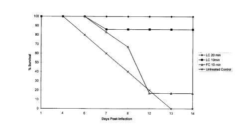
Treatment of mice against respiratory tularemia
Turning to Figure 1, the prophylactic efficacy of aerosolized free unencapsulated and
liposome-encapsulated ciprofloxacin to protect mice against a respiratory infection against
Francisella tularensis was evaluated. Mice were pr~llea~ed with 10 or 20 minutes exposures to
aerosol containing either PBS (control group), free ~-nen~psulated (FC) or liposome-
encapsulated ciprofloxacin (LC). At 24 hours post aerosol exposure, the mice were intranasally
infected with 10 times 50% lethal doses of F. tularensis. The survival rates in these groups of
mice at day 14 post infection were compared. Untreated control mice began to succumb to the
infection as early as day 5 post infection and by day 13, all mice in the group were dead. Little or
no protection was observed in mice treated with aerosolized free unencapsulated ciprofloxacin.
All but one of the mice in that group died by day 12 post infection. In mice exposed to 10
CA 0221~716 1997-09-17
minutes of aerosolized liposome-enc~psul~ted ciprofloxacin, the survival rate was significantly
higher than the untreated control group (83% versus 0%, P<0.05). The highest level of
protection was observed in mice exposed to 20 mimltes of aerosolized liposome-encapsulated
ciprofloxacin (100% vs. 0%, P<0.01). These results suggest liposome-encapsulated
ciprofloxacin delivered by the aerosol inhalation was highly effective in the prevention of
respiratory F. tularensis infection in mice.
Referring to Figure 2, the treatment of respiratory infection against Francisella tularensis
using aerosolized liposome-encapsulated ciprofloxacin and aerosolized unencapsulated
ciprofloxacin were compared. Groups of mice were intranasally infected with 10 times the 50%
lethal dose of F. tularensis. At 24 hours postinfection, the mice were treated with 20 minl]tes
exposures to aerosolized unencapsulated ciprofloxacin or aerosolized liposome-encapsulated
ciprofloxacin. The survival rates for these groups of mice at day 14 postinfection are shown in
Figure 2. The mice in the untreated control group began to succumb to the infection as early as
day 5 postinfection, and by day 9, all mice in the group were dead. Little or no protection was
observed in mice treated with aerosolized unencapsulated ciprofloxacin. All the mice in that
group died by day 9 postinfection. Among the mice exposed to aerosolized liposome-
encapsulated ciprofloxacin, all the mice survived (P<0.01 versus the control, unencapsulated
ciprofloxacin group). These results suggest that liposome-encapsulated ciprofloxacin delivered
by aerosol inhalation is highly effective in the treatment of respiratory F. tularensis infection in
mice.
16
CA 02215716 1997-09-17
Bacteria load of organs from infected and treated mice
The spleens, livers and lungs from the untreated and pretreated mice were isolated at days
7 and 14 post infection, respectively. These organs were homogenized and assayed for the
presence of F. tularensis growth in cysteine heart agar plates. The results are shown in Table 4.
CA 022l57l6 l997-09-l7
Table 4: Recovery of Francisella tularensis from organs of mice pretreated with aerosolized
liposome-enc~ps~ tçd ciprofloxacin
Group Organ CFU
Untreated control~ Lung 4 x 10⁷
Spleen 4 x 10⁶
Liver 3 x 10⁷
Pretreated~ Lung 2 x 10⁵
Spleen o
Liver 0
CFUs determined at approximately day 7 post infection, before mice were moribound from
infection
CFUs were determined at day 14 post infection
18
CA 0221~716 1997-09-17
The presence of bacteria was only found in the lung at day 14 post infection of mice
which were treated with aerosolized liposome-encapsulated ciprofloxacin. In contrast, the lung,
spleen, and liver of mice from the control group all had a high amount of bacteria at day 7 post
infection. These results suggest that aerosolized liposome-encapsulated ciprofloxacin is potent in
the eradication of F. tularensis from these tissues.
19