Note: Descriptions are shown in the official language in which they were submitted.
CA 02216913 1997-09-29
W096129~30 PCTrUS96JO4~84
METHOD AND APPARATUS FOR DETERMINING BONE DENSITY---
This application i~ a ~Gntinuation-in-part of U.S.
patent application Serial Number 08/025,941 filed March
3, 1993, entitled, "Method and Apparatus for Detel ;nin~
~ Bone Density and Diagnosing Osteoporosis".
5 Backqround and Summary of the Invention
Studies of bone strength in vitro have
demonstrated that decreases in bone strength in both the
spine and femur are directly proportional to bone m;ineral
content. For this reason, bone densitometry has been
10 used extensively for the determination of bone loss in
clinical diagnosis and monitoring. A variety of methods
have been used, such as single and dual photon
absorptiometry and quantitative computer tomography.
However, these methods are time-consuming, dependent upon
15 the availability of sophisticated and expensive
equipment, and thus expensive and ill-suited for
widespread implementation. As a result, there has been a
long-felt need in the art for a simple, efficient, and
low cost methodology for measuring bone density as
20 density is an effective indicator of the onset of
osteoporosis, a debilitating disease l_ ly found in
post-pregnancy and post-menopausal women. Treatment of
osteoporosis is most effective if the disease is detected
early whereupon hoL - ~l treatment may be ~ -n~.
25 However, because of the increased risk of side effects,
it is undesirable to begin ho~ ~n~l treatment until the
disease has been detected. Additionally, measurement of
bone density over time may be used to determine the
effectiveness of treatment, leading to adjustments in the
30 treatment protocols balanced against the atten~nt side
effect risk.
Still another medical situation in which bone
integrity is important is the healing process of bone
CA 02216913 1997-09-29
W O 96/29930 PC~r~US96/04384
fractures. There is a ph~n- -non known as non-union
heal ~ng in which a bone fracture fails to knit properly
to return the bone to its pre-fracture integrity.
Obviously, x-rays may be used to monitor the healing
5 process, but this methodology is expensive and
undesirable in that it repeatedly exposes a body part to
radiation. The inventor has previously participated in
studies which noted the relationship of the vibrational
response, and specifically the determination of the
10 natural frequency shift and phase angle shift as being
related, indirectly, to the progress of fracture healing.
This previous experimental work used cadaver bones and
its application to living patients is limited for the
reasons discussed in his previously published article.
lS See "Monitoring of Fracture Healing Bilateral and Axial
Vibration Analysis," Journal of Biomechanics, Vol. 23,
No. 4, 1990.
In order to solve these and other problems in the
prior art, the inventor has succ~e~ed in developing a
20 method and apparatus for determin;~g bone integrity by
measuring the vibrational response of the bone to a
stimulus and determining the modal damping factor of the
bone from the response.
In general, the inventor has developed two
25 t~hn;ques for measuring the modal damping factor of any
bone or other hard tissue. Both techniques include the
basic method of coupling a mechano-electrical vibration
transducer to the bone. The transducer senses the
vibrational response and produces an electrical output
30 which is proportional to the vibrational response. A
programmed electronic logic device such as a computer may
be used to determine the modal damping factor from the
electrical output of the transducer.
CA 02216913 1997-09-29
W 096/29930 PCT/u~ 4
In a first implementation of this method, a,n
impulse of energy is applied to the bone, such as by
striking the flesh surrolln~ ng the bone, in order to
generate a vibration in the bone at the lowest natural
5 frequency of the bone. This vibration has a decreasing
amplitude which may be measured and used to calculate the
modal damping factor. In a second implementation of this
same method, a continuous input of energy is applied to
the bone, such as by driving a speaker or other electro-
?~h~n;cal vibration transducer with a frequencygenerator and coupling the speaker to the bone, suc:h that
a continuous vibrational input is provided at about; a
natural frequency of the bone. Ideally, the frequency of
the energy output from the frequency generator is
15 adjustable so that it may be tuned to a natural freiquency
of the bone. The same transducer and computer may then
be used to calculate the modal damping factor through a
different mathematical analysis which depends upon the
half power bandwidth and center frequency of the
20 vibrational response of the bone.
One of the difficulties found in implementing the
methods described above is that not only does the bone
vibrate in response to the input, but also the flesh
surrol~n~ing the bone vibrates. The sensors used to
25 measure the bone vibration also pick up the vibration of
the flesh. Because the bone and flesh vibrate at
different natural frequencies and the vibration of each
is affected by the other, the resulting output sign~l
does not behave like a theoretical one degree of freedom
30 ~y~e",. Rather, the output signal has noise which can
mask the desired signal. Thus, the computational
analysis of the results bP -c somewhat unwieldy.
The inventor has overcome this problem by
developing a vibrator apparatus which compresses the
CA 022l69l3 l997-09-29
W 096/29930 PCTrUS9G/01~84
flesh while vibrating the bone. Compressing the flesh
raises its natural frequency and ~lr _._llS its response
sufficiently to reduce the amplitude of the vibrational
response of the flesh without affecting the amplitude of
5 the vibrational response of the bone. Therefore, a
relatively noise-free bone response which approaches a
theoretical one degree of freedom system is produced when
the bone is excited with the vibrator apparatus of the
present invention. Further, the vibrator only vibrates
10 when sufficient pressure is applied to the flesh to
sufficiently reduce its response and includes an
indicator for ~; gn~l ing the user if too great a pressure
which could cause injury or discomfort is applied to the
flesh. Vibration and force sensors are also built into
15 the apparatus of the preferred embodiment to improve the
control of the apparatus and reduce the need for some
external instrumentation.
Although the dynamic response of the flesh may be
significantly reduced, it is not completely eliminated
20 with the vibrator apparatus of the present invention.
Therefore, the response received from the bone contains
some noise even though the vibrator apparatus is used to
excite the bone. Further, digital computer analysis
techniques typically sample data at time intervals. This
25 discrete sampling presents inaccuracies and computational
difficulties in analyzing the bone response as is well
known in the art and further compounds the analysis
difficulties due to noise.
In order to solve these problems caused by noise
30 and discrete sampling, the inventor has succeeded in
developing a computer algorithm which estimates the modal
damping factor from discrete vibration data received from
the bone. The algorithm matches the measured response
with a theoretical one degree of system response and
CA 02216913 1997-09-29
WO !J6/29930 PCT/US961043~4
varies the theoretical system parameters until a suitable
correlation between the theoretical and actual responses
is achieved. When the suitable correlation is ach;ieved,
the actual bone modal damping factor is estimated ~to be
5 that of the theoretical ~y:~- . In proving the efficacy
of the methodology disclosed and claimed herein, the
inventor has conducted several experiment~ on bones. In
doing so, the inventor has discovered that the change in
the modal damping factor is one order of magnitude
lO greater than the corresponding change in bone dens:i.ty.
Thus, measurement of the modal damping factor is
sensitive to and useful in determ; n; ng bone density.
Furthermore, although it is desirable to locate
the vibration transducer close to the bone in order to
15 increase the gain of the measured vibrational response
and decrease noise from other structures to thereb~
; n; ; ze measurement errors, it has been found tha1, the
flesh which surrounds the bone has little affect on the
measurement because the bone ~om;~tes the vibrational
20 response and therefore the modal damping factor at the
lower natural frequencies. Thus, the vibration
transducer may be mounted on the skin and the
measurements may be taken through the flesh surrounding
the bone. Thus, no penetration of the skin is required
25 to make the measule"-ellt.
Still further, as the modal damping factor .is a
measure of the loss of strain energy during one vibratory
cycle in the stress bearing part of the bone, it i.c:
relatively insensitive to boundary conditions such as the
30 support given the bone and the characteristics of t:he
surro~ ;~g flesh. On the other hand, other dynamic
properties, such as the natural frequency used in the
inventor's prior published article, vary significan.tly
when the boundary conditions change, and are not ne.arly
CA 02216913 1997-09-29
W096/29930 PCTrUS96/04384
as sensitive to changes in density, which makes these
dynamic properties impractical to use in measuring and
monitoring bone density levels and changes in the density
over time.
The inventor's approach of using the modal damping
factor as an indicator of bone density is also
intellectually satisfying in that there is a rationale
for the experimentally measured variations in the modal
damping factor. It is generally understood and believed
10 that loss of mass, or decrease in density, of a bone is
due to the loss of minerals and a resultant void
nucleation that, in turn, results in stress co~c~ntration
and premature fracture. This void nucleation is detected
by a change in the modal damping factor as the bone
15 becomes more porous which decreases the bone stiffness
and increases the bone damping. Thus, the measurement of
the modal damping factor is seen to be a direct
measurement of this void nucleation and, hence, a direct
indication of the integrity of the bone.
The inventor's techniques may be readily applied
to the diagnosis and treatment of osteoporosis. In the
first instance, the density of a particular bone of a
patient may be estimated by measuring the modal damping
factor, and the modal damping factor may be determined in
25 the same ~nn~ at various intervals of time as the
patient is treated. These modal damping factors taken at
various time intervals may be compared to detect any
changes which would indicate a change in bone density. A
decrease in bone density in medically significant amounts
30 indicates the onset of osteoporosis. Alternately, it
would be hoped that treatment, perhaps through exercise,
would be helpful in increasing or at least forestalling
the decrease in bone density. Thus, these techniques may
be useful in measuring the effectiveness of treatments so
CA 02216913 1997-09-29
WO 96/29930 PCI/US9C/04384
that treatment protocols may be altered over time as the
patient is treated. This methodology and use of the
modal damping factor ~r~nA~ upon a relative ~ _-rison
of modal damping factor measurements for the same ~one in
5 the same patient over time.
An alternate methodology takes advantage of
st~A~rdized modal damping factors or bone density
v;~lues, yet to be detel ;neA~ for patients and bones
having various characteristics such as age, sex, f:itness
10 level, bone type. Using this alternate method, a
particular patient's bone density or modal damping factor
measurement may be compared to the st~n~rdized va:Lues in
order to determine their potential for having
osteoporosis. As the inventor has recently developed the
15 present invention, there has not been an opportuni~y to
determine these st~ ~dized values. However, it :is
believed to be a straightforward matter for one of
ordinary skill in the art to use the present invenlcion
and measure a statistically significant group of
20 individuals in order to determine these st~nA~rdized
values and the particular factors ; ,~ol~ant in
differentiating members of the group.
While the principal advantages and features of the
present invention have been described above, a more
25 complete and thorough underst~nAi ng of the inventic~n may
be attained by referring to the drawings and descriLption
of the preferred embodiment which follow.
CA 02216913 1997-09-29
W O 96/29930 PC~rrUS96/04384
Brief Descri~tion of the Drawin~s
Further objects and features of the present
invention are revealed in the following Detailed
Description of the Preferred Embodiment of the invention
5 and in the drawing figures wherein:
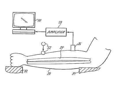
Figure 1 is a diayl -tic view of a first
ter-hn;que for measuring bone density by inputting an
impulse of energy to induce a vibration into the bone;
Figure 2 is a graph of the harmonic response of
10 vibrations induced in the bone through the technique
shown in Figure l;
Figure 3 is a diayLl- -tic view of a second
t~chnique for measuring bone density through the coupling
of a continuous energy source to the bone;
Figure 4 is a graph of the vibrational response
induced in the bone using the t~rhnique of Figure 3;
Figure 5 is a cross-sectional view of a vibrator
apparatus used as a continuous energy source to excite a
bone in vivo; and
Figure 6 is a logic listing of a computer program
used to estimate the bone modal damping factor.
Detailed DescriDtion of the Preferred Embodiment
As shown in Figures 1 and 2, the inventor's first
technique for measuring bone density includes the step of
25 inducing a vibration in the bone which is desired to be
measured, for example by striking the flesh of a
patient's arm 20 with a blunt instrument such as a rod 22
to induce vibrations in a bone 24 within the patient's ;
arm. For convenience, the opposite ends of the patient's
30 arm 20 may be supported by a pair of Sup~Ol Ls 32, 34. A
m~.h~no-electrical vibration transducer 26 measures the
induced vibration of the bone 24 and produces an
electrical output which may be amplified by an amplifier
CA 02216913 1997-09-29
W096129930 PCTnUS96~04384
28 and then input to a computer 30 for calculation of the~
modal damping factor.
As shown in Figure 2, the vibration induced by the
input of an impulse of energy into the arm 20 will have a
5 different initial amplitude corresponA~g to varying
input force levels. However, the ratio of the amplitudes
of the first and second cycles of vibration (Al/A2) is
invariant with respect to the level of the force input to
the bone. Thus, the modal damping factor may be
10 calculated by comparing the amplitudes of successive
cycles of vibration induced by any of these input ~orce
levels. As shown in Figure 2, the intensity of the blow
to the arm does not affect the measurement of the modal
damping factor as the modal damping factor is dete~ ;neA
15 by comparing two successive amplitudes and the ratio of
two successive amplitudes is constant regardless of' their
size. Whether the initial amplitude has an intensity of
a, b, or c, there is no variation in the measured modal
damping factor. Instead, the modal damping factor of the
20 bone 24 is preAom;n~ntly dependant on the characteristics
of the bone.
As shown in Figures 3 and 4, an alternate
te~hn;que for measuring bone modal damping factor may be
used. As before, the patient's arm 20 has a bone 24
25 surroll~A;ng the flesh to which a --h~no-electrical
vibration transducer 26 is mounted for converting t:he
sensed vibrational response to an electrical signal which
may be amplified by an amplifier 28 and input to a
computer 30. However, the initial energy input to the
30 patient's arm 20 is achieved by way of a frequency
generator 36 which produces an electrical output at; a
particular frequency which is amplified by a power
amplifier 38 which amplifies the output from the
frequency generator to a particular amplitude. The
CA 02216913 1997-09-29
W O 96/29930 PCTAUS96/04384
output from the power amplifier 38 is fed to a second
transducer 40, which may be a speaker or Qh~k~ or other
such electro- -ch~nical vibration transducer coupled to
the patient's arm 20. The frequency generator 36 is then
5 tuned to frequencies sweeping through a range of the
lower natural frequencies of the particular patient's
particular bone 24 being measured to produce a continuous
vibrational response as shown in Figure 4. A ~-xi ~m
amplitude of one of the several natural frequencies
10 induced in the patient's bone 24 is chosen for
measurement of the modal damping factor. As is well
known in the art, the modal damping factor is equal to
the half power bandwidth, ~F, or F2 - F1, divided by the
center frequency Fc. The center frequency, Fc, is the
15 frequency at which the ~xi ~m amplitude occurs. The
half power frequencies, Fl and F2, are those frequencies
at which the amplitude is (~2)/2, or about .707 times the
amplitude.
The inventor has conducted two separate
20 experiments which prove the efficacy of utilizing the
modal damping factor for measuring bone density. In a
first experiment, chicken femoral bones were treated with
hydrochloric acid for varying lengths of time, their mass
was measured, and then their modal damping factor was
25 determined using techniques similar to those disclosed
herein. Their modal damping factors were then compared
with the modal damping factors of untreated chicken
femoral bones. The modal damping factor directly
correlated with the number of hours of acid treatment of
30 the chicken femoral bones. This was to be expected as
the longer the bones were immersed in the acid, the
greater their porosity, the greater the reduction in
their mass and hence the greater the reduction in their
density. Furthermore, the change in modal damping factor
CA 02216913 1997-09-29
W 096/29930 PCTrUS96/04384
was nearly one order of magnitude greater than the change
in the measured density of the bone. Hence, the modal
damping factor was considered to be highly sensitive to
changes in density and thus a good parameter for
5 measuring density as smaller changes in density could be
readily detected.
In a ~on~ experiment, rat bones were used. More
particularly, tibiae of two groups of rats were compared,
one group having undergone an extensive training program.
lO It being understood that training increases bone density
which should cause a reduction in modal damping fac:tor.
Both groups included members which were relatively young,
as well as members which were relatively old. In
comparing the bones for the younger rats, it was found
15 that tr~;ni~g resulted in slightly lower bone density and
higher damping ratio, but these changes were considered
tc~ be statistically insignificant. However, in the older
rats, the average change in modal damping factor due to
training was about forty percent while the change in
20 density was about 23 percent. These experimental results
subject the beneficial results of physical exercise or
training in older individuals of maint~ining the mineral
content and hence the density of the individual's bones.
Again, the modal damping factor measurement was found to
25 significantly correlate with bone density.
Although various electro-~ech~nical vibration
transducers 40 may be used in the second method desrribed
above and shown in Figure 3, the inventor has succeeded
in developing a vibrator apparatus 100 shown in Figure 5
30 which is particularly well-suited to the task. The
apparatus 100 is generally comprised of an electro-
ch~n~cal vibration transducer 102 mounted between ahousing 104 and a pad 106 for vibrating the pad rel.~tive
to the housing. Further, the apparatus 100 has several
CA 02216913 1997-09-29
W 096/29930 PCTrUS~.-f~ 4
internal control and measurement circuits 110, 112, 114,
116 which determine when the transducer 102 vibrates,
which signal the user, which measure the force with which
the pad 106 is pressed against the patient and which
5 measure the amplitude at which the pad vibrates relative
to the housing 104, respectively. A connector 118
mounted on the housing 104 is used to communicate signals
between the control and measurement circuits 110, 112,
114, 116 and various external systems such as frequency
10 generators, amplifiers and computers.
The vibration transducer 102 of the preferred
embodiment includes two conc~ntric annular magnets 120,
122 joined by a flat magnetic plate 124 which in
combination produce a complex magnetic field about the
15 magnets. An electrical coil or coil driver 126 is
positioned in the space 128 formed between the inner and
outer annular magnets 122, 120 and is held in place by a
resilient elastic diaphragm 130 positioned adjacent the
magnets and opposite the magnetic plate 124. The
20 electrical coil 126 is a single strand of wire wound
about an axis as is well-known in electro-~?ch~nical
devices. Electrical leads 132 are connected to each end
of the single strand of wire in the electrical coil 126
and extend through the housing 104 to the connector 118
25 so that the coil may be energized from a source external
to the vibrator apparatus 100. Note that Figure 5
represents each electrical lead with a single thickness
curling line. Nonetheless, all of the leads in the
apparatus 100, including leads 132, are two-wire
30 insulated parallel conductors in the best mode.
Due to what is known as the Faraday effect, when
an electrical current passes through the coil 126, a
magnetic field is induced about the coil. Depending upon
the polarity of the electrical current, the magnetic
CA 02216913 1997-09-29
W 096/29930 PCT~US96J~43~4
field induced about the coil 126 will either attract or
repel the coil toward or away from the complex magnetic
field surrolln~;ng the permanent magnets 120, 122, ~L24.
If an alternating current passes through the coil ~L26,
5 the coil will alternately be attracted and repelled by
the magnetic field of the magnets 120, 122, 124. ~3ecause
the coil 126 is mounted on the resilient diaphragm 130,
the coil displaces toward and away from the magnets 120,
122, 124 as the alternating current passes through the
10 coil. Thus, the coil 126 oscillates back and forth in
the space 128 between the inner and outer magnets 122,
120 as the polarity of the alternating current changes
o~er time. The fre~uency and amplitude of the coi:L 126
03cillation varies with changes in the frequency and
15 amplitude of the alternating current. Dep~n~i ng upon the
~y~el-- dynamic characteristics, the frequency of the coil
126 oscillation may be equal to the frequency of the
alternating current, but need not be. Likewise, the coil
126 displ~c~ ~t amplitude may linearly vary with respec1
20 to the amplitude of the alternating current, but need
not. Although the particular structure described above
is used in the preferred embodiment, other embodiments as
are well known in the art are also within the scope of
this invention. For instance, the magnets 120, 122, 124
25 may be replaced with a ferrous material provided that the
mean of the alternating current is shifted to produce a
constant polarity alternating current. Further, t]ne
entire structure may be replaced with a c~ ~ "off-the-
shelf" acoustical speaker.
As shown in Figure 5, an annular disk 140 is
attached to the diaphragm 130 opposite the coil 12~ and a
hollow cylinder 142 extends from the disk toward t~he
center of the housing 104 where the cylinder connects to
a collar 144 and a circular ring 146. A hollow rod 148
CA 022l69l3 l997-09-29
W096/29930 PCTrUS96/04384
14
is cnnn~rted to the collar 144 with a pin 150 and extends
through the housing 104 opposite the magnetic plate 124.
Each of these connections may be made in any of a variety
of ways as are well known in the art, such as by screw
5 fast~n~ng, brazing, welding, adhesively bonding, etc.
Likewise, although a pin 150 is used in the preferred
embodiment to ~o~nect the rod 148 to the collar 144,
other fastening means may be used and are within the
scope of this invention. The pad 106 is positioned at
10 the distal end of the rod 148 and has a circular
configuration with rounded corners. Because the magnets
120, 122, 124 are connected to the housing 104 and the
pad 106 is connected to the coil 126, the pad oscillates
relative to the housing when the coil oscillates relative
15 to the magnets. A bl-~h;ng 154 seals the space between
the housing 104 and the rod 148 to prevent cont~in~ntS
from entering the housing. The bll~hi ng 154 also prevents
lateral mov.~ -nt and wear between the rod 148 and the
housing 104 to i ,love the life and performance of the
20 apparatus 100.
The housing 104 is comprised of a cylindrical
portion 160 which is sized to fit in the palm of the
user's hand and a conical portion 162, the apex of which
is adjacent the pad 106. The shape of the conical
25 portion 162 gives a better line of sight to the pad 106
than would otherwise be available if a non-tapered shape
where used. The line of sight permits visual
confirmation of the location of the pad 106 as the
cylindrical portion 160 is held in the user's hand.
30 Although the housing 104 of the preferred embodiment is
sized and shaped to be hand-held, the housing may
alternatively be sized and shaped to be mounted in a
robotic or stationary fixture without departing from the
scope of this invention.
CA 02216913 1997-09-29
WO 96/29930 PCT~US9. ~o ~?~
The electro-mech~nical vibration transducer 102
and vibrator apparatus 100 described above is fairly
typical of those found in the prior art. However, the
control and measurement circuits llO, 112, 114, 116 of
5 the vibrator apparatus described below depart from
typical vibration devices and make this vibrator
apparatus exceptionally well suited to the task of
exciting a patient's bone in vivo.
Two annular plate springs 170, 172 spaced by an
10 annular spacer 174 are att~che~ to the cylinder 142 and
to the housing 104. ~he cylinder 142 is att~h~ t:o the
inner diametral edges 175, 176 of the annular springs
170, 172 and the housing 104 is attached to the out:er
diametral edges 177, 178 of the springs. As shown in
15 Figure 5, the springs 170, 172 are positioned
intermediate the ends of the cyl;n~r 142 and near the
intersection of the cylindrical and conical portions 160,
1~2 of the housing 104. Although any elastic material
may be used for the springs 170, 172, a generally
20 linearly elastic material is used in the preferred
~mh~ nt so that the spring properties and dynamic
characteristics are easily evaluated. The springs 170,
172 bias the cylinder 142 toward an equilibrium position
with respect to the housing 104 wherein both sprinsrs are
25 substantially planar and undeflected. However, the pad
106 may be pushed toward the housing to deflect the
springs 170, 172 away from their equilibrium positions.
Because the springs have a generally linear spring
constant, the displ~ nt of the pad 106 relative to the
30 housing 104 is proportional to the force with which the
pad is pushed toward the housing. The pad 106 has a
constant surface area 179 configured to be pressed
against a patient's flesh. When the pad 106 is pressed,
the normal pressure against the pad is equal to the force
CA 02216913 1997-09-29
W 096/29930 PCTrUS~
with which the pad is pushed multiplied by the surface
area 179. Thus, because the displ~c- -nt of the pad is
~- ~OL Lional to the force and the pad surface area 179 is
constant, the displ~l- -nt is also ~Lo~ulLional to the
5 pressure with which the pad is pressed against the
patient's flesh.
A switch 180 is positioned within the housing 104
and adjacent the ring 146 so that the ring actuates the
switch when the ring travels a predetermined distance
10 relative to the housing. Because the distance traveled
is proportional tû the pressure with which the pad 106 is
pressed, the switch 180 may be set to actuate when a
~; n; 1~ predetermined pressure is achieved between the
pad and the patient's flesh. Actuation of the switch 180
15 closes the circuit within the switch to permit electrical
current to pass through the switch. Leads 182 extend
from the each side of the circuit within the switch 180
and may extend to the connector 118 mounted on the
housing. By placing the switch 180 in series with the
20 electro- ?ch~n;cal vibration transducer 102, the
apparatus 100 may be configured to only vibrate when a
predetermined ~; n; ]~ pressure is achieved between the
patient's flesh and the pad 106. Together the switch 180
and springs 170, 172 form the pressure-vibration control
25 110 which detel ;n~s when the transducer 102 vibrates.
As appreciated by those in the art, the control 110 is a
?ch~no-electrical force transducer. The value of the
minimum predetermined pressure may be changed or adjusted
by moving the switch 180 relative to the housing. Thus,
30 the switch 180 may be adjustably mounted to the housing
in any of several ways which are well-known in the art.
The over-pressure control 112 works similarly to
the pressure-vibration control 110 and is a second
mechano-electrical force transducer. The control 112
CA 02216913 1997-09-29
WO 96/29930 PCT/US96/04.~84
17
includes a ~ro~A switch 190 mounted within the housing
104 and ad;acent the ring 146 so that the switch c~.oses
when a maximum predetermined pressure is applied to the
pad 106. Leads 192 ext~n~ing from the switch 190 conn~ct
5 the switch to a light emitting diode (LED) 196 or c)ther
display or signal device to alert the user that the
Q~ predetel ~ n~ pressure has been re~ch~ ancl/or
~r~ . In this way, the user may be alerted that the
pressure between the pad 106 and the patient's flesh is
10 higher than needed and may be so high as to cause
contusions or lacerations or other undesirable side
effects. Similarly to switch 180, switch l9Q may be
adjustably mounted to permit the m~ ~ predetermined
pressure to be adjusted.
The displ~e~?nt sensor 114 includes a ~~h;qn~-
electrical vibration transducer 200 positioned bet~een
the housing 104 and the ring 146 to measure the
displ~- ~ t of the pad 106 relative to the housing. As
is well known in the art, -~hA~o-electrical vibrat,ion
20 transducers sense displacement and output an electrical
signal which is line~ly proportional to the sensed
displacement. Leads 202 extend from the ?ch~no-
electrical vibration transducer 200 to the connector 118
so that the amplitude of the vibratory oscillations of
25 the pad 106 may be monitored external to the vibrator
apparatus 100. Thus, the ~ys~ dynamic characteristics
o~ the apparatus 100 need not be known or even linear
with respect to the alternating current input to
determine the frequency and amplitude of the pad 106
30 oscillations relative to the housing 104.
The force sensor 116 includes a force transclucer
210 positioned between the rod 148 and pad 106 so that
the force transmitted through the rod may be measured.
Leads 212 extend from the transducer 210 to the ronn~rtor
CA 022l69l3 l997-09-29
W096/29930 PCTAUS96/04384
18
118 to permit the sensed force data to be transmitted
external to the housing 104. As previously described,
the force in the rod 148 is ~LO~L ~ional to the pressure
with which the pad 106 is applied to the flesh of the
5 patient.
To operate the vibrator apparatus 100 described
above, a user such as a nurse or medical technician holds
the cylindrical portion 160 of the housing 104 in the
palm of their hand and presses the pad 106 against the
10 patient's flesh. As mentioned previously, the conical
shape of the conical portion 162 of the housing 104
permits the user to visually confirm where the pad 106 is
being pressed with respect to the patient's flesh. As
the pressure between the pad 106 and the patient's flesh
15 increases, the ring 146 deflects toward the switches 180,
190 (upward as shown in Figure 1) until the ring actuates
switch 180 which is in series with the electro-mechanical
vibration transducer 102 and the circuit within the
switch closes to permit electrical current to pass
20 through the coil 126. Because the electrical current is
an alternating electrical current, the coil 146 and pad
106 oscillate as previously described when the pad is
pressed against the patient's flesh with a pressure equal
to or greater than the ~; ni ~m predetermined pressure.
25 When the user presses the pad 106 against the patient's
flesh with an increased pressure, the pressure may exceed
the predeteL ; nF~(9 pressure at which complications or
discomfort may begin. When this maximum predetermined
pressure is achieved, the other switch 190 is actuated to
30 energize and illuminate the LED 196. This signals the
user to reduce the pressure in order to avoid these
unwanted complications.
The advantage of pressing the pad 106 against the
patient's flesh is that the flesh is compressed which
CA 02216913 1997-09-29
W O 96/29930 PCTrUSS~ g
19
reduces the dynamic response of the flesh to the
vibration input. When the flesh is ~ essed, the
dynamic response of the flesh is ~ _cd and the natural
frequency of the flesh is increased because the effective
5 stiffness of the flesh is increased. In addition, the
vibrator apparatus 100 oscillates the pad 106 at
freguencies which are lower than the natural frequencies
of the compressed flesh but which are within the range of
the lower natural frequencies of the bone being measured.
10 Thus, the flesh is not excited by the vibratory input
from the vibration transducer 102 but the bone is
excited. Further, the flesh response is ~ _cd so its
amplitude is small relative to the amplitude of the~ bone
response. In this way, the response of the bone is
"decoupled" from the response of the flesh. Thus, the
noise in the response which occurred when using prior art
vibrator apparatus due to the flesh vibrating
independently of the bone is virtually eliminated,
leaving a relatively clean signal which may be easily
20 analyzed to determine the modal damping factor of the
bone alone.
The signals output through the ~o~n~ctor 118 may
be transmitted to an external control (not shown) cmd/or
to a computer for detailed analysis of both the
25 excitation and the response. Alternatively, the signals
may be stored in a data collection device (not sho~m) for
later and/or remote data reduction and analysis. ]:n the
preferred embodiment, the response to the excitation is
analyzed using a microprocessor c~n~ted to the vibrator
30 apparatus 100 external to the housing 104.
The mi~~ ocessor of the preferred embo~; ?nt
uses the algorithm shown in Figure 6 to analyze the
response to the continuous excitation. The
microprocessor may be based on a PENTIUM processor chip
CA 02216913 1997-09-29
W 096/29930 PCTAUS96/04384
or the like as is well known in the art. PENTIUM is a
U.S. registered trademark of the Intel Corporation.
The frequency of the input is varied over time and
the response is sampled over a range of input
5 frequencies. These responses are stored as values
representing the amplitude of displ~c~ ?nt taken at
spaced time intervals correspo~;ng with the sampling
rate. These stored amplitude values lay on a curve like
that shown in Figure 4 and may be processed as described
10 above with respect to the second method of determ~ n~ ng
modal damping factor. Because of minor noise in the
response signal as well as the discrete rather than
continuous sampling used, the center frequency and half
power frequencies of the stored data are difficult to
15 determine with the precision desired. Thus, the data is
matched to an idealized theoretical response and the
modal damping factor of the response is estimated as the
modal damping factor of the theoretical system.
The system dynamics calculations are simplified by
20 idealizing the behavior of the bone and flesh as a one
degree of freedom system with simple second order
response as is well understood in the art. This
idealization is fairly accurate if an excitation source
such as the vibrator apparatus 100 is used because the
25 bone and flesh are decoupled and the bone dominates the
response at the lower natural frequencies. For such an
idealized system, the amplitude of the response, Yi,
varies with the input frequency, ~i. as follows:
Yi= 2x~5 ~
~ [( - )2-1]2+(25 i)2
were xmax equals the maximum theoretical amplitude which
30 occurs at the center frequency, Fc, of the particular mode
-
CA 02216913 1997-09-29
WO 96/2993~ PCTnUS96104;384
being analyzed. As described earlier, ~ is the mo~al
damping factor of the bone for the mode being anal~zed,
and ~n is the natural frequency in radians per ~c,r~ of
the bone at the =~; theoretical amplitude, x~. It
5 is readily apparent that ~n and Fc are different
e~pressions for the same quantity; however, one is
expressed in cycles per cecon~ (Fc) and the other is
expressed in radians per second (~n ) ~
The computer algorithm shown in Figure 6 minimizes
10 the difference between the measured amplitudes and the
theoretical amplitudes by varying the maximum ampliitude
(x~), the modal damping factor (~), and the natural
frequency (~n)~ AS is ~ in the art, the difference
or error between the samples and the theoretical
15 amplitudes is evaluated by sl i n~ the squares of 1he
differences of the values over the entire range of
samples. Although many different iterative numerical
analysis techn;gues could be used, the inventor uses the
Newton-Raphson iteration method to r; n; m; ~e the error.
20 The Newton-Raphson method, also known as Newton's method,
is an iterative process whereby an initial value or guess
for a solution is assumed and a next value or ; ~ vv~d
guess is calculated by adjusting the previous guess by an
in~L -nt equal to the quantity of the function at the
25 last guess divided by the slope of the function at the
last guess. In other words,
f ( Xn )
This iteration method is used because of its simplicity
and relative speed of convergence to a solution. As with
any iterative analytical approach, the process of
30 improving the guess is repeated until the guess converges
to a solution having an error less than some specii-ied
CA 02216913 1997-09-29
W 096/29930 PCTAUS96/04384
value. Because of the nr nCl ature used in many ~
computer languages, each iterative process is commonly
referred to as a "do-loop".
In the first do-loop of the algorithm shown in
5 Figure 6, the Newton-Raphson method is used to obtain a
good initial guess for the modal damping factor. The
natural frequency and -xi amplitude are held constant
while the modal damping factor is varied until the amount
of change in modal damping factor is within a specified
10 value (i.e., "Icorrectionl < preset value"). Once this
improved initial guess is found, the algorithm contin
to a second do-loop.
In the second do-loop, the Newton-Raphson method
is again used; however, this time the modal damping
15 factor, natural frequency and ~xi ~m amplitude are each
varied and a separate value for the error and the change
in error with respect to the change in each of the three
parameters is calculated. The parameters are varied
until the sum of the squares of the differences of the
20 errors is mini~ized~ When the sum is ~inimized~ the
theoretical amplitudes are close to the sample amplitudes
over the entire range of samples. Thus, the theoretical
modal damping factor and the theoretical natural
frequency when the sum of the squares of the differences
25 in the errors is i ni i zed are a good estimate of the
actual modal damping factor and natural frequency of the
bone being analyzed. Therefore, a modal damping factor
may be accurately estimated for the system even though
the system response has small noise fluctuations and is
30 only discretely sampled.
It should be readily appreciated that other
iterative analysis and/or curve fitting techniques may
also be used to minim~ ze the difference between the
measured and theoretical amplitudes and modal damping
CA 02216913 1997-09-29
W 096/29930 PCTrUS96104.384
23
factors. Further, the Newton-Raphson method may be used
in different ways to arrive at a solution. For instance,
the first do-loop for fin~;ng an improved initial guess
for modal damping factor could be eliminated if desired
5 or supplanted by other do-loops in which improved initial
guesses for natural frequency or ~ amplitude ,are
found. Likewise, other error evaluation tPrhniques can
be used to measure the error between the theoretical and
measured amplitudes. Yet another variation of the
lO analysis technique is to use a simplified higher de!~ree
of freedom system or higher order system approximation
for the theoretical amplitude calculation. Each of these
variations in analysis technique are within the scope of
this invention.
In addition, there are various other changes and
modifications which may be made to the invention as would
be apparent to those skilled in the art. However, these
changes or modifications are included in the teaching of
the disclosure, and it is int~n~ that the invention be
20 limited only by the scope of the claims appended hereto.