Note: Descriptions are shown in the official language in which they were submitted.
CA 02216943 1997-09-29
WO 96/32907 PCT/IB96/(10272
-1-
DRUG RELEASE COATED STENT
BACKGROUND OF THE INVENTION
I. Field of the Invention
The present invention relates generally to elastic, self-expanding :dent
prostheses for lumen, e.g., vascular, implantation andu more particularly, to
the
provision of biostable elastomeric coatings on such stents which incorporate
elutable or diffusible biologically active species for controlled release
directly iru the
coating structure.
II. Related Art
to In surgical or other related invasive medicinal procedures, the insertion
and
expansion of stent devices in blood vessels, urinary tracts or other difficult
to
access places for the purpose of preventing restenosis, providing vessel or
lumen
wall support or reinforcement and for other therapeutic or restorative
functions
have become a common form of long-term treatment. Typically, such prostheses
are applied to a location of interest utilizing a vascular catheter, or
similar
transluminal device, to carry the stent to the location of interest where it
is
thereafter released and expanded in situ. These devices are designed primarily
as
permanent implants which may become incorporated in the vascular or other
tissue which they contact at implantation.
2o Stent devices of the self expanding tubular type for transluminai
implantation, then, are generally known. One type of such device includea a
flexible tubular body which is composed of several individual flexible thread
elements each of which extends in a helix configuration with the centerline of
the
body serving as a common axis. A plurality of elements having the same
direction
of winding but which are displaced axially relative to each other are provided
which .
meet under crossing a like number of elements also so axially displaced but
having
the opposite direction of winding. This configuration provides a sort of
braided
tubular structure which assumes a stable dedicated diameter upon the
relaxation
but which can be reduced as for insertion by the application of axial tension
which,
3o in turn, produces elongation of the body with a corresponding diameter
contraction
that allows the stent to be conducted through the vascular system as a narrow
elongated device and thereafter allowed to expand upon relaxation at the
location
of interest. Prostheses of the class including a braided flexible tubular body
are
CA 02216943 1997-09-29
WO 96/32907 PCT/IB96100272
-2-
illustrated and described in U.S. Patents 4,655,771 and 4,954,126 to Wallsten
and
5,061,275 to Wallsten et al.
The general idea of utilizing implanted stents to carry medicinal agents,
such as thrombolytic agents, also have been devised. U.S. Patent 5,163,952 to
Froix discloses a thermal memoried expanding plastic stent device which can be
formulated to carry a medicinal agent by utilizing the material of the stent
itself as
an inert polymeric drug carrier. Pinchuk, in U.S. Patent 5,092,877, discloses
a
stent of a polymeric material which may be employed with a coating associated
with the delivery of drugs. Other patents which are directed to devices of the
class
1o utilizing bio-degradable or bio-sorbable polymers include Tang et al, U.S.
Patent
4,916,193, and MacGregor, U.S. Patent 4,994,071. A patent to Sahatjian, Patent
No. 5,304,121, discloses a coating applied to a stent consisting of a hydrogel
polymer and a preselected drug in which possible drugs include cell growth
inhibitors and heparin. A further method of making a coated intravascular
stent
carrying a therapeutic material in which a polymer coating is dissolved in a
solvent
and the therapeutic material dispersed in the solvent and the solvent
thereafter
evaporated is described in European patent application 0 623 354 A1 published
09
November 1994.
An article by Michael N. Helmus (a co-inventor of the present invention)
2o entitled "Medical Device Design--A Systems Approach: Central Venous
Catheters", 22nd International Society for the Advancement of Material and
Process Engineering Technical Conference (1990) discloses surfactant-heparin
complexes to be used as controlled release heparin coatings. Those polymer/
drug/membrane systems require two distinct layers of function.
While many attempts have been made to incorporate drug delivery in
conjunction with long-term catheter or implanted stent placement, for example,
the
associated release time has been generally, relatively short, measured in
hours
and days, and success has been limited. There remains a need for a
comprehensive approach that provides long-term drug release, i.e., over a
period
of weeks or months, incorporated in a controlled-release system. In addition,
there
is a further need with respect to incorporating a drug release coating on a
metallic
stent. Polymeric stents, although effective, cannot equal the mechanical
properties
of metal stents of a like thickness. For example, in keeping a vessel open, a
CA 02216943 2002-06-05
60950-329
-3-
metallic stmt is superior because stem s braided of
relatively fine metal can provide a good deal of strength to
resist circumferential pressure. In order for a polymer'
material to provide the same strength characteristics, a.
much thicker-walled s~~ructure or heavier, denser filament
weave is required. This, in turn, reduces the area
available for flow through the stmt and/or reduces the
amount of porosity available in the stmt. In addition,
when applicable, it is more difficult to load such a stmt
onto catheter delivery systems for conveyance through the
vascular system of the patient to the site of interest.
SLTM~IARY OF THE INVENT7:ON
Many of the limitations of prior art implanted
prolonged drug delivery systems associated with deployed
stmt prostheses are overcome by the provision of a
relatively thin overla.yer of biostable elastomeric material
in which an amount of a biologically active material is
dispersed as a coating on the surfaces o.f the st mt. ThE=_
preferred stmt is a self-expanding, open-ended tubular
stmt prosthesis, with a thin flexible elastic sidewall
structure having openings therein. Although other materials
can be used
CA 02216943 1997-09-29
WO 96/32907 PCT/1B96I00272
-4-
including polymer materials, in the preferred embodiment, the tubular body is
formed of an open braid of fine single or polyfilament wire which flexes
without
collapsing and is axially deformable for insertion using a catheter or other
such
device but which resumes a predetermined stable diameter and length upon
s relaxation.
The coating layer is preferably applied as a mixture of polymeric precursor
and finely divided biologically active species or a solution or partial
solution of such
species in the polymer solvent or vehicle which is thereafter cured in situ.
The
coating may be applied by dipping or spraying using evaporative solvent
materials
of relatively high vapor pressure to produce the desired viscosity and coating
thickness. The coating further is one which adheringly conforms to the surface
of
the filaments of the open structure of the stent so that the open lattice
nature of the
structure of the braid or other pattern is preserved in the coated device.
The elastomeric material that forms a major constituent of the stent coating
should possess certain properties. It is preferably a suitable hydrophobic
biostable
elastomeric material which does not degrade and which minimizes tissue
rejection
and tissue inflammation and one which will undergo encapsulation by tissue
adjacent the stent implantation site. Polymers suitable for such coatings
include
silicones (e.g., polysiloxanes and substituted polysiloxanes), polyurethanes,
2o thermoplastic elastomers in general, ethylene vinyl acetate copolymers,
polyolefin
elastomers, and EPDM rubbers. The above-referenced materials are considered
hydrophobic with respect to the contemplated environment of the invention.
Agents suitable for incorporation include antithrobotics, anticoagulants,
antiplatelet agents, thrombolytics, antiproliferatives, antinflammatories,
agents that
inhibit hyperplasia and in particular restenosis, smooth muscle cell
inhibitors,
growth factors, growth factor inhibitors, cell adhesion inhibitors, cell
adhesion
promoters and drugs that may enhance the formation of healthy neointimal
tissue,
including endothelial cell regeneration. The positive action may come from
inhibiting particular cells (e.g., smooth muscle cells) or tissue formation
(e.g.,
3o fibromuscular tissue) while encouraging different cell migration (e.g.,
endothelium)
and tissue formation (neointimal tissue). '
The preferred materials for fabricating the braided stent include stainless
steel, tantalum, titanium alloys including nitinol (a nickel titanium,
thermomemoried
CA 02216943 2002-06-05
60950-329
-5-
alloy material), and certain cobalt alloys including cobalt-chromium-nidcef
alloys
such as Elgiloy~ and Phynox~. Further details cohceming the fabrication arid
details of other aspects of the stents-themselves, may be gleaned from the
above
referenced U.S. Patents 4,656,771 and 4,954,126 to Wallsten and 5,061,275 to
s Wallsten et al.
Various combinations of polymer coating materials cars be coordinated with
.biologically active species of interest to produce desired effects when
coated on
ix stents to be implanted in accordance with the invention. Loadings of
therapeutic
materials may vary. The mechanism of incorporation of the biologically active
species into the surface coating, and egress mechanism depend both on the=
nature of the surface coating polymer and the material to be incorporated. The
mechanism of release also depends on the mode of incorporation. The material
is may elute via interpartide paths or be administered via transport or
diffusion
through the encapsulating material itself.
The desired release rate profile can be tailored by varying the coating
thickness, the radial distribution of bioactive materials, the mixing method,
the
amount of bioactive material, and the crosslink density of the polymeric
material.
2o The crosslink density is related to the amount of crosslinking which takes
place
and also the relative tightness of the matrix created by the particular
crosslinking
agent used. This, after the curing process, determines the amount of
crosslinking
and so the crosslink density of the polymer material. For bioactive materials
released from the crosslinked matrix, such as heparin, a denser crosstink
structure
25 will result in a longer release time and small burst effect.
BRIEF DESCRIPTION OF THE DRAWINGS
In tfie drawings, wherein tike numerals designate like parts throughout the
same:
FIGURES 1A and 1B depict greatly enlarged views of
30 a fragment of a medical stent for use with the coating of
the invention;
FIGURES 2A and 2B depict a view of a stmt section
as pictured in Figures 1A and 1B as stretched or elongated
for insertion;
CA 02216943 1997-09-29
WO 96/32907 PCT/IB96/00272
-6-
FIGURE 3 is a light microscopic photograph of a typical uncoated stent
structure configuration (20X);
FIGURE 4A is a scanning electron microscope photograph (SEM) of a ,
heparin containing poly siloxane coating on a stent in accordance with the
invention (X20) after release of heparin into buffer for 49 days;
FIGURE 4B is a higher powered scanning electron microscopic photograph
(SEM) of the coating of Figure 4A (X600);
FIGURE 5A is another scanning electron microscopic photograph (SEM) of
a different stent coated with coating as produced with heparin incorporated
into the
to polysiloxane (X20);
FIGURE 5B is an enlarged scanning electron microscopic photograph
(SEM) of the coating of Figure 5B (X600);
FIGURE 6A is a light microscopic picture (X17.5) of a histologic cross-
section of a silicone/heparin coated stent implanted in a swine coronary for 1
day;
FIGURE 6B depicts a pair of coated filaments of tfie stent of Figure 6A
(X140) showing the open porous structure of the silicone;
FIGURE 7A is a scanning electron microscope photograph (SEM) that
depicts a polysiloxane coating containing 5% dexamethasone (X600);
FIGURE 7B depicts the coating of Figure 7A (SEM X600) after
2o dexamethasone release in polyethylene glycol (PEG 400/H20) for three
months;
FIGURE 8 is a plot showing the total percent heparin released over 90 days
from a coated stent in which the coated layer is 50% heparin (based on the
total
weight of the coating) in a silicone polymer matrix; release took place in
phosphoric
buffer (pH=7.4) at 37°C; and
FIGURE 9 is a plot of the total percent dexamethasone released over 100
days for two percentages of dexamethasone in silicon coated stents; release
took
place in polyethylene glycol (PEG), MW=400 (PEG 400/H20, 40/60, vol/vol) at
37°C.
DETAILED DESCRIPTION
A type of stent device of one class designed to be utilized in combination
with coatings in the present invention is shown diagrammatically in a side
view and
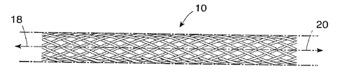
an end view, respectively contained in Figures 1A and 1B. Figure 1A shows a
broken section of a generally cylindrical tubular body 10 having a mantle
surface
CA 02216943 1997-09-29
WO 96/32907 PCT/IB96/00272
formed by a number of individual thread elements 12, 14 and 13, 15, etc. of
theae
elements, elements 12, 14, etc. extend generally in an helix configuration
axially
displaced in relation to each other but having center line 16 of the body 10
a;s a
common axis. The other elements 13, 15, likewise axially displaced, extend in
- 5 helix configuration in the opposite direction, the elements extending in
the two
directions crossing each other in the manner indicated in Figure 1A. A tubular
member so concerned and so constructed can be designed to be any convenient
diameter, it being remembered that the larger the desired diameter, the larger
the
number of filaments of a given wire diameter (gauge) having common composition
1o and prior treatment required to produce a given radial compliance.
The braided structure further characteristically readily elongates upon
application of tension to the ends axially displacing them relative to each
othier
along center line 16 and correspondingly reducing the diameter of the device.
This
is illustrated in Figures 2A and 2B in which a segment of the device 10 of
15 Figures 1A and 1 B has been elongated by moving the ends 18 and 20 away
from
each other in the direction of the arrows. Upon the release of the tension on
the
ends, the structure 10, if otherwise unrestricted, will reassume the relaxed
or
unloaded configuration of Figures 1A and 1B.
The elongation/resumption characteristic flexibility of the stent device
2o enables it to be slipped or threaded over a carrying device while elongated
for
transportation through the vascular or other relevant internal luminal system
of a
patient to the site of interest where it can be axially compressed and thereby
released from the carrying mechanism, often a vascular catheter device. At the
site of interest, it assumes an expanded condition held in place by
25 mechanical/frictional pressure between the stent and the lumen wall against
which
it expands.
The elongation, loading, transport and deployment of such stents is well
known and need not be further detailed here. It is important, however, to note
that
when one contemplates coatings for such a stent in the manner of the present
3o invention, an important consideration resides in the need to utilize a
coating
material having elastic properties compatible with the elastic deforming
properties
residing in the stent that it coats. The material of the stent should be rigid
and
elastic but not plastically deformable as used. As stated above, the preferred
CA 02216943 1997-09-29
WO 96/32907 PCT/1896/00272
_g_
materials for fabricating the metallic braided stent include stainless steel,
tantalum,
titanium alloys including nitinol and certain cobalt-chromium alloys. The
diameter
of the filaments may vary but for vascular devices, up to about 10 mm in
diameter
is preferable with the range 0.01 to 0.05 mm.
s Drug release surface coatings on stents in accordance with the present
invention can release drugs over a period of time from days to months and can
be
used, for example, to inhibit thrombus formation, inhibit smooth muscle cell
migration and proliferation, inhibit hyperplasia and restenosis, and encourage
the
formation of health neointimal tissue including endothelial cell regeneration.
As
to such, they can be used for chronic patency after an angioplasty or stent
placement. It is further anticipated that the need for a second angioplasty
procedure may be obviated in a significant percentage of patients in which a
repeat
procedure would otherwise be necessary. A major obstacle to the success of the
implant of such stents, of course, has been the occurrence of thrombosis in
certain
1s arterial applications such as in coronary stenting. Of course,
antiproliferative
applications would include not only cardiovascular but any tubular vessel that
stents are placed including urologic, pulmonary and gastro-intestinal.
Various combinations of polymer coating materials can be coordinated with
the braided stent and the biologically active agent of interest to produce a
2o combination which is compatible at the implant site of interest and
controls the
release of the biologically active species over a desired time period.
Preferred
coating polymers include silicones (poly siloxanes), polyurethanes,
thermoplastic
elastomers in general, ethylene vinyl acetate copolymers, polyolefin rubbers,
EPDM rubbers, and combinations thereof.
2s Specific embodiments of the present invention include those designed to
elute heparin to prevent thrombosis over a period of weeks or months or to
allow
the diffusion or transport of dexamethasone to inhibit fibromuscular
proliferation
over a like period of time. Of course, other therapeutic substances and
combinations of substances are also contemplated. The invention may be
30 implanted in a mammalian system, such as in a human body.
The heparin elution system is preferably fabricated by taking finely ground x
heparin crystal, preferably ground to an average particle size of less than 10
microns, and blending it into a liquid, uncured poly siloxane/solvent material
in
CA 02216943 1997-09-29
WO 96/32907 PCT/IB96/00272
-9-
which the blend (poly siloxane plus heparin) contains from less than 10% to
;as
high as 80% heparin by weight with respect to the total weight of the material
and
typically the layer is between 10% and 45% heparin.
This material is solvent diluted and utilized to coat a metallic braided
stent,
which may be braided cobalt chromium alloy wire, in a manner which applies a
thin, uniform coating (typically between 20 and 200 microns in thickness)of
the
heparin/polymer mixture on the surfaces of the stent. The polymer is then heat
cured, or cured using low temperature thermal initiators (<100°C) in a
room
temperature vulcanization (RTV) process in situ on the stent evaporating
solvent,
1o typically tetrahydrofuran (THF) with the heparin forming interparticle
paths in the
silicone sufficiently interconnected to allow slow but substantially complei:e
subsequent elution. The ultrafine particle size utilized allows the average
pore size
to be very small such that elution may take place over weeks or even months.
A coating containing dexamethasone is produced in a somewhat different
manner. A poly siloxane material is also the preferred polymeric material.
Nominally an amount equal to 0.4% to about 45% of the total weight of the
layer of
dexamethasone is used.
The dexamethasone drug is dissolved in a solvent, e.g., THF first. The
solution is then blended into liquid uncured poly siloxane/solvent (xylene,
THF,
2o etc.) vehicle precursor material. Since the dexamethasone is also soluble
in the
solvent for the polysiloxane, it dissolves into the mixture. The coating is
then
applied to the stent and upon application, curing and drying, including
evaporation
of the solvent, the dexamethasone remains dispersed in the coating layer. It
is
believed that the coating is somewhat in the nature of a solid solution of
recrystallized particles of dexamethasone in silicone rubber. Dexamethasone,
as a
rather small molecule, however, does not need gross pores to elute and may bra
transported or diffused outward through the silicone material over time to
deliver its
anti-inflammatory medicinal effects.
The coatings can be applied by dip coating or spray coating or even, in
3o some cases, by the melting of a powdered form in situ or any other
technique to
which the particular polymer/biologically active agent combination is well
suited.
It will be understood that a particularly important aspect of the present
invention resides in the technology directed to the incorporation of very fine
CA 02216943 1997-09-29
WO 96/32907 PCT/IB96/00272
- I 0-
microparticles or colloidal suspensions of the drug into the polymer matrix.
In the
case of a crystalline drug, such as heparin, the drug release is controlled by
the
network the drug forms in the polymer matrix, the average particulate size ,
controlling the porosity and so the ultimate elution rate.
Figure 4A depicts a stent which has been spray coated with a solvent
containing a cured polysilicone material including an amount of heparin
crystals to
provide a thin, uniform coating on all surfaces of the stent. The coated stent
was
cured at 150°C for 18 minutes; The sample was eluted in PBS for 49 days
at 37°C
and the stent was rinsed in ethanol prior to taking the scanning electron
1o microscope picture of Figure 4A. Figure 4B shows a greatly enlarged (600X)
scanning electron microscope photograph (SEM) of a portion of the coating of
Figure 4A in which the microporosity is evident. The coating thickness may
vary
but is typically from about 75 to about 200 microns.
Figures 5A and 5B show scanning electron microscope photographs of a
heparin containing polysiloxane stent. The Figure shows the coating prior to
elution of the heparin. The coating was cured at 150° for 18 minutes.
Figure 5B is
greatly enlarged photograph (SEM) of a fragment of the coated surface of
Figure 5A showing the substantially non-porous surface prior to elution.
Figures 6A and 6B show the posture of a stent in accordance with the
2o invention as implanted in a swine coronary. The blemish shown in Figure 6A
represents a histological artifact of unknown origin. As can be seen in Figure
6B,
the general texture of the heparin-containing silicone material appears as a
relatively open matrix containing a large number of gross pores.
The substantially non-porous surface of Figure 7A typically occurs with an
incorporation of an amount of non-particulate material such as dexamethasone
which partially or entirely dissolves in the solvent for the poly siloxane
prior to
coating and cure. Upon curing of the polymer and evaporation of the solvent,
depending on the loading of dexamethasone, the dexamethasone reprecipitates in
a hydrophobic crystalline form containing dendrite or even elongated hexagonal
3o crystals approximately 5 microns in size.
As can be seen in Figure 7B, even after release of the incorporated
material or three months, the coating surface remains substantially non-porous
indicating the transport or diffusion of the drug outward through the silicone
CA 02216943 1997-09-29
WO 96/32907 PCT/IB96/00272
-11-
material neither requires nor produces gross pores. The dexamethasone is
incorporated in its more hydrophobic form rather than in one of the relatively
more
hydrophilic salt forms such as in a phosphate salt, for example.
Figures 8 and 9 depict plots of total percent drug release related to long
s term drug release stent coating layers. Figure 8 depicts the release of
heparin
from a 50% heparin loading in silicone. The silicone was cured at 90°C
for 16
hours. The heparin release took place in a phosphoric buffer (pH=7.4) for 90
daiys
at 37°C. The heparin concentration in the phosphoric buffer was
analyzed by
Azure A assay.
Figure 9 depicts a graphical analysis, similar to that depicted for heparin in
Figure 8, for the release of dexamethasone at two different concentrations,
i.e., ~i%
and 10% in silicone polymer. The coated stents were cured at 150°C for
20
minutes and the release took place in a polyethylene glycol (PEG),
MW=400/wai~:er
. solution at 37°C ((PEG 4001H20) (40/60, vol/vol)). The dexamethaso~ne
concentrations were analyzed photometrically at 241 Nm.
Figures 8 and 9 illustrate possible stent layer polymer/bioactive species
combinations for long-term release. As stated above, the release rate profile
c,sn
be altered by varying the amount of active material, the coating thickness,
the
radial distribution of bioactive materials, the mixing method, and the
crosslink
density of the polymer matrix. Sufficient variation is possible such that
almost any
reasonable desired profile can be simulated.
As stated above, while the allowable loading of the elastomeric material
with heparin may vary in the case of silicone materials, heparin may exceed
60%
of the total weight of the layer. However, the loading generally most
advantageously used is in the range from about 10% to 45% of the total weight
of
the layer. In the case of dexamethasone, the loading may be as high as 50% or
more of the total weight of the layer but is preferably in the range of about
0.4% to
45%.
s It will be appreciated that the mechanism of incorporation of the
biologically
active species into a thin surtace coating structure applicable to a metal
stent is an
important aspect of the present invention. The need for relatively thick-
walled
polymer elution stents or any membrane overlayers associated with many prior
drug elution devices is obviated, as is the need for utilizingr biodegradable
~or
CA 02216943 1997-09-29
WO 96/32907 PCT/IB96/00272
-12-
reabsorbable vehicles for carrying the biologically active species. The
technique
clearly enables long-term delivery and minimizes interference with the
independent
mechanical or therapeutic benefits of the stent itself.
Coating materials are designed with a particular coating technique,
coating/drug combination and drug infusion mechanism in mind. Consideration of
the particular form and mechanism of release of the biologically active
species in
the coating allow the technique to produce superior results. In this manner,
delivery of the biologically active species from the coating structure can be
tailored
to accommodate a variety of applications.
to Whereas the polymer of the coating may be any compatible biostable
elastomeric material capable of being adhered to the stent material as a thin
layer,
hydrophobic materials are preferred because it has been found that the release
of
the biologically active species can generally be more predictably controlled
with
such materials. Preferred materials include silicone rubber elastomers and
i5 biostable polyurethanes specifically.
This invention has been described herein in considerable detail in order to
comply with the Patent Statutes and to provide those skilled in the art with
the
information needed to apply the novel principles and to construct and use
embodiments of the example as required. However, it is to be understood that
the
2o invention can be carried out by specifically different devices and that
various
modifications can be accomplished without departing from the scope of the
invention itself.