Note: Descriptions are shown in the official language in which they were submitted.
CA 02217266 1998-07-17
PATENT
ATTORNEY DOCKET NO: 08472/721001
CO-CULTIVATION OF CELLS IN A MICROPATTERNED CONFIGURATION
Cross-Reference to Related Application
5This application claims priority under 35 U.S.C.
119 from U.S. Serial No. 60/046,413, filed May 14, 1997.
Statement as to Federally SPonsored Research
This invention was produced, at least in part, with
funds from the United States Government under National
Institutes of Health Grant DK5270. Therefore, the United
States Government may have certain rights in the invention.
Backqround of the Invention
The invention relates to methods for co-cultivating
cells in micropatterned formations (e.g., for the production
of bioartificial organs).
Co-cultures of hepatocytes with another cell type
have been recognized to prolong cell survival rates,
maintain phenotype, and induce albumin secretion in
hepatocytes. Such co-cultures have been limited by the
inability to manipulate or control the interaction of the
two cell types in the culture. Generally, to prepare
conventional co-cultures, cells of one type are seeded onto
a substrate and allowed to attach; cells of a second type
then are seeded on top of the cells of the first type. In
such co-cultures, parameters such as cell number are
controllable, but the spatial orientation of cells within
the co-culture is not controlled (Clement, B., et al. "Long-
Term Co-Culture of Adult Human Hepatocytes with Rat Liver
Epithelial Cells: Modulation of Albumin Secretion and
Accumulation of Extracellular Material" Hepatology 4(3):
373-380 (1984); Schrode, W., et al. "Induction of Glutamine
Synthetase in Periportal Hepatocytes by Cocultivation with a
Liver Epithelial Cell Line" Euro. J. Cell Biol. 53: 35-41
CA 02217266 1998-07-17
(1990); Michalopoulos, G., et al., In Vitro 15(10): 796-806
(1979); Guguen-Guillouzo, C., et al. "Maintenance and
Reversibility of Active Albumin Secretion by Adult Rat
Hepatocytes Co-Cultured with Another Liver Epithelial Cell
Type" Experimental Cell Research 143: 47-54 (1983); Begue,
J. et al. "Prolonged Maintenance of Active Cytochrome P-450
in Adult Rat Hepatocytes Co-Cultured with Another Liver Cell
Type" Hepatology 4(5): 839-842 (1984); Agius, L.
"Metabolic Interactions of Parenchymal Hepatocytes and
Dividing Epithelial Cells in Co-culture" Biochem. J. 252:
23-28 (1988); and Reid, L. et al. "Culturing Hepatocytes
and Other Differentiated Cells" Hepatology 4(3): 548-559
(1984)).
Summary of the Invention
The invention provides methods for producing co-
cultures of cells in which at least two types of cells are
configured in a micropattern on a substrate. By using
micropatterning techniques to modulate the extent of
heterotypic cell-cell contacts, it is now possible to
modulate (e.g., upregulate or downregulate) metabolic and/or
synthetic functions of cells.
Accordingly, the invention provides a method for
producing a micropatterned co-culture containing at least
two cell types; the method entails:
i) providing a protein-coated substrate, wherein a
protein coating the substrate defines a micropattern on the
substrate;
ii) contacting the protein-coated substrate with
cells of a first cell type suspended in a first cell medium
under conditions such that cells of the first cell type bind
the protein of the protein-coated substrate, thereby
producing a micropatterned cell-coated substrate; and
CA 02217266 1998-07-17
iii) contacting the micropatterned cell-coated
substrate with cells of a second cell type suspended in a
second cell medium under conditions such that cells of the
second cell type bind the substrate, thereby producing the
micropatterned co-culture, wherein one of the cell media is
a selective medium and one of the cell media is an
attachment medium.
Typically, in practicing the invention, the cells of
the first and second cell types are mammalian cells,
although the cells may be from two different species (e.g.,
pigs, humans, rats, mice, etc). The cells can be primary
cells, or they may be derived from an established cell line.
In an alternative method, one of the cell types is
mammalian, and a second cell type is microbial in origin,
e.g., fungi or bacteria such as Streptococcus ssp.,
Staphylococcus aureus, or Staphylococcus epidermis.
Examples of suitable combinations of cells for producing the
co-culture include, without limitation:
a) hepatocytes (e.g., primary hepatocytes) and
fibroblasts (e.g., normal or transformed fibroblasts, such
as NIH 3T3-J2 cells);
b) hepatocytes and at least one other cell type,
particularly liver cells, such as Kupffer cells, Ito cells,
endothelial cells, and biliary ductal cells;
c) endothelial cells and smooth muscle cells;
d) tumorigenic parenchymal cells and mesenchymal
cells;
e) hematopoietic cells and bone marrow cells (e.g.,
adipocytes, fibroblasts); and
f) skin cells (e.g., keratinocytes) and fibroblasts.
Other combinations of cells also are within the invention.
The substrate on which the cells are grown can be
any biologically compatible material to which cells can
-- 3
CA 022l7266 l998-07-l7
adhere, such as glass, polymers (such as fluoropolymers,
fluorinated ethylene propylene, polyvinylidene,
polydimethylsiloxane, polystyrene, polycarbonate, and
polyvinyl chloride), and silicon substrates (such as fused
silica, polysilicon, or single silicon crystals).
To produce a micropattern of the co-cultured cell
types, protein (i.e., a peptide of at least two amino acids)
is first adhered to the substrate in order to define (i.e.,
produce) a micropattern. The micropattern produced by the
protein serves as a "template" for formation of the cellular
micropattern. Typically, a single protein will be adhered
to the substrate, although two or more proteins may be used
to define the micropattern (for example, one micropatterned
protein may be used to attract one cell type, while a second
micropatterned protein is used to attract a second cell
type). In practicing the invention, a variety of techniques
can be used to foster selective cell adhesion of two or more
cell types to the substrate. Included, without limitation,
are methods such as localized protein adsorption,
organosilane surface modification, alkane thiol self-
assembled monolayer surface modification, wet and dry
etching techniques for creating three-dimensional
substrates, radiofrequency modification, and ion-
implantation (Lom et al., 1993, J. Neurosci. Methods 50:385-
397; Brittland et al., 1992, Biotechnology Progress 8:155-
160; Singhvi et al., 1994, Science 264:696-698; Singhvi et
al., 1994, Biotechnology and Bioengineering 43:764-771;
Ranieri et al., 1994, Intl. J. Devel. Neurosci. 12(8):725-
735; Bellamkonda et al., 1994, Biotechnology and
Bioengineering 43:543-554; and Valentini et al., 1993, J.
Biomaterials Science Polymer Edition 5(1/2):13-36).
Proteins that are suitable for producing a
micropattern are those proteins to which one of the cell
-- 4
CA 02217266 1998-07-17
types of the co-culture specifically binds under the cell
culture conditions used to cultivate the co-culture (i.e.,
conventional cell culture conditions). For example,
hepatocytes are known to bind to collagen. Therefore,
collagen is well-suited to facilitate binding of hepatocytes
in a micropattern. Other suitable proteins include
fibronectin, gelatin, collagen type IV, laminin, entactin,
and other basement proteins, including glycosaminoglycans
such as heparan sulfate. Combinations of such proteins also
can be used.
Typically, in practicing the invention, the cells of
the first cell type (e.g., hepatocytes) initially are
suspended in an "selective" cell culture medium (e.g.,
serum-free medium and media that lack "attachment factors"),
while the cells of the second cell type are suspended in an
"attachment" medium [e.g., a cell culture medium that
contains serum (typically 1-10% (e.g., 5-10%)), or one or
more "attachment factors" (typically at least 1 ng/ml (e.g.,
5-100 ng/ml)) such as fibronectins and other extracellular
matrix, selectins, RGD peptides, ICAMs, E-cadherins, and
antibodies that specifically bind a cell surface protein
(for example, an integrin, ICAM, selectin, or E-cadherin)].
In another method of practicing of the invention,
the cells of the second type have intrinsic attachment
capabilities, thus eliminating a need for the addition of
serum or exogenous attachment factors. Some cell types will
attach to electrically charged cell culture substrates and
will adhere to the substrate via cell surface proteins and
by secretion of extracellular matrix molecules. Fibroblasts
are an example of one cell type that will attach to cell
culture substrates under these conditions. Thus, the
invention also includes a method for producing a
CA 02217266 1998-07-17
micropatterned co-culture containing at least two cell types
where the method entails:
i) providing a protein coated substrate wherein a
protein coating the substrate defines a micropattern on the
substrate;
ii) contacting the protein-coated substrate with cells
of a first cell type suspended in a first cell medium under
conditions such that the cells of the first cell type bind
the protein of the protein-coated substrate, thereby~0 producing a micropatterned cell-coated substrate; and
iii) contacting the micropatterned cell-coated
substrate with cells of a second cell type suspended in a
second cell medium under conditions such that the cells of
the second cell type bind to the substrate, thereby
producing the micropatterned co-culture, wherein the first
cell type (e.g., dermal fibroblasts of skin) is in
non-attachment medium and the second cell type has natural
attachment capabilities to attach it to the substrate. A
charged substrate is particularly useful in practicing this
variation of the invention.
In yet another variation, the micropatterned co-
culture can be produced by
i) providing a repellent-coated substrate wherein a
repellent coating the substrate defines a micropattern on
the substrate;
ii) contacting the repellent-coated substrate with
cells of a first cell type suspended in a first cell medium
under conditions such that cells of the first cell type bind
the substrate, thereby producing a micropatterned
cell/repellent-coated substrate; and
iii) contacting the micropatterned cell/repellent-
coated substrate with cells of a second cell type suspended
in a second cell medium under conditions such that cells of
-- 6
CA 02217266 1998-07-17
the second cell type bind the repellent, thereby producing
the micropatterned co-culture.
As used herein, a "repellent" is a composition that,
relative to the substrate to which it is applied, inhibits
adhesion of the first-applied cells, thereby causing the
first-applied cells to adhere preferentially to the
substrate. Agarose, hyaluronic acid, and alginate are
examples of suitable repellents. In this variation, the
cells of the first cell type (e.g., hepatocytes) can be
suspended in a selective medium or in a selective medium.
If desired, binding of cells of the first cell type to the
substrate can be facilitated by using a substrate that is
coated with a protein to which the cells of the first type
specifically bind, as described above. The cells of the
lS second cell type (e.g., fibroblasts) can be suspended in
attachment medium to facilitate binding to the repellent.
Alternatively, the second-applied cells can be cells that
naturally adhere to a component of the repellent; for
example, fibroblasts will naturally adhere to hyaluronic
acid. This method thus exploits differences in selectivity
exhibited by the two cell types. Relative to fibroblasts,
hepatocytes are selective in their ability to adhere to
surfaces. Fibroblasts are generally promiscuous in their
ability to bind to surfaces, and thus typically will serve
as the second cell type in this variation of invention.
In a variation of these methods for producing
micropatterned co-cultures, cells of one of the cell types
(typically the first cell type) is genetically engineered
using conventional techniques to produce a desired gene
product that acts upon cells of a second cell type. For
example, the first cell type can enable the second cell type
to reproduce and grow, or signal the cells to express other
functionality, such as causing the cells to divide more
-- 7
CA 02217266 1998-07-17
frequently (e.g., by expressing a growth factor) or undergo
apoptosis (e.g., by expressing an ICE gene). For example,
3T3-Ras cells, which express basic fibroblast growth factor,
can be co-cultivated with keratinocytes to induce the
keratinocytes to grow faster.
By using micropatterning techniques, such as those
described herein, the first and second cell types define a
micropattern (i.e., are configured into a pattern having a
resolution on a micron scale). In the micropattern of the
co-culture, cells of either the first or second cell type
are surrounded by (i.e., substantially (>95%), though not
necessarily completely, enclosed by) cells of either the
second or first cell type, respectively. For example, the
cells of the co-culture can be configured such that
"islands" of hepatocytes (cells of a first cell type) are
surrounded by fibroblasts (cells of a second cell type).
Such islands need not be perfectly circular in shape. For,
example, the islands can be produced as stripes or
rectangles. Regardless of the shape of the island, the
spatial configuration that provides optimal growth
conditions can readily be determined. In general, and when
hepatocytes and fibroblasts are co-cultured for example, it
is preferred that at least 30% of the cells of the island
are within 100 ~m of an interface between the island of
cells (e.g., hepatocytes) and the surrounding cells (e.g.,
fibroblasts). More preferably, at least 50%, 80%, or 90% of
the cells of the island are within 100 ~m of the interface.
Where the island is essentially circular, the island
typically will have a diameter of 25-1,000 ~m (preferably,
30-500 ~m (or 100-500 ~m)).
In a variation of the above methods, the invention
provides a method for upregulating a metabolic or synthetic
function of a cell of a first cell typei the method entails:
-- 8
CA 02217266 1998-07-17
i) providing a protein-coated substrate, wherein a
protein coating the substrate defines a micropattern on the
substrate;
ii) contacting the protein-coated substrate with
cells of a first cell type suspended in a first cell medium
under conditions such that cells of the first cell type bind
the protein of the protein-coated substrate, thereby
producing a micropatterned cell-coated substrate; and
iii) contacting the micropatterned cell-coated
substrate with cells of a second cell type suspended in a
second cell medium under conditions such that cells of the
second cell type bind the substrate, thereby producing the
micropatterned co-culture, wherein:
a) one of the cell media is a selective medium and
one of the cell media is an attachment medium; and
b) the cells of the first and second cell types
define a micropattern wherein cells of the second cell type
surround cells of the first cell type, and at least 30% of
the cells of the first cell type are within 100 ~m of an
interface between the cells of the first cell type and the
cells of the second cell type,
thereby producing a micropatterned co-culture,
wherein a metabolic or synthetic function of a cell of the
first cell type is upregulated relative to cells of the
first cell type in an unpatterned co-culture that comprises
cells of the first and second cell types.
This method derives from the observation that, by
using micropatterning techniques to modulate the level of
heterotypic cell-cell contact in a co-culture, it is
possible to upregulate a synthetic or metabolic function of
a cell in the co-culture. For example, DNA synthesis, mRNA
synthesis, and/or protein synthesis can be upregulated with
this micropatterning method. In a micropatterned co-culture
g
CA 02217266 1998-07-17
where islands of hepatocytes are surrounded by fibroblasts,
the upregulation of cell function can be detected as an
increase in intracellular or secreted albumin of a
hepatocyte. Alternatively, or in addition, upregulation of
cell function can be detected as an increase in urea
synthesis in a hepatocyte.
As in the above-described methods for co-cultivating
cells in a micropatterned configuration, examples of
suitable combinations of cells for the co-culture include,
without limitation,
a) hepatocytes (e.g., primary hepatocytes) and
fibroblasts (e.g., NIH 3T3-J2 cells);
b) hepatocytes and at least one other cell type,
particularly liver cells, such as Kupffer cells, Ito cells,
endothelial cells, and biliary ductal cells;
c) endothelial cells and smooth muscle cells;
d) tumorigenic parenchymal cells and mesenchymal
cells;
e) hematopoietic cells and bone marrow cells (e.g.,
adipocytes, fibroblasts); and
f) skin cells (e.g., keratinocytes) and fibroblasts.
Referring to the above list, the invention typically will be
practiced such that an island of the first-named cell type
in each of these combinations is surrounded by cells of the
second-named cell type, and the function of the first-named
cell type is upregulated. In producing the micropatterned
co-culture, it is not necessary to adhere to the substrate
the cells in which cell function will be upregulated prior
to adhering the other cells. However, when producing a co-
culture of hepatocytes and fibroblasts, the hepatocytestypically will be adhered to the protein-coated substrate
prior to contacting the substrate with the fibroblasts.
Other parameters of this aspect of the invention (e.g.,
-- 10 --
CA 02217266 1998-07-17
island size, attachment factors, substrate, etc.) are
essentially as described above.
Typically, the metabolic and/or synthetic function
of cells of the first cell type is modulated at least 1.5-
fold in micropatterned co-cultures, relative to a metabolic
or synthetic function of cells of the first cell type in an
unpatterned co-culture. As shown by the experiments
described below, a change of at least 5-10-fold also is
achievable. To detect the modulation of a metabolic or
synthetic function, conventional molecular and biochemical
assays can be used, such as those described below.
In practicing this method, not only is cell function
upregulated to a higher absolute level (e.g., of albumin
production) in the micropatterned co-cultures (relative to
unpatterned co-cultures), but also the kinetics of this
upregulation are increased. In other words, the rate at
which a metabolic or synthetic function is upregulated to a
particular level in the micropatterned co-culture is
increased relative to the rate at which a metabolic or
synthetic function is upregulated in an unpatterned co-
culture. Thus, the invention also provides a method for
modulating the kinetics at which metabolic or synthetic
functions of a cell are upregulated in a co-culture. From a
bioengineèring perspective, this increase in the kinetic of
cell function upregulation is advantageous, since it
decreases the cultivation time needed for cells to reach a
particular level of metabolic or synthetic function. In
practice, an unpatterned co-culture may take 1-2 weeks to
reach a particular level of cell function, whereas a
micropatterned co-culture could be upregulated to that level
in a single day.
Also included within the invention are the
micropatterned co-cultures produced according to the methods
-- 11 -
CA 02217266 1998-07-17
described herein. Such micropatterned co-cultures of cells
can be used as bioartificial organs for in vivo, ex vivo, or
in vi tro purposes. For example, a micropatterned co-culture
of hepatocytes combined with fibroblasts can be used as an
implantable (in vivo) or extracorporeal (ex vivo) artificial
liver for replacement of liver function (e.g., in response
to diseases, infections, or trauma), or in in vi tro assays
of liver function (for example, for toxicology or basic
research purposes). Similarly, such micropatterned co-
cultures can be used as a bioreactor or as a means tomanufacture peptide compounds such as protein, enzymes, or
hormones (e.g., albumin or clotting factors produced from
hepatocytes). In this regard, the invention provides an
advantage over cell-free methods of producing proteins,
because intracellular post-translational modifications that
occur in the co-cultures of the invention will provide a
properly modified (e.g., glycosylated) protein.
As used herein, the term "micropattern" refers to a
pattern formed on a substrate (e.g., by a protein, cell, or
combination of cells of two or more types), which has a
spatial resolution (e.g., 1-5 ~m) that permits spatially
controlling cell placement at the single-cell level. Thus,
using micropatterning methods, one can precisely manipulate
cell-cell interactions. In contrast, in an "unpatterned"
co-culture of cells, the cells are randomly distributed.
As used herein, an "island" of cells is a single
cell, or typically a group of cells, of one cell type that
is surrounded by cells of another cell type (e.g., a group
of hepatocytes surrounded by fibroblasts). Thus, an
interface is formed where cells at the periphery of the
island meet the surrounding cells. An island need not be
circular in shape; for example, rectangular islands, and
islands of other, amorphous shapes can be used in the
- 12 -
CA 02217266 1998-07-17
invention. The size of the island can be adjusted to
provide optimal growth conditions for the particular
combination of cells in the co-culture. For example, for
islands of hepatocytes surrounded by fibroblasts, at least
30% (preferably at least 50%, 80%, or 90%) of the cells in
the island typically are within 100 ~m from an interface
between the cell types. Thus, where the island is
essentially circular in shape, islands that are less than
1,000 ~m in diameter are suitable. Typically, the island
will be 30-500 ~m in diameter.
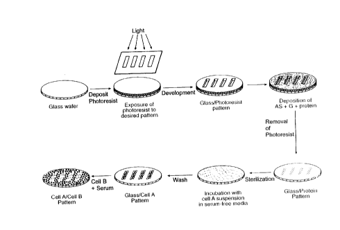
Brief Description of the Drawinqs
Fig. 1 is a schematic representation of a process to
generate micropatterned co-cultures.
Fig. 2 is a schematic representation of a method for
determining X, the heterotypic interaction parameter.
Fig. 3 is a schematic representation of a method to
obtain separation of cell populations.
Detailed DescriPtion
The working examples are provided to illustrate, not
limit, the invention. Various parameters of the scientific
methods employed in these examples are described in detail
below and provide guidance for practicing the invention in
general.
In these particular working examples, hepatocytes
are co-cultured with fibroblasts; as is described herein,
similar methods can be used to co-culture other combinations
of cells. These experiments demonstrate that two cell types
can be co-cultured in a micropattern configuration. In
other words, the two cell types can be used to define a
pattern having a resolution on a micron scale. These
experiments also show that, by using micropatterning to
- 13 -
CA 02217266 1998-07-17
optimize the extent of heterotypic cell-cell contacts in the
co-culture, the metabolic and synthetic functions of cells
of the micropatterned co-culture are upregulated relative to
cells in an unpatterned configuration.
PART I
MATERIALS AND METHODS
Microfabrication techniques were used to modify
glass substrates with biomolecules. These modified
substrates were utilized to pattern a single cell type or
10 micropattern co-cultures in various configurations. Fig. 1
schematically depicts the overall process for producing
micropatterned co-cultures.
Microfabrication of Substrates
The experimental substrates were produced utilizing
15 standard microfabrication techniques. Chrome masks of the
desired dimensions were generated on a pattern generator
(Gyrex), which transferred the pattern to a chromium coated
quartz plate using a contact printer and a developer.
Round, 2" diameter X 0.02" thickness borosilicate wafers
20 (Erie Scientific) were cleaned in a piranha solution (3:1
H2SO4: 30% H2O2) for 10 minutes, rinsed, and blown dry with a
N2 gun. Wafers were then dehydrated by baking for 60
minutes at 200~C. Discs were subsequently coated with
positive photoresist (OCG 820-27 centistokes) on a Headway
25 spin-coater with vacuum chuck as follows: dispense
photoresist at 500 RPM for 2 seconds, spread photoresist at
750 RPM for 6 seconds, spin at 4000 RPM for 30 seconds,
resulting in a 1 ~m coating (Step A, Fig. 1). Wafers were
then pre-baked for 5 minutes at 90~C to remove residual
30 solvent and anneal any stress in the film. Wafers were
- 14 -
CA 02217266 1998-07-17
exposed in a Bottom Side Mask Aligner (Karl Suss) to
ultraviolet light through the desired chromium mask to
create a latent image in the resist layer. Exposure
occurred under vacuum-enhanced contact for 3 seconds.
Exposed photoresist was then developed to produce the final
three-dimensional relief image for 70 seconds in developer
(OCG 934 1:1), rinsed three times under running deionized
water and cascade rinsed for 2 minutes (Step B, Fig. 1).
Subsequently, discs were hard-baked for 30 minutes at 120~C
to remove residual developing solvents and promote adhesion
of the film. Finally, substrates were exposed to oxygen
plasma at 250 W for 4 minutes to remove unwanted resist in
areas to be subsequently modified. Wafers were stored at
room temperature for up to 2 months. Substrates were
subsequently re-exposed to oxygen plasma 24 hours prior to
further processing to ensure availability of borosilicate
for surface modification on a Plasma Day Etcher at a base
vacuum of 50 mTorr and ~2 pressure of 100 mTorr at a power
of 100 W for 2-4 minutes.
Surface Modification of Substrates
Substrates were modified using experimental methods
similar to those developed by Lom et al. and Britland et al.
(Step C, Fig. 1) (Stenger et al., 1992, J. American Chemical
Society 114:8345-8442; Lom et al., 1993, J. Neurosci.
Methods 50:385-397). Briefly, substrates were rinsed twice
in distilled, deionized (DD) water and allowed to air dry.
Silane immobilization onto exposed glass was performed by
immersing samples for 30 seconds in freshly prepared, 2% v/v
solution of 3-[(2-aminoethyl)amino] propyltrimethoxysilane
(AS, Huls America) in water followed by 2 rinses in 200 mL
DD water. Wafers were then dried with nitrogen gas and
baked at 120 ~C for 10 minutes. Next, discs were immersed
- 15 -
CA 02217266 1998-07-17
in 20 mL of 2.5~ v/v solution of glutaraldehyde in PBS (pH
7.4) for 1 hour at 25 ~C. Substrates were then rinsed twice
in fresh PBS, and immersed in a 4 mL solution of a 1:1
solution of 1 mg/mL collagen I (Dunn et al., 1991,
Biotechnology Progress 7:237-245): DD water for 15 minutes
at 25~C. Discs were subsequently immersed in acetone and
placed in a bath sonicator (Bransonic) for 15 minutes to
remove residual photoresist ultrasonically (Step D, Fig. 1).
Wafers were then rinsed twice in DD water, and soaked
overnight in 70~ ethanol for sterilization (Step E, Fig. 1).
Surface Characterization of Substrates
Autofluorescence. Wafers were observed using a
Nikon Diaphot microscope equipped with a Hg lamp and power
supply (Nikon). The autofluorescence of photoresist
(excitation: 550 nm, emission: 575 nm) was used to visualize
micropatterned substrates prior to surface modification.
Absence of autofluorescence after sonication was taken to
indicate removal.
ProfilometrY. Profilometry was performed to
characterize surface topology on a Dektak 3 Profilometer
(Veeco Instruments) with a 12.5 ~m radius probe at a scan
rate of 100 ~m/s.
Atomic Force MicroscoPY (AFM). AFM was performed in
order to characterize the spatial distribution of
immobilized groups. AFM was performed with a Nanoscope 3
(Digital Instruments) equipped with a standard 117 ~m
silicon cantilever operating in tapping mode with a scan
size of 100 ~m.
- 16 -
CA 02217266 1998-07-17
Indirect Immunofluorescence of Collaqen I.
Collagen-derivatized substrates were incubated at
37~C with undiluted Rabbit Anti-Rat Collagen I Antisera
(Biosciences) by inverting substrates onto parafilm that
contained a droplet (50 ~L) of antisera for 1 hour.
Substrates were then washed thoroughly in PBS and placed on
a rotating shaker at 25~C for 30 minutes. This washing
procedure was repeated twice. Next, discs were inverted
onto parafilm with 50 ~L (1:20) of Dichlorotriazinylamino
Fluorescein (DTAF)-conjugated Donkey Anti-Rabbit IgG
(Jackson) in blocking solution. Blocking solution consisted
of 3% w/w bovine serum albumin, 1% donkey serum, 0.04 %
sodium azide, pH 7.4. Finally, substrates were washed in
PBS overnight, and observed by fluorescence microscopy
(excitation: 470 nm, emission: 510 nm).
Cell Culture
Hepatocyte Isolation and Culture. Hepatocytes were
isolated from 2- to 3-month-old adult female Lewis rats
(Charles River) weighing 180-200 g (Seglen et al., 1976,
Methods in Biol. 13:29-83; Dunn et al., 1989, FASEB J.
3:174-177). Routinely, 200-300 million cells were isolated
with viability between 85% and 95%, as judged by Trypan blue
exclusion. Non-parenchymal cells, as judged by their size
(c 10 ~m in diameter) and morphology (nonpolygonal or
stellate), were less than one percent. Culture medium was
Dulbecco's modified Eagle's Medium (DMEM, Gibco)
supplemented with 10% fetal bovine serum (FBS, JR
Scientific), 0.5 U/mL insulin, 7 ng/mL glucagon, 20 ng/mL
epidermal growth factor, 7.5 (g/mL hydrocortisone, 200 U/mL
penicillin , 200 (g/mL streptomycin and 50 (g/mL gentamycin
('hepatocyte media with serum'). Serum-free culture medium
was identical except for the inclusion of 40 (g/mL of
- 17 -
CA 022l7266 l998-07-l7
L-Proline (Sigma) and exclusion of FBS (Lee et al., 1993,
Biomaterials 14:12) (' serum-free hepatocyte media').
NIH 3T3-J2 Culture. NIH 3T3-J2 cells, grown to
pre-confluence, were trypsinized in 0.01% trypsin (ICN
Biomedicals)/0.01% EDTA (Boehringer Mannheim) solution in
PBS for 5 minutes and then resuspended in 25 mL media.
Approximately 10% of the cells were inoculated into a fresh
tissue culture flask. Cells were passaged at pre-confluency
no more than 12 times. Cells were cultured in 75 cm3 flasks
(Corning) at 10% CO2, balance moist air. Culture medium
consisted of DMEM (Gibco) with high glucose, supplemented
with 10% bovine calf serum (BCS, JR~I Biosciences) and 200
U/mL penicillin and 200 ~lg/mL streptomycin.
Cell Culture on Modified Surfaces. Wafers were
rinsed in sterile water, and incubated in 0. 05 % W/w bovine
serum albumin in water at 37(C for 1 hour to pre-coat
substrates with a non-adhesive protein. Substrates were
then washed twice with serum-free media. Next, hepatocytes
were seeded at high density (4 x 10⁵/mL) in serum-free media
for 1. 5 hours at 37 ~C, 10% CO2, balance air (Step E,
Fig. 1). Surfaces were then rinsed twice by pipetting and
then aspirating 4 mL of serum-free media, re-seeded with
hepatocytes for 1. 5 hours, rinsed with 4 mL of serum-free
media, and incubated overnight (Step F, Fig. 1). The
following day, 3T3 cells were trypsinized as described
above, counted with a hemocytometer and plated at lx106 /mL
in 2 mL of serum-containing, high glucose DMEM, and allowed
to attach overnight (Step G, Fig. 1).
'Randomly-distributed' (i.e., unpatterned)
co-cultures consisted of hepatocyte seeding in the desired
number (usually 250,000) on a uniformly collagen-derivatized
surface followed by 3T3 seeding after 24 hours.
- 18 -
CA 02217266 1998-07-17
Immunofluorescent Staininq
Cultures were washed 2 times with 2 mL PBS, fixed
and permeabilized with 10 mL of acetone at -20 ~C for 2
minutes, and washed twice in 10 mL PBS. Cultures on wafers
were incubated at 37 ~C with undiluted Rabbit Anti-Rat Pan
Cytokeratin Antisera (Accurate Chemical), by inverting
substrates onto parafilm containing a 50 ~L droplet of
antisera for 1 hour. Substrates were then washed, incubated
with secondary antibody, and washed (as described above for
indirect immunofluorescence of collagen). Secondary
antibody also included rhodamine-phalloidin (1:100,
Molecular Probes) for fluorescent labeling of F-actin.
Specimens were observed and recorded using a Nikon Diaphot
microscope (Nikon) equipped with a Hg lamp and power supply
(Nikon), light shuttering system (Uniblitz D122), CCD camera
(Optitronics CCD V1470), and MetaMorph Image Analysis System
(Universal Imaging) for digital image acquisition.
Imaqe Ana 1YS i S
To quantitatively describe the extent of heterotypic
interactions, the fraction of cell perimeter in contact with
adjacent cells of a different cell type (X) was measured.
For example, X=1 for a single cell island whereas X=0 for a
cell amidst hepatocyte neighbors. Images were acquired as
described above and analyzed with MetaMorph Image Analysis
System. Cells were sampled from each field and manually
outlined to obtain individual cell perimeters, P.
Subsequently, the regions of heterotypic cell-cell contact
were similarly delineated, F. Each cell was assigned its
characteristic X = F/P and these values of X were averaged
over 20-50 cells for each condition. For population
distributions, individual values of X were assigned to an
appropriate 'bin', and histograms were generated.
-- 19 --
CA 02217266 1998-07-17
RESULTS
As is discussed below, surface characterization
studies on substrates in the absence of cells were first
performed to first exemplify spatially-defined surface
5 chemistries. Subsequently, the ability to micropattern
single cell cultures and co-cultures including two different
cell types was shown, as is described below.
Surface Characterization
Topological and spatial uniformity of photoresist
10 patterns were assessed using profilometry and
autofluorescent properties of photoresist. The photoresist
coating was approximately 1.35 ~m thick, as determined using
the specified spin-coating parameters. Furthermore, the
thickness of photoresist varied <5% within each scan.
15 Autofluorescence of photoresist was utilized to examine
integrity and distribution of photoresist prior to and
during processing. Autofluorescent regions corresponding to
-1 ~m variations in thickness were detected. Absence of any
cont~m'n~nt fluorescence in the dark lanes indicates
20 complete, uniform removal of exposed photoresist during
development.
To demonstrate regional aminosilane (AS)
modification of borosilicate, substrates were exposed to AS,
followed by removal of photoresist. Aminosilane
25 modification has been previously reported to modify the
three-phase contact angle of water with the surface (Lom et
al., 1993, J. Neurosci. Methods 50:385-397); therefore, the
perimeter of a single water droplet was used to display
microscopic undulations on patterns of varying
30 hydrophilicity. These undulations were observed; 20 llm AS
modified lanes exhibit differential wetting properties
relative to the adjacent 50 ~Lm unmodified lanes. Therefore,
-- 20 --
CA 02217266 1998-07-17
selective AS modification of exposed glass was demonstrated
in the pattern of the original 20 ~m/50 ~m striped
photoresist pattern, indicating that photoresist can serve
as a 'chemical mask' to AS modification of underlying
glass.
Collaqen immobilization via qlutaraldehYde derivatization of
patterned AS surfaces was also characterized.
Fluorescence micrographs were obtained, showing the
results of indirect immunofluorescent staining of areas of
presumed collagen immobilization. Fluorescent regions,
corresponding to regions of collagen localization, were
patterned uniformly with spatial resolution on the micron
level. Furthermore, fluorescent patterns corresponded to
initial photoresist patterns without evidence of
undercutting. Despite processing in acetone and 70%
ethanol, collagen retained sufficient immunoreactivity for
antibody binding.
Collagen-derivatized surfaces were also analyzed
with AFM to determine differences in topology between
unmodified and modified borosilicate. Modified regions with
a width of 20 ~m were found to have an average height of
4 nm above the unmodified, 50 ~m lanes. These data can be
utilized to approximate the number of collagen monolayers
atop AS.
MicroPatterning of Co-Cultures
The aforementioned experiments demonstrate the
ability to reproducibly utilize photoresist patterns to
generate immobilized collagen patterns; the following
experiments illustrate the applicability of these techniques
to cellular micropatterning. Seeding of the first cell
type, hepatocytes, resulted in localization to
- 21 -
CA 02217266 1998-07-17
collagen-derivatized regions and normal polygonal
morphology. The cellular configurations were dictated by
the positioning of collagen on glass, the pattern of which
was in turn controlled by the choice of chromium mask in the
microfabrication procedure. In addition, hepatocytes
conformed to the edges of the collagen pattern on the
modified glass. The typical hepatocyte diameter in
suspension is 20 ~m, whereas, upon attachment and
unconstrained spreading, cell diameters increase to
30-40 microns. Therefore, after attachment to 20 ~m lines,
cells elongated in the axial direction upon spreading.
Similar cytoskeletal changes were observed in cells on
corners of larger patterns or on the perimeter of circular
patterns.
The versatility of this technique was seen in
phase-contrast micrographs. Initial hepatocyte patterns of
20 ~m and 200 ~m were modified by the addition of
fibroblasts in serum-containing media. Fibroblasts
localized solely to unmodified (glass) regions of patterned
substrates resulting in micropatterned co-cultures of
20 ~m/50 ~m and 200 ~m/500 ~m. This approach is adaptable
to both micropatterning of single cell cultures and
co-cultures of two different cell types.
Spreading of the primary cell type typically
resulted in negligible residual sites of
collagen-derivatization. Therefore, attachment of the
secondary cell type is limited either to unmodified glass or
the surface of the primary cell type. 3T3 fibroblasts do
not undergo significant attachment to hepatocyte surfaces,
as shown in plating experiments of fibroblasts on monolayers
of hepatocytes which showed no attachment even after a
4 hour incubation (data not shown). In addition, fibroblast
attachment and spreading on glass was characterized by
- 22 -
CA 02217266 1998-07-17
seeding cells in serum-containing media on glass coverslips
where they attached and spread with high efficiency within
4 hours (data not shown).
Indirect immunofluorescence was used to stain
selectively cell populations and aid in visual
discrimination between different cell types. The presence
of cytokeratin, an intermediate filament expressed in
hepatocytes but absent in mesenchymal cells, was compared
with F-actin, a cytoskeletal protein present in all
mammalian cells. A patterned co-culture of 200 ~m/500 ~m
was also compared with a 'randomly distributed' co-culture
with identical attached cell numbers of both cell
populations. The level of homotypic hepatocyte interaction
in a 200 ~m stripe of micropatterned cells was compared with
the level in a random distribution of cells. Hepatocytes in
the 200 ~m stripe had primarily homotypic contacts, whereas
those in the random distribution had predominantly
heterotypic contacts. Furthermore, the distribution of
heterotypic interaction over the patterned cell population
was greatly reduced over that of random co-cultures, where
hepatocytes were present in single cell islands, doublets,
and triplets.
To describe quantitatively the extent of heterotypic
contact, image analysis and perimeter tracing were used to
define the fractional cell perimeter engaged in heterotypic
cell contact as X, as described above. Fig. 2 schematically
depicts sample perimeter tracings (black lines) with
high-lighted interfaces of heterotypic contacts
corresponding to hepatocytes in a digitally-acquired phase
micrograph. This particular pattern (200 ~m/500 ~m) has
very little heterotypic contact, as was visually observed;
therefore, the average X over the population is small due to
the majority of cells with X=0. The mean value of X over a
- 23 -
CA 02217266 1998-07-17
cell population can be changed from 0.7 in the randomly
distributed culture to 0.08 using micropatterning.
Moreover, different patterns (20/50? produce distinct mean
values of X (X = 0.55). Variations of X from the mean were
also examined for randomly distributed cultures as compared
to defined patterns (20/50). As observed microscopically,
hepatocytes in randomly distributed cultures experience
heterogeneous microenvironments - single hepatocytes,
doublets, and multicellular aggregates can be observed
within a given culture. Quantitative analysis of population
distributions corroborate the variability in X in randomly
distributed cultures as compared to micropatterns (20/50 and
50/50), which exhibited a relatively small variance around
the mean value of X. Thus, variations in cellular
microenvironment, both in amount and variability, were
achieved without varying the numbers of cells in each
sub-population.
DISCUSSION
Many conventional co-culture systems have been
limited by the inability to vary local cell seeding density
independently of the cell number, as well as inherent
variations in the distribution of cell contacts over a
population of cells. The invention provides a versatile
technique for the micropatterning of two different cell
types derived from conventional strategies for surface
modification with aminosilanes linked to biomolecules and by
manipulating the serum content of cell culture media, as
described above. This co-culture technique allows the
manipulation of the initial cellular microenvironment
without variation of adhered cell number. Specifically, it
was possible to control both the degree and type of initial
cell-cell contact. Differences in homotypic and heterotypic
- 24 -
CA 022l7266 l998-07-l7
interaction were demonstrated, allowing variations in
exposure to cell-surface receptors, locally secreted
extracellular matrix, and local concentrations of soluble
factors.
In these patterning methods AS was applied after
photoresist patterning but before photoresist lift off. The
integrity of the photoresist was preserved throughout the
surface modification process and removed the photoresist
after the deposition of collagen. This was achieved by
deposition of AS in water, which does not normally attack
photoresist. AS is known to oligomerize in aqueous solution
(Arkles et al., 1991, Huls America, N.J., 65-73), but is
stable at least for a period of hours. In this way,
photoresist was used to mask the borosilicate from
non-specific protein adsorption and it was not necessary to
rely on protein denaturation and desorption or on AS
deposition prior to photoresist patterning.
Atomic force microscopy was utilized to approximate
the depth of the immobilized collagen layer. Modified
regions were -4 nm above the unmodified regions. AS
molecules have been estimated to have a height of 1.2 nm
end-to-end (Lom et al., 1993, J. Neurosci. Methods 50:385-
397). In the helical configuration, collagen I fibrils have
dimensions of 300 nm in length and 1. 2 nm in diameter
(Darnell et al., 1990, Molecular Cell Biology, 904-905).
These data suggest that there were 1-2 layers of collagen
fibrils, configured lengthwise, corresponding to an upper
limit of 0.1 ~g/cm2 per monolayer of 'side-on' packed
fibrils (Deyme et al., 1986, J. siomedical Materials
Research 20:39-45). Therefore, achievable collagen surface
concentrations are within an order of magnitude those
observed in adsorbed collagen systems (O. 37 ~g/cm2) (Deyme
et al., 1986, J. Biomedical Materials Research 20:39-45).
- 25 -
CA 02217266 1998-07-17
Another consideration is the bioactivity of biomolecules
after exposure to acetone and ethanol. The preservation of
bioactivity of collagen I via cell attachment and spreading
as well as by antibody binding for indirect
immunofluorescence has now been demonstrated. Proteins
sensitive to acetone may benefit from adaptation of the
photoresist lift-off procedure.
Using primary rat hepatocytes and 3T3 fibroblasts,
the initial heterotypic (X) interactions were varied over a
wide range while preserving the ratio of cell populations in
culture. Thus, co-culture interactions now can be
manipulated in an entirely new dimension. If desired,
three-phase co-cultures can be established by patterning of
two different, cell-specific biomolecules. The
micropatterned co-cultures had less variation in the level
of heterotypic contacts (X) than did random co-cultures.
Therefore, measurement of macroscopic biochemical quantities
in micropatterned co-cultures will provide better
representations of specific cell-cell interactions than
those seen in unpatterned co-cultures.
SU~RY
The invention provides a simple, versatile technique
for controlling homotypic versus heterotypic interactions of
at least two cell types in culture. One can vary X without
changing the number of cells in each sub-population and
therefore vary the ratio of cell types in a given culture.
- 26 -
CA 02217266 1998-07-17
PART II
The following experiments demonstrate that the
methods of the invention can be used to upregulate metabolic
and/or synthetic functions (e.g., liver-specific functions)
of cells in the micropatterned co-culture.
MATERIALS AND METHODS
Substrates for micropattern formation were prepared
essentially as described above.
He~atocYte Isolation and Culture
Hepatocytes were isolated from 2- to 3-month-old
adult female Lewis rats (Charles River Laboratories,
Wilmington, MA) weighing 180-200 g, by a modified procedure
of Seglen (1976). Detailed procedures for isolation and
purification of hepatocytes were previously described by
Dunn et al (FASEB, 1989). Routinely, 200-300 million cells
were isolated with viability between 85% and 95%, as judged
by trypan blue exclusion. Nonparenchymal cells, as judged
by their size (~10 ~m in diameter) and morphology
(nonpolygonal or stellate), were less than 1%. Culture
medium was Dulbecco's modified eagle's medium (DMEM, Gibco)
supplemented with 10% fetal bovine serum (FBS, Sigma,
St.Louis, MO), 0.5 U/mL insulin, 7 ng/mL glucagon, 20 ng/mL
epidermal growth factor, 7.5 mg/mL hydrocortisone, 200 U/mL
penicillin, and 200 mg/mL streptomycin ('hepatocyte media
with serum'). Serum-free culture medium was identical
except for the exclusion of FBS.
NIH 3T3-J2 Culture
NIH 3T3-J2 cells were provided by Howard Green
(Harvard Medical School). Cells grown to preconfluence were
- 27 -
CA 02217266 1998-07-17
passaged by trypsinization in 0.01% trypsin (ICN
Biomedicals, Costa Mesa, CA)/0.01% EDTA (Boehringer
Mannheim, Indianapolis, IN) solution in PBS for 5 minutes,
diluted, and then inoculated into a fresh tissue culture
flask. Cells were passaged at pre-confluency no more than
10 times. Cells were cultured in 175 cm2 flasks (Fisher,
Springfield, NJ) at 10% CO2, balance moist air. Culture
medium consisted of DMEM (Gibco, Grand Island, NY) with high
glucose, supplemented with 10% bovine calf serum (BCS, JRH
Biosciences, Lenexa, KS) and 200 U/mL penicillin and 200
mg/mL streptomycin ('fibroblast media'). In some cases,
growth arrested cells were obtained for DNA analysis by
incubation with 10 mg/mL mitomycin C (Boehringer Mannheim)
in media for 2 hours (reconstituted just prior to use)
followed by three washes with media. Mitomycin C-treated
fibroblasts were shown to have constant levels of DNA for
10 days of culture, verifying the lack of fibroblast growth
under these conditions.
Cell Culture on Modified Surfaces
Wafers were sterilized by soaking for 2 hours in 70%
ethanol in water at room temperature. Subsequently, wafers
were rinsed in sterile water and incubated in 0.05% w/w
bovine serum albumin (BSA) in water at 37~C for 1 hour to
precoat substrates with a nonadhesive protein. Substrates
were then placed in sterile P-60 tissue culture dishes
(Corning, Corning, NY), and rinsed in sterile water followed
by a final rinse with serum-free media. Next, hepatocytes
were seeded at high density (1-2 x 10⁶/mL) in 2 mL
serum-free media for 1.5 hours at 37~C, 10% CO2, balance air
followed by two rinses with serum-free media. This process
was repeated twice to ensure confluence of hepatocytes,
especially on larger dimension patterns. The following day,
- 28 -
CA 02217266 1998-07-17
3T3 cells were trypsinized as described above, counted with
a hemocytometer, plated at 3.75 x 10⁵/mL in 2 mL of
serum-containing, high-glucose DMEM, and allowed to attach
overnight. Subsequently, 2 mL hepatocyte culture media
(described above) was sampled and replenished daily.
Experimental Desiqn
Spatial configurations of micropatterned co-cultures
were manipulated by varying mask dimensions. Transparent
circular areas (or 'holes') on chrome masks correspond to
derivatized, and ultimately hepatocyte-adhesive, areas of
glass substrates. In order to achieve identical hepatocyte
numbers across varying micropatterned configurations, the
total surface area of all 'holes' was kept constant across
all masks despite changes in hole diameter and
center-to-center spacing. All arrays were hexagonally
packed with the exception of the largest dimension hole
which consisted of a single unit of 17800 ~m diameter.
Thus, pattern dimensions varied as follows (hole diameter
(in microns), center-to-center spacing (in microns)): 36,
90; 100, 250; 490, 1229; 6800, 16900; and a single unit of
17800 ~m diameter, where the resulting total
hepatocyte-adhesive area on 2" diameter glass substrates
would be identical in all cases.
AnalYtical AssaYs
Media samples were collected daily and stored at 4~C
for subsequent analysis for urea and albumin content. Urea
synthesis was assayed using a commercially available kit
(Sigma Chemical Co., kit No. 535-A). Reaction with diacetyl
monoxime under acid and heat yields a color change detected
at 540 nm. Albumin content was measured by enzyme-linked
immunosorbent assays (ELISA) as described previously (Dunn
- 29 -
CA 02217266 1998-07-17
et al., 1991, Biotechnology Progress 7:237-245). Rat
albumin and anti-rat albumin antibodies were purchased from
Cappel Laboratories (Cochranville, PA).
DNA analysis was adapted from a method of MacDonald
and Pitt (1991). Cells were sacrificed at the indicated
time of culture by washing with PBS, removal and immersion
of wafer into PBS to eliminate dead cells underneath the
substrate, and subsequent incubation with 0.05% (w/v) type
4 collagenase (Sigma) in Kreb's Ringer Buffer at 37~ C for
30 minutes to release the cell layer from the underlying
substrate. Next, cells were removed with a rubber policeman
and the cell/collagenase mixture was removed. The substrate
was washed with PBS which was then combined with the above
solution. The resulting solution was combined with an
equivalent volume of hepatocyte media for neutralization of
collagenase, followed by centrifugation at 1000 RPM for
5 minutes. The supernatant was aspirated, and ceIls were
resuspended in 2 mL PBS. Subsequently, the samples were
frozen at -80~C for up to 1 month.
For analysis, the frozen samples were rapidly thawed
in a 37~ C water bath to promote membrane rupture.
Freeze-thaw protocols have been established as an effective
way to rupture the cell membrane in order to gain access to
cellular contents. To ensure complete cell lysis, samples
were then sonicated using a probe sonicator (Branson) for
10 seconds at 4~C. Samples were vortexed and 20 ml samples
were placed into a 96-well plate (NUNC, Denmark). Similarly,
20 ml of DNA standard (double stranded Calf Thymus DNA,
Sigma) in PBS from 100 to 0 mg/mL were vortexed and placed
on each plate. This volume was combined with 100 ml
salt/dye buffer (2 M NaCl, 10 mM Tris, 1 mM EDTA, 1.6 mM
Hoechst 33258 (Molecular Probes, Eugene, OR)). Samples and
standards were allowed to incubate with salt/dye buffer at
- 30 -
CA 02217266 1998-07-17
room temperature in the dark for 30 minutes before reading
on a Spectrofluorometer (Millipore, Bedford, MA) Excitation
360 nm, 1/2 bandwidth 40 nm, Emission, 460 nm, 1/2 bandwidth
40 nm.
Analysis of DNA Content
The total DNA content in cultures with
growth-arrested fibroblasts was assayed as follows.
Mitomycin C was utilized to growth arrest fibroblasts (as
described above) and 1.5 X 10⁶ fibroblasts were counted with
a hemocytometer and added to micropatterned hepatocyte
cultures. Replicate cultures were either sacrificed 6 hours
after fibroblast seeding or after 9 days of co-culture and
assayed for total DNA as described above.
Immunohistochemistry
Cultures were washed twice with PBS, fixed with
4% paraformaldehyde in PBS for 30 minutes, and permeabilized
for 10 minutes with 0.1% Triton in PBS. Endogenous
avidin-binding activity of hepatic tissue was blocked by
20 minute incubations with 0.1% avidin and 0.01% biotin in
50 mM Tris-HCl respectively (Biotin Blocking System X590,
DAKO, Carpinteria, CA). Endogenous peroxidase activity was
blocked by 30 minute incubation with a hydroxgen peroxide
and sodium azide solution (Peroxidase Blocking Reagent,
DAKO). Rabbit anti-rat albumin antibodies (Cappell) were
utilized with horse-radish peroxidase visualization by use
of a biotinylated secondary antibody to rabbit IgG,
streptavidin-labelled horse radish peroxidase, and hydrogen
peroxide in the presence of 3-amino-9-ethylcarbazole as a
substrate (Rabbit Primary Universal Peroxidase Kit #K684,
DAKO).
CA 02217266 1998-07-17
Functional Bile Duct Staininq
Cultures were washed three times with media and
incubated for 5 hours with 2 ~M Carboxyfluorescein diacetate
(Molecular Probes) in an adapted method of LeCluyse et al.
(1994). Subsequently, cultures were washed again three
times and examined microscopically. Digital images were
obtained on a Nikon Diaphot microscope equipped with Hg lamp
and excited at 470 nm excitation and 510 nm emission.
Imaqe Acquisition and Analysis
Specimens were observed and recorded using a Nikon
Diaphot microscope equipped with a CCD camera (Optronics CCD
V1470), and MetaMorph Image Analysis System (Universal
Imaging, Westchester, PA) for digital image acquisition.
Image analysis on immunostained images was performed
utilizing the thresholding function in MetaMorph and visual
correlation with brightfield images.
Statistics and Data Analysis
Experiments were repeated two to three times with
duplicate or triplicate culture plates for each condition.
Two duplicate wells were measured for biochemical analysis.
One representative experiment is presented where the same
trends were seen in multiple trials but absolute rates of
production varied with each animal isolation. Each data
point represents the mean of three dishes. Error bars
represent standard error of the mean. Statistical
significance was determined using one-way ANOVA (analysis of
variance) on Statistica (StatSoft) with Tukey HSD (Honest
Significant Difference) Post-Hoc analysis with p ~ 0.05.
- 32 -
CA 02217266 1998-07-17
RESULTS
Micropatterned co-cultures were generated with
variations in heterotypic interface but with identical
surface area (i.e., cell numbers) dedicated to both
hepatocyte and fibroblast adhesion. Five different
configurations ranging from maximal heterotypic contact
(smallest islands) to minimal heterotypic contact (single
island) were characterized for expression of liver-specific
function by use of: two biochemical markers (albumin and
urea synthesis), immunohistochemistry (intracellular albumin
staining), transport across apical surface (bile duct
excretion), and DNA content. These results show that
micropatterning can be used to optimize the degree of
heterotypic interactions and thereby optimize cell function.
In this case, an increase in heterotypic interactions is
correlated with an increase in liver-specific functions.
Characterization of Initial Cell Distribution
All 5 micropatterns were designed to have similar
levels of hepatocyte-adhesive surface area (2.5 cm2), which
is expected to correspond to identical number of attached
hepatocytes. Variations in spatial configurations were
utilized to generate differences in total perimeter of
hepatocyte islands from 5.6 cm to 2800 cm, which, upon
addition of fibroblasts, generally correspond to variations
in the total heterotypic interface. Micropatterns ranged
from many single hepatocyte islands of 36 ~m diameter to a
single island of 17.8 mm diameter (100 ~m, 490 ~m, and 6800
~m islands also were detected). Micropatterned hepatocytes
adhered predominantly to collagen-modified areas in all 5
conditions with close agreement between theoretical and
observed values for total initial hepatocyte island
perimeter (data not shown).
- 33 -
CA 022l7266 l998-07-l7
To verify similar numbers of attached hepatocytes
across various spatial configurations, the DNA content of
micropatterned hepatocyte cultures was measured 24 hours
after hepatocyte seeding (i.e. prior to fibroblast seeding).
All cultures had statistically similar levels of DNA
(8 i 1.8 ~g) with the exception of increased DNA content
(18 i 3.3 llg) on the smallest island (36 ~m diameter)
micropatterns.
The smallest islands were designed to produce single
10 cell islands. The dimensions of these islands (36 ,um
diameter) was chosen to correspond with the experimentally
determined projected surface area of a single, spread
hepatocyte on immobilized collagen I of 1000 ~m2 (data not
shown); however, isolated rat hepatocytes have a diameter of
15 approximately 20 ~lm, allowing the potential for individual
islands to retain more than one hepatocyte upon seeding with
a concentrated cell suspension. In addition, hepatocytes
have been shown to have an increased mitotic index at low
seeding densities (Nakamura et al., 1983, J. Biochemistry
20 94 :1029-1035), which may have contributed to increased
hepatocyte DNA in this condition. To distinguish between
increased cell number as compared to increased ploidy, image
analysis of one thousand 36 ,um micropatterned islands was
completed at 6 hours after initiation of cell seeding. This
25 analyses demonstrated more than one cell per island in
57% of cases, with an average of 1.9 i 1. 2 cells per island.
Therefore, increased DNA was due to increased hepatocyte
number on the smallest pattern.
Addition of 3T3-J2 fibroblasts to micropatterned
30 hepatocytes resulted in micropatterned co-cultures with
marked alterations in initial heterotypic interface despite
similar numbers of fibroblasts and hepatocytes across
conditions. Phase contrast micrographs of 4 of the 5
- 34 -
CA 02217266 1998-07-17
configurations (36 ~m, 100 ~m, 480 ~m, and 6800 ~m islands)
demonstrated the significant variation in hepatocyte
microenvironment which was achieved by altering micropattern
dimensions.
Biochemical Analysis of Liver-SPecific Function
To demonstrate the effect of modulation of the local
hepatocyte environment on liver-specific function, albumin
secretion and urea synthesis were measured as markers of
differentiated function. These two markers were measured as
a function of micropattern dimensions in the presence and
absence of fibroblasts. In cultures of fibroblasts alone,
albumin secretion and urea synthesis by fibroblasts was
found to be undetectable; therefore, changes in these
markers in co-cultures were attributed to differences in
hepatocyte metabolism.
Albumin secretion for five different spatial
configurations was determined for pure hepatocyte cultures.
A rapid decline in liver-specific functions was detected for
all five conditions (36 ~m, 100 ~m, 490 ~m, 6800 ~m, and
17,800 ~m islands), from initial values of 8.8 + 0.9 ~g/day
to undetectable levels.
Albumin secretion for the same five micropatterns
with the addition of fibroblasts was also measured. Albumin
synthesis increased over time in culture in all
configurations from less than 10 ~g/day to greater than
34 ~g/day, indicating up-regulation of this liver-specific
function due to co-culture with fibroblasts. These
micropatterned co-cultures had decreasing amounts of initial
heterotypic contact with maximal levels occurring at the
smallest hepatocyte island dimension (36 ~m) and minimal
levels occurring at the single large hepatocyte island
(17.8 mm). Smaller islands with high levels of heterotypic
- 35 -
CA 02217266 1998-07-17
contact demonstrated greater albumin secretion than larger
islands (less heterotypic contact) after day 5 of culture.
Two fundamental patterns of up-regulation were observed:
(1) dramatic up-regulation to similar levels of albumin
secretion in the three smallest island configurations (19 to
26-fold of initial levels) and (2) relatively modest
up-regulation (~7-fold) in the two larger island
configurations. Therefore, a three-fold increase in albumin
production was achieved in certain pattern configurations by
modulation of the initial cellular microenvironment.
Analysis of urea synthesis in micropatterned
co-cultures revealed similar qualitative results. Urea
synthesis was either constant over culture or increased from
less than -100 ~g/day to 160 ~g/day indicating up-regulation
of another liver-specific function due to co-cultivation -
with fibroblasts. In addition, two patterns of
up-regulation were observed using this marker of
differentiated function: (1) up-regulation of urea
synthesis to similar levels in the three smallest island
configurations (up to 2-fold increase), and (2) relatively
little up-regulation in the two larger island
configurations. Therefore, a two-fold increase in urea
synthesis production was achieved in certain pattern
configurations by modulation of the initial cellular
microenvironment. Asterisks indicate pcO.05 in Tukey
post-hoc analysis of variance.
Hepatocyte Function In Si tu: Immunostaining of Intracellular
Albumin
In order to further examine the observed variations
in liver-specific function exhibited by various
micropatterned co-cultures, the hepatocyte phenotype in si tu
was examined by immunostaining of intracellular albumin.
- 36 -
CA 02217266 1998-07-17
Specifically, these studies first focused on the
distribution of albumin staining as it related to the
heterotypic interface in one representative pattern, 490 ~m
hepatocyte islands (at days 2 and 6). In addition, in order
to distinguish between homotypic effects on differentiation
and the effects arising from varying the heterotypic
interface, immunostaining on micropatterned pure hepatocyte
cultures was performed at days 2 and 6. Hepatocytes
cultured alone stained uniformly for intracellular albumin
at 48 hours after isolation. The level of protein declined
subsequently on the order of days. In comparison,
micropatterned co-cultures displayed a more complex
behavior. They also displayed initial uniform staining for
intracellular albumin. Over 6 days, however, hepatocytes
close to the heterotypic interface stained for high levels
of intracellular albumin, whereas protein levels in
hepatocytes far from the heterotypic interface (> 3-4 cells)
continued to decline as in the pure hepatocyte cultures. To
ensure that this 'ring' of intense staining was due to
variations in intracellular albumin content of hepatocytes,
as opposed to the detachment of hepatocytes or fibroblasts
from the lightly-stained areas, phase contrast microscopy of
these cultures was performed. The presence of fibroblasts
in the periphery of the hepatocyte island and cellular
structures in the center of the hepatocyte island was
clearly depicted. This peripheral 'ring' of intense
staining observed across a 490 ~m micropatterned co-culture
was reproducible.
In order to correlate the pattern of immunostaining
with the variations that were observed using biochemical
analysis of secreted products in media, the distribution of
high levels of intracellular albumin in comparatively small
(100 ~m) and large (6800 ~m) micropatterned co-cultures was
- 37 -
CA 02217266 1998-07-17
examined. These micrographs demonstrate uniform intense
staining in smaller islands (initial island size 100 ~m), a
well-demarcated ring of ~120 ~m in intermediate size islands
(initial size 490 ~m), and a well-demarcated ring of -380 ~m
in larger islands (initial size 6800 ~m), indicating a
negative correlation between differentiated hepatocyte
phenotype and distance from the heterotypic interface.
Hepatocyte Function in Situ: Bile Duct Excretion
Another in situ marker of liver-specific function is
the formation of functional bile caniliculi between
hepatocytes. Carboxyfluorescein diacetate (CFDA) is taken
up by hepatocytes, cleaved by intracellular esterases, and
in the presence of normal biliary transport, excreted across
the apical membrane of the hepatocyte. The presence of
normal biliary transport of the dye as well as functional
integrity of the tight-junctional domains bounding the
caniliculus, causes illumination of visibly fluorescent bile
duct structures between hepatocytes. Two patterns were
probed: one from a highly functioning co-culture (490 ~m
circles) and one from a poorly functioning group (17800 ~m
circle), as determined by albumin and urea production, in
order to examine this marker of liver-specific function.
Phase contrast micrographs of both cultures were produced.
The 490 ~m patterns developed functional bile caniliculi,
especially in the island periphery, while fluorescent bile
duct staining was not observed on 17800 ~m islands.
DISCUSSION
This set of experiments demonstrates that
liver-specific tissue function can be modulated by
controlling initial heterotypic cell-cell interactions,
despite the use of identical cellular components.
- 38 -
CA 02217266 1998-07-17
Furthermore, these differences in bulk tissue properties as
a function of cellular microenvironment were generated by
induction of spatial heterogeneity in the hepatocyte
phenotype. Hepatocytes in the vicinity of the heterotypic
interface had a relative increase in levels of
liver-specific function; therefore, spatial configurations
with maximal initial interface exhibited superior function.
Cellular Microenvironment Modulated Liver-sPecific Functions
Evidence that liver-specific function could be
controlled by variations in initial cell-cell interactions
is seen in the functional differences between predominantly
heterotypic co-cultures ~smallest islands of 36 ~m diameter)
and predominantly homotypic co-cultures (largest island of
17800 ~m diameter) as assessed by markers of metabolism
(urea synthesis), synthetic function (albumin secretion and
cytoplasmic content), and apical transport (biliary
excretion). These cellular microenvironments significantly
altered liver-specific functions as follows: increasing
hepatocyte island size correlated with a relative decline in
urea synthesis, albumin secretion, intracellular albumin
staining, and effective biliary excretion. Smaller
hepatocyte islands of 36, 100, and 490 ~m initial diameter
yielded three-fold steady-state increases in albumin
secretion and two-fold steady-state increases in urea
synthesis over 6800 and 17800 ~m islands. Similarly, a
smaller pattern (490 ~m initial diameter) exhibited
functional biliary excretion as assessed by accumulation of
a fluorescent compound within bile canilicular structures
between hepatocytes whereas larger islands (17,800 ~m
initial diameter) showed reduced functional biliary
excretion with no evidence of focal fluorescence. The
presence of fluorescent biliary structures between
- 39 -
CA 02217266 1998-07-17
hepatocytes has been correlated to biliary structures
observed on electron microscopic analysis (LeCluyse et al.,
1994, American Physiological Society). The absence of
fluorescent biliary structures was attributed to either
(1) low rate of excretion across apical domain (2) absence
or loss of function of tight junctions at borders of apical
membrane or (3) decreased uptake of dye by hepatocytes. The
lack of fluorescent biliary structures in 17800 ~m pattern
indicates some such functional deficit. Therefore,
hepatocytes in smaller island co-cultures have improved
biliary transport as well as relative improvements in other
liver-specific functions due to alterations in the initial
cellular microenvironment (as compared with larger island
co-cultures).
In concluding that bulk tissue function (secreted
albumin and urea) was modulated by initial cellular
microenvironment, hepatocyte numbers were measured to ensure
that changes in these liver-specific markers were due to
changes in level of hepatocellular function (as opposed to
differences in cell division). In order to assess the
relative contribution of hepatocyte division, as compared to
up-regulation of functions, fibroblasts were
growth-arrested, and total DNA in co-cultures was measured.
Thus, changes in total DNA could be attributed solely to
hepatocytes. Total DNA of co-cultures was measured at
6 hours of co-culture and compared to DNA content at 9 days
of co-culture. This analysis demonstrated that no
significant increase in total DNA occurred in co-cultures
over 9 days, indicating increases in hepatic functions were
due to up-regulation of synthesis rather than a marked
increase in hepatocyte population (data not shown). These
data correlate well with reports of minimal hepatocyte
division under various co-culture configurations
- 40 -
CA 02217266 1998-07-17
(Guguen-Guillouzo, 1986, John Libbery Eurotext, INSERM:259-
284; Kuri-Harcuch and Mendoza-Figuera, 1989, Differentiation
410:148-157; Donato et al., 1990, In Vitro Cell and
Developmental Biology 26:1057-1062). Furthermore, this
result correlated well with visual observation of larger
micropatterns (490 micron island diameter and greater),
where hepatocyte island size was observed to be relatively
constant over the course of culture, indicating a lack of
significant cell division. Taken together, these data
indicate that variations in hepatic functions between
culture configurations were due predominantly to relative
levels of hepatic upregulation, as opposed to hepatocyte
~ . . .
alvl s lon .
The conclusion that bulk tissue function was
modulated by variation of the cell-cell interactions at the
heterotypic interface prompted confirmation of similar
initial hepatocyte numbers to confirm that changes in
secreted products were due to up-regulation of
liver-specific functions, rather than differences in numbers
of initial hepatocytes. Comparison of initial total
hepatocyte DNA in all five micropatterns showed this to be a
valid approximation (8 + 1.8 ~g DNA) with, perhaps, the
exception of the smallest (36 ~m) islands which were found
to have two-fold elevated levels of DNA. This may be due to
the potential for more than one unspread hepatocyte (20 ~m
diameter) to adhere to 36 ~m islands. In any event, in
these studies, the trend to increased long-term liver
specific function resulting from maximal initial heterotypic
interface remained a consistent finding.
The experiments described above were conducted with
the same surface area dedicated to fibroblasts in all
conditions. This allowed examination of the local cellular
environment as an isolated variable, without differences in
- 41 -
CA 02217266 1998-07-17
cell numbers and resultant variations in concentrations of
potential signaling factors (such as humoral factors in
media). In addition, these experiments allowed simultaneous
control over both oxygen delivery to hepatocytes, as well as
amount of media. In contrast, variation of culture plate
area necessitates either a change in media volume to
preserve the depth of media above the cell population (and
the diffusion of oxygen) or a change in media depth to
preserve media volume. Therefore, these methods for
controlling cellular environment have definitively
demonstrated the importance of local cellular
microenvironment as an isolated modulator of liver-specific
function (i.e., metabolic and synthetic functions).
Cellular Microenvironment Induced SPatial Heteroqeneity in
Hepatocyte PhenotYPe
In addition to demonstrating that liver-specific
tissue function can be modulated by controlling initial
heterotypic cell-cell interactions, the experiments
described herein demonstrate that spatial heterogeneity in
the induction of the hepatocyte phenotype was the primary
cause of these variations in function. In si tu
immunostaining of intracellular albumin on micropatterned
hepatocyte/fibroblast co-cultures displayed increased
staining in the vicinity of the heterotypic interface,
indicating up-regulation of this marker of differentiated
function. Specifically, smaller (100 ~m islands) stained
throughout hepatocyte regions, whereas larger islands
(490 ~m and greater) exhibited intense staining in a
well-demarcated ring in the periphery. This pattern of
staining was highly reproducible both spatially and across
various conditions. The differentiated hepatocyte phenotype
appeared to dominate within 100-400 ~m of the heterotypic
- 42 -
CA 02217266 1998-07-17
interface; therefore, these data suggest that patterns with
greater interfacial regions displayed superior tissue
function.
To confirm that variations in intracellular albumin
represented variations in hepatocyte phenotype due to
heterotypic interactions, the effect of homotypic hepatocyte
interactions on the spatial distribution of intracellular
albumin in a representative micropattern (490 ~m island) was
assessed. These experiments revealed uniform staining in
pure hepatocyte cultures with decreased staining over a
period of one week, consistent with the observed decline in
secreted albumin and previous studies showing residual
albumin mRNA hepatocyte immediately after isolation with
decline of mRNA over 1 week; therefore, patterns of
immunostaining in co-cultures were indeed due to heterotypic
interactions with fibroblasts, rather than homotypic
interactions.
Imaqe AnalYsis: In order to correlate intracellular
albumin staining with albumin secretion data, image analysis
was performed on immunostained co-cultures. Specifically,
the fraction of hepatocytes contributing to albumin secreted
into the media was estimated. Image analysis of
intracellular albumin staining revealed -100% of hepatocytes
stained intensely in 100 ~m patterns, ~65% in 490 ~m
2s patterns, and -20% in 6800 ~m patterns. By assuming a
negligible contribution of weakly staining hepatocytes to
albumin production, hepatocytes adjacent to the heterotypic
interface in larger patterns were estimated to have produced
35-50% more albumin per cell than those in 100 ~m
micropatterns. These data suggest there may be a further
increase in albumin production in hepatocytes adjacent to
relatively undifferentiated homotypic neighbors.
- 43 -
CA 02217266 1998-07-17
Micropatterning co-cultures, as described above,
allowed the creation of larger hepatocyte colonies than
those that come about by random aggregation and cell
migration; therefore, these assays was able to demonstrate a
finite penetration length of a differentiation signal to the
interior of a large hepatocyte colony. This result
contradicts the notion that hepatocytes are able to
communicate effectively throughout a hepatocyte colony.
Related Observations on Control of Cell-Cell Interactions
While the ability to micropattern co-cultures
provides the ability to modulate tissue function via the
initial cellular microenvironment, the inherent dynamics of
cell adhesion and motility may further modify these
engineered tissues in insubstantial ways. The degree of
morphogenesis depended upon hepatocyte island size. In
these experiments, hepatocyte islands of 490 ~m with
center-to-center spacing of 1230 ~m produced a relatively
stable configuration whereas hepatocytes in islands of 100
~m and smaller underwent some reorganization into cord-like
structures. Reorganization of tissue may be prevented by
cytoskeletal toxins such as cytochalasin D. Despite the
tendency for some spatial configurations to reorganize, the
perturbations which were achieved in initial cellular
microenvironment had significant long-term impact on tissue
function.
SUMM~RY
These experiments show that micropatterning can be
used as a vehicle to control heterotypic cell-cell
interactions without significant variations in cell numbers.
Indeed, modulation of heterotypic interface as an
independent variable was achieved. This modulation of the
- 44 -
CA 02217266 1998-07-17
heterotypic interface over three orders of magnitude
dramatically altered levels of detectable liver-specific
function in the resulting composite tissues as measured by
markers of metabolic, synthetic, and excretory function.
Variations in function were due to modulation of the
hepatocyte phenotype: specifically, epithelial
differentiation varied inversely with distance from the
heterotypic interface, causing cultures with a relative
increase in cell interaction to exhibit superior function.
The ability to control heterotypic cell-cell interactions
and probe the resulting tissue for evidence of cell
communication has applications both in basic science (e.g.,
in vi tro assays of tissue function) and development of
functional tissue constructs for medical applications. From
a fundamental perspective, these co-culture techniques can
be exploited in assays for determining the mechanisms by
which cells communicate. In the area of tissue engineering,
the ability to co-cultivate two or more cell types in a
micropattern and modulate cell function provides an
unprecedented level of control over the in vi tro
reconstruction of skin, bone marrow, muscle, and many other
tissues.
PART III
In the following experiments, microfabrication
techniques as well as conventional culture methodologies
were used to further examine the mechanism of induction of
hepatocyte differentiation at the heterotypic interface.
These experiments indicate that the biological signal for
the observed induction of hepatic functions is
"cell-associated" (broadly defined to include membrane-bound
receptors, locally secreted extracellular matrix, and local
- 45 -
CA 02217266 1998-07-17
matrix or cell-bound growth factors), rather than "freely
secreted" (broadly defined to include humoral factors such
as soluble cytokines and growth factors). Thus, the
micropatterning techniques described herein can be used to
modulate metabolic and/or synthetic cell functions.
MATERIALS AND METHODS
Examination of the modes of cell communication in
hepatocyte/3T3 co-culture was conducted using in situ
immunostaining to assess the contribution of homotypic
hepatocyte interactions, and various methods of probing the
class of signal(s) responsible for induction of the
hepatocyte phenotype in hepatocytes proximal to the
heterotypic interface. These techniques included
pre-treated media to probe for soluble factors (conditioned
media), separation of cell populations to probe for labile
soluble factors and to eliminate contribution of fibroblast
adhering to the hepatocyte surface (spacer), and cultures
conducted with continual disturbance of overlying media to
probe for transport limitations (agitation).
General Techniques
Methodology for micropatterned substrate
preparation, hepatocyte isolation and culture, NIH 3T3-J2
fibroblast culture, immunohistochemistry, analytical assays,
and image acquisition are presented in detail above.
Immunostaininq of Micropatterned Cultures
To assess the contribution of hepatocyte homotypic
interaction on spatial patterns of albumin immunostaining,
various sizes of micropatterned hepatocytes were probed both
in the presence and absence of additional fibroblasts.
Micropatterned cultures of hepatocytes alone and
- 46 -
CA 02217266 1998-07-17
hepatocyte/fibroblast co-cultures were generated as
described above in the following hepatocyte island
dimensions: 36, 100, 490, 6800, and 17800 ~m. Hepatocytes
were either cultured alone or co-cultured with 750,000 NIH
3T3-J2 fibroblasts. Culture media (2 mL) was replaced
daily. Cultures were fixed and stained at 48 hours and 144
hours.
Conditioned Media
Conditioned media experiments were performed in
unpatterned configurations. Glass substrates were modified
by aminosilane, glutaraldehyde, and collagen I as described
in above, resulting in collagen I immobilization over the
entire wafer. Hepatocytes were seeded in 'hepatocyte media
with serum' as described previously, at a density of 250,000
per P-60. Four different culture configurations were
investigated. First, in order to control for baseline
degradation of biochemical compounds in media at 37~C,
hepatocytes were fed daily with 2 mL of media which had been
previously incubated for 24 hours in tissue culture plastic.
Second, in order to examine the effects of fibroblast
secreted products, hepatocytes were fed daily with 2 mL of
media which had previously incubated for 24 hours with
(750,000 initially seeded) NIH 3T3-J2 cells on an unmodified
glass wafer. Third, in order to probe the effects of
fibroblast secreted products which require hepatocyte
interaction for their up-regulation, hepatocytes were fed
daily with 2 mL of media which had been previously incubated
for 24 hours with a co-culture of (750,000 initially seeded)
NIH 3T3-J2 cells and 250,000 hepatocytes on an, unpatterned,
collagen-modified wafer. Last, in order to generate a
'positive control' to compare the above conditions to
co-culture induced up-regulation of liver-specific
- 47 -
CA 02217266 1998-07-17
functions, hepatocytes were co-cultured with NIH 3T3-J2
fibroblasts by the addition of 750,000 NIH 3T3-J2 cells on
day 2 of culture. Media was collected daily and stored
at 4~C.
PhYsical separation of cell tYPes
Hepatocytes and fibroblasts were separated by the
following general protocol: placement of a polymer annulus
on glass substrate, surface modification of glass within the
annulus by adsorption of collagen I, attachment of
hepatocytes to central, collagen-immobilized region,
'capping' of hepatocyte population during fibroblast seeding
to prevent access of fibroblasts to top surface of
hepatocytes, and removal of cap and annulus. Differential
spacing was achieved by variation in annulus width resulting
identical inner diameter (and therefore size of hepatocyte
island) and larger outer diameter (resulting in larger
separation between cell populations). Fig. 3 depicts a
schematic overview of method.
Annuli were fabricated with polydimethysiloxane
(PDMS) (Sylgard 184, Dow Corning, Lansing, MI). Stock
sheets of 500 ~m thickness were prepared by casting polymer
solution (mixed as described by the manufacturer) in
polystyrene tissue culture plastic for 2 hours at 65~C.
Annuli were fabricated with inner diameter of 0.6 cm and
various outer diameters using disposable skin punch biopsy
cutting tools. To limit potential cytotoxicity, PDMS annuli
were then coupled to collagen I with
aminoethylaminopropyltrimethoxysilane and glutaraldehyde
using conventional methods.
'Caps' were fabricated from sheets of polyethylene
teraphthalate (PET) by use of a standard paperpunch to
generate 0.6 cm disks from 7 mil thickness mylar film
- 48 -
CA 022l7266 l998-07-l7
(Kodak). Discs were soaked in 70% ethanol in water for 2
hours followed by rinsing in media.
Annuli were affixed to clean, 2 " diameter,
borosilicate wafers, and subsequently 'heat-fixed' to
5 prevent detachment via three consecutive exposures to a heat
gun at a distance of 10 cm for 5 seconds. Collagen
adsorption to the inner circular region of exposed glass was
achieved by addition of 200 ~1 of collagen I: water in 1:1
ratio, pH 5 . 0, and incubation at 37~C for 45 min. Wafers
were then sterilized overnight in 70% ethanol in water,
rinsed in water, exposed to 0. 05% bovine serum albumin and
rinsed with serum-free hepatocyte media (as previously
described). Hepatocytes were seeded in serum-free media as
previously described and allowed to spread overnight.
The following day, PET caps were applied to PDMS
annuli under sterile conditions, growth-arrested (mitomycin
C treatment described above) fibroblasts were seeded and
allowed to attach for 1 hour, rinsed twice with 'fibroblast
media' , followed by removal of annuli and cap. The
20 separated co-culture was rinsed once more with fibroblast
media and fibroblasts were allowed to spread for 6 hours
prior to replacement of fibroblast media with 'hepatocyte
media with serum'. Control co-culture was performed by
methods described previously on 0.68 cm hepatocyte island
25 patterns (as described above). Briefly, glass was modified
by immobilization of collagen I, hepatocytes were seeded
followed by fibroblasts. No cap or polymer annulus was
applied in this condition.
Finally, absence of overlying fibroblasts on
30 hepatocyte island was confirmed using fluorescent labels
CMFDA (chloromethylfluorescein diacetate, C-2925, Molecular
Probes) and CMFTR (chloromethylbenzoylaminotetramethyl
rhodamine, C-2927). Cells were loaded by incubation in 25
-- 49 -
CA 02217266 1998-07-17
~M dye in media for 45 minutes, rinsed, and incubated for 30
minutes prior to a final rinse. Fibroblasts were then
trypsinized as previously described and utilized in the
above protocol. Separated co-cultures were rinsed and
imaged 7 hours after initial fibroblast seeding.
Aqitation
In order to examine the influence of fluid
convection on heterogeneity in hepatocyte phenotype,
co-cultures were conducted in static and 'shaken'
conditions. One representative pattern was utilized for
this study. Micropatterned co-cultures were generated
utilizing 490 ~m hepatocyte islands with 1230 ~m
center-to-center spacing as described previously. 750,000
NIH 3T3-J2 fibroblasts were added 24 hours after initial
hepatocyte seeding. Replicate cultures were then cultured
under two different conditions: (1) under static culture
conditions as previously described and (2) under 'shaken'
conditions by culturing on a rocking platform at
approximately 1 Hz within a separate incubator. Media
(2 mL) was replaced daily. Cultures were fixed and stained
for intracellular albumin at indicated times.
RESULTS
Effect of Homotypic Hepatocyte Interactions on SPatial
Pattern of Immunostaininq
In these experiments, the potential contribution of
homotypic hepatocyte interaction to spatial heterogeneity
was examined by studying micropatterns with different levels
of homotypic interaction both in the presence and absence of
fibroblasts. Patterns of intracellular albumin for five
different micropatterned hepatocyte configurations were
- 50 -
CA 02217266 1998-07-17
compared after 48 and 144 hours of culture. Uniform
distribution of intracellular albumin was detected at
48 hours in all patterns, which diminished over the time in
micropatterned hepatocytes alone. Micropatterned
co-cultures (i.e., addition of fibroblasts at 24 hours of
culture) displayed a uniform distribution of intracellular
albumin similar to that observed in micropatterned
hepatocyte cultures. After 6 days of co-culture, however,
hepatocytes display differential levels of staining.
Hepatocytes far from the heterotypic interface exhibit a
similar behavior to hepatocytes cultured in the absence of
fibroblasts, low levels of staining. In contrast,
hepatocytes proximal to the heterotypic interface exhibit
relatively high levels of intracellular albumin. Thus,
homotypic hepatocyte interactions do not seem to be the sole
contributor to the observed spatial heterogeneity in
hepatocyte phenotype.
Use of Conditioned Media
In order to examine the possible induction of
hepatic differentiation by secreted fibroblast products,
experiments were conducted with hepatocytes treated with
'conditioned media'. Urea synthesis was measured as a
marker of liver-specific function in a variety of such
culture conditions. Media was 'conditioned' by 24 hours
incubation with (1) tissue culture plastic as a control
(hepatocytes + media), (2) fibroblasts alone (hepatocytes +
fibroblast conditioned media), or (3) co-culture of
fibroblasts and hepatocytes (hepatocytes of co-culture
conditioned media). These data were compared to co-cultured
fibroblasts and hepatocytes which served as a positive
control for the expected level of liver-specific function
(co-culture + media).
- 51 -
CA 02217266 1998-07-17
These data indicate an expected decline in
liver-specific function in pure hepatocyte over the first
week of culture to less than 50 ~g/day. A similar decline
in liver-specific function was observed in cultures treated
with fibroblast conditioned media indicating insufficient
concentration of humoral factors for induction of hepatic
differentiation. In contrast, co-cultures of hepatocyte and
fibroblasts displayed up-regulation of urea synthesis from
-60 ~g/day to ~175 ~g/day over 10 days of culture followed
by stable production of urea. Some cultures were treated
with co-culture conditioned media to probe for humoral
factors present only when both cell types were allowed to
communicate. These did not display any further induction of
liver-specific function over that observed in co-culture
controls, indicating insufficient concentration of humoral
factors for induction of hepatic differentiation (detection
of urea in this media was due to production of urea by the
co-culture utilized for conditioning media - any induction
of urea synthesis in the target hepatocyte population would
therefore have generated a further increase in urea
production over control co-cultures).
Physical Separation of Cell Populations
Hepatocyte and fibroblast populations were
co-cultured in the same dish yet separated by an annulus of
bare glass to probe the role of labile, freely secreted
factors in induction of hepatic functions. Phase contrast
micrographs were produced of two different initial annuli
dimensions translating to two different achievable
separation widths. Growth-arrested fibroblasts migrated
towards the central hepatocyte region at a rate of
approximately 500 microns per day. After 3 days, the
1500 ~m initial separation was observed to have diminished
- 52 -
CA 02217266 1998-07-17
completely and cell contact occurred at the periphery of the
hepatocyte island. Subsequently, cells were allowed to
interact for 8 days ('contact' condition). In contrast,
initial cell separation of 6000 microns narrowed to
500 microns over the same time frame ('non-contact~). This
experimental design allowed the examination of the role of
cell proximity/cell contact in induction of hepatic
functions as well as the elimination of overlying
fibroblasts as confirmed by fluorescent dye labeling.
Hepatocytes in the 'contact' condition exhibited an
intense staining pattern in the periphery of the hepatocyte
island similar to the peripheral ring of staining observed
in the control co-culture. In contrast, hepatocytes in the
'non-contact' condition lacked significant staining for
intracellular albumin. These results indicated the
importance of cell proximity (< 500 ~m) for differentiation
of hepatocytes. Furthermore, spatial heterogeneity in
hepatocyte phenotype persisted despite absence of fibroblast
adhesion to surface of hepatocytes, indicating that regional
differences in hepatocyte staining is not due to overlying
fibroblasts.
Aqitation of Co-Cultures
Another method of examining the potential role of
secreted products by fibroblasts was the addition of fluid
convection to co-cultures. Under these 'shaken' conditions,
humoral factors which theoretically require a high local
concentration for their bioactivity would be diluted in the
bulk fluid phase and the resulting pattern of hepatocyte
differentiation would differ from static conditions. In
addition, agitation of culture media would allow mixing of
nutrients (oxygen, glucose) and thereby alleviate potential
CA 02217266 1998-07-17
transport limitations to the center of large hepatocyte
islands.
The effect of agitation of one representative
micropatterned co-culture, 490 ~m, as compared to static
conditions was measured. Phase contrast micrographs
demonstrate that agitation did not cause any overt
fibroblast damage due to mechanical shear. In addition, low
magnification, bright field images of cultures stained for
intracellular albumin demonstrated no significant
differences in patterns of spatial heterogeneity. The
'penetration' length of the signal for hepatocyte
differentiation from the heterotypic interface did not vary
significantly when compared to static cultures. These data
suggested (1) spatial heterogeneity of hepatocyte phenotype
in static cultures was not caused by significant nutrient
limitation due to diffusional transport and (2) dilution of
secreted factors by mixing did not modulate the observed
pattern of spatial heterogeneity.
DISCUSSION
The experiments summarized above use both
conventional and microfabrication techniques to probe in
detail the mechanisms by which cells interact. These
experiments focused on classification of the signal(s)
broadly defined as cell-associated or freely secreted. In
addition, these experiments examined potential contributors
to the finite 'penetration' length of this signal leading to
spatial heterogeneity in the hepatocyte phenotype. The
results of these experiments are summarized below.
- 54 -
CA 02217266 1998-07-17
A Cell-Associated Siqnal is Implicated in Induction of
Hepatic Function
Experiments to classify the differentiation signal
as free versus bound provided evidence that the signal(s) is
cell-associated. Taken together, the results of these
experiments (use of conditioned media, separation of cell
populations within a co-culture, and agitation of
micropatterned co-cultures) point towards cell-associated
molecules. Neither fibroblast conditioned media nor
co-culture conditioned media were able to induce
hepatocellular functions in target hepatocytes, indicating
the absence of a freely soluble signaling molecule.
The experiments described above indicate that it is
implausible that a freely soluble labile signal mediates the
induction of hepatic cell function. These data indicated
that cell contact (or very close proximity, ~ 5 ~m)
correlated with induction of liver-specific function in
hepatocytes, whereas lack of contact ( ~ 500 ~m) did not
induce an observable signal as measured by immunostaining of
intracellular albumin. With the exception of some unique
biochemicals such as nitric oxide, other highly labile
signals would be expected to signal hepatocytes across
500 ~m in this separated culture configuration.
These studies also elucidated the morphology of
hepatocytes separated from underlying fibroblasts by a 1 mm
thick collagen I hydrogel and observed fibroblastic,
de-differentiated morphology after a few days of culture,
further suggesting the lack of a freely soluble, highly
labile signal (data not shown).
The potential role of freely soluble factors whose
bioactivity depends on a high local concentration was also
found to be minimal by the combined results of conditioned
media and agitation experiments. Any soluble factor which
- 55 -
CA 02217266 1998-07-17
did not induce a signal in conditioned media due to its
dilution in the larger media volume, would also be diluted
in agitation experiments due to fluid convection in the
media. Therefore, if fluid mixing causes reduction of the
concentration of some putative soluble signaling factor
below its bioactive concentration, one would not expect
local induction of hepatocyte function in agitation
experiments. In fact, similar patterns of local induction
of intracellular albumin were found in hepatocytes in
micropatterned co-cultures and static controls, indicating
that the dilution of soluble factors was not a critical
limitation in induction of hepatocellular function.
Analysis of the Finite Penetration Lenqth of Differentiation
Siqnal
With respect to the potential contributors to the
spatial heterogeneity observed in the hepatocyte phenotype
in co-cultures, the experiments described herein identify
three potential contributors thee effects of which can be
discounted: (i) inadequate delivery of oxygen or other
nutrients to the center of hepatocyte islands, (ii) a
primary homotypic effect wherein lack of hepatocyte
neighbors in island periphery induced up-regulation of
functions, and (iii) heterogeneous signaling from
fibroblasts attached to the top surface of hepatocytes.
The role of primary hepatocyte homotypic
interactions in induction of spatial heterogeneity of
hepatocyte phenotype was not significant. Specifically,
when hepatocytes were cultured alone, no spatial variation
in intracellular albumin was observed as a result of
variations in homotypic interaction. Hepatocytes in small
islands exhibited intense, uniform staining similar to
staining patterns of hepatocytes both in the periphery and
- 56 -
CA 02217266 1998-07-17
center of larger islands, followed by a spatially uniform
decline in liver-specific function at day 6. In contrast,
micropatterned co-cultures exhibited marked variations in
hepatocyte phenotype where hepatocytes adjacent to the
heterotypic interface expressed greater levels of albumin,
indicating that spatial heterogeneity is not an artifact of
homotypic interactions.
The adequacy of diffusive transport of oxygen and
other transport was determined by comparison of static and
agitated micropatterned co-cultures. In both cases, a
similar pattern of induction was observed at day 4,
indicating convective mixing of media did not modify
hepatocyte behavior. Finally, the contribution of overlying
fibroblasts in the observed spatial heterogeneity was also
determined to be minimal. Fibroblasts were noted to adhere
to the top surface of spread hepatocytes at larger
dimensions of hepatocyte islands with dual label vital dyes
and fluorescent microscopy (data not shown); however,
experiments performed to separate cell populations
effectively prevented fibroblast attachment to the surface
of hepatocytes under these conditions. Therefore, the
presence of spatial heterogeneity resulting from highly
characterized initial conditions was assessed. After 8 days
of contact between cell types, intracellular albumin
immunostaining indicated the presence of peripheral staining
and persistence of the heterogeneous hepatocyte response.
Spatial heterogeneity could not be attributed to variations
in signals arising from overlying fibroblasts.
Morphoqene s i s
The role of tissue reorganization on spatial
heterogeneity in hepatocyte phenotype was addressed.
Notably, reorganization of cultures (both hepatocytes alone
- 57 -
CA 02217266 1998-07-17
and co-cultures) was observed in smaller pattern dimensions
and was significantly diminished in large hepatocyte islands
(greater than 490 ~m). In these studies, pattern
configuration at later time points was perturbed by
morphogenesis in the tissue, i.e., observed patterns of
staining were determined not only by initial pattern
configuration but also by the long-term conformation adopted
by the culture. For example, 100 ~m islands did not display
spatial heterogeneity in albumin staining, presumably
because they reorganized to a pattern where all hepatocytes
were proximal to the heterotypic interface. In contrast,
36 ~m islands reorganized to larger dimension 'cord-like'
hepatic structures where some hepatocytes were a greater
distance from the heterotypic interface, resulting in
spatial heterogeneity of hepatocyte phenotype. Despite the
existence of some reorganization in these tissues, the
fl]n~mental pattern of spatial heterogeneity remained
constant; hepatic structures larger than 100 ~m exhibited
spatial heterogeneity in hepatocyte phenotype whereas
hepatocytes far from the heterotypic interface exhibited low
levels of intracellular albumin. Therefore, the conclusions
reached above remain well founded.
SUMMARY
In these experiments, conventional culture
techniques were combined with microfabricated co-cultures to
show that the primary signal for differentiation of
hepatocytes in hepatocyte/fibroblast co-cultures is tightly
fibroblast-associated. Taken together, the results of
conditioned media, separated co-culture, and agitation
experiments indicated a 'cell-associated' signal promotes
modulation (e.g., up-regulation) of liver-specific
functions. The observed finite penetration of the
- 58 -
CA 02217266 1998-07-17
differentiation signal may be due to gap junctional
communication, 'tissue phase' diffusion of signaling
molecules, and/or physical penetration of fibroblast
processes.
With respect to the implications for the design of a
co-culture-based bioreactor, evidence that the signal is
fibroblast-associated suggests that fibroblasts and
hepatocytes should have direct contact (i.e., occupy the
same compartment in a bioreactor) to produce adequate levels
of liver-specific function. These co-cultures will allow
the examination of candidate biochemical signals as well as
spatial configurations which minimize the fraction of
hepatocytes far from the heterotypic interface, creating the
potential for further improvements in bulk tissue function.
Other Embodiments
It is to be understood that, while the invention has
been described in conjunction with the detailed description
thereof, the foregoing description is intended to illustrate
and not limit the scope of the invention, which is defined
by the scope of the appended claims. Other aspects,
advantages, and modifications are within the scope of the
following claims.
What is claimed is:
- 59 -