Language selection

Search

Patent 2217481 Summary

Third-party information liability

Some of the information on this Web page has been provided by external sources. The Government of Canada is not responsible for the accuracy, reliability or currency of the information supplied by external sources. Users wishing to rely upon this information should consult directly with the source of the information. Content provided by external sources is not subject to official languages, privacy and accessibility requirements.

Claims and Abstract availability

Any discrepancies in the text and image of the Claims and Abstract are due to differing posting times. Text of the Claims and Abstract are posted:

  • At the time the application is open to public inspection;
  • At the time of issue of the patent (grant).
(12) Patent Application: (11) CA 2217481
(54) English Title: IMPROVED DEVICES FOR REMOVING FIBRIN SHEATHS FROM CATHETERS
(54) French Title: SYSTEMES AMELIORES D'ENLEVEMENT DES DEPOTS FIBRINEUX DES CATHETERS
Status: Dead
Bibliographic Data
(51) International Patent Classification (IPC):
  • A61M 25/088 (2006.01)
  • A61B 17/22 (2006.01)
  • A61M 25/00 (2006.01)
(72) Inventors :
  • SUHOCKI, PAUL V. (United States of America)
  • RIDINGER, MARK T. (United States of America)
(73) Owners :
  • DUKE UNIVERSITY (United States of America)
(71) Applicants :
  • DUKE UNIVERSITY (United States of America)
(74) Agent: BORDEN LADNER GERVAIS LLP
(74) Associate agent:
(45) Issued:
(86) PCT Filing Date: 1996-04-05
(87) Open to Public Inspection: 1996-10-10
Availability of licence: N/A
(25) Language of filing: English

Patent Cooperation Treaty (PCT): Yes
(86) PCT Filing Number: PCT/US1996/004799
(87) International Publication Number: WO1996/031162
(85) National Entry: 1997-10-03

(30) Application Priority Data:
Application No. Country/Territory Date
08/417,018 United States of America 1995-04-05

Abstracts

English Abstract


A medical device (10) adapted especially to remove biological material (e.g.,
fibrin sheath) from the distal end of a patient internal catheter that
includes a tubular element (12), a central wire (14) positioned within the
tubular element, a snare wire (16) attached at one end (16a) to the central
wire so as to form an acute angle therewith, and at its other end (16b) to the
tubular member. The snare wire includes a proximally extending segment which
follows a course of about 360 degrees about the distal end (12a) of the
tubular member, and thus may be positioned adjacent the catheter's distal end.
By effecting relative rotation between the central wire and the tubular member
(for example, by rotating the central wire about its longitudinal axis while
maintaining the tubular member stationary) will cause the snare wire segment
to wrap around the catheter's distal end. Thereafter, manipulation of the
device so as to advance the wrapped snare wire segment in a distal direction
will strip the biological material from the distal end of the catheter.


French Abstract

Dispositif à usage médical (10) spécialement conçu pour enlever les matières biologiques (par exemple un dépot de fibrine) de l'extrémité distale d'un cathéter dans l'organisme d'un patient. Ce dispositif comprend un élément tubulaire (12), un fil central (14) placé à l'intérieur de l'élément tubulaire, un fil formant un noeud coulant (16) attaché par l'une de ses extrémités (16a) au fil central, de manière à former un angle aigu avec celui-ci et, par l'autre extrémité (16b), à l'élément tubulaire. Le fil formant un noeud coulant comporte un secteur proximal qui suit un cheminement d'environ 360 degrés autour de l'extrémité distale (12a) de l'élément tubulaire et peut ainsi se placer de manière contiguë par rapport à l'extrémité distale du cathéter. En réalisant une rotation entre le fil central et l'élément tubulaire (par exemple en faisant tourner le fil central selon son axe longitudinal tout en maintenant l'élément tubulaire fixe), on obtient un enroulement du noeud coulant autour de l'extrémité distale du cathéter. Après quoi, une manoeuvre du dispositif tendant à faire avancer dans le sens distal le noeud coulant permet d'obtenir un enlèvement de la matière biologique de l'extrémité distale du cathéter.

Claims

Note: Claims are shown in the official language in which they were submitted.


WHAT IS CLAIMED IS:

1. A medical device comprising:
an elongate tubular member;
a central wire positioned within said tubular member;
a snare wire having one end attached to a terminal end
of said central wire to form an acute angle therewith,
and another end attached to a distal end of said
tubular member, said snare wire between said one
and another ends being wrapped about an exterior
surface of said distal end of said tubular
member, wherein relative rotation between said
central wire and said tubular member causes said
snare wire to wrap around said distal end of said
tubular member.

2. A medical device as in claim 1, wherein said snare wire is
wrapped about 360° about said distal end of said tubular member.

3. A medical device as in claim 1, wherein said central wire is
axially and rotationally moveable within said tubular member.

4. A medical device as in claim 1, wherein said snare wire has
a length sufficient to establish a segment between said one and
another ends which extends proximally of said distal end of said
tubular member.


5. A medical device as in claim 1 or 4, wherein said one end of
said snare wire is bonded to a terminal end of said central wire.

6. A medical device as in claim 1 or 4, wherein said terminal
end of said central wire includes an eyelet, and wherein said one end
of said snare wire is attached to said eyelet.

7. A medical device as in claim 6, wherein said one end of said
snare wire is bonded to said eyelet.

8. A medical device as in claim 1, wherein said central wire
and said snare wire are unitary.

9. A medical device as in claim 1, wherein said central wire
and said snare wire each have a diameter in the range between about
0.001 to about 0.040 inch.

10. A medical device as in claim 9, wherein a ratio of the
central wire diameter to the snare wire diameter is between about
1.1:1 to about 10.0:1.

11. A medical device for removing biological material from a
distal end of a patient-internal catheter comprising:
an elongate tubular member for intraluminal insertion
within the patient-internal catheter and having a
length sufficient to allow a distal end portion of said


11


tubular member to extend beyond the distal end of
the patient-internal catheter;
a central wire element positioned within said elongate
tubular member and having a terminal end portion
which extends distally beyond said distal end of said
tubular member;
a snare wire having one end attached to said central
wire to form an acute angle therewith, and another
end attached to said distal end of said tubular
member at a position proximally of said one end;
wherein
said snare wire includes a segment thereof between said
one and another ends which follows a course which is
wrapped about said tubular member and extends in a
proximal direction so as to be located adjacent the
distal end of the patient-internal catheter; and
wherein
relative rotation between said central wire and said
tubular member causes said snare wire segment to be
wrapped around said distal end of the
patient-internal catheter such that distal
advancement of said wrapped snare wire segment:
relative to said distal end of the patient-internal
catheter strips the biological material therefrom.

12


12. A medical device as in claim 11, wherein said segment of
said snare wire follows a course so as to be wrapped about 360° about
the tubular member.

13. A medical device as in claim 11, wherein said one end of
said snare wire is bonded to a terminal end of said central wire.

14. A medical device as in claim 11, wherein said central wire
includes an eyelet at a terminal end thereof, and wherein said one
end of said snare wire is attached to said eyelet.
15. A medical device as in claim 14, wherein said one end of
said snare wire is bonded to said eyelet.

16. A medical device as in claim 11, wherein said central wire
and said snare wire are unitary.

17. A medical device as in claim 11, wherein said central wire
and said snare wire each have a diameter between about 0.001 to
about 0.040 inch.

18. A medical device as in claim 17, wherein a ratio of the
central wire diameter to the snare wire diameter is between about
1.1:1 to about 10.0:1.

19. A procedure for removing a fibrin sheath from a distal end
of a venous catheter comprising:

13

(i) intraluminally advancing a medical device having a
tubular member, a central wire positioned within
said tubular member and a snare wire connected at
one end to said central wire and at another end to
said tubular member;
(ii) manipulating said medical device to position a
segment of said snare wire proximally of said
another end thereof adjacent the fibrin sheath at
the distal end of the venous catheter;
(iii) effecting relative rotation between said central wire
and said tubular member to cause said snare wire
segment to wrap around the distal end of the
venous catheter; and then
(iv) causing said wrapped snare wire segment to
advance in a distal direction relative to the distal
end of the venous catheter to thereby strip the
fibrin sheath therefrom.

20. The procedure as in claim 19, wherein step (iii) is
practiced by rotating said central wire about its longitudinal axis
while maintaining said tubular member stationary.

21. The procedure as in claim 19, wherein step (iv) is practiced
by advancing cat least one of said central wire and said tubular
member in a distal direction.

14


22. The procedure as in claim 19, wherein step (iv) is practiced
by advancing said central wire in a distal direction while maintaining
said tubular member stationary.

Description

Note: Descriptions are shown in the official language in which they were submitted.


CA 02217481 1997-10-03

W O96~1162 PcT/u~tO~799


IMPROVED DEVICES FOR REMOVING FIBRIN SHEATHS
FROM CATI~ETERS

RELATED APPUCATIONS
5This aprlir~on may be deemed to be related to U.S. Patent
Aprlic~ion Serial No. ---,--- (Atty. Dkt. 1579-79) filed even date
h~l~wil~ in the name of the same il~v~ uls as the present
aprlic~hon, the entLre cn~ . of which is expressly incorporated
he. ~i. Lo by lefe. ~..ce.

FIELD OF INVENTION
The present l--v~--Lion relates generally to the field of medical
devices. More particularly, the present illve~Lion relates to the field
of snares used during medical procedures to remove nl~tP~l from a
15 patient. ~ its ~ler~.led embodiments, the present ive~Lion is
especially adapted to remove fibrin .she~th.s from the distal ends of
dvascular catheters.

BACKGROUND AND SUMMARY OF ~HE INVENTION
20Catheters formed of a biocompatible plastics m~t~ri~l are
sometimes imp~nted in p~h~n~s to relieve various by~u..~.s and/or
to assist in medical procedures. For ~mple, central venous
catheters _ave been imp]~nted into a p~h~nt;'s vein during vascular
y. One problem associated with such im~l~nted catheters,
25 how~ve., is that a fibrin .she~fh (which is a deposit of fibrin and
platelets) may form on the imrl~nted catheter, initially at the
entrance site into the vein and then along the length of the catheter.

-
CA 02217481 1997-10-03

W O96/31162 PCTrUS96/04799


W~ile it us l~lly takes weeks to m~nth.c for the fibrin ch~t~ to form,
it has been reported to form in as little as 24-48 hours following
im~ n1~

The fibrin .ch~th can cause catheter dysft.n~h-m, usually being
manifested by the physician being able to infilse through, but not to
aspirate from, the catheter. Intralllmin~l urolrin~.ce may then be
:~mini.ctered several times to e2clude the ps.c.cihility of intrall.min:~l
clotting. If intral~rnin~l urokinase tre~ nt is ineffective,
fluoroscopy may then be performed to allow the physician to evaluate
catheter tip location and to obts~in evidence of fibrin .che~th
fo~rn~ on

Once the presence and e2tent of the fibrin she~th have been
i~n~ified, the physician must take the necessary steps to remove the
.qhe:~h from the implanted catheter. W~ile it is conceivable that the
imrl~n~ed catheter may be removed and replaced surgically, it is
more desirable for the fibrin .ch~h to be removed without surgical
removal of the impl~nterl catheter.
Presently, there are b~.cic~lly two approaches which may be
employed without removal of the impl~nted catheter. The first
approach involves introducing p~ . .eously a goose-neck snare
(e.g., a snare device generally disclosed in U.S. Patent No. 5,171,233
to Amplatz et al, the entire co. . ~elLt of which is incorporated expressly
hereinto by lef~l~..ce) into the patient's groin area. The snare is then
advanced through the patient's femoral vein to the catheter impl~nt
-

CA 02217481 1997-10-03

W O96131162 PCTrUS96/04799


site, at which time it is manipulated so that the snare encircles the
distal end of the imrl~nte~l catheter so that the fibrin she~t~ may be
stripped the.~eLu~. While the fibrin .sh~th which is stripped from
the distal end ûf the imr1~nted catheter travels to the patienPs lullg,
5 surgical removal has been shown to result in emb~ hon as well.

Another technique that has been employed to strip fibrin
ehe~th.e from the distal ends of imr1~nted catheters is to introduce a
J-tipped wire intralllmin~lly through the imrl~nted catheter.
10 Rotation of the J-tipped wLre about the distal end of the imrl~nted
catheter will thus strip a portion of the fibrin sheath thel ~L o.~ ..
VV~ile this te~hnique is adv~lageous since the imrl~nted catheter
serves as a guide p~CC~eway (i.e., separate in~ jon.C to access the
femoral vein are unnecessaIy), the J-tipped wire is typically or~ly
5 capable of removing less than all of the fibnn she~th from the
irnrl~nte~ catheter due to its size lin~it~t.it~n.c.

What has been needed in this art, therefore, is a medical
device which is capable of being guided intral11min~11y through an
20 imrl~nted catheter, but which is capable of removing subst~nf:i~11y
all of the fibrin sh~th that may have formed at the catheter's distal
end. It is towards fi11fi11in~ such a need that the present invention is
directed.
1. .
Broadly, the present invention is embodied in medical devices
having a snare loop for removing patient-internal biological m~tP~1
from an jmr1~nted catheter (e.g., a fibrin ~he~t.h which may form at

CA 02217481 1997-10-03

W O96~1162 PCTrUS96/04799


the distal end of a venous catheter) which may be inserted
intrall~minAlly through the c~thetPr during a me~icAl procedure. The
tubular member has a length sl7ffiriPnt to allow its distal end portion
e~tend beyond the distal end of the patient-intPrnAl catheter. A
5 central wire ~lPmPnt is movably po.cition~rl wi~hin the elongate
tubular member and has a sl~ffiri~nt length so that its ~ Al end
portion e~tends distally beyond said distal end of said tubular
member.

0 Impol L~lly, a snare wire is provided such that one of its ends
is attached to the central wire with the other end At~rh~d to the
distal end of said tubular member after completing a~ x;..-:~tPly
360~ wrap around the tubular member between the ends. The snare
wire, between its Att~rhPd ends, will include a se~mPnt which
15 extends ~. vx...~Ally at an acute angle and may therefore be located
upon m~nipulation of the device adjacent the distal end of the
patient-internal catheter. Relative rotation between the central wire
and the tubular member ~e.g., by lo~ g the central wire about its
longitll~in~l ~Yis while m~i.~l~i..i,lF the tubular member stationary
20 or vice versa) causes said snare wire segTnent to be wrapped around
said distal end of the patient-internal catheter. Distal advancPm~nt
of this wrapped snare wire seFrnPnt relative to said distal end of the
patient-internal catheter will thelerwe strip the biological matPriAl
the. eL o


,

CA 02217481 1997-10-03

W O96~1162 PCTrUS96/04799


Fur~er aspects and a.lv~tages of this ~v~ Lion will become
more clear after r~reful con.ci-~eration is given to the following
det~iled ~Ps~rr~ption of the ~l~r~ d P~Pmrl~ry embo~imPnt. thereo~

BRIEF DESCRIPTION OF THE DRAWINGS

Reference will hereinafter be made to the ~ccomr~nying
drawings wherein like l er~ ce numerals throughout the various ~ .
FIGURES denote like structural P1ement~, and wherein;
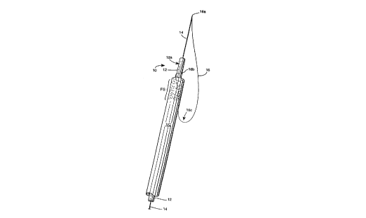
FIGURE 1 is a srh~m ~hc perspective view of a ~ lled
m e~irzll snare device embodying the present i I'v~- ~Li~n; and

FIGURES 2A-2C are srhPm~hc elev:~tion~l views showing a
sequence of the device depicted in FIGURE 1 during use.


DETAILED DESCRlPTlON OF THE PREFERRED
EXEMPLARY EMBODIMENTS
One preferred embo~im~nt of a medical snare device 10
accol.1;l,g to the present i~ vellLion is shown in accompanying
FIGURE 1. The snare device 10 is depicted srhPm~c~11y as being
positioned intral1~min~11y wi~ a venous cat~eter VC, it being
- 25 understood that the distal end region of venous catheter VC will inuse be impl~nted Wil~lill a patient's vein. The dist~l end section of
the venous catheter VC is depicted in accompanying FIGURE 1 as

CA 02217481 1997-10-03
W O96~1162 PCTrUS96/04799
,


having a fibrin chP~h FS Ç~.n~ing ~luxi~ lly along the catheter's
rinr snrf~e

The snare device 10 is generally co...". i.ced of an elongate
5 tubular member 12 and a central wire PlPmPnt 14 which is movably
pociti~ned wil~li.l the lumen of the tubular member 12. Each of the
tubular member 12 and central w1re PlPmPnt 14 is of sllffiriPnt length
to allow the physician to intralllm;n~lly insert them as a unit
t.hrough the venous catheter VC so that the distal end 12a of the
0 tubular body 12 is capable of e~tPn~ing dista~ly beyond the distal end
of the venous catheter VC, and so that the ~eL~ al end 14a of the
cenhral wire PlP~mPnt 14 is c~p~hle of being e~t~n~e~ beyond the
distal end 12a of the tubular member (e.g., to achieve relative
posihnning as show.. in FIGURE 1).
1!;
Important to the present ...v~.~l;nn, the snare device 10
includes a snare wire 16 formed of a flP~ihle metal or plastics wire,
thread or the like. The snare wile 16 has its distal end 16a
physically ~ rhed to the cenhral wire PlPm~nt 14 so as to form an
20 acute angle therewith and its pro~im~l end 16b physically attached
(e.g., via biocompatible epo~y, heat-welding, imbedding or the like) to
the tubular member 12 at or near its distal end 12a. The ends 16a,
16b are thus ~nally separated from one another along the length of
the device 10 so as to form a snare loop collectively with the terminal
2~ end 12a of the tubular member and that length of the central wire
~lPmPnt 14 egt~n~lin~ therebeyond. As shown, the snare wire 16,
between the ends 16a and 16b is preferably wrapped appro~im~ ly

CA 02217481 1997-10-03

W O 96/31162 PCTnUS96/04799


360~ around the tubular member 12 so that the end 16b faces
distally.

The relative diameters of the central wire 14 and the snare
5 wire 16 are depentlPnt. in large part upon the particl lar medical
procedure in which the device 10 of this i~lv~:..Lion is in~n~ed to be
employed. It is ~lerelled~ how~v~:l, that the diameters of the central
wire 14 and the snare wire 16 each be within the range of about
0.001 to about 0.040 inch. Moreover, it is ~lefel. ed that the snare
0 wire 16 have a lesser diameter as comp~red to the central wire
m~nt 16 so that the former is relatively more fle~ible, while the
latter is relatively more stiff. Thel~role, it is ~ler~lled that the ratio
of the central wire diameter to the snare wire dia~neter be between
about 1.1:1 to about 10.0:1.
The snare wire 16 is of s~.ffiri~nt length between its ends 16a,
16b such that a segm~nt 16c thereof may be positioned pro~imzllly of
the end 16b adjacent the distal end of the venous catheter. With the
snare wire segm~nt 16c po.cihoned in such a m~nn~r, the physician
2~ may rotate the central wire el~m-ont 14 about its longitl~in~ is
wi~ the lumen of the tubular member 12 as showrl by arrow Al in
FIGURE 2A. Relative rotation bet~,veen the central wire element 14
and the tubular member 12 (e.g., rotation of the central wire element
14 while ~nz?int~ining the tubular member 12 stationary) will thereby
25 cause the snare wire se~ nt 16c to be wrapped or twisted more or
less h~lir~lly about the e~terior surface of the venous catheter's distal
end as shown in FIGURE 2B. The several turns of the wrapped

CA 02217481 1997-10-03

W O96~1162 PCT/U'9G/01799


snare wire se~rn~nt 16c Will thus be brought into contact with the
fibrin .che:~h FS at the distal end of the venouc catheter VC. Ac
such, adv~nl Pm~nt of the central wire 14 and/or the tubular member
12 in a distal direction (arrow A2 in FIGURE 2C) wi~l, in turn, cause
5 the wrapped snare wire segm~nf; 16c to be moved distally along the
e2terior surface of the distal end of the venous catheter VC thereby
sLi~i. g the fibrin che~t.h FS thel~:L.~

Although the central wire element 14 has been depicted in the
0 ~rComranying drawing FIGURES as including an eyelet 14a at its
~e....i..al end, it will be appreciated that the eyelet 14a is not
critically necessary since the end 16a of the snare wire 16 may be
bonded to the central wire's Lel,..;..al end via biocompatible epoxy,
solder, or the like. Furthermore, the central wire 14 and the snare
15 wire 16 may be formed as a single (unitary) mnnnfil~m~nt wire,
instead of the separate, but cormected, wires as shown in the
accompanying drawing FIGURES.

Thelefo.e, while the i. v~Lionhas been describedin conn~c~on
20 with what is presently considered to be the most pr~ cz~l and
preferred embor~imen~ it is to be understood that the i~vellLion is not
to be limited to the disclosed embo~im~nt but on the co..l~ , is
int~ncled to cover various mo~ific~t~inn.c and equivalent arrang~ment-
~included wiL~i~ the sp~Iit and scope of the appended cl~im.c.
Representative Drawing
A single figure which represents the drawing illustrating the invention.
Administrative Status

For a clearer understanding of the status of the application/patent presented on this page, the site Disclaimer , as well as the definitions for Patent , Administrative Status , Maintenance Fee  and Payment History  should be consulted.

Administrative Status

Title Date
Forecasted Issue Date Unavailable
(86) PCT Filing Date 1996-04-05
(87) PCT Publication Date 1996-10-10
(85) National Entry 1997-10-03
Dead Application 2004-04-05

Abandonment History

Abandonment Date Reason Reinstatement Date
2003-04-07 FAILURE TO PAY APPLICATION MAINTENANCE FEE
2003-04-07 FAILURE TO REQUEST EXAMINATION

Payment History

Fee Type Anniversary Year Due Date Amount Paid Paid Date
Application Fee $150.00 1997-10-03
Registration of a document - section 124 $100.00 1997-12-29
Maintenance Fee - Application - New Act 2 1998-04-06 $50.00 1998-03-31
Maintenance Fee - Application - New Act 3 1999-04-06 $50.00 1999-04-06
Maintenance Fee - Application - New Act 4 2000-04-05 $50.00 2000-03-24
Maintenance Fee - Application - New Act 5 2001-04-05 $75.00 2001-03-23
Maintenance Fee - Application - New Act 6 2002-04-05 $75.00 2002-03-26
Owners on Record

Note: Records showing the ownership history in alphabetical order.

Current Owners on Record
DUKE UNIVERSITY
Past Owners on Record
RIDINGER, MARK T.
SUHOCKI, PAUL V.
Past Owners that do not appear in the "Owners on Record" listing will appear in other documentation within the application.
Documents

To view selected files, please enter reCAPTCHA code :


To view images, click a link in the Document Description column. To download the documents, select one or more checkboxes in the first column and then click the "Download Selected in PDF format (Zip Archive)" or the "Download Selected as Single PDF" button.

List of published and non-published patent-specific documents on the CPD .

If you have any difficulty accessing content, you can call the Client Service Centre at 1-866-997-1936 or send them an e-mail at CIPO Client Service Centre.


Document
Description 
Date
(yyyy-mm-dd) 
Number of pages   Size of Image (KB) 
Representative Drawing 1998-01-20 1 4
Cover Page 1998-01-20 2 67
Abstract 1997-10-03 1 50
Description 1997-10-03 8 304
Claims 1997-10-03 6 153
Drawings 1997-10-03 2 27
Fees 2001-03-23 1 22
Fees 1999-04-06 1 34
Fees 1998-03-31 1 36
Assignment 1997-10-03 4 125
PCT 1997-10-03 40 1,504
Correspondence 1997-12-16 1 30
Assignment 1997-12-24 6 225