Note: Descriptions are shown in the official language in which they were submitted.
CA 02218092 1997-10-10
WO 96/32145 PCT/US95/04531
SELECTIVE AORTIC PERFUSION SYST$M
Field of the Invention
The present invention relates generally to
treatment of a patient during cardiopulmonary resuscitation
(CPR) and more particularly to a process and apparatus for
aortic occlusion along with oxygen carrying fluid infusion
and/or aortic balloon counterpulsation for use during CPR.
Background of the Invention
Cardiopulmonary resuscitation has not fulfilled
its original expectations, and the prognosis for patients
remaining in cardiac arrest more than ten minutes remains
poor (Becker AB, Ann Emerg Med, 20:355 (1991)). indeed,
cardiopulmonary resuscitation has recently been termed a
"spectacular failure" in which only a small minority of
patients have been successfully resuscitated (Barsan WG,
JAMA, 265:3115-3118 (1991)). Standard advanced cardiac life
support (ACLS) has only limited efficacy after the first few
minutes of cardiac arrest. Studies in animal models have
shown that vital organ blood flow, and thus oxygen delivery,
during CPR is poor (Ditchey RV, et al, Circ, 66:297-
302(1982); Ditchey RV et al, Cardiovasc Res, 19:419-425
(1985); and Taylor RB, et al, Resuscitation, 16:107-
118(1988)). Indeed, CPR generally provides only a small
fraction of normal oxygen supply to the brain and heart, and
even less to other organs. Recent human studies have
confirmed that perfusion pressures, the driving force for
organ blood flow, are inadequate in humans during CPR
(Paradis NA, et al, Circ, 80:361-368 (1989); Paradis NA, et
al, JAMA, 263:1106-1113 (1990); and Martin GB, et al, Ann
Emerg Med, 15:125-130(1986)). High-dose epinephrine, open
chest CPR, and cardiopulmonary bypass increase perfusion
pressure (Paradis NA, et al, JAMA, 265:1139-1144 (1991);
Martin GB, et al, Ann Emerg Med, 16:628-636 (1987); and
Howard MA, et al, Ann Emerg Med, 15:664-665 (1986)).
However, these are not effective in all patients, or require
significant resources not generally available.
CA 02218092 1997-10-10
WO 96/32145 PCT/13S95/04531
- 2 -
In an effort to find simple but effective methods
to improve perfusion during CPR, a number of mechanical
intravascular based therapies have been investigated. Among
these are arterial and venous volume infusion and aortic
occlusion (Gentile NT, et al, Crit Care Med, (1990) (in
press); Abu-Nema T et al, Cirg Shock, 4:55-62 (1988); Suzuki
A et al, Jpn J Anesthesiol, 29:677-682 (1980); Spence PA, et
al, J Surg Res, 49:217-221 (1990)); and Manning JE et al,
Ann Emerg Med, 19:212 (1990). These techniques, however,
have failed to improve outcome. Standard aortic
counterpulsation that is without distal aortic occlusion,
may improve perfusion, but not enough to significantly
improve outcome (Emerman CL, et al, Am J Emerg Med, 7:378-
383 (1989)). Simple balloon occlusion, with or without
volume_infusion, does not appear to be effective.
It is known to provide oxygenated fluorocarbon
emulsions to transport oxygen to oxygen deprived brain
tissue (see U.S. Patent 4,927,623 to Long, Jr.).
Balloon catheter devices and methods are known for
directing blood toward the heart during spontaneous
circulation (see, for example, U.S. Patents 4,407,271 to
Schiff; 4,804,358 to Karcher et al; 4,601,706 to Aillon; and
4,459,977 to Pizon et al).
Such catheter devices with two or more balloons
are also known (see, for example, U.S. patents 4,531',936 to
Gordon; 4,527,549 and 4,741,328 to Gabbay; 4,697,574 to
Karcher et al.; 5,176,619 to Segalowitz; and 4,771,765 and
4,902,273 to Choy et al.). None of these devices were
designed for use during cardiac arrest.
Summary of the Invention
It is a primary object of the present invention to
provide a process and apparatus for carrying out aortic =
occlusion along with oxygen carrying fluid infusion to the
heart and brain, and/or aortic balloon counterpulsation, as
a therapy for cardiac arrest.
It is a further object of the present invention to
increase the period of time for possible successful
resuscitation during cardiac arrest.
CA 02218092 1997-10-10
WO 96/32145 PCT/US95/04531
- 3 -
The above objects are accomplished through the use
of a specially constructed balloon catheter which, when
inflated, occludes the aorta such that fluids infused
through the catheter will be restricted to that part of the
aorta above the balloon occlusion. An oxygenating fluid
(such as either oxygenated fluorocarbons or stroma-free
polyhemoglobin or recombinant hemoglobin) is used as the
infused fluid either in a pre-oxygenated state or after an
oxygenator has oxygenated the fluid.
In a preferred embodiment of the present
invention, the occluding balloon is moved away from the tip
of the catheter to make room for a counterpulsation balloon.
The counterpulsation balloon is connected to a drive unit
outside of the patient by an additional lumen in the
is catheter. The drive unit is preferably a portable unit
which can be carried in ambulances or to various locations
in the hospital. A sensor means is added to the tip of the
catheter and connected to the external drive unit by a small
u:ire .bedded in the catheter-wall. The drive unit inflates
the counterpulsation balloon when it senses the relaxation
phase of CPR, thereby increasing the aortic relaxation
pressure which is the primary determinant of outcome during
CPR. Because the distal aorta is continuously occluded, all
of the increased perfusion is directed cephalad primarily to
the heart and brain. In a further preferred embodiment, the
balloon is inflated first at its base (caudal end) and that
inflation moves cephalad toward the tip. This accelerates
oxygen-carrying fluid, either blood or infusate, toward the
heart and brain. The occlusion balloon is preferably as
close as possible to the caudal end of the counterpulsation
balloon.
The combination of the occlusion balloon and the
, counterpulsation balloon may accomplish the objects of the
present invention even without the concomitant infusion of
oxygenating fluids through the catheter. Thus, the
embodiment of the present invention in which the occlusion
balloon is combined with a counterpulsation balloon may be
used with or without concomitant oxygen-carrying fluid
infusion.
CA 02218092 1997-10-10
WO 96/32145 PCT/US95/04531
- 4 -
Aortic occlusion with oxygen-carrying fluid
infusion will significantly improve oxygen supply for short
periods of time to the heart and brain. The infusion of
oxygen-carrying fluid will be retrograde up the descending
aorta toward the head resulting in preferential infusion of the coronary and
carotid arteries. Attempts at
defibrillation after this period of improved perfusion of the cardiac muscle
will result in significantly higher rates
of return of spontaneous circulation when compared to
standard CPR.
Furthermore, aortic occlusion, in combination with
standard cardiopulmonary resuscitation (CPR) techniques
which trigger counterpulsation by a counterpulsation balloon
will also result in preferential circulation of blood into
coronary and carotid arteries so that attempts at
defibrillation will result in significantly higher rates of
return of spontaneous circulation when compared to standard
CPR. The combination of aortic occlusion and relaxation
phase counterpulsation will result in even better coronary
perfusion pressures and myocardial oxygen supply, perhaps
even greater than during normal circulation. After return
of spontaneous circulation, counterpulsation and/or
occlusion, can be continued in its standard mode to improve
myocardial perfusion during the initial unstable period that
often follows cardiac arrest.
Placement of a balloon catheter in the descending
aorta through the femoral artery is not difficult (Bregman
D, et al, Am J Cardiol, 46:261-264 (1980)) and it may be
possible to accomplish this even in a prehospital setting.
The catheter which is used for the present
invention is specially designed so as to have a large
infusion port. Known balloon catheters have an infusion
lumen which is relatively small, designed for the delivery
of drugs or small amounts of fluid or for measurement of
blood pressure. The catheter of the present invention must
be designed to permit the flow of large amounts of perfusion
fluid.
The catheter which is used in accordance with the
present invention for simultaneous aortic occlusion and CPR
CA 02218092 1997-10-10
WO 96/32145 PCT/US95/04531
- 5 -
relaxation phase counterpulsation is also specially
designed. The occluding balloon is moved away from the tip
of the catheter to make room for the counterpulsation
balloon. One or more additional lumens serve the
counterpulsation balloon to cause inflation. In a preferred
embodiment, these lumens are also used for active balloon
deflation. Active deflation will augment flow from the left
ventricle during compression phase of CPR and improve
cardiac output. The occlusion balloon is preferably as
close as possible to the base of the counterpulsation
balloon. If the counterpulsation balloon is segmented in
order to cause a wave-like motion during inflation which
would cause a directional pumping activity toward the
carotid and cardiac arteries, the occlusion balloon may,
indeed, be the first segment of the counterpulsation
balloon, which segment is not deflated during the
counterpulsation pumping cycle.. Unlike counterpulsation
devices intended for use during spontaneous circulation, the
portion of the present device serving as the occlusion
balloon must be capable of inflating to the extent of
occluding the aorta so as to maximize perfusion volume and
pressure.
A sensor, such as a micromanometer, is preferably
added to the tip of the catheter in order to sense the chest
compression phase of CPR. Such a micromanometer will
trigger off the changes in intravascular pressure generated
by chest compression. Alternative, the central infusion
lumen may be used, when infusion is not occurring, to
transmit central aortic pressure to an external pressure
transducer, which may control the drive unit. A drive unit
controlled by a signal from the micromanometer or external
transducer is designed to inflate the counterpulsation
balloon when it senses the drop in pressure at the beginning
relaxation phase of CPR. The counterpulsation balloon is
preferably designed in such a way that the balloon is
inflated first at its base and that inflation moves cephalad
toward the tip. Because the drive unit need not be
triggered by variable and complicated electrocardiograph
signals, but only a simple signal from a micromanometer, the
CA 02218092 1997-10-10
WO 96/32145 PCT/US95/04531
- 6 -
drive unit may be greatly simplified from normal
cardiopulmonary balloon assist drive units. Only one
pattern of inflation, cephalad relaxation phase inflation
and compression phase deflation, may be required. This will
simplify downsizing of such a drive unit to permit
portability. This drive unit may be incorporated in
existing devices intended to inflate external vests that
provide CPR. A combination of the two drive units would
coordinate optimization of external CPR and internal
intravascular circulation.
As the success of defibrillation in return to
spontaneous circulation will be greatly improved after the
selective aortic perfusion or circulation of the present
invention, it is expected that the process and apparatus of
the present invention will supplant CPR alone and will be
used in all emergency departments and other critical care
areas, and potentially in all advanced life support
ambulances.
The present invention further comprehends a kit
for use in performing the process of the present invention,
which kit will include a catheter, a supply of stroma-free
polyhemoglobin (or other oxygen-carrying perfusion fluid)
and, optionally, an oxygenator to cause oxygenation of the
fluid prior to perfusion through the catheter. The kit will
preferably also include all of the other paraphernalia for
carrying out the process of the present invention.
Brief Description of the Drawings
The above and other objects will be shown in more
detail in the following detailed description of the
preferred embodiments when read in conjunction with the
attached drawings, in which:
Fig. 1 is a schematic representative cross-section
of a balloon catheter which can be used in accordance with
the present invention;
Fig. 2 is a schematic representative cross-section
of a pressurizable container for use in dispensing
oxygenatable fluid;
CA 02218092 1997-10-10
WO 96/32145 PCT/US95/04531
- 7 -
Fig. 3 is a schematic representative cross-section
of an oxygenating system for use with the present
invention;
Fig. 4 is a diagrammatic representation of a kit
in accordance with the present invention;
Fig. 5 is a diagrammatic representation showing
the catheter in appropriate position during use;
Fig. 6 is a schematic representative cross-section
of a balloon catheter which can be used in accordance with a
second embodiment of the present invention and which
includes aortic balloon counterpulsation;
Fig. 7 is a schematic representative cross-section
of another embodiment of an aortic balloon counterpulsation
catheter; and
is Fig. 8 is a schematic representative cross-section
of yet a further embodiment of an aortic balloon
counterpulsation catheter.
Detailed Description of the Preferred Embodiments
In a first embodiment of the present invention,
the selective aortic perfusion system of the present
invention has three major components. The first is a
specially constructed balloon catheter 12 as shown in Fig.
1. This catheter is sized and dimensioned to permit
insertion through the femoral artery and feeding up into the
aorta until the balloon 14 is located in the descending
aorta. Generally, the medical or paramedical personnel
using this invention in an emergency setting will know when
the balloon is in appropriate position when the tip of the
catheter impacts the top of the aortic arch and cannot
easily be maneuvered further through the aorta. Preferably,
however, the catheter 12 will include markings 22, 24 which
signal the distance from the marking to the tip 20 of the
catheter 12. For example, a double mark 24 may mean that
the distal tip is 70 cm away with each single mark 22 being
in 2 cm increments. The person inserting the catheter 12
will know the position of the balloon 14 in the aorta from a
consideration of the markings at the proximal end of the
catheter. When the balloon 14 is in position and is
CA 02218092 1997-10-10
WO 96/32145 PCTlUS95/04531
- 8 -
inflated, it will occlude the aorta above the level of the
diaphragm (see Fig. 5). Thus, infused fluids will be
restricted only to the volume of the aorta and associated
arteries above the balloon occlusion.
The catheter 12 is constructed to have two lumens
16, 18. The smaller lumen 16 is used for inflating the
balloon. This lumen 16 can be attached to, for example, a
30 cc syringe (not shown) filled with saline. In the rare
event of a balloon failure, only saline fluid would be
released into the aorta. The larger lumen 18 opens distal
to the balloon 18 at a point 20 and is attached to the
system for infusion of oxygen containing fluid. The lumens
16, 18 may be side by side or coaxial.
The larger lumen 18 distinguishes the catheter of
the present invention from all prior art balloon catheters.
The cross-section of this lumen must be large enough to
permit sufficient infusion of oxygenating fluid to oxygenate
the myocardium and the cerebrum. It has been calculated
that to completely replace all of the oxygen deficit which
occurs after eight minutes of cardiac arrest, including the
ongoing deficit thereafter, a total of four liters of fully
oxygenated stroma-free polyhemoglobin would have to be
infused over the course of two minutes. It should not be
necessary, however, to infuse 1000 of the oxygen deficit.
Thus, it is fully expected that a replacement of 50!~ of the
oxygen deficit, i.e., two liters of fluid over a course of
two minutes, will provide results which are substantially
better than standard CPR and yet avoid a possible volume
overload when spontaneous circulation returns. Indeed, CPR
is occasionally successful despite providing significantly
less oxygen supply. Thus, 0.25-1.5 liters will most
probably be sufficient in practice, up to a maximum of 3
liters (when fully oxygenated SFPH is used as the fluid) =
over the course of about one to three minutes.
The appropriate size lumen to permit this much infusion over the specified
time period can be designed
using standard engineering formulas, such as Poiseuille's
Law, and/or empirical testing. The optimum diameter is on
the order of about 2-3 mm so as to permit infusion without
CA 02218092 1997-10-10
WO 96/32145 PCT/US95/04531
- 9 -
using excessive feed pressure while maintaining the catheter
as a whole sufficiently small to permit insertion through
the femoral artery. It is believed that the largest
existing balloon catheters have an inner lumen of less than
' 5 about 1 mm. For some applications, such as pediatric
applications, the diameter may be as small as 1.5 mm and
could be as large as 4 mm. Catheters of this large size can
be easily placed using existing guide wire/introducer sheath
techniques.
The most important function of the oxygenating
fluid is to perfuse the myocardium, thereby permitting a
vastly improved chance of return to spontaneous circulation
after defibrillation. It is a secondary function to
oxygenate the brain in order to prevent damage to the brain
during the period of cardiac arrest. However, a prompt
return to spontaneous circulation after perfusion of the
myocardium and defibrillation will serve the purpose of
oxygenating the cerebrum much better than the perfusion of
oxygenating fluid in accordance with the present invention.
In order to increase the amount of perfusion of the
myocardium and cerebrum, it may be desirable to actually
prevent flow of the perfusion fluid to the upper extremities
by applying pressure to the appropriate arteries during
infusion of the fluid. Furthermore, while it is permissible
to continue CPR throughout the procedure of the present
invention, it is permissible to suspend CPR once the
catheter has been inserted, as the perfusion being caused by
the infusion of oxygenating fluids will be much greater than
that caused by the CPR. It may be desirable to leave the
balloon fully or partially or partially inflated after
return of spontaneous circulation to continue preferential
perfusion of the brain and heart.
The second component of the system of the present
invention is oxygenating fluid. Currently, there are two
types of artificial fluids which are used to carry oxygen
for use in humans. The first is oxygenated fluorocarbons
and the second is stroma-free polyhemoglobin (SFPH) or
recombinant hemoglobin. SFPH has recently been approved for
CA 02218092 1997-10-10
WO 96/32145 PCTNS95/04531
- 10 -
preliminary human testing and is available from the Biopure
Company.
The oxygenating fluid solution is preferably
packaged in a special container capable of being 5 pressurized, as shown in
Fig. 2, so that it can be infused
at pressures necessary to overcome those during the
compression phase of CPR and necessary to provide a
sufficient flow rate of oxygenating fluid. The oxygenating
fluid is stored in a pressure bag 32, preferably in a
quantity of 500 cc or more. The bag is placed in a
container 30 having a lid 31 sealed thereto. An inlet valve
34 in the lid 31 of the container includes a regulator
mechanism (the details of which are well known and not
shown) which permits entry of pressurized fluid at a
specified maximum pressure regardless of the pressure of the
fluid at the inlet of the valve 34. The valve 34 includes
an inlet nozzle 36 which is connectable to the source of
standardized pressurized oxygen available in all emergency
and critical care settings. While pressurized oxygen is the
preferred source of pressurized fluid to drive the feeding
of the oxygenating fluid, it is to be understood that any
other source of pressurized gas or liquid could be used for
this purpose.
An outlet tube 37 connected to the interior of the
bag 32 extends=from the container 30 and includes a
connector 38 for connection to the oxygenating fluid feeding
lumen 18 of the catheter 12.
While the special container described above is the
preferred means of dispensing the oxygenating fluid
solution, it should be understood that this particular means
is not critical and that any manner of supplying the
oxygenating fluid under pressure sufficient to provide the
desired amount of infusion over the predetermined period of
time can be used in the method of the present invention.
The oxygenating solution may also include the
simultaneous infusion of other drugs or agents to improve
myocardial and cerebral outcome. Any agent demonstrated to
be effective when given intravenously may be more effective
when administered to the heart and brain selectively by
CA 02218092 1997-10-10
WO 96/32145 PCT/US95/04531
- 11 -
means of the present invention. Tissue salvaging agents in
particular may be included in the oxygenated infusion fluid.
Examples of agents which may be included in the oxygenated
infusion fluid of the present invention are: epinephrine or
other adrenergic agonists and pressors; antioxidants and
free-radical scavengers such as the 21-amino steroids
(lazaroids); anti-inflammatory agents including steroids and
non-steroidal anti-inflammatory drugs such as a ibuprofen;
calcium channel blockers such as lidoflazine, nimodipine,
nicardipine, flunarizine, etc.; excitatory neurotransmitter
blockers (NMDA receptor agonists) such as MK801, etc.;
anticoagulants such as heparin; iron and heavy metal
chelators such as deferoxamine; osmotic agents such as
mannitol; anti-acidosis agents such as bicarbonate or
is dichloroacetate; insulin; antibodies such as anti-
neutrophile antibody; and allopurinol. This list is
intended to be exemplary only and not limiting.
The third component of the system of the present
invention is an apparatus to oxygenate the oxygenating fluid
before infusion. This component is optional as the
oxygenating fluid may be supplied already oxygenated and
ready to use. However, as the oxygenating fluid may lose
its oxygen over storage time it is preferred that the fluid
be freshly oxygenated immediately prior to perfusion.
Simple infusion of the solution into the arterial side of
the circulation without pre-oxygenation would-not improve
the delivery of oxygen to the myocardium and brain. As
shown in Fig. 3, a hollow fiber membrane oxygenator 40
having hollow fibers 41 can be placed in the system between
the pressurized container 30 of oxygenating fluid, at in-
port 42 and the lumen 18 of the balloon catheter 12 at out-
port 44. These systems allow the blood to flow around
numerous hollow fibers 41 which have been specially
constructed to allow diffusion of gas phase components
without leakage of oxygenating fluid or blood. Oxygen is
forced in at in-port 46 and exits at out-port 48. The
mechanism of oxygenation is preferably countercurrent, which
should result in oxygenation of the stroma-free
polyhemoglobin, or other oxygenating fluid, to its maximum
CA 02218092 1997-10-10
WO 96/32145 PCTfUS95/04531
- 12 -
saturation. This device will also be constructed so that
standard oxygen tanks or other emergency room oxygen supply
can be used to supply it. The same oxygen can be used to
inflate the pressure bag and drive the infusion as is used
to oxygenate the fluid.
The components of the present invention are
preferably packaged in kit form for use by medical and
paramedical personnel. The components of the kit are
preferably packaged in a sealed container which is compact
and portable and suitable for use by emergency room or
ambulance medical or paramedical personnel. As shown in
Fig. 4, the components may be packaged in a container 50,
preferably a rigid plastic material molded to provide
compartments for the various components of the kit.
Compartment 52 holds the catheter 12, compartment 54 holds
the container 30 with the sack 32 of oxygenating fluid (or
compartment 54 may comprise the container 30 and hold only
the sack 32), and compartment 56 holds the oxygenator 40.
Compartments for other paraphernalia necessary to implement
the process of the present invention may also be present.
For example, compartment 58 may be present for holding a
syringe 60 to be used for inflation of the balloon and
compartment 62 may be present to hold a catheter insertion
sheath 64. Other components such as oxygenation tubing,
guide wires, instructions for use, etc., may also be
present.
The preferred kit form for use in the present
invention is one which is the simplest and easiest to use in
an emergency situation, which is often somewhat chaotic.
Thus, the connections between the oxygenator and the source
of oxygenating fluid may be built-in so that the source of
oxygenating fluid and the oxygenator need never be removed
from the kit. Similarly, the connection between the oxygen
output of the oxygenator and the pressure chamber of the
oxygenating fluid compartment may be built into the kit., as
may the connection between the balloon syringe and the
catheter lumen leading to the balloon and the connection
between the oxygenator fluid output and the catheter lumen
for feeding the oxygenated fluid.
CA 02218092 1997-10-10
WO 96/32145 PCT/US95/04531
- 13 -
For example, as shown in Fig. 4, the oxygenator 40
may be sealed within the kit 50 with only an appropriate
oxygen input nozzle 70 extending from the container. Nozzle
70, which extends from container 50 may be a standard
connector for connection to a source of pressurized oxygen
such as a standard oxygen tank or other emergency room
oxygen supply. The nozzle 70 is connected within the
container 50, by means of tube 71, to a Y-junction 80 which
divides the oxygen input into line 82, which leads to the
oxygen in-port 46 of the oxygenator, and line 84, which
leads to the pressurizable container 30. The oxygen out-
port 48 of the oxygenator may be vented to the atmosphere.
All of these lines may be sealed within the container 50.
The regulator valve 34 is also present within the container
50 so as to regulate the maximum pressure of fluid entering
the container 30 for pressurizing the sack of oxygenating
fluid 32. A rubber or plastic,pressure bladder, not shown,
may be placed between the sack 32 and the pressure chamber
30 to diminish risk of damage to sack 32. A second
regulator valve 86 may also be built-in at the oxygen in-
port 46 of the oxygenator 40 in order to regulate the
pressure of oxygen entering the oxygenator 40.
The output port 37 from the sack 32 of oxygenating
fluid may be directly connected to the oxygenating fluid
inlet port 42 of the oxygenator 40 by means of a tube 74
sealed within the container 50. A valve 76, accessible from
the outside of the container 50, may be used to open or
close access of the oxygenating fluid from the sack 32 to
the oxygenator input 42. The output from the oxygenator 40
through the out-port 44 may be directly connected to the
fluid input cannula 18 of the catheter 12. Thus, no
physical connections need be made by the emergency personnel
except for attachment of an oxygen source to nozzle 70.
In use, the catheter 12 is removed from the
compartment 52 of the kit. Lumen 18, which is much longer
than is schematically shown in Fig. 4, is already connected
to the output 44 of the oxygenator. Lumen 16 is also
already connected to the syringe 60. Syringe 60 is
preferably one which contains exactly the right amount of
CA 02218092 1997-10-10
WO 96/32145 PCT/17S95/04531
- 14 -
fluid to cause inflation of the balloon and is constructed
so as to permit one-time use only. The use of such a pre-
packaged syringe will eliminate the possibility of over-
inflation and rupture of the balloon. A source of
pressurized oxygen is connected to the nozzle 70 of the
container 50.
The femoral artery 90 (see Fig. 5) will be
punctured by a needle, through which a guide wire is
advanced into the descending aorta 92. The guide wire may
be any standard flexible guide wire or it could be a
specially designed guide wire of increased stiffness to
facilitate placement directly up the aorta. An introducer
sheath 64 is removed from the compartment 62 of the
container 50 and then advanced over the wire into the
femoral artery. The central trochar and the guide wire are
removed from the introduced sheath and the distal end 20 of
the catheter 12 is introduced through the sheath into the
femoral artery and fed until the balloon reaches the
appropriate position in the aorta as determined by the
markings 22 and 24 on the catheter or by other known means.
Alternatively, the guide wire can be left in place to
facilitate placement of the catheter 12, the guide wire
being removed after placement of the catheter 12. The
balloon 14 is then inflated by means of the syringe 60 and
the valve 76 is opened in order to permit the feeding of the
oxygenating fluid through the oxygenator 40 and into the
lumen 18 and opening 20 into the aorta at the predetermined
flow rate. At the same time, oxygen will flow through the
oxygenator, countercurrent to the flow of oxygenating fluid,
and into the pressure chamber 30 to drive the flow of
oxygenating fluid at the predetermined rate, determined by
the regulator valve 34. The oxygen output from the
oxygenator may include a bleed valve to ensure flow of
oxygen through the oxygenator even when not necessary for
pressurization of the chamber 30.
Another embodiment of the present invention is
illustrated in Figs. 6-8. In the embodiment illustrated in
Fig. 6, the catheter 112 is substantially similar to the
catheter 12 of Fig. 1. With respect to the occlusion
CA 02218092 1997-10-10
WO 96/32145 PCT/US95/04531
- 15 -
balloon 114 which corresponds to the balloon 14 of Fig. 1,
smaller lumen 116, which is used for inflating the occlusion
balloon 114 and corresponds to the smaller lumen 16 of the
catheter 12 of Fig. 1 and the larger lumen 118, which
corresponds to the larger lumen 18 of Fig. 1. However, in
the embodiment of Fig. 6, the occlusion ballobn 114 is moved
away from the tip 120 of the catheter and a counterpulsation
balloon 108 is disposed on the catheter 112 between
occlusion balloon 114 and the distal tip 120. Occlusion
balloon 114 preferably has a diameter sufficient to fill the
aortic lumen when inflated. A third lumen 106 is also made
part of the catheter 112 to inflate the counterpulsation
balloon 108. The three lumens 116, 118 and 106 may be side-
by-side or coaxial.
At the distal tip of the catheter 112 is a
micromanometer 104 capable of sensing the chest compression
phase of CPR. A wire 102 is connected to the micromanometer
104 and is embedded in the wall of the catheter 118.
A drive unit, schematically shown at 110, drives
the operation of the counterpulsation balloon 108. Thus,
the lumen 106 which feeds inflating fluid (either liquid or
gas) to the counterpulsation balloon 108 is connected to
this drive unit as is the wire 102 which is connected to the
micromanometer. The drive unit is designed such that, when
the micromanometer senses the relaxation phase of CPR, the
drive unit causes inflating fluid to pass through the lumen
106, thereby inflating the counterpulsation balloon 108. In
a preferred embodiment, the drive unit also causes active
deflation of the counterpulsation balloon 108 by actively
removing fluid therefrom when the micromanometer senses the
beginning of the compression phase of CPR. In the
embodiment shown in Fig. 6, the occlusion balloon 14 is also
connected to the drive unit 110. The drive unit 110
maintains the occlusion balloon 14 continuously in an
inflated position. The occlusion balloon may also be filled
separately by syringe.
When the counterpulsation balloon is being used in
combination with CPR, it is not necessary to infuse
oxygenating fluid through the catheter 118 in order to
CA 02218092 1997-10-10
WO 96/32145 PCT/US95/04531
- 16 -
improve oxygenation of the heart and brain. Thus, it is not
necessary that the catheter 118 be as large as is described
above for the embodiment of Fig. 1. Indeed, this lumen may
be of standard size and used only for injection of drugs.
Alternatively, oxygenating fluid can be perfused
simultaneously with the counterpulsation CPR in order to
even more greatly increase the volume and oxygenation
content of the fluid reaching the heart and the brain during
this operation. In another embodiment, the micromanometer
104 may be omitted and the balloon 108 used only as a pump
mechanism to assist in the infusion of the oxygenating fluid
through the lumen 119 without external CPR. For this
embodiment, it is important only that the pulsation balloon
108 be driven in such a way as to allow alternating cardiac
output and aortic pulsation. In this embodiment, it is
being used solely as a pump.
In another preferred embodiment of the present
invention, the counterpulsation balloon 108 is designed so
as to preferentially inflate, beginning at the caudal end,
i.e., the end closest to the occlusion balloon 114, and then
continuously inflate toward the distal end. This will cause
the fluidto be pumped unidirectionally in a cephalad
direction, primarily to the heart and brain. This will also
prevent excessive pressure build-up in the aorta between the
counterpulsation balloon 108 and the occlusion balloon 114.
This unidirectional inflation of the counterpulsation
balloon 108 may be accomplished in any of various manners.
In the embodiment shown in Fig. 6, it is accomplished by
placing the outlet of the lumen containing the inflation
fluid at the caudal end of the balloon so that inflation
will commence at that end and proceed cephalad toward the
distal end of the catheter.
Other means may also be used to achieve this
unidirectional pumping function. For example, as shown in
Fig. 7, the counterpulsation balloon may be segmented into
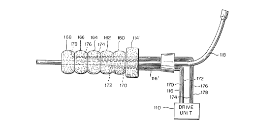
segments 160, 162, 164, 166 and 168. Each segment is served
by a respective one of a plurality of lumens 170, 172, 174,
176, 178. The lumens are supplied with fluid in such a
programmed way that the caudal segments inflate before the
CA 02218092 1997-10-10
WO 96/32145 PCT/US95/04531
- 17 -
next cephalad segment inflates. In this embodiment, the
occlusion balloon 114' may be the first segment of such a
segmented balloon, or the first balloon of a series of
balloons, which first segment remains continuously inflated.
The remaining segments are inflated in a wave-like motion to
cause unidirectional wave-like inflation in a cephalad
direction. Thus, segment 160 is first inflated by means of
its lumen 170, followed by inflation of segment 162 by means
of its lumen 172, followed in turn by inflation of segment
164, 166 and 168 by means of their respective lumens 174,
176 and 178. Proper order and speed inflation is all
controlled by drive unit 110 by means which will be readily
apparent to those of ordinary skill in this art. The active
deflation may be simultaneous or sequential for segments
other than the segment which which serves as the occlusion
balloon 114.
Another way of obtaining unidirectional inflation
is shown in Fig. 8. In this embodiment, the lumen 106'
extends throughout the length of the counterpulsation
balloon 108. The lumen 106' has a plurality of openings
142, 144, 146, 148, 150, 152. In order to permit
unidirectional inflation, the multiple openings are designed
with variable resistance, such that the openings at the
caudal end have the least resistance and the openings at the
cephalad end have the most resistance. As shown in Fig. 8,
the opening 142 at the proximal or caudal end of the lumen
106' within the balloon 108 is larger than the next distal
opening 144, which, in turn, is larger than the next distal
opening 146, etc. Thus, fluid will fill the caudal end of
the balloon 108 first, causing unidirectional inflation.
A further way of achieving unidirectional
inflation is by causing progressively greater reinforcement
to the balloon from the caudal to the cephalad end thereof,
so that the non-reinforced caudal end will present the least
resistance to inflation and will inflate first with the more
reinforced cephalad end, having a greater resistance to
inflation, inflating last. In another preferred
embodiment of the present invention, the fluid is removed
from the counterpulsation balloon by active deflation. That
CA 02218092 1997-10-10
WO 96132145 PCT/US95/04531
- 18 -
is, the fluid is actively withdrawn from the
counterpulsation balloon during deflation from outside of
the catheter, such as by means of the drive unit 110, by
pulling a suction through the lumen 106 to cause the fluid
to be removed from the balloon and enhance or speed up the
deflation. In this way, deflation need not depend solely on
the elasticity of the balloon material and, indeed, the
balloon material need not even be elastic. Even if the
balloon material is elastic, active deflation will further
assist in the rapid and total deflation of the
counterpulsation balloon 108, thereby improving
circulation.
In the embodiment shown in Fig. 6, active
deflation may cause occlusion of the outlet opening of lumen
106 by the balloon before all of the fluid is removed
therefrom. Accordingly, an embodiment such as that of
Fig. 7 or Fig. 8 is preferred when active deflation is being
used.
In the embodiment of Fig. 8, active deflation may
cause the caudal end of the balloon 108 to deflate first,
but this should not be a problem, as directionality or non-
directionality is not as important during deflation as it is
during inflation. in the embodiment of Fig. 7, active
deflation can be programmed in any order so that all of the
segments may be deflated simultaneously or the cephalad
segment may be deflated first in order to cause a wave-like
deflation opposite to the wave-like inflation, if so
desired.
Another advantage of active deflation is that the
movement of blood from the left ventricle during the
compression phase of CPR may be augmented by the antegrade
flow caused by the active deflation of the balloon. In
other words, deflation of the balloon will actually help to
draw liquid from the left ventricle, further augmenting the
effectiveness of the CPR. =
The fluid serving the counterpulsation balloon or
balloons is preferably a gas, such as COZ or helium. For
the reasons discussed above, a liquid such as saline would
be preferred for safety purposes, but because of the large
CA 02218092 1997-10-10
WO 96/32145 PCT/US95/04531
- 19 -
volume of fluid which must pass through the openings of the
lumens in a short period of time during counterpulsation,
there is usually too much resistance when using a liquid.
Carbon dioxide, helium or any other gas that is rapidly
absorbed into blood, are the preferred gases because a leak
of such gases into the aorta would not cause catastrophic
effects, as would occur if air were leaked in.
While the embodiments of Figs. 6-8 show the lumen
116 which serves the occlusion balloon 114 to be driven by
the drive unit 110, it should be understood that this lumen
may be separately controlled, as there is no need for
programmed inflation and deflation of the occlusion
balloon.
It should further be understood that any type of
sensor which can sense the pressure being applied to the
chest during CPR may be used, as the sensor 104 in place of
a micromanometer. Those of ordinary skill in the art will
be aware of other types of sensor devices which can serve
the gamt_ ~ nrr~n~c. - - - . -
the same - ------
In operation, the catheter 112 will be inserted
into the femoral artery in the same manner as discussed
above with respect to Fig. S. In order to prevent damage to
the aorta, the distal tip of the catheter 112 should be
placed just short of the aortic arch. Partially inflating
the counterpulsation balloon(s) during insertion may
facilitate placement in the aortic arch, should this be
desired, although there may be some risk of entering a
second order artery, such as the carotid. The occlusion
balloon 114 may be disposed anywhere in the descending aorta
which is cephalad to the celiac arteries. The
counterpulsation balloon 108 disposed between the occlusion
balloon 114 and the distal tip of the catheter is preferably
longitudinally extended in order to create as much power as
possible in the unidirectional pumping action.
Once in place, the occlusion balloon 114 is
inflated, either by means of the drive unit 110 or by means
of a syringe as described with respect to the embodiment of
Fig. 1. When the micromanometer 104 senses the relaxation
phase of CPR, the drive unit 110 causes inflating fluid to
CA 02218092 1997-10-10
WO 96/32145 PCT/I7S95/04531
- 20 -
pass through the lumen 106, thereby inflating the
counterpulsation balloon 108. When the micromanometer
senses the commencement of the compression phase, the drive
unit actively withdraws the fluid from the counterpulsation
balloon 108, thus actively removing the fluid therefrom,
causing rapid deflation, thereby assisting in the removal of
blood from the left ventricle during the compression phase
of CPR. Throughout the CPR, oxygenating fluid may be fed
through the larger lumen 118. In a preferred embodiment,
the infusion of oxygenating fluid will be timed to
correspond to the relaxation phase of CPR, while the
counterpulsation balloon is unidirectionally inflating, thus
forcing a larger volume of blood and oxygenating fluid into
the cardiac and carotid arteries. This phased infusion of
the oxygen carrying material may be accomplished by the
external drive unit or by coordinating with it.
Once spontaneous beating of the heart is resumed,
the occlusion balloon 114 is deflated in order to permit
blood to be delivered throughout the body. The
counterpulsation balloon may continue to operate in the
normal mode of a circulatory assist pump, particularly when
the embodiment shown in Fig. 7 is used, which has greater
control over the manner of inflation and deflation of the
counterpulsation balloon. In the normal mode of
cardiocirculatory assist of a spontaneously beating heart,
the unidirectional motion may be abandoned and uniform
inflation and deflation adopted so that blood will be forced
in both directions during operation of the counterpulsation
balloon. For this embodiment, the drive unit 110 may
include electrocardiograph leads in order to drive the
counterpulsation balloon in the normal manner of a
cardiocirculatory assist pump once spontaneous beating has
commenced.
The system of each of the embodiments of the
present invention will be efficacious in the treatment of
cardiac arrest and its potential application is quite
extensive. As stated previously, standard techniques for
the treatment of cardiac arrest are useful only in the
initial few minutes. It is believed that rapid application
CA 02218092 1997-10-10
WO 96/32145 PCT/1JS95/04531
- 21 -
of the selective aortic perfusion system, or the aortic
occlusion and CPR counterpulsation technique, of the present
invention will extend the period during which successful
resuscitation could be obtained. The system should prove
efficacious and it is believed that emergency departments
and other critical care areas, and, potentially, life
support ambulances, will be able to easily stock this
particular piece of equipment and use the system of the
present invention.
The foregoing description of the specific em-
bodiments will so fully reveal the general nature of the
invention that others can, by applying current knowledge,
readily modify and/or adapt for various applications such
specific embodiments without departing from the generic
concept, and, therefore, such adaptations and modifications
should and are intended to be comprehended within the
meaning and range of equivalents of the disclosed
embodiments. It is to be understood that the phraseology or
terminology employed herein is for the purpose of
description and not of limitation.
35