Note: Descriptions are shown in the official language in which they were submitted.
CA 02218255 1997-10-14
WO 96/39226 PCT/US96/07470
-1-
METHOD OF TREATMENT USING ELECTROPORATION
MEDIATED DELIVERY OF DRUGS AND GENES
TECHNICAL FIELD
The present invention relates to the treatment of ailments in humans and other
mammals, and more particularly, to an improved method and apparatus for the
application of controlled electric fields for in vivo delivery of genes and
pharmaceutical
compounds into live cells of a patient by electroporation.
BACKGROUND ART
In the 1970's it was discovered that electric fields could be used to create
pores
in cells without causing permanent damage to them. This discovery made
possible the
insertion of large molecules into cell cytoplasm. It is known that genes and
other
molecules such as pharmacological compounds can be incorporated into live
cells
through a process known as electroporation. The genes or other molecules are
mixed
with the live cells in a buffer medium and short pulses of high electric
fields are applied.
The cell membranes are transiently made porous and the genes or molecules
enter the
cells. There they can modify the genome of the cell.
Electroporation has been recently suggested as one approach to the treatment
of
certain diseases such as cancer. For example, in the treatment of certain
types of cancer
with chemotherapy it is necessary to use a high enough dose of a drug to kill
the cancer
cells without killing an unacceptable high number of normal cells. If the
chemotherapy
drug could be inserted directly inside the cancer cells, this objective could
be achieved.
Some of the best anti-cancer drugs, for example, bleomycin, normally cannot
penetrate
the membranes of certain cancer cells. However, electroporation makes it
possible to
insert the bleomycin info the cells.
One therapeutic application of electroporation is for cancer treatment.
' Experiments on laboratory mammals have been carried out and reported as
follows:
Okino, M., E. Kensuke, 1990. The Effects of a Single High Voltage Electrical
Stimulation with an Anticancer Drub on in vivo Growing Malignant Tumors. Jap.
1 CA 02218255 1997-10-14 _
P:\WP60\USERS\TT~MP2',PATENTS\t'C'P,GEhE55.RPL iZEPLACL,1WE1~T SHEr.T
-2-
Journal of Surgery. 20: 197-204. Mir, L.M., S. Orlowski, J. Belehradek Jr.,
and C.
Paoletti. 1991. Electrochemotheragy Potentiation of Antitumor Effect of
Bleomycin by
Local Electric Pulses. Eur. J. Cancer. 27: 68-72. Clinical trials have been
conducted
and reported by Mir, L. M., M. Belehradek, C. Domenge, S. Orlowski, B.
Poddevin, et
al. 1991. Electrochemotheraw, a novel antitumor treatment: first clinical
trial. C.R.
Acad. Sci. Paris. 313: 613-618.
This treatment is carried out by infusing an anticancer drug directly into the
tumor and applying an electric field to the tumor between a pair of
electrodes. The field
strength must be adjusted accurately so that electroporation of the cells of
the tumor
occurs with minimal or no damage to any normal or healthy cells. This can
normally
be carried out with external tumors by applying the electrodes to opposite
sides of the
tumor so that the electric field is between the electrodes. The distance
between the
electrodes can be measured and a voltage according to the formula E=V/d can
then be
applied to the electrodes (E=electric .field strength in V/cm; V=voltage in
volts; and
d=distance in cm). It is not easy to position electrodes to treat internal
tumors and
measure the distance between them. U. S. Patent, No. 5,439,440 discloses
apparatus
for in vivo electroporation wherein electrodes needles inserted into a body.
In U. S.
Patent, No. 5,273,525 a syringe for injecting molecules and macromolecules for
electroporation utilizes needles for injection which also function as
electrodes. This
enables the subsurface placement of electrodes into or adjacent tumors so that
electric
fields can be generated in the tissue for electroporation of the cells of the
tumor.
Document WO-A 94/22526 discloses a device that includes a plurality of needle
electrodes to be inserted into tissue to be treated and define a treatment
volume. The
needles form pairs and a switch successively directs pulse from a generator
into the
different needle pairs.
Studies have also shown that large size nucleotide sequences (up to 630 kb)
can
be introduced into mammalian cells via electroporation (Eanault, et al., Gene
(Amsterdam), 144(21:205, 1994; Nucleic Acids Research, 15(3):1311, 1987;
Knutson, et
al., Anal. Biochem., 164:44, 1987; Gibson, et al., EMBO J., 6(8):2457, 1987;
Dower,
et al., Genetic Engineering, 12:275, 1990; Mozo, et al., Plant Molecular
Biology,
16:917, 1991 ), thereby affording an efficient method of gene therapy, for
example.
a
Q,:'~~Yll~'
CA 02218255 1997-10-14
P:\WP60~USERS\TEMPS'.PATEN'tS''PC'I'.GENESS.RPL REPLACEMENT SHEET
. .
-3-
DISCLOSURE OF INVENTION
Accordingly, it is a primary object of the present invention to provide an
improved apparatus that can be conveniently and effectively positioned to
generate '
predetermined electric fields in pre-selected tissue.
It is another principal object of the present invention to provide an improved
a
apparatus that provides an effective and convenient means for positioning
electrodes into
tissue for the injection of therapeutic compounds into the tissue and
application of
electric fields to the tissue.
In accordance with a primary aspect of the present invention an electrode
apparatus for the application of electroporation to a portion of the body of a
patient,
comprises a support member, an array of a plurality of opposed pairs of needle
electrodes adjustably mounted on the support member for insertion into tissue
at selected
positions and distances from one another, and means including a signal
generator and
switch means for applying an electric signal to selected pairs of the
electrodes for
1 ~ generating an electric field of a predetermined strength.
Another aspect of the invention includes needles that function for injection
of
therapeutic substances into tissue and function as electrodes for generating
electric fields
for portion of cells of the tissue.
In yet another aspect of the invention is provided a therapeutic method
utilizing
the needle array apparatus for the treatment of cells, particularly tumor
cells.
BRIEF DESCRIPTION OF DRAWING
The objects, advantages and features of this invention will be more readily
appreciated from the following detailed description, when read in conjunction
with the
accompanying drawing, in which:
Fig. 1 is a side elevation view, in section of a needle assembly in accordance
with a preferred embodiment of the invention.
Fig. 2 is a bottom view of the embodiment of Fig. 1.
Fig. 3 is an assembly drawing showing a perspective view of an alternate
embodiment of the invention .
~~T
yy
CA 02218255 2001-O1-08
y'.
PCT/US96/07470
WO 96/39226
Fig. 4 is a perspective view of the embodiment of Fig. 3 shown assembled.
Fig. 5 is a perspective view of a selector switch for the electrode assembly
of
Fig.4.
Figs. 6a-6c is a diagrammatic illustration of selected contact positions of
the
switch of Fig. 5.
Fig. 7 is a perspective view of a further embodiment of the invention.
Fig. 8 is a perspective view of a still further embodiment of the invention.
Figs. 9a-9d is a top plan view, illustrating a preferred form of electrodes
and
sequence of use.
Figs. l0a and l Ob show the tumor volume after 43 days of ECT with bleomycin
in Panc-3 xenografted nude mice. (D=drug; E=electroporation)
Fig. 11 is an illustration of tumor growth of Panc-3 cells after ECT with
bleomycin in nude mice.
Figs. 12a and 12b show the tumor volume after 20 and 34 days of ECT with
1 S bleomycin, respectively, in non-small cell lung carcinoma (NSCLC)
xenografted nude
mice. (D=drug; E=electroporation)
Fig. 13 shows the tumor volume after 34 days of ECT with bleomycin in non-
small cell lung carcinoma (NSCLC) xenografted nude mice. The arrow indicates
retreatment of one mouse at day 27. (D=drug; E=electroporation)
Figs. 14a and 14b show pre-pulse dosing with neocarcinostatin in Panc-3 and
NSCLC, respectively, in the nude mouse model.
Figs. 14c and 14d show post-pulse dosing with neocarcinostatin in Panc-3 in
the
nude mouse model.
BEST MODE FOR CARRYING OUT THE INVENTION
As used herein the term "molecules" includes pharmacological agents, genes,
antibodies or other proteins. One human therapeutic application of
electroporation
consists of infusion of an anticancer drug and electroporation of the drug
into the tumor
by applying voltage pulses between electrodes disposed on opposite sides of
the tumor,
called electrochemotherapy (ECT). The present invention was devised primarily
for
enabling ECT such as that reported by Okino and Mir et al to be carried out on
non-
?; CA 02218255 1997-10-14
:~.WP60'.USERS\TEMP.'.PATENTS~f'CT'GENESS.ftPL FZEPLACLl'VI~S1~IT SHEET
-5-
surface tumors such as those inside the body. However, it may be utilized for
other
therapeutic applications.
Referring to Fig. 1 of the drawings, a needle assembly for illustrative
purposes
is illustrated and designated generally by the numeral 10. The needle assembly
comprises an elongated tubular support body 12 which is preferably in the form
of a
hollow stainless steel shaft. A center needle mount 14 is mounted on the lower
end of
the shaft 12 and has a central bore 16 for receiving and guiding a center
needle 18. The
shaft 12 includes a needle exit slot 20 through which the needle electrode 18
extends
from the interior thereof to the exterior where it is secured by a clamp 22 to
the outside
of the tube 12.
The upper end of the electrode 18 may be secured to a screw 24 for connection
to an electrical circuit. The lower end of the tubular holder 12 includes
threats 26 for
threatably receiving a collar 28 for mounting a plurality of needles and a
stop collar 30
for stopping or locking the collar 28 in position.
A plurality of needles 32 are mounted in grooves 3~. equally spaced around the
outer surface of the needle collar 28. This provides a circular array of
equally spaced
needles, eight in number in the illustrated embodiment. The needles are held
in place
by a band clamp 36, having the ends clamped together by a screw or nut and
bolt 38
which also serves as an electrical connection for the needles. The band clamp
36
directly engages and holds the needles in place.
This electrode assembly is designed to apply electrical energy to living
tissue
when the needles are inserted into the tissue. The center needle 18 acts as
one electrode,
such as an anode or cathode, and the other or annular arrangement of needles
32
functions as the opposite electrode. All of these needles are held in fixed
positions when
the clamps are installed and secured. One or more of the needles may be
cannular or
tubular in form for injecting molecules of genes, pharmaceutical or other
substances into
the tissue.
In one mode of operation the center needle should be adjusted in order to
achieve
the desired tissue penetration. This is done by releasing the pressure of the
center needle
30- clamp 22 and sliding the center needle 18 outwardly or inwardly, as seen
in Fig. 1, so
that it extends from the center needle guide 14 to desired penetration
distance. The
u;~ ~'
CA 02218255 1997-10-14
F:\WP60\USERS\1'EMP2\PATENTS\I'C11GENESS,RPL REPLACEMENT SHEET
-6-
needle is then clamped in position. Thereafter the annular needles 32 are
adjusted to
achieve the desired penetration into the tissue. This can be accomplished by
releasing
the pressure of the band clamp 36 and sliding the needles 32 into the desired
position.
Minor adjustments can also be made by moving the needle collar 28 toward and
away
from the end of the shaft 12. A therapeutic substance may be injected into the
tissue
through one or more of these needles or by a separate means.
After all needles are adjusted to the proper penetration, the shaft 12 is
grasped
and the needles are inserted into the tissue to the desired depth. Thereafter,
a suitable
pulse generator is connected to the electrode assembly and the appropriate
voltage
applied to the electrodes. A suitable quantity of therapeutic substance such
as genes or
molecules of a suitable chemical or pharmaceutical for treatment of the tissue
is injected
into the tissue before the voltage is applied.
A modification to this electrode assembly could include a solid non-
penetrating
electrode (not shown) in place of the center needle. The non-penetrating
center
electrode could be any suitable shape conductor such as a button or plate
attached to the
end of the shaft 12 to contact the surface tissue. The annular needle
arrangement would
be adjusted to penetrate the tissue at the desired depth when the center
electrode is
resting on a tissue surface. Electrical energy would flow from the penetrating
needles
through the tissue and to the central electrode on the surface. These
arrangements can
be utilized to treat near surface tumors where the circular array of
electrodes are
designed to encircle the tumor. The central electrode is positioned such that
the
electrical energy flows through the tumor to the central electrode.
Other advantages of this electrode assembly are that all needles 18 and 32 can
be independently adjusted to achieve the desired penetration. The needle 28
collar can
also be adjusted to position it from the end of the shaft 12 so that insertion
of the center
and annular needles can be directly observed. In addition, the needle collar
28 can have
any size or configuration to encircle the tissue area to be treated. The
needles can also
be energized in pairs as described relative to Figs. 3-6.
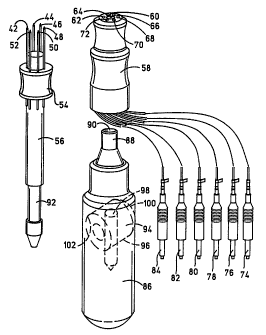
Referring to Figs. 3 and 4 an alternate embodiment of a circular array needle
electrode assembly is illustrated and designated generally by the numeral 40.
This
needle assembly comprises a circular array of needles 42 through 52, which are
mounted
in equally spaced relation in a hub 54 mounted on an elongated cylindrical
shaft 56.
~.;,
CA 02218255 1997-10-14
WO 96/39226 PCT/US96/07470
_'7_
The hub 54 is preferably of a suitably selected diameter to provide the
desired diameter
of the arrays to position around a tumor or other tissue to be treated. One or
more of
the needles may be hollow to enable the injection of molecules of a
therapeutic
a substance, as will be more fully described hereinafter.
An electrical connector socket assembly comprises a body member 58 having a
central opening or bore 60 for receipt of shaft 56 and an annular array of a
plurality of
sockets 62 through 72 for receipt of the ends of needles 42 through 52. The
sockets 62
through 72 electrically connect the needles to leads 74 through 84 which
connect to a
distributing switch, as will be subsequently described.
The electrical connector socket 58 fits onto shaft 56 with the end of the
needles
extending into the electrical sockets 62 through 72 for connecting to the
leads 74
through 84. The shaft 56 which mounts the needle array hub 54 and the socket
assembly 58 mounts onto a holder 86 adapted to be held in the hand. The holder
86 has
an elongated cylindrical configuration adapted to be held in the hand for
manipulation.
The holder 86 has a forward socket and including a forwardly extending tubular
shaft
88 having a bore 90 into which shaft 56 extends while the shaft 88 extends
into a bore
(not shown) within the connector member 58. The shaft 56 extends into bore 90
and
has a annular groove or recess 92 which is engaged by a retainer latch which
comprises
a transverse plug 94 in a bore 96 biased to one side and including a bore 98
in which
the annular slot 92 extends and is retained in the holder. A spring 102
mounted in bore
96 biases plug 94 to the latched position. The shaft 56 may be released for
removal by
pressing on end 100 of plug 94.
The holder when assembled as shown in Fig. 4 may be grasped in the hand and
the needles inserted into a selected tissue area. The needles 42-52 are
preferably spaced
and positioned to surround the selected tissue of treatment. One or more of
the needles
42-52, as previously explained, may be hollow to enable the injection of the
desired
~ therapeutic substance. The electrode leads 74-84 are then connected in a
preferred
arrangement to a rotatable switch assembly, as shown in Fig. 5, which enables
the
selection of opposed pairs of the needles for activation or the application of
the electrical
potential.
CA 02218255 1997-10-14
WO 96/39226 PCT/US96/07470
_g_
The switch assembly designated generally by the numeral 104 comprises a
stationary housing 106 which, in the illustrated embodiment, is generally
cylindrical in
configuration and in which is mounted a rotor 108 with spaced contacts 110 and
112
connected by a pair of conductors 114 and 116 to a pulse power generator 115.
The
rotor contacts l 10 and 112 are positioned within housing 106 to engage
annular contacts
118, 120, 122, 124, 126 and 128 to which leads 74-84 are connected.
Referring to Figs. 6a, b and c, the rotor 108 has an internal portion having
contacts 110 and 112 each of which bridge between two contacts 118-128 to
which the
leads 74 through 84 are connected to connect the source of power. The internal
contacts
110 and 112 rotate with the rotor 108 and can be selectively positioned in
conductive
relation with pairs of the internal contacts 118-128 to thereby activate
opposed pairs of
the needle electrodes. This enables the operator to selectively position the
electrodes
surrounding a selected tissue and to selectively apply the direction of the
electrical field
as desired for optimum treatment. The rotor 108 enables the field to be
selectively
generated around or across the tissue from all directions.
Referring to Fig. 7 an alternate embodiment of an electric field generating
array
of parallel adjustably positionable electrodes, as disclosed in the parent
application, is
illustrated. The electrode assembly designated generally by the numeral 130
includes
a pair of spaced apart arrays 132 and 134 of conductive needle electrodes 136
and 138
mounted on a dielectric carrier or support member 140. The needle array 132 is
held
in a fixed clamp 142 which allows the needles 136 to be adjusted in depth
relative to
the support 140.
The needles 138 are mounted in a moveable clamp 146 which is adjustably
mounted on support member 140 by a clamp screw 148. The needles 136 and 138
are
each provided with a penetration stop 144. The gap spacing clamp screw 148
secures
the clamp 146 in selected positions on the support 140. A gap spacing sensor
150
senses the distance between the needle arrays 132 and 134 and generates a
signal that
is sent to the pulse generator via conductor cable 152. A pulse generator is
connected
to the needle electrodes by means of cables 154 and 156. '
Referring to Fig. 8, details of a needle holder or template for various
arrangements for establishing a spaced pair or parallel arrays of needles is
illustrated.
CA 02218255 1997-10-14
WO 96/39226 PCT/US96/07470
-9-
This embodiment comprises a base holder member 158 having a plurality of
adjacently
positioned parallel slots 160 into which selected needles 162 and 164 may be
positioned
in selected spaced relation. This holder may serve to mount a pair of
oppositely
" polarized needle electrodes 162 and 164, as illustrated. These can be
selectively
positioned in selected space relationship to be disposed on opposite sides of
a selected
tissue. The needles are clamped into the slots by a clamp or plate 159. In
addition, the
holder may be used in combination with an additional holder for provision of
multiple
arrays on opposite sides of a selected tissue. The illustrated needles may be
connected
by conductors 166 and 168 to a suitable pulse generator.
Referring to Figs. 9a through 9d, an additional aspect of the invention is
illustrated. As more clearly illustrated, the combination electrodes may take
the form
of separate needles 170 and 172 which may be first inserted into or beside a
selected
tissue area such as on opposite sides of a tumor 194 as illustrated.
Thereafter the
needles may be connected to a syringe or other source of molecules and used to
inject
a selected molecular solution into the tissue area. The needles may be non-
conductive
and a pair of electrodes 176 and 178, as illustrated in Fig. 9b, are
selectively fed through
the bore or lumen of the respective needles into the tissue, as illustrated,
and thereafter
the needle is removed, as shown in Fig. 9c. The electrodes 176 and 178 are
each
provided with an elongated insulated conductor 180 and 182 with conductive
tips 184
and 186.
A pair of conductors 188 and 190 from a suitable power generator may then be
connected to the ends of the conductors of the electrodes by micro clamps 192
and 194,
as shown in 9d, and an electric potential applied across the electrodes. This
generates
a field in the tissue and electroporates the cells of the selected tissue,
such as a tumor
or the like. This electroporation enables the selected molecules to enter the
cells of the
tissue and more efficiently kill or alter the cells as desired. This form of
needle and
electrode may be used with any or all the above described assemblies.
These needle electrode assemblies, as above described, enable the in vivo
" positioning of electrodes in or adjacent to subsurface tumors or other
tissue. While the
focus of the present application has been on electrochemotherapy, the
embodiment of
CA 02218255 1997-10-14
WO 96/39226 PCT/US96/07470
-10-
the subject invention may be applied to other treatments, such as gene therapy
of certain
organs of the body.
The nature of the electric field to be generated is determined by the nature
of the
tissue, the size of the selected tissue and its location. It is desirable that
the field be as
homogenous as possible and of the correct amplitude. Excessive field strength
results
in lysing of cells, whereas a low field strength results in reduced efficacy.
The
electrodes may be mounted and manipulated in many ways including but not
limited to
those in the parent application. The electrodes may be conveniently
manipulated on and
by forceps to internal position.
The waveform of the electrical signal provided by the pulse generator can be
an
exponentially decaying pulse, a square pulse, a unipolar oscillating pulse
train or a
bipolar oscillating pulse train. The electric field strength can be 0.2kV/cm
to 20kV/cm.
The pulse length can be ten ~,s to 100 ms. There can be one to one hundred
pulses. Of
course, the waveform, electric field strength and pulse duration are also
dependent upon
the type of cells and the type of molecules that are to enter the cells via
electroporation.
The various parameters including electric field strengths required for the
electroporation of any known cell is generally available from the many
research papers
reporting on the subject, as well as from a database maintained by
Genetronics, Inc., San
Diego, California, assignee of the subject application. The electric fields
needed for in
vivo cell electroporation, such as ECT, are similar in amplitude to the fields
required for
cells in vitro. These are in the range of from 100 V/cm to several kV/cm. This
has
been verified by the inventors own experiments and those of others reported in
scientific
publications. The first in vivo application of pulsed electric fields in the
chemotherapy
field to treat tumors was reported in 1987 by Okino in Japan.
Pulse generators for carrying out the procedures described herein are and have
been available on the market for a number of years. One suitable signal
generator is the
ELECTRO CELL MANIPULATOR Model ECM 600 commercially available from
GENETRONICS, INC. of San Diego, California, U.S.A. The ECM 600 signal
generator
generates a pulse from the complete discharge of a capacitor which results in
an
exponentially decaying waveform. The electric signal generated by this signal
generator
is characterized by a fast rise time and an exponential tail. In the signal
generator, the
CA 02218255 1997-10-14
WO 96/39226 PCT/US96/07470
-11-
electroporation pulse length is set by selecting one of ten timing resistors
marked R1
through R10. They are active in both High Voltage Mode (HVM) (capacitance
fixed
at fifty microfarads) and Low Voltage Mode (LVM) (with a capacitance range
from 25
to 3,175 microfarads).
The ECM 600 signal generator has a control knob that permits the adjustment
of the amplitude of the set charging voltage applied to the internal
capacitors from 50
to 500 volts in LVM and from 0.05 to 2.SkV in the HVM. The amplitude of the
electrical signal is shown on a display incorporated into the ECM 600 signal
generator.
This device further includes a plurality of push button switches for
controlling pulse
length, in the Low VM mode, by a simultaneous combination of resistors
parallel to the
output and a bank of seven selectable additive capacitors.
The ECM 600 signal generator also includes a single automatic charge and pulse
push button. This button may be depressed to initiate both charging of the
internal
capacitors to the set voltage and to deliver a pulse to the outside electrodes
in an
automatic cycle that takes less than five seconds. The manual button may be
sequentially pressed to repeatedly apply the predetermined electric field.
Preferably, the therapeutic method of the invention utilizes a square wave
pulse
electroporation system. For example, the ElectroSquarePorator (T820), also
available
from GENETRONICS, INC., can be used.
Square wave electroporation systems deliver controlled electric pulses that
rise
quickly to a set voltage, stay at that level for a set length of time (pulse
length), and
then quickly drop to zero. This type of system yields better transformation
efficiency for
the electroporation of plant protoplast and mammalian cell lines than an
exponential
decay system.
The ElectroSquarePorator (T820) is the first commercially available square
wave
electroporation system capable of generating up to 3000 volts. The pulse
length can be
adjusted from 5 ,sec to 99 msec. The square wave electroporation pulses have a
gentler
effect on the cells which results in higher cell viability.
' The T820 ElectroSquarePorator is active in both the High Voltage Mode (HVM)
(100-3000 volts) and the Low Voltage Mode (LVM)(SO-500 volts). The pulse
length for
CA 02218255 1997-10-14
WO 96/39226 PCT/US96/07470
-12-
LVM is about 0.3 to 99 msec and for HVM, 5 to 99 ~csec. The T820 has multiple
pulsing capability from about 1 to 99 pulses.
Mir and others have used square wave pulses for electrochemotherapy, which
allows the insertion of chemotherapeutic agents into cancerous tumors. Mice
were _
injected with a low dose of bleomycin. The cancerous tumors were then
electroporated
resulting in the reduction or complete remission of the tumors (Mir, L.M.,
Eur. J.
Cancer 27( 1 ):68, 1991 ) .
Saunders has compared the square wave with exponential decay pulses in the
electroporation of plant protoplast. Square wave electroporation produced
higher
transformation efficiency than the exponential decay pulses. He also reported
that the
optimization of electroporation parameters is much easier with square wave
pulses since
sufficient transformation efficiency can be produced over a larger range of
voltages
(Saunders, Guide to Electroporation and Electrofusion, pp.227-247, 1991).
The therapeutic method of the invention includes electrotherapy, also referred
to
herein as electroporation-mediated therapy, using the apparatus of the
invention for the
delivery of macromolecules to a cell or tissue. As described earlier, the term
"macromolecule" or "molecule" as used herein refers to drugs (e.g.,
chemotherapeutic
agents), nucleic acids (e.g., polynucleotides), peptides and polypeptides,
including
antibodies. The term polynucleotides include DNA, cDNA and RNA sequences.
Drugs contemplated for use in the method of the invention are typically
chemotherapeutic agents having an antitumor or cytotoxic effect. Such drugs or
agents
include bleomycin, neocarcinostatin, suramin, and cisplatin. Other
chemotherapeutic
agents will be known to those of skill in the art (see for example The Merck
Index). The
chemical composition of the agent will dictate the most appropriate time to
administer
the agent in relation to the administration of the electric pulse. For
example, while not
wanting to be bound by a particular theory, it is believed that a drug having
a low
isoelectric point (e.g., neocarcinostatin, IEP=3.78), would likely be more
effective if
administered post-electroporation in order to avoid electrostatic interaction
of the highly
charged drug within the field. Further, such drugs as bleomycin, which have a
very '
negative log P, (P being the partition coefficient between octanol and water),
are very
large in size (MW--1400), and are hydrophilic, thereby associating closely
with the lipid
CA 02218255 1997-10-14
WO 96/39226 PCT/US96/07470
-13-
membrane, diffuse very slowly into a tumor cell and are typically administered
prior to
or substantially simultaneous with the electric pulse. Electroporation
facilitates entry of
bleomycin or other similar drugs into the tumor cell by creating pores in the
cell
membrane.
It may be desirable to modulate the expression of a gene in a cell by the
introduction of a molecule by the method of the invention. The term "modulate"
envisions the suppression of expression of a gene when it is over-expressed,
or
augmentation of expression when it is under-expressed. Where a cell
proliferative
disorder is associated with the expression of a gene, nucleic acid sequences
that interfere
with the gene's expression at the translational level can be used. This
approach utilizes,
for example, antisense nucleic acid, ribozymes, or triplex agents to block
transcription
or translation of a specific mRNA, either by masking that mRNA with an
antisense
nucleic acid or triplex agent, or by cleaving it with a ribozyme.
Antisense nucleic acids are DNA or RNA molecules that are complementary to
at least a portion of a specific mRNA molecule (Weintraub, Scientific
American, 262:40,
1990). In the cell, the antisense nucleic acids hybridize to the corresponding
mRNA,
forming a double-stranded molecule. The antisense nucleic acids interfere with
the
translation of the mRNA, since the cell will not translate a mRNA that is
double-
stranded. Antisense oligomers of about 15 nucleotides are preferred, since
they are
easily synthesized and are less likely to cause problems than larger molecules
when
introduced into the target cell. The use of antisense methods to inhibit the
in vitro
translation of genes is well known in the art (Marcus-Sakura, Anal.Biochem.,
172:289,
1988).
Use of an oligonucleotide to stall transcription is known as the triplex
strategy
since the oligomer winds around double-helical DNA, forming a three-strand
helix.
Therefore, these triplex compounds can be designed to recognize a unique site
on a
- chosen gene (Maher, et al., Antisense Res. and Dev., 1 3 :227, 1991; Helene,
C.,
Anticancer Drug Design, 6 6 :569, 1991 ).
Ribozymes are RNA molecules possessing the ability to specifically cleave
other
single-stranded RNA in a manner analogous to DNA restriction endonucleases.
Through
the modification of nucleotide sequences which encode these RNAs, it is
possible to
CA 02218255 1997-10-14
WO 96/39226 PCT/US96/07470
-14-
engineer molecules that recognize specific nucleotide sequences in an RNA
molecule and
cleave it (Cech, J.Amer.Med. Assn., 260:3030, 1988). A major advantage of this
.
approach is that, because they are sequence-specific, only mRNAs with
particular
sequences are inactivated.
There are two basic types of ribozymes namely, tetrahymena-type (Hasselhoff,
Nature, 334:585, 1988) and "hammerhead"-type. Tetrahymena-type ribozymes
recognize
sequences which are four bases in length, while "hammerhead"-type ribozymes
recognize
base sequences 11-18 bases in length. The longer the recognition sequence, the
greater
the likelihood that the sequence will occur exclusively in the target mRNA
species.
Consequently, hammerhead-type ribozymes are preferable to tetrahymena-type
ribozymes
for inactivating a specific mRNA species and 18-based recognition sequences
are
preferable to shorter recognition sequences.
The present invention also provides gene therapy for the treatment of cell
proliferative or immunologic disorders mediated by a particular gene or
absence thereof.
Such therapy would achieve its therapeutic effect by introduction of a
specific sense or
antisense polynucleotide into cells having the disorder. Delivery of
polynucleotides can
be achieved using a recombinant expression vector such as a chimeric virus, or
the
polynucleotide can be delivered as "naked" DNA for example.
Various viral vectors which can be utilized for gene therapy as taught herein
include adenovirus, herpes virus, vaccinia, or, preferably, an RNA virus such
as a
retrovirus. Preferably, the retroviral vector is a derivative of a marine or
avian
retrovirus. Examples of retroviral vectors in which a single foreign gene can
be inserted
include, but are not limited to: Moloney marine leukemia virus (MoMuLV),
Harvey
marine sarcoma virus (HaMuSV), marine mammary tumor virus (MuMTV), and Rous
Sarcoma Virus (RSV). When the subject is a human, a vector such as the gibbon
ape
leukemia virus (GaLV) can be utilized. A number of additional retroviral
vectors can
incorporate multiple genes. All of these vectors can transfer or incorporate a
gene for "
a selectable marker so that transduced cells can be identified and generated.
Therapeutic peptides or polypeptides may also be included in the therapeutic
method of the invention. For example, immunomodulatory agents and other
biological
response modifiers can be administered for incorporation by a cell. The term
CA 02218255 1997-10-14
WO 96/39226 PCT/US96/07470
-15-
"biological response modifiers" is meant to encompass substances which are
involved
in modifying the immune response. Examples of immune response modifiers
include
such compounds as lymphokines. Lymphokines include tumor necrosis factor,
interleukins 1, 2, and 3, lymphotoxin, macrophage activating factor, migration
inhibition
factor, colony stimulating factor, and alpha-interferon, beta-interferon, and
gamma-
interferon and their subtypes.
Also included are polynucleotides which encode metabolic enzymes and proteins,
including antiangiogenesis compounds, e.g., Factor VIII or Factor IX.
The macromolecule of the invention also includes antibody molecules. The term
"antibody" as used herein is meant to include intact molecules as well as
fragments
thereof, such as Fab and F(ab')Z.
Administration of a drug, polynucleotide or polypeptide, in the method of the
invention can be, for example, parenterally by injection, rapid infusion,
nasopharyngeal
absorption, dermal absorption, and orally. In the case of a tumor, for
example, a
chemotherapeutic or other agent can be administered locally, systemically or
directly
injected into the tumor. When a drug, for example, is administered directly
into the
tumor, it is advantageous to inject the drug in a "fanning" manner. The term
"fanning"
refers to administering the drug by changing the direction of the needle as
the drug is
being inj ected or by multiple inj ections in multiple directions like opening
up of a hand
fan, rather than as a bolus, in order to provide a greater distribution of
drug throughout
the tumor. As compared with a volume that is typically used in the art, it is
desirable
to increase the volume of the drug-containing solution, when the drug is
administered
(e.g., injected) intratumorally, in order to insure adequate distribution of
the drug
throughout the tumor. For example, in the EXAMPLES herein, one of skill in the
art
typically injects 50 p,l of drug-containing solution, however, the results are
greatly
improved by increasing the volume to 150 ~.1. Preferably, the injection should
be done
~ very slowly and at the periphery rather than at the center of the tumor
where the
intertidal pressure is very high
' Preferably, the molecule is administered substantially contemporaneously
with
the electroporation treatment. The term "substantially contemporaneously"
means that
the molecule and the electroporation treatment are administered reasonably
close together
CA 02218255 1997-10-14
WO 96/39226 PCT/US96/07470
-16-
with respect to time. The administration of the molecule or therapeutic agent
can at any
interval, depending upon such factors, for example, as the nature of the
tumor, the
condition of the patient, the size and chemical characteristics of the
molecule and half
life of the molecule.
Preparations for parenteral administration include sterile or aqueous or non-
aqueous solutions, suspensions, and emulsions. Examples of non-aqueous
solvents are
propylene glycol, polyethylene glycol, vegetable oils such as olive oil, and
injectable
organic esters such as ethyl oleate. Carriers for occlusive dressings can be
used to
increase skin permeability and enhance antigen absorption. Liquid dosage forms
for oral
administration may generally comprise a liposome solution containing the
liquid dosage
form. Suitable forms for suspending the liposomes include emulsions,
suspensions,
solutions, syrups, and elixirs containing inert diluents commonly used in the
art, such
as purified water. Besides the inert diluents, such compositions can also
include
adjuvants, wetting agents, emulsifying and suspending agents. Further,
vasoconstrictor
agents can be used to keep the therapeutic agent localized prior to pulsing.
Any cell can be treated by the method of the invention. The illustrative
examples
provided herein demonstrate the use of the method of the invention for the
treatment of
tumor cells, e.g., pancreas and lung. Other cell proliferative disorders are
amenable to
treatment by the electroporation method of the invention. The term "cell
proliferative
disorder" denotes malignant as well as non-malignant cell populations which
often
appear to differ from the surrounding tissue both morphologically and
genotypically.
Malignant cells (i. e., tumors or cancer) develop as a result of a multi-step
process. The
method of the invention is useful in treating malignancies or other disorders
of the
various organ systems, particularly, for example, cells in the pancreas and
lung, and also
including cells of heart, kidney, muscle, breast, colon, prostate, thymus,
testis, and
ovary. Preferably the subject is human.
The following examples are intended to illustrate but not limit the invention.
While they are typical of those that might be used, other procedures known to
those
skilled in the art may alternatively be used.
CA 02218255 1997-10-14
WO 96/39226 PCT/US96/07470
-17-
EXAMPLES
The following examples illustrate the use of electrochemotherapy (ECT) of a
poorly differentiated human pancreatic tumor (Panc-3) xenografted
subcutaneously on
the left flank of nude mice. The single treatment procedure involved injection
of
bleomycin (0.5 units in 0.15 ml saline) intratumorally, using fanning, as
described herein
followed by application of six square wave electrical pulses, ten minutes
later, using
proprietary needle array electrodes arranged along the circumference of a
circle 1 cm in
diameter. Needle array of variable diameters (e.g., 0.5 cm, 0.75 cm and 1.5 cm
can also
be used to accommodate tumors of various sizes. Stoppers of various heights
can be
inserted at the center of the array to make the penetration depth of the
needles into the
tumor variable. A built-in mechanism allowed switching of electrodes for
maximum
coverage of the tumor by the pulsed field. The electrical parameters were:
1300 V/cm
and 6 x 99 ~.s pulses spaced at 1 sec interval.
Results showed severe necrosis and edema in nearly all the mice at the
treatment
1 G ~;+o ~IT4,;lo +l.,o..o .~ ~..1.~+.,.,+:..1 ..,.".1....+:..... :... +t,..
+.-.r..- ....~._~_ i_~__ _ _m-L~ :~
1J J1W. vvaum. uram vva~ a JuuJ1.a11ua1 lcuuGL1V11 111 utc L11111V1 VV111111C
~4.1LCI~ 'd Sll~IlL lIlltlW1
increase due to edema) of the mice in the treated group (D+E+; D=Drug,
E=Electrical
field), those in the control group (D+E-) increased dramatically. Nearly
complete tumor
regression was observed in 90% of the mice treated by ECT after 28 days. No
response
was seen in 10% of the mice. A complete regression with no palpable tumor has
been
observed in 60% of the cases 77 days after the initial treatment. However,
there was
tumor regrowth in 20% of the mice 35 days after treatment but at a much slower
growth
rate compared to the control. This observation has been linked to incomplete
treatment
of large primary tumors where the needle depth was lower than the Z dimension
of the
tumor. Histological analysis of tumor samples showed necrotic tumor cell
ghosts in
D+E+ group compared to a mixture of viable and necrotic cells in D+E- group.
Preliminary studies with human non-small cell lung cancer (NSCLC) tumors
xenografted
onto nude mice have also shown very encouraging results with ECT treatment
with
bleomycin.
CA 02218255 1997-10-14
WO 96/39226 PCT/CJS96/07470
-18-
Example 1:
The tumor cell line Panc-3, a poorly differentiated adenocarcinoma cell line
of
the pancreas, was supplied by Anticancer, Inc., San Diego. For ECT
experiments, tissue
taken from the stock mice, where the tumor line was maintained, was thawed and
cut
into very small pieces about 1 mm each, and 8-10 pieces were surgically
xenografted
in a subcutaneous sac made in left flank of nude mice, and then closed with
6.0 surgical
suture. After the average tumor size reached about 5 mm, mice with palpable
tumors
were divided randomly, 10 mice for control group (D+E-; D=Drug, E=Electric
field) and
mice for ECT treatment, namely bleomycin injection followed by pulsing (D+E+)
10 from a BTX Square Wave T820 Generator. The tumor dimensions were measured
and
the tumor volume calculated using the formula:
(II/6)xaxbxc
where a, b, and c are, respectively, the length, width and thickness of the
tumor. 0.5
units Bleomycin (Sigma Chemicals) was dissolved in 0.15 ml of 0.9% NaCl and
was
injected in each mice intratumorally by fanning for both the control (D+E-)
and the
treated (D+E+) groups. Ten minutes after the injection, each mouse in the D+E+
group
was pulsed from a BTX T820 square wave electroporator with a set of needle
array
electrodes as described in the present invention. Electrical parameters used
were as
follows: field strength 1300 V/cm, 6 pulses of 99 ~s each, at 1 sec interval.
The mice were monitored every day for mortality and any signs of a diseased
state were noted. The tumor dimensions were measured at regular intervals and
tumor
growth regression/progression monitored. Another set of nude mice with
xenografts of
non-small cell lung cancer line was also treated by the same procedure as for
the Panc-3
tumors.
Figures l0a and l Ob show the analysis of the tumor volume determined over a
43 day period after ECT using bleomycin for the Panc-3 tumors. There was a
dramatic
difference between the untreated and treated mice in terms of tumor volume.
There was
essentially no detectable tumor after approximately 24 days of treatment. The
results of
Figure 10 are also summarized in Table 1 below. An illustration of the actual
regression
of the tumor is shown in Figure 11.
CA 02218255 1997-10-14
WO 96/39226 PCT/US96/07470
-19-
TABLE 1
ELECTROCHEMOTHERAPY OF PANC-3 TUMORS IN NUDE MICE
Days after Tumor volume Tumor volume Tumor volume Tumor volume
- treatment (mm' ) C1 (mm3) C2 (mm3) Tl (mm3) T2
0 138.746 148.94 123.11 178.37
1 206.979 179.82 210.95 252.72
8 394.786 451.787 104.55 211.11
557.349 798.919 113.21 226.966
18 939.582 881.752 161.73 246.91
10 24 1391.057 1406.98 41.56 47.2228
28 1628.631 1474.21 0 0
35 2619.765 2330.31 0 0
38 2908.912 2333.967 0 0
43 3708.571 5381.759 0 0
15 Cell Line: poorly differentiated human pancreatic tumor (panc3)
Mouse model: nude mouse
Transplant: subcutaneous xenograft
Control mice: C1 and C2
Treated mice: T1 and T2
The Panc-3 experiment was repeated using a non-small cell lung cancer cell
line
(NSCLC), 177 (Anticancer, San Diego, CA). The results were similar to that
found with
bleomycin and Panc-3 as shown in Figures 12a and 12b. In one experiment, a
tumor that
had recurred was retreated at day 27 (Figure 13) and after 7 days, there was
no evidence
of tumor.
The Panc-3 and NSCLC models were utilized with the drug neocarcinostatin
' (NCS) following the same procedures as outlined above. As shown in Figure
14a and
14b, pre-pulse dosing with NCS in a manner similar to that used for the
bleomycin
studies, was not effective in reducing tumor size at all. It was believed that
due to the
low isoelectric point of NCS, electrostatic interaction prevented the drug
from entering
CA 02218255 1997-10-14
WO 96/39226 PCT/US96/07470
-20-
the tumor cell. Therefore, the experiment was repeated by pulsing first and
inj ecting
NCS post-pulse (pp).
Figure 14c shows the initial tumor volume (I) as compared to the final tumor
volume (F) at day 13 for 7 mice treated (Mouse ID 1-7). In several of the mice
(ID 1,
2, 4, and 7), an increase in tumor volume was observed, but appeared to be due
to
edema. However, as shown in Figure 14d, when a separate group of 5 mice were
examined at day 23, all mice showed a marked reduction in tumor volume.
A comparison of Figures 14 a and b with 14 c and d indicated that post-pulse
with NCS was more effective than pre-pulse administration for NCS.
Summary
The present Examples illustrate that a poorly differentiated Pancreatic cancer
(Panc-3) and Non-srriall cell lung cancer (NSCLC) xenografted subcutaneously
onto
nude mice can be effectively treated by the electrochemotherapy protocol using
bleomycin or NCS and needle array electrodes. Other similar chemotherapeutic
agents
can also be effective using the method of the invention.
The results show a complete regression of Panc-3 tumors was achieved in 60%
of the treated group with no palpable tumor seen even 77 days after the single
treatment.
Partial regression (80% reduction in tumor volume) was observed in 30% of
cases, while
only 10% did not respond (Table 2).
Histological studies clearly showed severe necrosis of the tumor region for
the
group subjected to ECT whereas no necrosis was apparent in the control group.
Intratumoral drug injection with larger volume of bleomycin, combined with
fanning to
maximize uniform drug distribution throughout the tumor volume, was found to
be very
effective as compared to the conventional mode of injecting the drug prior to
pulsing.
CA 02218255 1997-10-14
WO 96/39226 PCT/US96/07470
-21-
TABLE 2
Electrochemotherapy of Panc-3 with Bleomvcin
Days after 28 35 57 77
treatment
CR (100%) 6 6 6 6
PR (80%) 3
NR (%) 1 1 1 1
Death 2*
Tumor regrowth 2
Retreatment 2
Histology 1
Number of mice treated: 10
CR: Complete Regression
PR: Partial Regression
NR: No Response
* 1 mice died after retreatment
1 mice died after 64 days survival
Although the invention has been described with reference to the presently
preferred embodiment, it should be understood that various modifications can
be made
without departing from the spirit of the invention: Accordingly, the invention
is limited
only by the following claims.