Note: Descriptions are shown in the official language in which they were submitted.
CA 02218483 1997-10-16
MICROSPHERES WITH FLUORESCENT SPHERICAL ZONES
FIELD OF THE INVENTION
The invention describes polymer microspheres possessing at least one internal
spherical zone
labeled with one or more fluorescent dyes. The resulting microspheres are
useful as standards for
instrument calibration, particularly confocal microscope calibration, and as
tracers.
BACKGROUND TO THE INVENTION
Polymeric microspheres labeled with fluorescent dyes (fluorescent
microspheres) are most
commonly used in applications that can benefit from the use of monodisperse,
chemically inert,
biocompatible particles that radiate detectable fluorescence and that can be
coupled to members of
specific binding pairs so as to make them bind to a particular substance in a
sample. However,
fluorescent microspheres have also found use in a wide variety of other
applications, including
instrument calibration and tracing. There are predominantly two types of
labeled microspheres:
surface labeled or labeled throughout. For surface-labeled microspheres, a
monolayer of dye is
typically deposited on the microsphere surface using a fluorescent protein
coating or by covalent
attachment of the desired label. Fluorescent microspheres labeled essentially
throughout their entire
volume are prepared either by copolymerization of a fluorescent monomer (the
dye is covalently
bound to a monomer prior to polymerization) or by batch staining of preformed
microspheres with a
dye that is soluble in the polymer microsphere.
Recent advances in microscope instrumentation, such as confocal laser scanning
microscopy
and wide-field microscopy coupled with data deconvolution, allow the user to
analyze a sample in
three dimensions, to record a single optical section of the specimen having a
preferred thickness, and
to acquire and analyze information at multiple fluorescence emission
wavelengths (simultaneously or
sequentially), often using multiple excitation wavelengths. Computer
restoration permits the
reconstruction of essentially three-dimensional images of the sample. The
complexity of this
instrumentation is such that many of the optical parameters of the microscope
are not readily
calibrated using the methods and standards of conventional microscopy. In
particular, it is difficult
to calibrate dimensions along the z-axis of the sample (perpendicular to the
plane of the microscope
slide), which can be measured as the set of distances between the surface of
the microscope slide and
the coverslip. Users of such microscopes therefore employ a variety of
standards to evaluate their
CA 02218483 1997-10-16
instrument performance, such as etched test patterns, integrated circuits,
diatom frustules immersed
in a fluorescent solution, or biological cells stained with single or multiple
dyes.
Each type of commonly used calibration standard possesses some limitations.
The two-
s dimensional nature of etched patterns and integrated circuits offers poor
calibration along the z-axis.
Diatoms, as natural organisms, possess irregularity in size and structure.
Biological cells are also
irregular in shape and can exhibit very different fluorescence emissions at
different parts of the same
cell. In contrast, the unique microspheres of this invention, which when
viewed in a cross-section
that includes the center of the microsphere (hereafter referred to as an
equatorial cross-section),
display one or more distinct concentric rings of fluorescence, provide uniform
standards of known
geometry, fluorescence intensity and staining pattern that facilitate
instrument alignment and
computer-generated image reconstruction. In addition, the dyes inside the
microsphere are protected
from solvent effects, such as pH variation, making them both brighter and less
prone to
photobleaching than surface-stained microspheres. Furthermore the ability to
select fluorescent
excitation and emission spectra from ultraviolet to infrared wavelengths
permits the correction of
chromatic aberration and other optical artifacts. Also, the ability of
microscopes to reconstruct the
staining pattern of single microsphere makes it possible to distinguish and
identify a single
microsphere within a mixture of microspheres that have a wide variety of
patterns, colors,
fluorescence intensities and sizes.
BRIEF DESCRIPTION OF THE DRAWINGS
Figure l: Figures lA-1F depict several distinctive staining patterns suitable
for the microspheres of
the present invention. Each microsphere is shown in equatorial cross-section.
Figure lA depicts a microsphere having a single distinct fluorescent ring near
the
microsphere's outer surface.
Figure 1B depicts a microsphere having a single distinct fluorescent ring
located well within
the interior of the microsphere.
Figure 1C depicts a microsphere having a distinct fluorescent ring near the
microsphere's
outer surface in conjunction with an additional distinct fluorescent ring
located well within
the interior of the microsphere.
2
CA 02218483 2001-04-27
Figure ID depicts a microsphere having multiple distinct fluorescent rings
within the interior
of the microsphere, such that they are partially coincident.
Figure lE depicts a microsphere having a distinct fluorescent ring near the
microsphere's
outer surface in conjunction with a concentric fluorescent disk located at the
core of the
microsphere.
Figure IF depicts a microsphere having multiple distinct fluorescent rings in
conjunction
with a fluorescent disk at the core of the microsphere.
Figure 2: Figure 2A shows a cut-away view of a microsphere that has been
shallowly stained with a
fluorescent label, as indicated by cross-hatching. The dashed lines of Figure
2A indicate the
expanded view shown in Figure 2B, wherein dye molecules (indicated by dark
dots) are depicted as
incorporated into the polymeric matrix of the microsphere (indicated by
partially cross-linked lines).
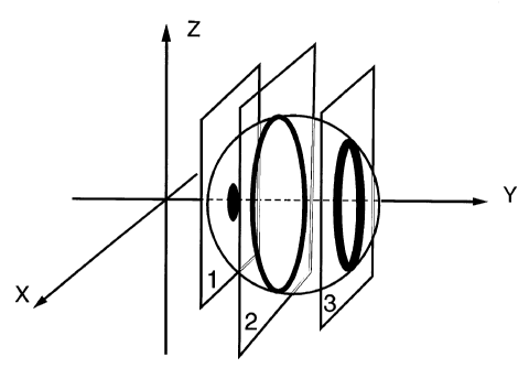
Figure 3: Figure 3A shows a microsphere of the present invention that has been
shallowly stained
with a fluorescent label. Planes I, 2 and 3 represent cross-sections of the
microsphere in the X-Z
plane and correspond to two-dimensional views 1, 2 and 3 shown in Figure 3B.
Figure 4: Figure 4 shows the use of three specific embodiments of the
invention to correct the
alignment of an instrument, wherein each of Figures 4A, 4B and 4C shows a
cross-section of the
microsphere before and after alignment. In each case, proper alignment of the
instrument is readily
evidenced by the restoration of the accurate representation of the
microsphere.
Figure 4A shows the use of a uniform fluorescent blue-stained microsphere that
is
additionally shallowly stained with a fluorescent orange label.
Figure 4B shows the use of a microsphere that has been shallowly stained with
three
fluorescent labels, yielding coincident spherical zones that possess crimson,
orange and green
fluorescence, respectively.
Figure 4C shows the use of a microsphere that is labeled with a fluorescent
blue internal disk,
a distinct fluorescent crimson ring located well within the interior of the
microsphere, and a
fluorescent yellow shallow stain.
CA 02218483 2001-04-27
SUMMARY OF THE INVENTION AND DESCRIPTION OF PREFERRED
EMBODIMENTS OF THE INVENTION
This invention provides a polymeric microsphere that is labeled such that
following
excitation of said microsphere at an appropriate wavelength, an equatorial
cross-section of said
microsphere displays a first distinct fluorescent ring that is concentric with
and within said
microsphere.
The invention describes novel fluorescent microspheres that are labeled so as
to possess at
least one fluorescent spherical zone, such that following excitation of the
particle at an appropriate
wavelength, an equatorial cross-section displays a distinct fluorescent ring
that is concentric with and
within said microsphere. Individual microspheres may exhibit multiple rings,
or may optionally
exhibit a fluorescent disk, that may or may not overlap with each other when
viewed in cross-section.
Fluorescent spherical zones can also differ in excitation and emission
properties and fluorescence
intensity. The polymeric microspheres can differ in diameter and polymer
composition. The size,
number and color characteristics of the rings and disks is controlled by
varying parameters in the
staining protocol or by changing the methods used to prepare the microspheres.
The invention further describes methods for preparing and using the
spherically labeled
microspheres. Methods of using the patterned microspheres for calibrating
microscopes and as labels
and tagging agents are described. Also described are kits that include
multiple fluorescent
microspheres that can be distinguished from each other using optical imaging
of the staining patterns
in three-dimensions.
The microspheres of this invention possess one or more distinct, fluorescently
labeled,
spherical zones. By "spherical zone" is meant a fluorescent zone that is
present within the polymer
microsphere and concentric with the microsphere, such that any equatorial
cross-section of the
microsphere evidences a distinct fluorescent ring, the diameter of which ring
is less than the diameter
of the microsphere. Optionally, the microspheres of the present invention are
stained with additional
fluorescent spherical zones, or with an internal solid spherical zone such
that an equatorial cross-
section evidences a distinct fluorescent disk. The staining patterns of the
microspheres of the
invention are necessarily isotropic, so that any equatorial cross-section is
equivalent to any other
equatorial cross-section. Therefore, for ease of illustration, the spherical
zones are described as
either rings or disks. It is understood, however, that these terms encompass
the three-dimensional
configuration that corresponds to a ring (i.e. a spherical shell) or a disk
(i.e. a solid sphere).
The microspheres of this invention are spherical in shape, and have a diameter
of between
CA 02218483 1997-10-16
about 2-100 pm, and are preferably less than 50 ~m in diameter. More
preferably, the microspheres
have a diameter of about 4-16 p,m, most preferably about 8-16 pm. Typically,
the labeled
microspheres of the invention are of a size sufficient to obtain at least 10
optical cross-sections along
the z-axis. For example, performing this type of cross-sectional analysis on a
microsphere having a
diameter of 2 pm would generate ten optical sections each having an average
thickness of 0.2 pm,
well within the resolution of contemporary confocal laser scanning
microscopes.
For all embodiments of the invention, the microspheres possess at least one
distinct
fluorescent-labeled spherical zone, such that when the microsphere is viewed
in an equatorial cross
section, the cross-section displays a staining pattern that includes at least
one distinct fluorescent
ring. The distinct fluorescent ring is essentially circular and concentric
with the microsphere itself.
'Distinct', as used herein, means readily distinguishable. That is, the
fluorescent ring is
readily distinguishable either by spectral characteristics or by fluorescence
intensity from the
remainder of the cross-section, as well as from additional fluorescent rings
or disks that are
optionally present in the cross-section. The boundaries, or edges, of the ring
are distinguishable and
are not diffuse, so that the width of the ring is readily determinable.
Where a fluorescent ring is readily distinguishable by spectral
characteristics, typically the
fluorescence emission maximum of each distinguishable ring is separated by at
least 5 nm, preferably
by at least 10 nm and more preferably by at least 20 nm. In a preferred
embodiment, by utilizing
independent optical filters and optical channels of an instrument (such as a
confocal laser scanning
microscope) each distinct fluorescent spherical zone is capable of being
separately excited, and
separately detected, and the fluorescence emissions of each spherical zone can
be visibly
distinguished. However, the ability of advanced imaging equipment to
discriminate between closely
separated emissions and to accurately quantitate fluorescence intensities
makes it possible to utilize
such instruments to reliably differentiate between individual microspheres,
even when the distinct
staining patterns of the respective microspheres may not be distinguishable by
the human eye.
In one embodiment, the fluorescent ring is distinct because the maximum
fluorescence
intensity of the ring is at least 20% greater than the average background
fluorescence of the
microsphere, when measured at the wavelength of maximum fluorescence of the
ring, and when the
average background fluorescence of the microsphere excludes the contribution
of the fluorescence of
the ring. An average background fluorescence intensity calculated in this
manner is herein referred
CA 02218483 1997-10-16
to as an "adjusted average fluorescence intensity". In another embodiment, a
distinct fluorescent ring
is a ring wherein the concentration of the fluorescent dye or dyes present
within the fluorescent
spherical zone is at least 10-fold greater than the concentration of the same
dye or dyes in any region
outside the fluorescent spherical zone.
The "width" of a fluorescent ring, as described herein, means the shortest
measured distance
between the outside radius and the inside radius of the fluorescent ring. For
all embodiments, the
width of each fluorescent ring is at least 0.2 Vim, and is no greater than the
equivalent of 35% of the
radius of the labeled microsphere. That is, a fluorescent ring within a 4 pm
microsphere is between
0.2 pm and 0.7 pm wide; a fluorescent ring in a 15 pm microsphere is between
0.20 ~m and 2.63 ~m
wide. Preferably, the fluorescent rings have a width of at least 0.5 pm and
the width of each
fluorescent ring is no greater than 30% of the radius of the microsphere, more
preferably no greater
than 25% and yet more preferably no greater than 20% of the radius of the
microsphere. Typically,
the width of the fluorescent rings is less than 3 p.m, more typically less
than 2 pm.
The fluorescent ring may be at or near the polymer microsphere's interior
surface (as shown
in Figure lA) although the ring must be within the microsphere itself, rather
than on the exterior
surface, in order for the microsphere to possess the advantages arising from
internal incorporation of
the dye. In this embodiment, the ring has an outer diameter that is
essentially equal to the diameter
of the microsphere itself. This type of microsphere is also referred to herein
as 'shallowly stained'.
In another embodiment, the ring has an outer radius that is essentially equal
to the radius of the
microsphere itself and an inside radius greater than 75% of the radius of the
microsphere itself, more
typically, an inside radius greater than 80% of the radius of the microsphere.
In yet another
embodiment, the ring is present well within the interior of the microsphere,
in the zone between the
center of the cross-section and the surface (for example, as shown in Figure
1B).
In the simplest embodiment of the invention, the microsphere of the invention
is labeled with
a single distinct fluorescent spherical zone, yielding a single distinct
fluorescent ring. Preferably the
microspheres of this embodiments possess a fluorescent ring having an outer
diameter essentially
equal to the diameter of the microsphere itself.
In another embodiment of the invention, the microsphere possesses one or more
additional
fluorescent spherical zones, such that an equatorial cross-section possesses
two or more distinct
fluorescent rings. The rings are readily distinguishable from each other and
from the remainder of
CA 02218483 1997-10-16
the cross-section, and may be non-coincident, i.e. discrete rings (as shown in
Figure 1C) having the
same or different spectral properties, or may be fully or partially coincident
and have detestably
distinct spectral properties (an example of partially coincident multiple
rings is shown in Figure 1D).
In yet another embodiment of the invention, an equatorial cross-section of the
microsphere
possesses one or more distinct fluorescent disks in addition to at least one
distinct fluorescent ring.
The fluorescent disk is readily distinguishable either by spectral
characteristics or by fluorescence
intensity from the remainder of the cross-section, as well as from additional
fluorescent rings or disks
that are optionally present in the cross-section. The fluorescent disk may be
non-coincident with any
fluorescent rings having the same or different spectral properties, or may be
fully or partially
coincident with any fluorescent rings or additional disks and have detestably
distinct spectral
properties.
Each fluorescent disk is concentric with the microsphere itself, and each disk
has a diameter
of at least 0.4 p,m and as large as the diameter of the microsphere itself.
Where the diameter of the
fluorescent disk is essentially equal to the diameter of the microsphere, the
resulting microsphere
possesses essentially uniform fluorescent staining throughout the interior of
the microsphere. In one
embodiment of the invention, the fluorescent disk has a diameter no greater
than 70% of the diameter
of the microsphere itself. In another embodiment of the invention, a
microsphere possesses a single
distinct fluorescent disk in addition to a single distinct fluorescent ring
(as shown in Figure lE). In
another embodiment of the invention, a microsphere possesses a single
fluorescent disk in
conjunction with multiple distinct fluorescent rings (as shown in Figure 1F).
Where the disk
produces essentially uniform staining, the microsphere must also possess one
or more distinct
fluorescent spherical zones. Preferably, the essentially uniform staining is
combined with one or
more fluorescent spherical zones having an outer diameter essentially equal to
the diameter of the
microsphere itself (shallowly stained).
In one embodiment of the invention, more than one fluorescent spherical zone
is spatially
coincident and yet each spherical zone displays detestably distinct spectral
properties. In another
embodiment, the spectral properties of a single distinct fluorescent spherical
zone are due to the
presence of a series of fluorescent dyes selected so as to undergo significant
energy transfer. In this
embodiment, the series of dyes comprises an initial donor dye having a desired
excitation peak and
final acceptor dye having a desired emission peak, where each dye in the
series has a spectral overlap
sufficient to allow for significant energy transfer of excitation energy. In
one embodiment, the series
CA 02218483 2001-04-27
of dyes is selected so that excitation of the microsphere at 488 nm results in
a fluorescence emission
at between 630 and 680 nm. Microspheres of the invention that possess
fluorescent spherical zones
labeled in this manner possess extended and readily controllable Stokes
Shifts. Fluorescent
microspheres that are uniformly stained with such an energy transfer dye
series have been described
in U.S. Patent Nos. 5,326,692 to Brinkley et al. (1994) and 5,573,909 to
Singer et al. (1996).
In all embodiments of the invention, the microspheres of the invention
optionally further
comprise a member of a specific binding pair that is bound covalently or is
noncovalently adsorbed
onto the surface of the microsphere, or other surface modifications.
Suitable Microspheres
Preferably the microspheres of the present invention are highly uniform; that
is for a given
batch of microspheres, the individual microspheres within the batch will be
essentially identical.
This uniformity is typically measured using the standard deviation, or by the
coefficient of variation
(CV). The coefficient of variation for the microspheres of the invention is
typically about 1-3%,
depending upon the size of the particular microspheres.
The polymeric microspheres of the present invention may be prepared from a
variety of
compositions including, but not limited to, polymers and copolymers of
styrenes and divinyl
benzenes; an acrylate or methacrylate ester; an acrylic acid or methacrylic
acid; an acrylamide or
methacrylamido; an acrylonitrile or methacrylonitrile; vinyl and vinylidene
halides, esters and ethers;
alkenes, including ethylene, propylene, butadiene and isoprene; epoxides and
urethanes. Preferably
the microspheres are polystyrene or predominantly polystyrene microspheres
that are optionally
crosslinked such as by the incorporation of divinylbenzene during the
polymerization reaction.
In one embodiment, a polymeric microsphere core is optionally coated with a
polymer
having a different composition, so as to modify the surface properties of the
resulting microsphere, or
to modify the ability of the microsphere to absorb a desired dye composition.
Unstained microspheres in a variety of sizes and polymer compositions that are
suitable for
preparation of fluorescent microspheres of the invention are available from a
variety of sources,
including: Interfacial Dynamics Corporation (Portland, OR), Bangs Laboratories
(Carmel, IN),
Dynal (Great Neck, NY), Polysciences (Warrington, PA), Seradyne (Indianapolis,
IN), Magsphere
CA 02218483 1997-10-16
(Pasadena, CA), Duke Scientific Corporation (Palo Alto, CA), Spherotech Inc.
(Libertyville, IL) and
Rhone-Poulenc (Paris, France). Chemical monomers for preparation of
microspheres are available
from numerous sources.
Preparing the Fluorescent Microsphere
Fluorescent dyes have been incorporated into uniform microspheres in a variety
of ways, for
example by copolymerization of the fluorescent dye into the microspheres
during manufacture (U.S.
Patent No. 4,609,689 to Schwartz et al. (1975), U.S. Patent No. 4,326,008 to
Rembaum (1982)); by
entrapment of the fluorescent dye into the microspheres during the
polymerization process; or by
non-covalent incorporation of the fluorescent dye into previously prepared
microspheres (U.S. Patent
No. 5,326,692, supra). Each of these methods has previously been used to
produce microparticles
that are internally stained essentially throughout the interior of the
particle.
The two basic means of preparing the microspheres of the invention are as
follows: 1 ) bath
dying of unstained or selectively stained microspheres; 2) Copolymerization of
a fluorescent or non-
fluorescent monomer onto the surface of an unstained or selectively stained
microsphere. The above
two techniques, when used alone or in combination, produce a variety of
staining patterns within the
subject microspheres.
Bath Dying
Bath dying refers to the absorption of a dye or dyes into the microsphere
directly from
solvent. Somewhat hydrophobic fluorescent dyes, being freely soluble in
organic solvents and very
sparingly soluble in water, are readily introduced by solvent-based addition
of the dye to previously
formed microspheres.
Bath dying has previously been used to produce fluorescent (and colored)
microspheres
without regard to producing a specific spherical staining pattern of staining.
Novel bath-staining
techniques as described in the invention are required to prevent substantial
penetration of the dye into
the microspheres in order to produce a distinct spherical zone near the
surface of the microsphere. In
particular, a variety of parameters must be carefully controlled in order for
distinct shallow staining
to occur, including solvent polarity, the complete absence of water in the
staining solution, the
physical characteristics of the dyes utilized, the composition of the
microsphere, and the staining
CA 02218483 1997-10-16
duration.
The solvent combination utilized for the staining solution must swell
polymeric matrix of the
microspheres enough so that staining is controlled, but not enough to
permanently damage the
microsphere itself, or to allow excessive amounts of fluorescent dye already
present to 'bleed' from
the microsphere. The degree of swelling of the microspheres is typically
manipulated by controlling
the amount of chlorinated organic solvent present in the staining solution.
Chlorinated solvents
include, among others, methylene chloride and chloroform, preferably methylene
chloride. While for
uniform staining of the microspheres, the staining solution contains 25% or
more chlorinated solvent,
preferably greater than 30%, but for applying shallow staining the staining
solution must contain less
than 25% chlorinated solvent, preferably less than 20%.
The shallow staining must occur under strictly anhydrous conditions. The
presence of water
during the shallow staining procedure typically causes precipitation of the
fluorescent dye, and
agglutination of the microspheres. Compensation for the presence of water by
the addition of more
chlorinated solvent results in excessive swelling of the microspheres
resulting in a complete loss of
shallow staining, and permanent damage to the microsphere. All traces of water
must be carefully
excluded from the staining solution in order to achieve shallow staining.
Fluorescent dyes used for shallow staining must be selected to have
hydrophobicities and
steric properties consistent with the staining requirements. In particular,
the dyes must be largely
nonpolar (electrically neutral), and possess a structural geometry consistent
with intercalation into the
polymeric matrix. Extremely large or excessively bulky dyes will be prevented
from diffusing into
the microsphere interior, while dyes that are not sufficiently hydrophobic
will fail to be well-retained
after dye preparation, in both cases resulting in inferior shallow staining.
Similarly, a dye selected as a uniform stain for the interior of the
microsphere must be both
hydrophobic and sterically bulky enough to resist diffusion out of the
microsphere during the brief
exposure to solvents while the shallow staining is being performed.
In principle, the microspheres must remain in contact with the staining
solution long enough
for the suspension to become essentially homogeneous, and for the desired
degree of staining to
occur. While precisely defined staining times are not needed, staining times
of less than 10 minutes
are typically utilized, more typically less than 5 minutes, and preferably the
microspheres are kept in
CA 02218483 1997-10-16
the staining solution for about 1 minute.
Bath dying can utilize a single dye (Example 4-7) or, multiple dyes may be
used to produce
spherical zones that are partially or fully coincident (Examples 1-3).
Multiple dyes are also utilized
to produce extensive energy transfer within the stained region, mixing the
dyes in the dying solution
according to ratios selected to give desired combinations of spectral
properties.
Polymerization onto an existing core
It is common for unstained microspheres that have a uniform diameter to be
prepared
through multiple polymerization reactions, each successive step adding new
coating to the surface of
the microsphere (Bangs, UNIFORM LATEX PARTICLES, 1984, Seragen, Inc.). This
mufti-step
process is typically used to produce uniform microspheres having a diameter
greater than about 4
p,m. Modification of this method of microsphere preparation can be used to
produce microspheres
with one or more fluorescent spherical zones. This method is particularly
useful for producing one or
more discrete fluorescent zones that are well within the microsphere, for
example by selection of
either fluorescent or non-fluorescent monomers for additional polymerization
steps.
Preparing microspheres with nonfluorescent cores is analogous to preparing
those with
fluorescent cores except that the initial microsphere is essentially
nonfluorescent or already contains
a fluorescent spherical zone. When a nonfluorescent core is coated with a
fluorescent monomer (or
monomers) then a similar pattern of ring staining at or near the surface is
observed as is produced
using bath dying. Utilizing copolymerization of a fluorescent monomer on a
nonfluorescent core has
the advantage that the resulting spherical fluorescent zones do not diffuse
either deeper into (or out
of) the microsphere.
Combined Techniques
The two techniques may be combined to produce exceptionally powerful methods
for
producing a desired staining pattern in the subject microsphere. As bath dying
is typically utilized to
produce a shallowly stained microsphere, a microsphere may be bath dyed to
produce a narrow
spherical zone of fluorescent labeling, followed by copolymerization of
additional fluorescent or
nonfluorescent monomer to produce an internal fluorescent spherical zone. The
resulting
microsphere can then be subjected to bath dying again to produce additional
distinct shallow staining.
11
CA 02218483 1997-10-16
In an additional embodiment, polymeric cores that are relatively impermeant to
dye
absorption from solvent are coated in a subsequent polymerization step with a
second layer that is
more receptive to bath dying. The core of such a microsphere may be selected
so as to retard or
prevent subsequent migration of dye further into the interior of the
microsphere. In yet another
embodiment of the invention, the core of the microsphere is paramagnetic and
fluorescent or
nonfluorescent and also contains a shallow ring stain.
In each embodiment of the invention, the microparticles utilized for the
invention can be
prepared or purchased with a variety of surface properties, with functional
groups including, but not
limited to sulfate, phosphate, hydroxyl, carboxyl, ester, amide, amidine,
amine, sulfhydryl and
aldehyde. If required, some of these groups may be activated for coupling to
members of specific
binding pairs or other surfaces. The surface groups can also be selected so as
to give the particles
desired physical characteristics, such as varying degrees of hydrophilicity,
or to provide another
means of attachment for a member of a specific binding pair.
Dve Selection
Where the fluorescent microspheres of the invention are prepared by bath dying
a pre-formed
and unstained microsphere, the microspheres are typically stained using
electrically neutral dyes that
are generally hydrophobic. Where the microspheres of the invention are
prepared by
copolymerization of a fluorescent monomer with a nonfluorescent monomer (or
monomers) the dye
is required to have a functional group that will participate in the
polymerization reaction so as to
become covalently incorporated in the microsphere. These functional groups
include but are not
limited to fluorescent derivatives of styrenes and divinyl benzenes; acrylate
and methacrylate acids,
esters, amides and nitrites; vinyl and vinylidene halides, esters and ethers;
alkenes, including
ethylene, propylene, butadiene and isoprene; epoxides and isocyanates. In the
case of fluorescent
monomers while it is preferable that the dye be electrically neutral, it is
not strictly essential.
The dye or dyes selected for incorporation into the microparticles are
typically selected based
upon the desired excitation and emission spectral properties, that are readily
determined by
conventional means. The spectral properties of the fluorescent dyes should be
determined in the
polymeric materials in which they will be used. The excitation peaks) of a dye
can be
approximately determined by recording an absorption spectrum on an absorption
spectrophotometer
12
CA 02218483 1997-10-16
or, more exactly, by running a fluorescent excitation spectrum using a
scanning fluorescence
spectrophotometer. The emission peak of the dye may also be determined using a
fluorescence
spectrophotometer to get an emission spectrum using a scanning fluorometer.
The quantum yield of
a candidate dye is typically determined by measuring with a fluorometer the
total fluorescence
emission of the dye in the desired polymer matrix, along with that of a
reference dye with known
absorbances at the excitation wavelength. The extinction coefficient is
typically determined for a
free dye in solution by using a spectrophotometer to measure absorbance of a
solution with a
gravimetrically determined concentration and calculating the extinction
coefficient based on the
Beer-Lambert law.
Once the spectral characteristics of a dye are determined in polymeric
materials, as described
above, those characteristics can be used to select the optimal dye or dye
combination for a given
application, taking into account the excitation source to be used, the
available detection system, and
the environment in which the materials will be used. Dyes useful for the
invention generally have a
I S quantum yield of greater than about 0.2 in the microsphere, preferably
greater than about 0.5, as well
as an extinction coefficient of greater than about 20,000 cm-1M-l, preferably
greater than about
50,000 cm-1M-l. Dyes with lower quantum yields or lower extinction
coefficients may be useful
provided that sufficient concentrations can be incorporated within the
microsphere so as to yield
detectable fluorescent rings and/or disks.
Dyes that absorb light at the wavelengths of the principal excitation sources
used in
microscopy, and in particular those utilized for confocal laser scanning
microscopy, are of particular
importance for preparation of the fluorescent microspheres of the invention.
These preferred
absorbance wavelengths include those corresponding to the emission of the
argon-ion laser
(especially 350-360 nm, 454 nm, 488 nm and 514 nm), the krypton-ion laser
(especially 568 nm and
647 nm), helium-neon lasers (especially 543 nm, 592 nm and 633 nm), mercury
arc lamps (especially
near 365 nm and 545 nm) and various other excitation sources, including laser
diodes, frequency-
doubled lasers and other light sources.
The polymeric microspheres of the present invention that are efficiently
excited at
wavelengths from the ultraviolet region to about 480 nm can be prepared using
a wide variety of
electrically neutral dyes. Many of these are known and widely used as laser
dyes such as those
commercially available from Lambda Electronics (Melville, NY) and by Exciton.
Useful dyes
include, but are not limited to, naphthalenes, anthracenes, phenanthrenes,
indoles, carbazoles,
13
CA 02218483 1997-10-16
stilbenes, benzimidazoles, benzoxazoles, benzothiazoles, quinolines,
benzoxanthrones, oxazoles,
isoxazoles, oxadiazoles, benzofurans, pyrenes, perylenes, coronenes,
coumarins, carbostyryls,
bimanes, acridines, polyphenylenes such as terphenyl, alkenyl and polyalkenyl
dyes (including 1,6-
diphenyl-1,3,5-hexatriene and 1,1,4,4-tetraphenyl-1,3-butadiene) and others.
Other long wavelength dyes such as luminescent phenoxazones, oxazines and
pyronines
(including nile red); porphines, porphyrins, phthallocyanines and their
metallated complexes,
including complexes with rare earth ions such Eu3+ and Tb3+; xanthenes
(including fluoresceins and
rhodamines); cyanine, carbocyanines and merocyanines (including styryl dyes;
hydrocarbon
derivatives such as rubrenes and azulenes; are suitable provided that they are
either electrically
neutral; or their ionic charges are balanced by lipophilic counterions that
include but are not limited
to lipophilic ammonium salts (such as hexadecyltrimethylammonium or
benzyltrimethylammonium),
fatty acids, fatty sulfonic acids or fatty sulfates (such as sodium dodecyl
sulfate), detergents such as
anionic or cationic derivatives of cholic acids, tetraarylphosphonium or
tetraarylboride; or they
contain a suitable functional group (as described above) for copolymerization.
The derivatives of the polyaza-s-indacene family of dyes known as
dipyrrometheneboron
difluoride dyes possess advantageous spectral data and other properties that
result in superior
performance when incorporated into polymeric microspheres (U.S. Patent No.
5,326,692, supra).
These dyes are both electrically neutral and lipophilic and possess long
wavelength absorption and
emission bands that are easily tuned by chemical modifications to the dyes. A
wide range of
dipyrrometheneboron difluoride dyes are commercially available under the
trademark BODIPY
(Molecular Probes, Inc., Eugene OR); and their synthesis is now well-
documented in scientific and
patent literature, including US Patent No. 4,774,339 to Haugland, et al. (
1988); US Patent No.
4,916,711 to Boyer, et al. (1990); US Patent No. 5,187,288 to Kang et al.
(1993); US Patent No.
5,248,782 to Haugland, et al. (1993); US Patent No. 5,274,113 to Kang, et al.
(1993); US Patent No.
5,326,692 to Brinkley, et al. (1994); US Patent No. 5,338,854 to Kang, et al.
(1994); US Patent No.
5,433,896 to Kang, et al. (1995); US Patent No. 5,189,029 to Boyer et al.
(1993); and in US Patent
No. 5,446,157 to Morgan et al. (1995). A variety of dyes suitable for
copolymerization are
described, including polyaza-s-indacene dyes (Jones et al. PROC. INT. CONE
LASERS, 18, 375
(1996); PROC. SPIE 2968, 65 (1996)), coumarin dyes (U.S. Patent No. 5,286,803
to Lindsay et al.
(1994)), and rhodamines (U.S. Patent No. 5,136,005 to Hermes, (1992)), or are
readily prepared by
methods well-known in the art.
14
CA 02218483 1997-10-16
In one embodiment of the invention, novel fluorescent materials are prepared
from two or
more polyaza-s-indacene dyes, preferably diaza-s-indacene or triaza-s-indacene
(i.e. derivatives of
4,4-difluoro-4-bora-3a,4a-diaza-s-indacene or 4,4-difluoro-4-bora-3a,4a,8-
triaza-s-indacene).
Polyaza-s-indacene derivatives suitable for preparation of fluorescent polymer
microparticles
according to this invention have the general structure of formula (I):
R1 R6
R~
R2 ~ ~ ~ R5
N~B~N ~
R3 F ~ ~ F R4
(I)
wherein Rl-R6, which may be the same or different, are hydrogen, halogen,
nitro, sulfo, cyano,
alkyl, perfluoroalkyl, alkoxy, alkenyl, alkynyl, cycloalkyl, arylalkyl, acyl
(wherein the alkyl portions
of each contain fewer than 20 carbons, typically fewer than 10 carbons); or
substituted or
unsubstituted aryl or heteroaryl. Typically, no more than four of R1-R6, which
may be the same or
different, are non-hydrogen. If the polyaza-s-indacene dye is to be
incorporated by staining from a
bath then none of R1-R6 is sulfo. If the polyaza-s-indacene dye is to be
incorporated into the
microsphere during a copolymerization reaction then one of R1-R6 is required
to be modified so as
to incorporate a styrene; an acrylate or methacrylate acid, ester, amide or
nitrile; a vinyl or vinylidene
halide, ester or ether; an alkene or dime; an epoxide or an isocyanate.
R~ is nitrogen; or methine; or halogen-, cyano-, alkyl-, perfluoroalkyl-,
alkoxy-, alkenyl-,
alkynyl-, cycloalkyl-, arylalkyl-, acyl- (wherein the alkyl portions of each
contain fewer than 20
carbons, typically fewer than 10 carbons), aryl- or heteroaryl-substituted
methine. Typically R~ is
unsubstituted methine (C-H) or nitrogen.
Alternatively, R~ is methine; or alkyl-, perfluoroalkyl-, cycloalkyl-
substituted methine
(wherein the alkyl portions of each contain fewer than 20 carbons); or aryl-
or heteroaryl-substituted
methine; and adjacent substituents R1-R2, and RS-R6, taken in combination form
a fused benzo ring
according to the formula (II):
CA 02218483 1997-10-16
7
B
R3 F ~ ~ F R4
(II)
where each fused benzo ring optionally contains substituents, which may be the
same or different,
that are hydrogen, halogen, cyano, alkyl, perfluoroalkyl, alkoxy, alkenyl,
alkynyl, cycloalkyl,
alkylthio, alkylamido; or substituted or unsubstituted aryl, heteroaryl, aryl-
amido, heteroaryl-amido,
aryl-oxy, heteroaryl-oxy, aryl-amino, or heteroaryl-amino; or 1-2 additional
fused benzo or
heteroaromatic rings that are optionally unsubstituted or substituted as
described above for Rl-R6
substituents, including substituents that permit copolymerization of suitably
substituted fluorescent
monomers.
Where the dipyrrometheneboron difluoride dye of the invention has the
structure of formula
II, substituents R3 and R4 are independently alkyl, cycloalkyl,
perfluoroalkyl, aryl or heteroaryl.
As used herein, aryl is defined as an aromatic or polyaromatic substituent
containing 1 to 4
aromatic rings having 6 conjugated carbon atoms and no heteroatoms that are
optionally fused to
each other or bonded to each other by carbon-carbon single bonds and attached
by a single bond.
Heteroaryl is defined as a 5- or 6-membered aromatic heterocycle that is
optionally fused to
additional six-membered aromatic rings, or is fused to one 5- or 6-membered
heteroaromatic ring,
said heteroaromatic rings contain at least 1 and as many as 3 heteroatoms that
are selected from the
group consisting of O, N or S in any combination, where the heteroaryl group
is attached by a single
bond. Both aryl and heteroaryl groups are optionally substituted by additional
bathochromic
substituents that are 1-2 aryl or heteroaryl substituents bound in series,
that are separated by covalent
bonds or by ethenyl, butadienyl or hexatrienyl linkages. Polyaza-s-indacene
dyes having the
structure given in formula II that are further substituted by aryl or
heteroaryl groups that are
substituted by 1-2 additional bathochromic substituents possess very long-
wavelength fluorescence
emission properties. Such dyes typically possess emissions in the infrared
region.
Preferred dyes for the preparation of the microspheres of the present
invention are selected
from the following:
16
CA 02218483 1997-10-16
1,6-diphenyl-1,3,5-hexatriene
1,1,4,4-tetraphenyl-1,3-butadiene
nile red
coumarin 138
coumarin 314
coumarin 6
naphthalene
anthracene
phenanthrene
stilbene
benzimidazole
benzoxazole
benzothiazole
benzoxanthrone
pyrene
perylene
coronene
bimane
acridine
4,4-difluoro-1,3,5,7,8-pentamethyl-4-bora-3a,4a-diaza-s-indacene
4,4-difluoro-1,3-dimethyl-5,7-diphenyl-4-bora-3a,4a-diaza-s-indacene
4,4-difluoro-1, 3, 5, 7-tetraphenyl-4-bora-3 a,4a, 8-triaza-s-indacene
4,4-difluoro-1,3-diphenyl-5-(2-pyrrolyl)-4-bora-3a,4a-diaza-s-indacene
4,4-difluoro-1,3-dipropyl-4-bora-3a,4a-diaza-s-indacene
4,4-difluoro-1,3-diphenyl-5,7-dipropyl-4-bora-3a,4a-diaza-s-indacene
4,4-difluoro-1-phenyl-3-(4-methoxyphenyl)-5-(2-pyrrolyl)-4-bora-3a,4a-diaza-s-
indacene
difluoro( 1-((3-(4-methoxyphenyl)-2H-isoindol-1-yl)methylene)-3-(4-
methoxyphenyl)-1 H-
isoindolato-N1,N2)boron
difluoro(5-methoxy-1-((5-methoxy-3-(4-methoxyphenyl)-2H-isoindol-1-
yl)methylene)-3-(4-
methoxyphenyl)-1 H-isoindolato-N 1,N2)boron
4,4-difluoro-2-ethyl-1,3,5,7,8-pentamethyl-4-bora-3a,4a-diaza-s-indacene
4,4-difluoro-1,3-dimethyl-5-styryl-4-bora-3a,4a-diaza-s-indacene
4,4-difluoro-3,S-di(4-methoxyphenyl)-4-bora-3a,4a-diaza-s-indacene
17
CA 02218483 1997-10-16
3-decyl-4,4-difluoro-5-styryl-4-bora-3a,4a-diaza-s-indacene
4,4-difluoro-1,3-dimethyl-5-(4-methoxyphenyl)-4-bora-3a,4a-diaza-s-indacene
4,4-difluoro-1,3-dimethyl-5-(2-thienyl)-4-bora-3a,4a-diaza-s-indacene
difluoro( 1-((3-(2-(5-hexyl)thienyl)-2H-isoindol-1-yl)methylene)-3-(2-(5-
hexyl)thienyl)-1 H-
isoindolato-NI,N2)boron
4,4-difluoro-1,3,5,7-tetraphenyl-4-bora-3a,4a-diaza-s-indacene
4,4-difluoro-1,3-dimethyl-5-(2-(5-methoxycarbonyl-4-methyl-2-oxazolyl)ethenyl)-
4-bora-3 a,4a-
diaza-s-indacene
difluoro(5-methoxy-1-((5-methoxy-3-(2-(5-(4-methoxyphenyl))thienyl)-2H-
isoindol-1-
yl)methylene)-3-(2-(5-(4-methoxyphenyl))thienyl)-1H-isoindolato-Nl,N2)boron
Specific Binding Pair Members
In one aspect of the invention, the surface of the microsphere of the
invention is modified to
be covalently or noncovalently attached to a member of a specific binding
pair. Each specific
binding pair member has an area on the surface or in a cavity that
specifically binds to and is
complementary with a particular spatial and polar organization of its
complementary specific binding
pair member. A specific binding pair member can be a ligand or a receptor. As
used in this
document, the term ligand means any organic compound for which a receptor
naturally exists or can
be prepared. A receptor is any compound or composition capable of recognizing
a spatial or polar
organization of a molecule, e.g. epitopic or determinant site. Ligands for
which naturally occurring
receptors exist include natural and synthetic peptides and proteins, including
avidin and streptavidin,
antibodies, enzymes, and hormones; nucleotides and natural or synthetic
oligonucleotides, including
primers for RNA and single- and double-stranded DNA; polysaccharides and
carbohydrates.
Representative specific binding pairs are shown in Table 1.
Table l: Representative Specific Binding Pairs
antigen...................................................antibody
biotin.....................................................avidin (or
streptavidin)
IgG*......................................................protein A or protein
G
drug receptor.........................................drug
toxin receptor........................................toxin
carbohydrate.........................................lectin
peptide receptor....................................peptide
18
CA 02218483 1997-10-16
protein receptor....................................protein
carbohydrate receptor...........................carbohydrate
DNA (RNA).........................................aDNA (aRNA)~'
enzyme.................................................substrate
*IgG is an immunoglobulin
I aDNA and aRNA are the antisense (complementary) strands used for
hybridization
In one aspect of the invention, the specific binding pair member is an
antibody or antibody
fragment, avidin or streptavidin. In this embodiment of the invention, the
complementary binding
pair member is typically a hapten, including drugs, an antigen or a biotin.
Where the complementary
binding pair member is a hapten, the hapten typically has a molecular weight
less than 1000 daltons.
In another aspect of the invention, the specific binding pair member is an
oligonucleotide or nucleic
acid polymer. Optionally, the complementary binding pair member is present in
a cell, bacteria,
virus or yeast cell such as an Fc receptor. Alternatively, the complementary
member is immobilized
on a solid or semi-solid surface, such as a polymer, polymeric membrane (such
as polyvinylidene
difluoride or nitrocellulose) or polymeric particle (such as an additional
microsphere), a microchip
array, or in a semi-solid matrix (such as an electrophoretic gel).
Preferably, the microspheres of the invention that are derivatized with a
specific binding pair
member are useful for detecting and optionally quantifying the presence of the
complementary
specific binding pair member in a sample, by methods that are well known in
the art. Once the
complementary specific binding pair member has been labeled with the
microsphere of the invention,
the staining pattern of the microsphere serves as an identifying marker,
indicating which specific
binding pair members) exhibited specificity for the complementary member.
Microsphere Kits
The diversity of staining patterns that can be created using fluorescent
microspheres of the
invention makes it possible to prepare kits for tagging samples that comprise
at least two groups of
labeled microspheres, the contents of each group comprising microspheres
possessing a specific
combination of staining pattern (including ring width, ring intensity,
overlaps of rings, diameters of
disks, and spectral properties of each feature) and microsphere size.
Preferably all microspheres in
each group come from a single production lot of the labeled microspheres and a
sample of each group
is retained for later comparison with the original sample to verify a match.
Such sample groups or
19
CA 02218483 1997-10-16
microspheres are useful as tagging reagents, as detection reagents, for
combinatorial synthesis, or as
tracers such as for monitoring water or air flow. Any or all of the
microspheres optionally further
comprise a member of a specific binding pair whose presence is correlated with
the staining pattern
and is determined as part of the manufacturing process.
The separately determinable groups of microspheres are in a single container,
or are
optionally combined in known proportions within any container or containers in
the kit and it is a
combination of the staining patterns of the various microspheres and their
relative portions in the
mixture that permits subsequent identification of which group or groups was
used as a tagging
reagent, detection reagent or tracer.
Where the microspheres of the invention are used to tag a sample, the staining
pattern of a
specific microsphere is readily determined utilizing conventional three-
dimensional microscopy,
preferably confocal laser scanning microscopes.
Use of Microspheres as Ta~,gin~ Agents and Tracers
In one aspect of the invention the microspheres are used as tagging agents or
tracers so as to
be used to subsequently identify a material that has been labeled or a process
that is being traced. In
this application the presence of microspheres having a specified staining
pattern is determined using
a microscope that permits optical sectioning of the microsphere. As discussed
above, the availability
of a diverse array of microspheres possessing a variety of individually
distinct staining patterns
makes the microspheres useful, for instance, in detecting explosives or
counterfeit goods, such as
cosmetics, garments or currency. In another aspect of the invention, the
presence, location and
concentration of multiple highly distinctive microspheres are used to assess
whether the specified
aspects of a process were carried out, including whether the desired relative
proportions of
components were correctly combined during the process (e.g. assessing
manufacturing, testing, or
application of pesticides or herbicides on crops). Alternatively, distinctive
microspheres of the
invention are used to trace the flow of a fluid or gas, such as in ground-
water studies, assessment of
pollution sources or studies of inhalation or blood flow in animals.
When used as tagging or tracing agents, at least one distinctive microsphere
of the invention,
preferably a plurality of microspheres, is added to the material to be tagged
or traced. In one
embodiment, the microspheres are mixed to near homogeneity with the entire
material. In another
CA 02218483 1997-10-16
embodiment, the microspheres are applied to a specific portion of the
material, such as a particular
spot on a garment before its sale. The location of the spheres on a tagged
item can be a further
indication of authenticity of the item. The amount applied to the sample is
typically insufficient to be
visible to an unaided eye. The minimum amount required for such use is that
amount sufficient for
observation under a confocal laser scanning microscope (approximately 5 pL of
a sample containing
greater than about 100 beads). The sample is typically collected from the
tagged material by washing
the item with water, followed, if necessary, by centrifugation to concentrate
the sample. If required,
stained microspheres are separated from larger or smaller contaminants in the
sample by appropriate
filtration. Where the microspheres of the invention are polystyrene
microspheres, they are optionally
treated with a room temperature hydroxide solution to digest associated
organic matter; this treatment
typically does not affect the dyes incorporated within the microspheres.
However, such treatment is
typically useful when the microspheres are used for inhalation or blood flow
studies.
Labels for Combinatorial Analysis
The microspheres of the present invention are particularly useful as tagging
agents where a
large library of peptide or protein sequences, oligonucleotide sequences, or
potential drugs is being
screened for specificity with a particular binding site. Using conventional
combinatorial and
sequencing methods, a large variety of potential binding pair members for a
target of interest can be
prepared, e.g. by synthesis on the surface of the microsphere or by coupling
the binding pair member
to the microsphere post-synthesis. Each distinct potential binding pair member
is labeled with a
microsphere possessing a specific combination of distinct internal staining
patterns, intensities and
other distinguishable properties. It is then possible to add a large number of
potential binding pair
members to the target of interest (optionally immobilized on a surface), allow
sufficient time to form
a complex, and then remove those potential binding pair members that failed to
form a stable
complex by washing. A microscopic examination of the target reveals the
presence of any
microsphere-labeled binding pair members complexed with the target, while a
subsequent
examination of the bound microspheres in optical cross-section reveals the
distinctive "coding" that
particularly identifies the successful binding pair sequence.
Instrument Evaluation and Correction
The microspheres of the present invention possess utility for improving the
performance of
any instrument capable of three-dimensional spatial analysis. While confocal
laser scanning
21
CA 02218483 1997-10-16
microscopy is the most common instrument used for three-dimensional analysis,
any other method of
microscopy that yields three-dimensional information about a specimen, such as
wide-field
microscopy coupled with image deconvolution, can be evaluated and/or
calibrated using the
microspheres of the invention.
The microspheres essentially function as microscopic three-dimensional gauges.
Microspheres are isotropic, i.e. their staining pattern does not depend on the
orientation of the
microsphere with respect to the illumination utilized. Upon examination of the
microsphere, as
processed by the instrument to be evaluated, any deviation of the staining
pattern from the known
characteristics of the microsphere indicates inaccuracy in either the physical
optics of the instrument,
the data acquisition parameters, or in post-acquisition data analysis.
For evaluating and calibrating an instrument, the instrument is first used to
generate a three-
dimensional representation of one or more microspheres of the present
invention. The three-
dimensional representation can be, for example, an actual optical image, and
electronic image, a set
of optical cross-sections, or a three-dimensional data array. The three-
dimensional representation is
then compared with the expected three-dimensional representation, which is
based on knowledge of
the actual physical and spectral characteristics of the microspheres. In
comparing the experimental
data with the expected result, the performance of specific operating
parameters of the instrument can
be evaluated. Once the instrument has been evaluated, the operating parameters
of the instrument are
then adjusted so to make the three-dimensional representation more accurate
with respect to the
known physical and spectral characteristics of the microsphere (e.g.,
restoring the circularity of the
image, or correcting a lack of superimposition).
In one aspect of the invention, the microspheres are used to evaluate, align,
and calibrate the
optical elements of the instrument, from the objective lens to the detector.
By optical elements is
meant both the excitation and collection optics. Elements of the optical path
subject to adjustment or
evaluation include, for example, excitation sources, lenses, relay mirrors,
scanning mirrors, dichroics,
beamsplitters, filter wheels and filter blocks. Examples of the types of
evaluation and calibration
possible include, evaluation of the objective lens to aid in appropriate lens
selection, evaluation of the
flatness of the optical field (or spherical aberration), evaluation of the
chromatic registration in the
optical field, i.e. chromatic aberration in the x-y or x-z axis, and aiding in
identifying the need for
correction in the ultraviolet region or other wavelengths.
22
CA 02218483 1997-10-16
It has traditionally been especially difficult for users of confocal laser
scanning instruments
to detect and correct for chromatic aberration along the z-axis. Certain
microspheres of the invention
are particularly useful in this regard. There microspheres have at least one
fluorescent spherical zone
that contains multiple dyes, where each dye has a different emission maximum
and gives a distinct
ring. Such a microsphere should yield coincident fluorescent rings in the x-y
plane, at every position
along the z-axis. The appearance of multiple nonsuperimposed rings in
different fluorescence
channels indicates chromatic aberration or misalignment of optical components
and adjustments can
therefore be made to restore coincidence of the rings.
In another aspect of the invention, the microspheres of the invention are used
in conjunction
with evaluating data acquisition parameters. For example, evaluation of image
resolution, image
intensity, magnification and detector sensitivity allows for acquisition
parameters to be adjusted to
maximize image accuracy.
In another aspect of the invention, the microspheres of the invention are
utilized in
conjunction with post-acquisition data analysis. For example, the microspheres
of the invention are
useful for facilitating image deconvolution using wide-field microscopy.
Additionally, the
microspheres possess utility for facilitating image correction and image
reconstruction, with respect
to making the x, y and z axes coincident in each emission channel.
Alternatively, the microspheres
are used to identify inaccuracies in volume reconstruction calculations, or to
correct for errors in
post-acquisition color representation.
Similarly, the microspheres of the present invention facilitate the
determination of both the
magnitude and anisotropy of chromatic aberration or spherical aberration, and
can facilitate either
physical corrections, corrections to the data acquisition parameters, or
corrections to the post-
acquisition data analysis to compensate for such chromatic aberration or
spherical aberration.
In general, the microspheres of the invention are used to detect equipment
malfunction or
failure, to verify that collected data accurately represents the specimen of
interest, and in general to
"troubleshoot" every aspect of the instrument being utilized.
The examples below are given so as to illustrate the practice of this
invention. They are not
intended to limit or define the entire scope of this invention.
23
CA 02218483 2001-04-27
EXAMPLES
Example 1. Preparation of microspheres having fluorescent blue and orange
coincident rip stains'
The following stock solutions are prepared: 3-Decyl-4,4-difluoro-5-styryl-4-
bora-3a,4a-
diaza-s-indacene (5.0 mg; Molecular Probes Inc.) is dissolved in methylene
chloride to give a stock
solution having a concentration of 2.0 mg/mL (Stock solution A). 1,1,4,4-
Tetraphenyl-1,3-butadiene
(5.0 mg; Sigma Chemical) is dissolved in methylene chloride to give a stock
solution having a
concentration of 5.0 mg/mL (Stock solution B).
A 1.0 mL suspension (10% solids) of 15.0 pm microspheres (polystyrenel2%
divinylbenzene; Bangs Laboratories) is placed in a test tube. Approximately 12
mL of ethanol is
added to the test tube and the microspheres are resuspended. The suspension is
centrifuged at 2,000
x g and the supernatant liquid is carefully decanted from the pellet. This
wash step is repeated twice
more, taking care to prevent the microspheres from drying out, and to the
resulting pellet is
immediately added 1.0 mL of ethanol. To the resulting suspension is added a
magnetic stir bar, and
the suspension is stirred.
A ring staining solution is prepared by combining 35 p.L of stock solution A,
480 PL of stock
solution B, 450 p,L of ethanol, and 35 p.L of methylene chloride and mixing
thoroughly. The staining
solution is added to the stirring microsphere suspension, and the microsphere
suspension is stirred for
exactly 1 minute. The suspension is then quickly centrifuged for 5 seconds and
the supernatant
solution is discarded. The microspheres are then washed three times, as above,
using methanol in
place of ethanol, and with sonication of the suspension during the second wash
step. The resulting
microsphere pellet is then washed three more times using a 0.02% solution of
TWEEN-20~'(VWR
Scientific) centrifuging for 1 minute in each step and with sonication of the
suspension during the
first and third wash. After washing is complete, the microspheres are
suspended in approximately 5
mL of 0.02% TWEEN-20, carefully vacuum filtered using a polyester filter and
washed with
additional 0.02% TWEEN-20. The stained microspheres are then resuspended in
0.02% TWEEN-20
to the desired suspension concentration.
The resulting microspheres possess a well-defined region of shallow staining.
When viewed
in cross-section they display coincident ring staining that has both blue and
orange fluorescence.
Example 2. Preparation of microsnheres havin~~fluorescent green and dark red
coincident ring
*Trademark 24
CA 02218483 1997-10-16
stains:
The following stock solutions are prepared: 4,4-Difluoro-1,3-dipropyl-4-bora-
3a,4a-diaza-s-
indacene (5.0 mg; Molecular Probes Inc.) is dissolved in ethanol to give a
stock solution having a
concentration of 1.0 mg/mL (Stock solution C). 4,4-Difluoro-1,3,5,7-
tetraphenyl-4-bora-3a,4a,8-
triaza-s-indacene (5.0 mg; Molecular Probes Inc.) is dissolved in methylene
chloride to give a stock
solution having a concentration of 2.0 mg/mL (Stock solution D).
A 1.0 mL suspension of 10.0 pm microspheres is prepared for staining as
described in
Example 1.
The microspheres are stained and washed exactly as described in Example l,
except using a
ring staining solution prepared by combining 300 pL of stock solution C, 150
p,L of stock solution D,
100 pL of ethanol, and 200 pL of methylene chloride.
The resulting stained microspheres possess a well-defined region of shallow
staining within
the exterior surface of the microsphere. When viewed in cross-section they
display coincident ring
staining that has both green and dark red fluorescence.
Example 3. Preparation of microspheres having fluorescent green dark red and
orange coincident
ring stains:
Stock solutions A, C and D are prepared as in Examples 1 and 2.
A 1.0 mL suspension of 15.0 pm microspheres is prepared for staining as
described in
Example 1.
The microspheres are stained and washed exactly as described in Example 1,
except using a
ring staining solution prepared by combining 100 pL of stock solution A, 300
pL of stock solution C,
175 pL of stock solution D, 200 p,L of ethanol, and 45 p,L of methylene
chloride.
The resulting stained microspheres possess a well-defined region of shallow
staining within
the exterior surface of the microsphere. When viewed in cross-section they
display coincident ring
staining that displays green, dark red and orange fluorescence.
CA 02218483 1997-10-16
Example 4. Preparation of microspheres having uniform blue fluorescence and
fluorescent orange
ring stains:
Stock solution A is prepared as in Example 1. Stock solution E is prepared by
dissolving 5.0
mg of 1,6-diphenyl-1,3,5-hexatriene in methylene chloride to give a stock
solution having a
concentration of 5.0 mg/mL.
A 1.0 mL suspension of 15.0 pm microspheres is prepared for staining as
described in
Example 1.
The uniform staining solution is prepared by combining 150 pL of stock
solution E, 850 pL
of methylene chloride and 1.0 mL of ethanol. The uniform staining solution is
added to the stirring
microsphere suspension, and the suspension is stirred for 6 minutes. The
suspension is then quickly
centrifuged for 5 seconds and the supernatant solution is discarded. The
microspheres are then
washed twice, with methanol using sonication of the suspension during the
second wash step. The
resulting microsphere pellet is then washed three more times using a 0.02%
solution of TWEEN-20
with an increase in the centrifugation time to 1 minute and using sonication
of the suspension during
the first and third wash step.
The uniformly stained microspheres are then washed with ethanol and stained
using the
procedure described in Example l, using a ring staining solution prepared by
combining 100 pL of
stock solution A, 400 pL ethanol and 200 p,L of methylene chloride.
The resulting stained microspheres possess a well-defined region of shallow
orange
fluorescent staining, and uniform blue fluorescence throughout the
microsphere. When viewed in
cross-section they display blue fluorescent interiors and fluorescent orange
ring staining.
Example 5. Preparation of microspheres having uniform blue fluorescence and
fluorescent green
rin stains:
Stock solutions C and E are prepared as described in Examples 2 and 4
A 1.0 mL suspension of 15.0 pm microspheres is prepared for staining as
described in
Example 1.
26
CA 02218483 1997-10-16
The microspheres are stained and washed exactly as described in Example 4,
except using a
uniform staining solution prepared by combining 150 ~L of stock solution E,
1.0 mL ethanol and 850
pL of methylene chloride, and a ring staining solution prepared by combining
400 p,L of stock
solution C, 400 pL of ethanol, and 300 pL of methylene chloride.
The resulting stained microspheres possess a well-defined region of shallow
green
fluorescent staining, and uniform blue fluorescence throughout the
microsphere. When viewed in
cross-section they display blue fluorescent interiors and fluorescent green
ring staining.
Example 6. Preparation of microspheres having uniform dark red fluorescence
and fluorescent green
ring stains:
Stock solutions C and D are prepared as described in Example 2.
A 1.0 mL suspension of 15.0 pm microspheres is prepared for staining as
described in
Example 1.
The microspheres are stained and washed exactly as described in Example 4,
except using a
uniform staining solution prepared by combining 140 p,L of stock solution D,
1.0 mL ethanol and 1.1
mL of methylene chloride, and a ring staining solution prepared by combining
400 p,L of stock
solution C and 300 ~,L of ethanol.
The resulting stained microspheres possess a well-defined region of shallow
green
fluorescent staining, and uniform dark red fluorescence throughout the
microsphere. When viewed in
cross-section they display red fluorescent interiors and fluorescent green
ring staining.
Example 7. Preparation of microspheres having uniform green fluorescence and
fluorescent dark red
rind stains:
Stock solutions C and D are prepared as described in Example 2.
A 1.0 mL suspension of 6.0 ~m microspheres is prepared for staining as
described in
Example 1.
27
CA 02218483 1997-10-16
The microspheres are stained and washed exactly as described in Example 4,
except using a
uniform staining solution prepared by combining 300 p,L of stock solution C,
700 pL ethanol and 1.0
mL of methylene chloride, and a ring staining solution prepared by combining
200 p,L of stock
solution D, 500 pL of ethanol and 200 pL of methylene chloride.
The resulting stained microspheres possess a well-defined region of shallow
dark red
fluorescent staining, and uniform green fluorescence throughout the
microsphere. When viewed in
cross-section they display green fluorescent interiors and fluorescent red
ring staining.
Example 8. Preparation of shallowly stained microspheres having 505/690 nm
excitation and 690 nm
emission:
The following stock solutions are prepared:
Stock solution E: 4,4-Difluoro-1,3-dipropyl-4-bora-3a,4a-diaza-s-indacene (5
mg) is dissolved in
ethanol to give a stock solution having a concentration of 3.0 mg/mL.
Stock solution F: 4,4-Difluoro-1,3-diphenyl-5,7-dipropyl-4-bora-3a,4a-diaza-s-
indacene (5 mg) is
dissolved in methylene chloride to give a stock solution having a
concentration of 3.0 mg/mL.
Stock solution G: 4,4-Difluoro-1,3,5,7-tetraphenyl-4-bora-3a,4a-diaza-s-
indacene (5 mg) is
dissolved in methylene chloride to give a stock solution having a
concentration of 3.0 mg/mL.
Stock solution H: 4,4-Difluoro-1,3-diphenyl-5-(2-pyrrolyl)-4-bora-3a,4a-diaza-
s-indacene (5 mg) is
dissolved in methylene chloride to give a stock solution having a
concentration of 3.0 mg/mL.
Stock solution I: 4,4-Difluoro-1,3,5,7-tetraphenyl-4-bora-3a,4a,8-triaza-s-
indacene (5 mg) is
dissolved in methylene chloride to give a stock solution having a
concentration of 3.0 mg/mL.
A 1.0 mL suspension of 15.0 pm microspheres is prepared for staining as
described in
Example 1.
The microspheres are stained and washed exactly as described in Example l,
except using a
28
CA 02218483 2001-04-27
ring staining solution prepared by combining S00 uL of stock solution E, 70 ~L
of stock solution F,
70 ~L of stock solution G, 70 PL of stock solution H, and 1401tL of stock
solution I.
The resulting stained microspheres possess a well-defined region of shallow
staining,
wherein the incorporated series of dyes undergo significant energy transfer.
When viewed in cross-
section, they display ring staining that has green excitation and dark red
fluorescence (505 nm
excitation peak and 680 nm emission).
Example 9. Counline of microsoheres havine uniform blue fluorescence and
fluorescent shallow
orange staininr~ to NEUTRALITE avidin:
NEUTRALITE avidin (2 mg, Pierce) is placed in a test tube. To the avidin is
added 10 mL
of 100 mM sodium phosphate and 100 mM sodium chloride (pH 7.5). A stir bar is
added and the
solution is stirred.
Once the avidin is dissolved, a 10 mL suspension of I5.0 p.m microspheres
(prepared as in
Example 4) (0.4% solids in 0.02% TWEEN-20) is slowly added to the reaction
mixture. The mixture
is stirred for an additional 2 hours or more.
The resulting coated microspheres are separated from unbound avidin by
centrifugation at
2,000 x g for 1 minute. The supernatant fluid is drawn offwith a pipet and the
labeled microspheres
are resuspended in 10 mL of SO mM sodium phosphate, 50 mM sodium chloride (pH
7.4) containing
I% bovine serum albumin and 0.02% TWEEN-20 and centrifuged again. The
microspheres are
washed an additional 3 times by centrifugation with 50 mM sodium phosphate, 50
mM sodium
chloride (pH 7.4) containing 0.02% TWEEN-20 and resuspended the final time in
8 mL of this
buffer.
It is to be understood that, while the foregoing invention has been described
in detail by way
of illustration and example, numerous modifications, substitutions, and
alterations are possible
without departing from the spirit and scope of the invention as described in
the following claims.
*Trademark
29