Note: Descriptions are shown in the official language in which they were submitted.
CA 02219117 1997-10-24
W 096/34284 PCTrUS96/05777
5~ARRA7INE DYES AND DERIVATIVES FOR pH MEASUREMENT
Backqround of the Invention
This invention relates to carbazine dyes and
derivatives thereof for purposes of pH measurement.
More particularly, the invention relates to carbazine
dyes, compositions containing carbazine dyes bonded to
solid supports, and methods of using such carbazine dyes
and compositions for measuring pH.
Hydrogen ion concentration or pH is an extremely
important parameter in biological and many chemical
systems. Many chemical and biological reactions require
close regulation of pH for reactions to occur properly.
For example, a complex natural process for the control
of pH occurs in human blood, which normally has a pH of
about 7.4. Variations of even a few tenths of a pH unit
can cause serious illness or death. The carbon dioxide
concentration of the blood affects the pH significantly
because of the propensity of CO2 to combine with water to
form carbonic acid. Hemoglobin plays a crucial role in
regulation of blood pH by transporting carbon dioxide
from the capillaries to the lungs and also by playing a
role, with plasma proteins, as a buffer. The lungs
ordinarily remove carbon dioxide from the blood as fast
as it is formed, thus helping to maintain a constant pH.
The kidneys also have a primary role in regulating the
hydrogen ion concentration of the intracellular and
extracellular fluids by secreting acidic or basic
constituents when these deviate from normal and
restoring the balance thereof.
Although a variety of techniques have been
developed to measure pH, they generally are based on
either electrochemical or optical principles. A
standard laboratory pH meter, for example, comprises a
standard electrode of known potential, a special glass
electrode that changes potential depending on the
concentration of hydrogen ions in the solution into
which it is dipped, and a potentiometer that measures
the potential between the two electrodes. The
CA 02219117 1997-10-24
W 096t34284 PCT~US96/05777
potentiometer reading is automatically converted
electronically to a direct reading of the pH of the
solution being tested. Indicators, on the other hand,
are dyes that change optical properties, such as
absorbance or fluorescence, with changes in pH. The
greatest sensitivity of indicators to small changes in
pH occurs when the equilibrium constant between the
acidic and basic forms of the indicator, i.e. the pKa~ is
near the pH of the medium being measured.
As a broad generalization, optical pH measurement
is considered inferior to electrochemical techniques,
primarily because factors other than hydrogen ion
concentration, such as temperature, ionic strength, and
protein concentration, affect the dyes and interfere
with pH measurement. Nevertheless, optical techniques
have strong advantages where cost and size are
concerned. Among the optical techniques, methods based
on fluorescence are more sensitive than those based on
absorbance due to the well known sensitivity advantage
for measuring emitted versus absorbed light.
Unfortunately, fluorescence emission from typical dyes
is substantially more sensitive to interfering factors
than is absorbance. Measurement of pH-dependent
emission intensity in single cells or on fiber optics
with a single excitation wavelength suffer spurious
results related to dye concentration, photobleaching of
the dye, and cell thickness or path length.
A solution to the problem of dye concentration is
to determine the ratio of the amount of fluorescence at
a fixed wavelength with excitation at a pH-sensitive
wavelength to the amount of fluorescence at the same
wavelength with excitation at a relatively pH-
insensitive wavelength. This method is commonly used to
estimate the pH inside cells with fluorescein
derivatives, e.g., Paradiso et al., 325 Nature 477
(1987), and is practical for suspensions of cells and in
homogeneous fluids in a research fluorometer or
CA 02219117 1997-10-24
W 096134284 PCTrUS96/05777
microscope. It is usually impractical, however, to
produce two different wavelengths of light of known
intensity for exciting fluorescence in flow systems,
including flow cytometers and fiber optic systems, for
continuous monitoring of pH of flowing fluids, such as
blood. U.S. Patent No. 4,945,171 describes xanthene
dyes having a fused (c) benzo ring that exhibit the
advantages of being able to measure two emission maxima
with excitation at only one wavelength, selectivity in
exciting the acid and base forms independently and
measuring their emission at either single or dual
wavelengths, and measuring characteristic pH-dependent
absorption or fluorescence excitation spectra. Compared
to the carbazine dyes that are the subject of this
invention, these xanthine dyes exhibit lower
fluorescence, less stability, greater temperature
sensitivity, and smaller Stokes shift, and are difficult
to immobilize on a solid support.
R. Hill et al., The Phenol Dyestuff of Liebermann
as an Acridan Derivative, J. Chem. Soc. (C) 2462 (1970),
describes an acridan derivative, 7-
hydroxyspiro[acridine-9,1'-cyclohexa-2',5'-diene]-
2(9H),4'-dione, that has been used as an oxidation-
reduction indicator. This compound and related acridan
derivatives, 4',7-dihydroxyspiro[acridine-9,1~-
cyclohexane]-2(9H)-one; 7-hydroxy-2',3~,5',6~-
tetramethylspiro[acridine-9,1'-cyclohexa-2',5'-diene]-
2(9H),4'-dione; 9,9-diphenyl-7-hydroxyacridin-2(9H)-one;
and 9,9-dimethyl-7-hydroxyacridin-2(9H)-one, yield blue
solutions in sulfuric acid which turn red on dilution,
this color being due to protonation of the free base.
The neutral forms of the compounds are yellow in most
solvents. A method of synthesizing these compounds is
also disclosed.
In view of the foregoing, it will be appreciated
that pH-sensitive dyes and methods of use for
determining pH, with reduced sensitivity to potentially
CA 02219117 1997-10-24
W 096/34284 PCTrUS96/05777
interfering factors and substantially improved pH
measurement performance in biological systems, most of
which function in the pH range of 5 to 9, would be a
significant advancement in the art.
Obiects and Summary of the Invention
It is an object of the present invention to provide
pH-sensitive fluorescent dyes and methods of use thereof
for determining pH.
It is another object of the invention to provide
fluorescent dyes and a method of optical pH measurement
that greatly reduce the inhibitory effects of
temperature, ionic strength, and presence of other
molecules such as proteins.
It is also an object of the invention to provide
fluorescent dyes and a method of optical pH measurement
that substantially improve pH measurement in biological
systems in the range of pH 5 to 9.
It is still another object of the invention to
provide fluorescent dyes and a method of optical pH
measurement with the advantages of greater fluorescence,
greater stability, lower temperature sensitivity, and
larger Stokes shift than heretofore known.
It is yet another object of the invention to
provide fluorescent dyes immobilized on a solid support
and a method of optical pH measurement therewith.
It is a further object of the invention to provide
a fiber optic pH sensor using fluorescent carbazine
dyes.
It is a still further object of the invention to
provide fluorescent dyes and a method of pH
determination wherein all excitation and emission
wavelengths are in the visible range so that inexpensive
plastic ~iber optic materials can be used in a fiber
optic pH sensor.
These and other objects are achieved by providing
a composition for indicating pH of a solution into which
CA 02219117 1997-10-24
W O 96/34284 PCTrUS96/05777
S the composition is placed comprising a fluorescent
carbazine dye covalently bonded to a solid support, the
dye-support composition represented by the formula:
D-B-M
wherein M is any solid support containing or derivatized
to contain a functional group reactive with hydrazine
such that reaction with hydrazine forms a hydrazine-
derivatized solid support; D is any fluorescent
carbazine dye reactive with the hydrazine-derivatized
solid support at the 1-carbon of the spiro ring; and B
is the covalent linkage formed by reaction between the
hydrazine-derivatized solid support and the l-carbon of
the carbazine dye. The carbazine dye (D) of the
composition is represented by the formula
~ N
Rs~ / \ ,R3
ll l
~ ~R2 (Formula 1)
wherein R2, R3, R5, and R6 are each independently a member
selected ~rom the group consisting o~ H and alkyl.
Preferably, the carbazine dye is a single excitation,
dual emission dye. Preferably, B is a covalent linkage
selected from the group consisting of -NHNH-, =N-NH-,
and =N-N=. Preferably, M is a member selected from the
group consisting of periodate-oxidation-susceptible
polymers, epoxide-reactive supports, inorganic supports,
polyaldehydes, and poly(methyl ketones). Preferred
periodate-oxidation-susceptible polymers include paper,
starch, cellulose, amylose, rayon, cellophane, and
mixtures thereof. Preferred epoxide-reactive supports
CA 02219117 1997-10-24
W O 96/34284 PCT~US96105777
include supports containing a surface functional group
selected from the group consisting of hydroxyl, amino,
carboxylic acid, and anhydride. Preferred inorganic
supports include glass, glass fibers, sand, silica gel,
alumina, titania, nickel oxide, aluminum oxide,
zirconia, and mixtures thereof, with glass, glass
fibers, sand, silica gel, alumina, and mixtures thereof
being more preferred. Preferred polyaldehydes include
polyacrolein and polymerized glutaraldehyde.
A composition of matter for use as a pH indicator
comprises a fluorescent carbazine dye covalently bonded
to hydrazine or a substituted hydrazine, wherein the
composition is a member selected ~rom the group
consisting of
HO~O
Rs~/ \~ R3
l l l (Formula 3)
R6 ~,~ Rz
N
NHR 7
~ N ~/~
HO~j~/\ O
Rs~/ R3
Il 11 .
R6~ R z ( Formula 4)
NH
NHR 7
CA 02219117 1997-10-24
W 096/34284 pcTrus96lo5777
HO
Rs~ / \ ~ R3
I I ~
R 6~ ~ R 2 (Formula 5)
Il
N
N
CH 3 ~R 7
wherein Rz, R3, R5, and R6 are each selected from the
group consisting of H and alkyl, and R, is selected from
the group consisting of H and alkyl. Preferably, the
carbazine dye is a single excitation, dual emission
carbazine dye.
A fiber optic system for determining pH comprises:
(a) a probe for indicating pH of a solution into
which the probe is placed comprising a fluorescent
carbazine dye covalently bonded to a solid support, the
dye-support composition represented by the formula:
D-B-M
wherein M is any solid support containing or derivatized
to contain a functional group reactive with hydrazine
such that reaction with hydrazine forms a hydrazine-
derivatized solid support; D is any fluorescent
carbazine dye reactive with the hydrazine-derivatized
solid support at the 1-carbon of the spiro ring; and B
is the covalent linkage formed by reaction between the
hydrazine-derivatized solid support and the 1-carbon of
the carbazine dye;
CA 02219117 1997-10-24
W O 96/34284 PCT~US96/05777
(b) an optical fiber coupled to the probe for
receiving excitation light from a fluorometer and
conducting the excitation light to said probe and for
receiving emitted light from the probe and conducting
the emitted light to the fluorometer;
(c) a fluorometer coupled to the optical fiber for
generating excitation light at a selected wavelength and
delivering the excitation light to the fiber, for
receiving and measuring intensities of the emitted light
at a first selected wavelength and at a substantially
different second selected wavelength and generating an
electronic signal containing measurements of the
intensities; and
(d) means coupled to the fluorometer for receiving
the electronic signal, calculating a ratio of the
measured intensities, correlating the ratio to a
previously determined relationship of such ratios with
pH, and displaying the pH.
The fiber preferably comprises a plastic fiber, and
the probe is preferably in the form of a bead. The
2S selected wavelength of excitation light is preferably in
the range of about 480 to about 540 nm, the first
selected wavelength of emitted light is in the range of
about 570 to about 620 nm, and the second selected
wavelength of emitted light is in the range of about 650
to about 720 nm.
A method of determining pH of a solution comprises
the steps of:
(a) providing a composition comprising a
fluorescent carbazine dye covalently bonded to a solid
support, the dye-support composition represented by the
formula:
D-B-M
wherein M is any solid support containing or derivatized
to contain a functional group reactive with hydrazine
such that reaction with hydrazine forms a hydrazine-
derivatized solid support; D is any fluorescent
CA 02219117 1997-10-24
W O 96/34284 PCT~US96/05777
carbazine dye reactive with the hydrazine-derivatized
solid support at the 1-carbon of the spiro ring; and B
is the covalent linkage formed by reaction between the
hydrazine-derivatized solid support and the 1-carbon of
the carbazine dye;
(b) placing the composition in the solution for
which pH is to be determined;
(c) contacting the composition in the solution
with light of a selected wavelength for exciting
emission of fluorescent light by the carbazine dye;
(d) measuring intensities of the fluorescent light
at a first selected wavelength and at a substantially
different second selected wavelength;
(e) calculating a ratio of measured intensities at
the first selected wavelength and the second selected
wavelength; and
(f) correlating the ratio with a predetermined
relatlonshlp of such ratlos to pH
Brief Description of the Drawinqs
FIG. 1 shows a graphic representation of absorbance
of an illustrative carbazine dye at wavelengths in the
range of 400-800 nm at various pH levels.
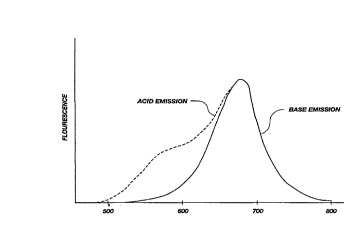
FIG. 2 shows a graphic representation of
fluorescence emission of acid and base forms of an
illustrative carbazine dye at wavelengths in the range
of about 500-800 nm.
FIG. 3 shows a graphic representation of
fluorescence emission of acid and base forms of a
carbazine azine at wavelengths in the range of about
500-800 nm.
FIG. 4 shows a graphic representation of emission
ratio (emission at S90 nm divided by emission at 680 nm)
as a fu,nction of pH for buffer samples analyzed with a
fiber optic pH measurement system according to the
present invention.
CA 02219117 1997-10-24
W O 96/34284 PCTrUS96/05777
FIG. 5 shows a graphic representation of emission
ratio as a function of pH for protein-containing buffer
samples with a fiber optic pH measurement system
according to the present invention.
FIG. 6 shows a schematic diagram of an optical
system of an illustrative fluorometer for use in
determining pH according to the present invention.
FIG. 7 shows a block diagram of an electronic
system of an illustrative fluorometer for use in
determining pH according to the present invention.
Detailed DescriPtion of the Invention
Before the present compositions and methods for
carbazine-dye-based pH measurement are disclosed and
described, it is to be understood that this invention is
not limited to the particular process steps and
materials disclosed herein as such process steps and
materials may vary somewhat. It is also to be
understood that the terminology and examples employed
herein are used for the purpose of describing particular
embodiments only and are not intended to be limiting
since the scope of the present invention will be limited
only by the appended claims and equivalents thereof.
It must be noted that, as used in this
specification and the appended claims, the singular
forms "a," "an," and "the" include plural referents
unless the context clearly dictates otherwise. Thus,
for example, reference to a composition containing "a
carbazine dye" includes reference to a mixture of two or
more such carbazine dyes, reference to "a solid support"
includes reference to one or more of such solid
supports, and reference to "a functional group'~ includes
reference to a mixture of two or more such functional
groups.
In describing and claiming the present invention,
the following terminology will be used in accordance
with the definitions set out below.
CA 02219117 1997-10-24
W O 96/34284 PCTrUS96/05777
As used herein, "periodate-oxidation-susceptible
polymer" means a polymer containing -OH groups attached
to adjacent carbon atoms such that upon oxidation with
periodic acid the carbon-carbon bond is cleaved and such
-OH groups are oxidized to aldehyde groups. Preferred
periodate-oxidation-susceptible polymers include paper,
starch, cellulose, amylose, rayon, cellophane, and the
like and mixtures thereof.
As used herein, "epoxide-reactive support" means a
solid support containing a functional group that is
reactive with an epoxide resulting in formation of a
covalent bond between the solid support and the epoxide.
Such functional groups that are reactive with an epoxide
include hydroxyl, amine, carboxylic acid, and anhydride
groups.
As used herein, "inorganic support" means a solid
support that is composed of an inorganic material.
Preferred inorganic supports include glass, glass
fibers, sand, silica gel, alumina, titania, nickel
oxide, aluminum oxide, zirconia, and mixtures thereof.
More preferred inorganic supports include glass, glass
fibers, sand, silica gel, alumina, and mixtures thereof.
As used herein, "fluorometer" means a device for
generating light at a selected wavelength for exciting
fluorescence of a carbazine dye, receiving and
measuring intensities of fluorescent light emitted by
such carbazine dye at a first selected wavelength and at
a substantially different second selected wavelength,
and generating an electronic signal containing
measurements of said intensities.
Carbazine DYes
A generalized structure for the carbazine dyes of
the present invention is shown in the following ~ormula:
.
CA 02219117 1997-10-24
W 096/34284 PCTrUS96/05777
HO
R5~ / \ , R3
l 1~ (Formula 1)
R6 ~ Rz
0
wherein R2, R3, R5, and R6 are independently selected ~rom
the group consisting of H and alkyl. No dye is formed
if R2, R3, R5, or R6 contains an oxygen atom, such as a
alcohol, ether, carbonyl, or a halogen atom.
These carbazine dyes are prepared by a modification
of the method of R. Hill et al., supra , hereby
incorporated by reference, and shown qualitatively in
the following reaction scheme.
CA 022l9ll7 l997-l0-24
W O 96/34284 PCTrUS96/05777
13
~ ~
G G
O--Z ~ O ~ C G
C G
~5
v
C ~ ~r
~ =Z 4~ ~ ~
.,~=< ~
~: C C
.,
~ G G ~
O--Z ~ O
Z
Z +
O o
t I /
~ C
~
~ G 0~
CA 022l9ll7 l997-l0-24
W 096134284 PCTrUS96/05777
14
S The sodium salt o~ indophenol is reacted with a
substituted phenol that has been modified by reacting
with sodium nitrite in sul~uric acid. The modi~ied
phenol/indophenol reaction is carried out in 90
sulfuric acid. The reaction mixture is maintained at
40~C and under slight vacuum to remove nitrogen oxides
as the dye is synthesized. Sul~uric acid concentration
is critical to the e~iciency o~ the reaction and must
be 90~ + 3~ ~or good yields. Very poor yields result
~rom sulfuric acid concentrations of less than 87~ or
greater than 93~. Dye ~ormation, ~luorescence, and
reactivity with a solid support depend on the nature of
the substituents on the phenol moiety.
Example 1
All reagents used in this and the ~ollowing
examples were purchased ~rom Aldrich Chemical Co.
(Milwaukee, Wisconsin), Sigma Chemical Co. (St. Louis,
Missouri), or Spectrum Chemical Co. (Gardena,
Cali~ornia), and were used without ~urther puri~ication.
One gram o~ the sodium salt o~ indophenol was thoroughly
mixed with 1.5 g o~ powdered phenol. This mixture was
added to 10 ml o~ 90.0~ sul~uric acid containing 600 mg
o~ dissolved sodium nitrite at 40~C in a 2000 ml side
arm ~lask containing about 100 ml of 1 cm diameter glass
spheres. The top o~ the ~lask was then sealed with a
stopper and a slight vacuum was applied to aid in
removal o~ nitrogen oxides that ~ormed immediately. The
~lask and contents were incubated ~or 15 min at 40~C
with intermittent shaking, and then an additional 10 ml
o~ 90~ sul~uric acid containing 600 mg of sodium nitrite
was added and mixed with shaking. The top was again
sealed, and the ~lask and contents were incubated
another 15 min at 40~C with occasional shaking. Then,
1 g o~ powdered phenol was added with shaking. The top
was again sealed and the reaction was permitted to
proceed ~or 30 minutes with occasional shaking.
CA 022l9ll7 l997-l0-24
W 096/34284 PCTrUS96/05777
The reaction mixture was then poured into about 2
liters of ice water with mixing. This mixture was then
exhaustively extracted with cold diethyl ether. The
ether extract was filtered and then extracted with cold
3~ sodium carbonate solution. The resulting highly
fluorescent carbonate solution was filtered, then a
slight current of air was passed through it to remove
dissolved ether. Then, 3.5 g of potassium ferricyanide
was slowly added to this solution and kept at room
temperature for 48 hours. The ferricyanide treatment
destroys by-products of the reaction. This solution,
containing fluorescent carbazine dye and decomposed
impurities, was filtered and then treated with
sufficient calcium chloride to precipitate the
carbonate. This turbid, yellow-green solution was
exhaustively extracted with diethyl ether. At this
stage of purification, the ether extract was highly
fluorescent orange. This ether solution was then
filtered and extracted with 3~ carbonate solution. The
resulting fluorescent blue carbonate solution was
acidified to pH 5 by addition of glacial acetic acid.
Upon standing at 5~C ~or several hours, the orange-
colored solid carbazine dye separated as a fine powder.
The solid dye was collected by filtration and dried
under vacuum. The resulting carbazine dye was 7-
hydroxyspiro[acridine-9,1'-cyclohexa-2', 5'- diene]-
2 (9H), 4'-dione having the structure of Formula 1 wherein
R2, R3, R5, and R6 were each H.
Example 2
The procedure of Example 1 was followed with the
exception that 3, 5-dimethylphenol (3, 5-xylenol) was
substituted for phenol. The resulting carbazine dye had
the structure of Formula 1 wherein R2 and R6 were H and
R3 and Rs were methyl.
CA 02219117 1997-10-24
W 096/34284 PCTrUS96/05777
Example 3
The procedure of Example 1 was followed with the
exception that 2,3,5-trimethylphenol (isopseudocumenol)
was substituted for phenol. The resulting carbazine dye
had the structure of Formula 1 wherein R6 was H and R2,
R3, and Rs were methyl.
Example 4
The procedure of Example 1 was followed with the
exception that durenol (2,3,5,6-tetramethylphenol) was
substituted for phenol. Durenol was synthesized by
exhaustive methylation of 3,5-dimethylphenol (3,5-
xylenol) by the method of Burawoy, J. Chem. Soc. 400
(1944), hereby incorporated by reference. The resulting
carbazine dye had the structure of Formula 1 wherein R2,
R3, Rs~ and R6 were each methyl.
Example 5
The procedure of Example 1 was followed with the
exception that 5,6,7,8-tetrahydro-1-naphthol was
substituted for phenol The resulting carbazine dye had
the structure of Formula 1 wherein R2 and R3 were 2,3-
cyclohexyl and Rs and R6 were each H.
Example 6
The procedure of Example 1 was followed with the
exception that 5-isopropyl-3-methylphenol was
substituted for phenol. The resulting carbazine dye had
the structure of Formula 1 wherein R2 and R6 were H, R3
was isopropyl, and Rs was methyl.
Example 7
The procedure of Example 1 was followed with the
exception that o-tert-butylphenol was substituted for
phenol. The resulting carbazine dye had the structure
of Formula 1 wherein R3, R5, and R6 were each H and R~ was
t-butyl.
CA 02219117 1997-10-24
W O 96/34284 PCT~US96/05777
17
Example 8
The procedure of Example 1 was followed with the
exception that m-tert-butylphenol was substituted for
phenol. No detectable amount of carbazine dye was
synthesized. It is thought that steric hindrance
prevented reaction of the substituted phenol with
indophenol.
Example 9
Certain properties of the carbazine dyes of
Examples 1-7 were determined. Spectral data were
obtained either with a Hewlett Packard Model 8452A Diode
Array Spectrophotometer or a Perkin Elmer Model LS 5OB
Luminescence Spectrometer. All of the dyes are similar
in their absorbance and emission spectra, with only
slight differences in peak locations, ratios of
absorbance of acid to absorbance of base, and pKas. A
typical absorbance curve versus pH is shown in FIG. 1.
The unmodified dyes show large separation between
absorbance peaks for acid and base ~orms. Typically,
the acid form has a peak absorbance at about 480 nm, and
the base form has a peak absorbance at about 660 nm. A
typical fluorescence emission curve of both acid and
base forms of the dyes is shown in FIG. 2. Fluorescence
emission of the base form has a well-defined single peak
at approximately 690 nm. The acid form o~ the dyes
exhibits an emission spectrum similar to that o~ the
base form, but has a definite shorter wavelength
emission component near 600 nm.
The pKa values, i.e. the approximate pH values o~ an
aqueous solution of the dye where the acid and base
forms of the dye are present in equal concentrations,
were derived ~rom the absorbance spectra and are listed
in the Table below. As mentioned above, indicator dyes
are generally most sensitive to pH changes near their
pK~s.
CA 022l9ll7 l997-l0-24
W 096/34284 PCTrUS96/05777
18
The relative susceptibilities of the dyes to
immobilization on a solid support were determined from
the intensity of dye covalently bound to regenerated
cellulose dialysis membrane. The immobilization data
summarized in the Table below were taken from
measurements in 2~ carbonate solution normalized to an
arbitrary scale of 1 to 10, where 1 represents no
detectable amount of dye bound and 10 represents the
greatest quantity of dye bound. The conditions of
coupling of the dyes to the regenerated cellulose were
according to immobilization Method 2 described below.
Carbazine dyes immobilized under these conditions yield
a uniform fluorescent blue dialysis membrane that
changes to fluorescent orange upon acidification. As a
point of reference, the carbazine dye of Example 3
immobilized on a "SPECTRAPOR 1" membrane (molecular
weight cutoff 6000 to 8000) yields a blue membrane with
an absorbance at 660 nm of greater than 1 at pH ~9.
The fluorescence ratings shown in the Table are
based on the maximum fluorescence obtainable from an
aqueous carbazine dye solution with excitation at the
maximum absorbance wavelength for the particular dye,
but with concentrations and pH constant between all
dyes. The fluorescence rating does not change with pH,
l.e. the dyes that are most fluorescent in base are also
most fluorescent in acid. Quantum yields of the acid
forms of the dyes appears as high as the base forms.
The carbazine dye of Example 4, sold commercially as
"CARBAZINE 720" (Exciton, Inc., Dayton, Ohio), has a
quantum yield of approximately 50~ in aqueous solution
containing base. The carbazine dye of Example 3 is as
fluorescent, can be produced in higher yield~ and has a
higher immobilization efficiency than "CARBAZINE 720."
Properties of the carbazine dyes of Examples 1-7
are summarized in the following Table:
CA 02219117 1997-10-24
W 096134284 PCTAUS96105777
Table
,. Dye'Yield~ Absorb. pR, Immobili- Fluores-
Ratio' zation~cence"
hi~h 0.426 6.5 lO 2
2 low 0.391 6.5 6 7
0 318~6 0.380 6.5 6 10
4 15~ 0.388 6.5 4 10
5 10~ 0.387 6.7 6 8
6 10~ 0.377 6.~ 6 7
low 0.324 7.2 1 6
a The number of the dye refers to the Bxamples.
b Based on the molar ratio of dye synth~c;~ to ;nrl~lph~n~
C D~.C.. 1~ = ratio of acid/base.
d Relative scale of 1 to 10 where 10 is best.
These data suggest that for high fluorescence
efficiency, some substitution in the spiro ring is
required. The chromophore portion of the dye molecule
comes from the indophenol molecule, which is virtually
nonfluorescent. It has been suggested that the
fluorescence of carbazine dyes is derived from the
tetrahedral carbon bridge causing the indophenol
chromophore to be rigid. Since all of the dyes
presented in the Table contain the tetrahedral carbon
bridge, substitution in the spiro ring appears to play
an important part in fluorescence.
The results presented in the Table also show a
trend relating immobilization efficiency to steric
effects near the 1 position of the spiro ring. The dye
of Example 7, containing a t-butyl group for R2, does not
react to any detectable extent in any of the
immobilization schemes disclosed herein, presumably due
to steric effects near the binding site to the solid
~ support. The dye o~ Example 4, further, does not bind
as effectively as does the dye of Example 3, presumably
due to the presence of a methyl group for R6. Finally,
the best dye for binding to a solid support in terms of
CA 02219117 1997-10-24
W 096/34284 PCTrUS96/05777
immobilization efficiency i8 the unsubstituted dye of
Example 1.
The optimum carbazine dye for pH measurement
applications is a compromise between yield,
fluorescence, and immobilization efficiency, each of
which is affected by the substituents on the spiro ring.
The dye of Example 3 is a preferred carbazine dye for pH
measurement because it represents an effective
compromise of the various factors that influence pH
measurement when immobilized on a fiber optic probe.
lS Modification of Carbazine DYes
The spiro ring of some of the carbazine dyes
described herein are reducible by nickel/aluminum alloy
in aqueous sodium hydroxide to the compounds shown
generically in the following formula:
2 5 HO ~ o
R5~ ~ \~ R3
R6 ~R2 (Formula 2)
OH
wherein R2, R3, R5, and R6 are independently selected from
the group consisting of H and alkyl. These saturated
compounds exhibit similar absorption and emission
spectra to the corresponding unsaturated compounds with
the exception of the absorption maxima for the basic
forms of the molecules. The basic forms of the
saturated compounds all show a blue shift of
CA 022l9ll7 l997-l0-24
W O 96/34284 PCTAUS96/05777
21
approximately 30 nm (from 660 nm to 630 nm) for the
absorption peak.
Example 10
The carbazine dye of Example 1 was reduced
- 10 according to the following procedure. Approximately 100
mg of dye was dissolved in 20 ml of 1 N NaOH in a beaker
fitted with a vacuum port. Approximately 500 mg of 50~
nickel/aluminum alloy was added, then the beaker was
sealed and immediately placed under vacuum. After a few
minutes, the blue dye solution became colorless as the
leuco compound formed. After a few minutes more, gas
bubbles began to form as hydrogen evolved by the action
of the base on the aluminum. After an additional
several minutes, the vacuum was removed, and the mixture
was rinsed from the beaker into approximately 500 ml of
0.1 N sodium bicarbonate. This solution was filtered
several times to remove aluminum hydroxide and the
rem~; n~ of the alloy. Air was slowly bubbled through
the filtered solution, containing the dissolved dye, to
oxidize the leuco compound. As this occurred, the
fluorescent color returned. This solution was then
treated with sodium phosphate until a yellow-green color
was obtained, and was then extracted with cold ether.
The ether extract was filtered, and then extracted with
cold 3~ carbonate solution. Air was then bubbled
through the ~luorescent blue carbonate solution to
remove the ether and then was acidified to pH 5 with
glacial acetic acid. The acidified solution was chilled
for several hours at 5~C, and then the precipitated
reduced dye was collected by filtration and dried under
vacuum. The resulting reduced carbazine dye had the
structure according to Formula 2 wherein R2, R3, Rs~ and
R6 are each H.
CA 02219117 1997-10-24
W O 96/34284 PCTrUS96/05777
Immobilizinq Carbazine D~es on Solid Sup~orts
Carbazine dyes according to the present invention
bind to hydrazine derivatives to form the compounds
shown in the following formulas:
~ \~
HO~/\ O
R5~/ \ , R3
1 1 (Formula 3)
R6 \ / R2
N
NHR 7
HO~J~ // ~\\~
Rs ~, R3
ll
R6 / Rz
(Formula 4)
NH
NHR7
CA 02219117 1997-10-24
W O 96/34284 PCT~US96/05777
HO ~ O
Rs ~ \ ~ Rl
- lo Jl 1
R6 ~ R2
N (Formula 5)
N
11
CH 3 -C-R 7
wherein R2, R3, R5, and R6 are independently selected from
the group consisting of H and alkyl, and R7 is a member
selected frolll the group consisting of H and alkyl.
Hydrazine and its derivatives react according to the
following reaction scheme in anhydrous protic or aprotic
solvents or in aqueous solution to form carbazine imines
as shown in Formula 3:
110 ~ ~~,u,O
R.J~ + ~H2--~HR7 R N R~
NHR ~
(Reaction 2)
c
These reactions occur best at between about pH 6.0 and
7.0, or simply in aqueo~s solution at a pH where the
carbazine dye remains green in color. The base ~orm o~
a carbazine dye is bright blue, whereas the acid ~orm is
yellowish orange. At the pKa of the dye, about pH 6.5,
CA 02219117 1997-10-24
W 096/342~4 PCTrUS96/0~777
24
there are equal numbers of molecules of ionized (basic
form, blue) and unionized (acid form, yellow) dye
molecules, thus resulting in a green to blue green
color. Substituted hydrazines are more reactive in
coupling to carbazine dye than is free hydrazine.
Carbazine imines, although not particularly stable,
are reducible to stable carbazine-substituted
hydrazines, as shown in Formula 4, by sodium
cyanoborohydride, as shown in the following reaction
scheme:
~ ~
N
NHR 7
NHR 7
(Reaction 3)
This reaction may be carried out simultaneously with the
imine formation reaction as a "one pot" reaction.
Carbazine dye, hydrazine or substituted hydrazine, and
sodium cyanoborohydride are dissolved or suspended in
water at pH 6.2. Cyanoborohydride is known to rapidly
and selectively reduce imine groups under these
conditions, and is relatively stable in aqueous
solutions above pH 6Ø
Carbazine-substituted hydrazines are highly
fluorescent compounds with spectra and pK~s similar to
the free dyes. The covalent bond formed between the
carbazine dye and the substituted hydrazine is
chemically stable, and the substituted hydrazine can be
CA 02219117 1997-10-24
W O 96/34284 PCTrUS96/05777
part of an insoluble support. Thus, carbazine dyes can
be covalently bonded to hydrazine-modified supports.
Carbazine azines, shown generically in Formula 5,
are prepared according to the following reaction scheme:
Ul~ 0 1}:, HO~o
R.~ + NH2-N=C-R,R~ N R~
O N
CH?~R7
(Reaction 4)
These compounds readily form in aqueous or organic
solution with wide latitude in reaction conditions. The
best coupling, however, seems to be in aqueous solution
at a pH of about 6 0 to 6.5.
Carbazine azines show unique fluorescence behavior
compared to other carbazine derivatives. While
absorbance data remain essentially unchanged, emission
spectra are as shown in FIG. 3. Further, the pKa of
these compounds is about 1 pH unit higher, i.e. about pH
7.5, than the unmodified dye. Base form emission
remains unchanged, but acid form emission is in a well
resolved, single peak centered around 590 nm. Carbazine
azines, thus, are dual excitation, dual emission dyes
with acid form excitation/emission of 480 nm/590 nm and
base form excitation/emission of 660 nm/690 nm. The
acid/base absorbance curves cross, as shown in FIG. 1,
at approximately 520 nm. An excitation wavelength can
be selected to suitably excite both the acid and base
forms of the carbazine azine to produce a single
excitation, dual emission pH indicator. As with the
CA 02219117 1997-10-24
W O 96/34284 PCTrUS96/05777
other dye forms, the carbazine azine is highly
fluorescent and chemically stable.
In the following methods, "R" is used to represent
the solid support onto which the carbazine dye is to be
immobilized. In some instances the solid support may be
modified, derivatized, or functionalized according to
the reaction schemes that follow. It is to be realized
that there must necessarily be some functional group or
groups on the solid support for a chemical reaction to
occur. These groups can be in the form of -OH, =O, -NH2,
-MgX, -COOH, ketones, and so forth, that can then be
further reacted by oxidation to aldehydes or acids,
derivatized with glycidol, GOPS, or reacted directly
with hydrazine or hydrazine derivative according to the
reaction schemes that follow. However, for purposes of
clarity and uniformity, the solid support will be simply
referred to as "R." It will be clear to one skilled in
the art what "R" represents according to the reaction
scheme utilized.
Polysaccharide Sup~orts - Method 1
In a first method, the dye-reactive
hydrazine/hydrazone groups are readily incorporated onto
the surface of a periodate-oxidation-susceptible polymer
support, such as paper, starch, cellulose, amylose,
rayon, cellophane, and the like and mixtures thereof.
Upon treatement with periodic acid, compounds containing
-OH groups attached to adjacent carbon atoms undergo
oxidation with cleavage of carbon-carbon bonds. R.
Morrison & R. Boyd, Orga~ic Chemistry 523-24 (4th ed.,
1983). The -OH groups are oxidized to aldehyde groups.
The aldehyde groups of the oxidized polysaccharide are
then reacted with hydrazine in the presence of sodium
cyanobor,ohydride to form an immobilized hydrazine. This
hydrazine-modified support is then reacted with a
carbazine dye as described above. This method results
in some polymer degradation due to breaking of the
CA 02219117 1997-10-24
W 096/34284 PCT~US96/05777
carbon-carbon bonds by periodate oxidation of the
polymer chain. This reaction scheme is illustrated as
follows:
O
. ll
R + NaIO4 R-C-H
o
Il
R-C-H + NH2NH2 R-CH=N-NH2 + H2O
R-CH----N-NH2 + NaCNBH3----~R-CH2-NH-NH2
R-CH2-NH-NH2 + D----~R-CH2-NH-N=D
R-CH2-NH-N=D + NaCNBH3 R-CH2-NH-NH-D
wherein R is a member selected from the group consisting
of paper, starch, cellulose, amylose, rayon, cellophane,
and other polymers that can be oxidized by periodate to
yield an aldehyde group and D is a carbazine dye
according to Formula 1 with R2, R3, R5, and R6
independently selected from H and alkyl. The covalent
bonds between the carbazine dye and the substituted
hydrazine are formed according to Formulas 3 and 4.
Polysaccharide Su~ports - Method 2
A second method of immobilizing carbazine dyes on
polymer supports does not require polymer degradation,
i.e. breaking of carbon-carbon bonds of the polymer by
periodate oxidation, and produces substantially higher
yields of immobilized dye than Method 1. This second
method is functional with any support material that
contains an epoxide-reactive group, such as a hydroxyl
group, amine group, carboxylic acid group, or anhydride
group. Reactions of various epoxide-reactive groups
with the epoxide, glycidol, are illustrated as follows:
O OH OH
/ \ NaOH l l
(1) R-OH + H2C CH-CH2-OH ~ R-C-CH2-CH - CH2
CA 02219117 1997-10-24
W 096/34284 PCT~US96/05777
28
O OH OH
/\ I I
(2)R--NH2 + H2C CH--CH2--OH ~ R--NH--CH2--CH--CH2
O O OH OH
/ \ 11 1 1
(3) R-COOH + H2C - CH-CH2-OH ~ R-C-O-CH - CH2
wherein R represents the solid support exclusive o~ the
epoxide-reactive group.
Glycidol reacts with the epoxide-reactive group of
the solid support, usually in an aqueous solution with
either acid or base catalysis, to ~orm a poly-
substituted product. The vicinal hydroxyls, i.e.
hydroxyl groups on adjacent carbon atoms, o~ the
glycidol residue are selectively oxidized by periodate
to the polyglyoxal (polyaldehyde) form, which is
sequentially reacted with hydrazine and a carbazine dye
as in the ~irst method described above. These reactions
are illustrated in the ~ollowing reaction scheme:
OH OH O
1 1 ll
R-O-CH2-CH - CH2 + NaIO4 R-O-CH2-C-H
11
R-o-cH2-c-H + NH2NH2 ? R-O-CH2-CH=N-NH2 + H2O
R--O--CH2--CH=N--NH2+ NaCNBH3----~R--O--CH2--CH2--NH--NH2
R-O-CH2-CH2-NH-NH2 + D 3 R-O-CH2-CH2-NH-N=D
R-O-CH2-CH2-NH-N=D + NaCNBH3 R-O-CH2-CH2-NH-NH-D
wherein R is the solid support containing the epoxide-
reactive group and D is a carbazine dye according to
Formula 1 with R2, R-, Rs, and R~ independently selected
~rom H and alkyl. The covalent bonds between the
CA 02219117 1997-10-24
W O 96/34284 PCTnUS96105777
carbazine dye and the ~ubstituted hydrazine are formed
according to Formulas 3 and 4.
Inorqanic Su~orts - Method 3
Many inorganic supports are "silanized" by
treatment with a silanizing reagent. These reactions
are generally thought to result in an organic molecule
covalently attached to a silanol (Si-oH) surface
functionality. This treatment both blocks the
reactivity of the surface silanol functionality and
imparts a reactive functionality to the surface of the
inorganic support corresponding to the organic portion
of the silanizing reagent.
Glycidoxypropyl trimethoxysilane (GOPS) reacts with
inorganic supports such as glass, glass fibers, sand,
silica gel, alumina, titania, nickel oxide, aluminum
oxide, zirconia, and other hydrophilic inorganic
supports and mixtures thereof to produce an organic
epoxy functionality on the inorganic surface, as is
illustrated in the following reaction scheme with silica
gel:
o
/ \ heat
(CH30)3Si(CH2)30CH2-CH - CH2 + silica gel
o
(silica gel-0)3Si(CH2)30CH2-CH - CH2
This epoxy functionality can be acid hydrolyzed to yield
a surface containing vicinal hydroxyl groups. These
vicinal hydroxyl groups can be further oxidized by
periodate to produce a surface aldehyde functionality.
These reactions are illustrated as follows:
CA 022l9ll7 l997-l0-24
W O 96/34284 PCTrUS96/05777
O
/ \ heat
(silica gel-O) 3Si (CH2)3OCH2-CH--CH2 + 2 H+
OH OH
(silica gel-O) 3Si (CH2) 30CH2-CH - CH2
OH OH
l l
(silica gel-O) 3Si (CH2)30CH2-CH - CH2 + NaIO4
o
(silica gel-O) 3Si (CH2)3OCH2-C--H
The surface aldehyde group can be coupled
sequentially to hydrazine and a carbazine dye as
described above.
Inorqanic Supports - Method 4
Glycidol will react with the vicinal hydroxyl
groups present on the compound o~ Method 3 resulting
~rom the reaction o~ silica gel or other appropriate
inorganic support to result in a periodate-oxidizable
sur~ace o~ greater hydrazine binding capacity, as
~ollows:
OH OH
(silica gel-O) 3Si (CH2) 30CH2-CH - CH2
/ \ NaOH
+ 2 H2C-- CH--CH2--OH
CH2-CH-CH2
l l
O OH OH OH OH
(silica gel-O) 3Si (CH2)30CH2-CH--CH2--O--CH2--CH--CH2
The vicinal hydroxyl groups can then be oxidized
with periodate to yield aldehyde groups, which can then
be sequentially coupled to hydrazine and a carbazine dye
as described above.
CA 02219117 1997-10-24
W O 96134284 PCTrUS96/05777
Polvaldehyde Su~orts - Method 5
Polyaldehydes, such as polyacrolein and
polyglutaraldehyde, react with hydrazine in the presence
of cyanoborohydride to yield substituted polyhydrazine
materials. Carbazine dyes can then be bonded to the
polyhydrazine supports according to the procedure of
Method 1. Reaction of the polyaldehydes with hydrazine
is illustrated as follows:
H H
l l
R-C=O + NH2NH2 ~ R-C- N - NH2 + H20
H H
R- 1=N-NH2 + NaCNBH3 , R-c-NH-NH2
Polymethylketone Supports - Method 6
Polymethylketones undergo a substitution reaction
with hydrazine hydrate at elevated temperatures to yield
polymethyl hydrazone. This product reacts directly with
carbazine dyes to produce highly fluorescent azine
polymers with unique optical and chemical
characteristics for PH measurement. Reactions of
polymethylketones with hydrazine and of polymethyl
hydrazone with carbazine dye are illustrated as follows:
O N - NH2
¦¦ heat 1l
R-C-CH3 + NH2NH2 , R-C-CH3 + H20
N-NH2 N-N=D
4 0 11 ll
R-C-CH3 + D ~ R-C-CH3 + H20
Example 17
Approximately l g of microcrystalline cellulose was
suspended in 50 ml of 1 N NaIO4 solution and reacted for
1 hour at room temperature. The cellulose was removed
CA 02219117 1997-10-24
W 096/34284 PCTrUS96/05777
32
by filtration, washed extensively with water and then
with anhydrous ethanol, and then dried under vacuum.
The cellulose was then suspended in a 10% (v/v) aqueous
solution hydrazine hydrochloride, pH 6.2, for 5 hours at
room temperature. The cellulose was then again removed
by filtration, washed extensively with water, and then
suspended in a solution of about 1 mg/ml sodium
cyanoborohydride in water, pH 6.2, and reacted for 5
hours at room temperature. The cellulose was then again
removed by filtration, washed extensively with water and
then with anhydrous ethanol, and then dried under
vacuum. The resulting compound was a hydrazine-modified
cellulose support prepared according to Method 1.
Example 18
About 1 g of microcrystalline cellulose was
suspended in about 20 ml of a 10% (w/v) solution of
glycidol in 1 N sodium hydroxide and permitted to react
overnight at room temperature. The solid material was
separated by filtration, washed extensively with water
and ethanol, and then reacted sequentially with
periodate, hydrazine, and sodium cyanoborohydride
according to the procedure of Example 17.
Example 19
About 1 g of silica gel was heated to 90~C in 50 ml
of a 10% (w/v) of GOPS and maintained for 1 hour while
the pH was maintained between 1 and 2 by addition of
HCl. The modified silica gel was then collected by
filtration, washed extensively in water, and then
reacted sequentially with periodate, hydrazine, and
sodium cyanoborohydride according to the procedure of
Example 17.
CA 02219117 1997-10-24
W O 96/34284 PCTrUS96/05777
33
Example 20
About 1 g of glass fibers was reacted with GOPS
according to the procedure of Example 19, and then
rinsed extensively. The modified glass fibers were then
reacted sequentially with glycidol, periodate,
~ 10 hydrazine, and sodium cyanoborohydride according to the
procedure of Example 18.
Example 21
About 1 g of polyacrolein was reacted with
hydrazine and cyanoborohydride according to the
procedure of Example 17.
Example 22
In this example, 5.0 g of poly(methyl vinyl ketone)
was suspended in about 50 ml of hydrazine hydrate and
heated in a steam bath for 48 hours. The swollen,
cross-linked gel was then removed by filtration and
washed extensively with water. The resulting gel was a
hydrazone-containing support.
Example 23
Cou~linq of Carbazine DYe to Hydrazine-modified
Support. About 1 g of hydrazine-modified support
prepared according to the procedure of Example 17 was
suspended in 1 ml of 0.1 N [N-(2-acetamido)-2-
aminoethane sulfonic acid] (ACES) buffer, pH 6.2. A
solution of 5 mg of carbazine dye, prepared according to
the procedure of Example 3, dissolved in 200 ~l of
dimethylformamide was added to the support-containing
solution, mixed, and permitted to react overnight at
room temperature. The support was then separated by
filtration, washed extensively with water, and suspended
in 5 ml of ACES buffer, pH 6.2, containing approximately
10 mg of sodium cyanoborohydride. The reaction was
permitted to proceed for 5 hours. Since this reaction
consumes hydrogen ions, acetic acid was added
CA 02219117 1997-10-24
W O 96134284 PCTrUS96/05777
occasionally to maintain the pH. Upon termination of
the reaction, a few drops of aqueous formaldehyde were
added to block any unreacted hydrazine groups, followed
by addition of about 5 mg more cyanoborohydride to
e~fect the formaldehyde reaction. This blocking
reaction was permitted to occur for 1 hour at room
temperature.
The two steps of (a) attachment of the carbazine
dye to the hydrazine to form a hydrazone, and (b)
reduction of the hydrazone with cyanoborohydride to form
the dye-substituted hydrazine can also effectively be
carried out simultaneously in a one-step reaction.
Since the dye is the most valuable reagent in this
synthesis, however, it is advantageous to be able to
recover the unreacted dye for recycling. The two-step
process allows for recovery of the dye without
cont~m;n~tion with cyanoborohydride.
Example 24
About 1 g of moist hydrazone-containing support,
prepared according to the procedure of Example 22, was
suspended in 1 ml of ACES buffer, pH 6.2. A solution of
5 mg of carbazine dye, prepared according to the
procedure of Example 3, dissolved in 200 ~l of
dimethylformamide was added to the support-containing
solution, mixed, and heated in a steam bath for 1 hour.
The support was collected by filtration, washed
sequentially with water, acetone, dilute acetic acid,
and aqueous carbonate, then stored in water. Dye-
substituted azines are chemically stable and require no
sodium cyanoborohydride reduction.
Fiber Optic pH Sensor
Fiber optic pH sensors using the materials and
methods described herein operate as single excitation,
dual emission sensors with excitation between about 480
and 540 nm, acid form emission at about 590 nm, and base
form emission at about 690 nm. A ratiometric technique
CA 02219117 1997-10-24
W O 96/34284 PCTrUS96/0~777
for determining pH with these fiber optic pH sensors
comprises exciting the fluorescent dye with a single
wavelength of light and simultaneously monitoring
fluorescence from the acid form and the base form of the
dye. The ratio of emission of the acid form to emission
of the base form correlates favorably with pH. The
carbazine dyes described herein show less sensitivity to
temperature and solvent changes than currently employed
fluorescent pH indicators. These fiber optic sensors
and method of use eliminate most of the problems
heretofore encountered in fiber-optic-based pH
measurement systems. Also, the compositions and methods
described herein permit the use of inexpensive plastic
fiber optic materials, since all wavelengths of light,
both excitation and emission, are in the visible part of
the spectrum. Most currently used dyes for fiber optic
pH measurement require ultraviolet excitation, thereby
requiring the use of quartz optical fibers.
To demonstrate the utility of these materials as pH
indicators, a fiber-optic-based pH probe was constructed
from a fluorescent dye, prepared according to Example 3,
bound to a 0.012 inch diameter ketone-containing
polyacrylate bead (XAD-7, Rohm & Haas) by the procedure
of Example 24 and 0.010 inch plastic optical fiber
(Polyoptical 1610 fiber). The bead was glued onto the
end of the fiber, without any end polishing or other
preparatory steps, with Norland Optical Adhesive #68.
This probe was optically coupled to a pulse fiber
fluorometer with excitation centered at about 520 nm,
acid emission centered at about 600 nm, and base
emission centered at about 680 nm. Interference filters
were used with bandwidths of about 40 nm to take
advantage of the large spectral separation of the dye.
The fluorometer was coupled to a computerized data
acquisition system for electronic data collection.
An illustrative pulse fiber fluorometer that can be
used in conjunction with the carbazine dyes of the
CA 022l9ll7 l997-l0-24
W 096/34284 PCT~US96/05777
36
present invention for measuring fetal pH is disclosed in
FIGS. 6 and 7. In FIG. 6 there is shown an optical
system 4 for producing an excitation wavelength centered
at 520 nm and for detecting emission wavelengths
centered at 600 nm and 680 nm. The optical system
comprises a f~lashlamp 8 for generating a high intensity
beam 12 of white light. The lamp 8 is selected for a
long useful life, e.g. 10 million flashes. Each ~lash
lasts about 100 microseconds. The electrical drive for
the lamp 8 is designed to m;n;m; ze electrical
interference with the other electronics.
The beam 12 of white light from each flash is
collected and focused to a focal point 16 by lenses 20
and 24. These lenses are aspheric to efficiently
collect as much light as possible. The focused light
thus formed is used to illuminate pinhole 28 in plate
32. The pinhole 28 has an approximate diameter of 0. 060
inch. The focused light can illuminate a substantially
larger area than the size of the pinhole 28, thus
allowing for some misalignment of the flashlamp 8,
lenses 20 and 24, and pinhole 28.
The beam 12 of light passing through the pinhole 28
is collimated by lens 36. The collimated light then
passes through filter 40. It is important to achieve
good collimation for filter 40 to perform correctly, as
will now be explained. Filter 40 is a highly selective
interference filter that transmits light of 520 nm + 20
nm. Any light that gets through filter 40 in
wavelengths at which fluorescence will be measured, 600
nm and 680 nm, will be ~alse fluorescence. The high
selectivity needed to substantially eliminate false
fluorescence can be achieved only with well collimated
light from lens 36.
The light transmitted through ~ilter 40, e.g. 520
nm light, then contacts lens 44, which focuses this
light on optical fiber 48. Optical fiber 48 conducts
the 520 nm light to a pH indicator probe comprising a
CA 02219117 1997-10-24
W O 96/34284 PCTrUS96/05777
fluorescent carbazine dye covalently bonded to a solid
support, as has been thoroughly explained above. Upon
being illuminated by the 520 nm light, the probe
fluoresces with dual emission fluorescence with
wavelengths centered at about 600 nm and 680 nm. A
portion of this emitted light is gathered by the optical
fiber 48, which conducts such emitted light back to the
optical system 4 of the fluorometer.
The fluorescent emitted light from the optical
fiber 48 passes through lens 44 and from there to filter
40. Filter 40 reflects this emitted fluorescent light
to mirror 52. Light contacting mirror 52 is then
reflected to filter 56. Filter 56 is another high
performance interference filter that transmits light of
680 nm and reflects light of 600 nm wavelength. This
transmitted light contacts lens 60, which focuses the
680 nm light onto pinhole 64 in plate 68. This pinhole
64 spatially removes scattered and stray light, since
only light emitted from the optical fiber 48 is focused
thereon. The focused 680 nm wavelength light that
passes through pinhole 64 contacts photodiode 72.
Light reflected by filter 56 contacts filter 76,
which is another high performance interference filter
that permits only light of about 600 nm to pass
therethrough. Such 600 nm light passing through filter
76 then contacts lens 80, which focuses this 600 nm
light on pinhole 84 in plate 88. As with pinhole 64,
pinhole 84 spatially removes scattered and stray light.
The 600 nm light passing through the pinhole 84 then
contacts photodiode 92, which detects this 600 nm light.
The angles of filter 40 and filter 56 are
maintained at 15 degrees from normal incidence. This
angle must be kept small for filter 40 and filter 56 to
function, properly in transmitting the selected
wavelengths of light and reflecting other wavelengths of
light. Filter 76 is not required to reflect light to
additional optical components of the system and is
CA 02219117 1997-10-24
W 096/34284 PCTrUS96/05777
therefore set at an incidence of 90 degrees. This
setting allows steeper edges to the transmission band of
the filter 76, which is important for the optical
channel with the least wavelength separation from the
illumination source.
This optical system 4, using well designed filters,
allow photodiodes 72 and 92 to detect fluorescent
signals many orders of magnitude lower in intensity than
that of the illumination source (flashlamp 8). In
operation, the fluorescence channels detect essentially
no light in the absence of an optical fiber 48. Even
placing a reflector at the position of optical fiber 48
results in essentially no detection of false
fluorescence.
A schematic diagram of the electronics system 100
that accompanies the optical system 4 (FIG. 6) of the
fluorometer is shown in FIG. 7. All functions of the
fluorometer are under microprocessor control. There are
two detector channels, one for 600 nm and another for
680 nm light. Each detector is composed of a high
sensitivity, low noise photodiode connected to suitable
amplifiers, analog to digital (A/D) converter then to
the microprocessor. Each channel is identical. A
description of one such channel follows.
Light contacting the 600 nm photodiode 92 is
converted to a weak electrical current, with the current
proportional to the intensity of the light. This
current is amplified through two separate
transimipedence amplifiers 104 and 108 into a positive
and a negative voltage. These voltages are conducted
into a true differential amplifier 112 where the voltage
signals are combined into a higher voltage signal, with
the voltage out proportional to the illumination
intensity on the photodiode 92. This scheme of
differential amplification is used to reduce common mode
noise that may be present due to the small signals
generated by the photodiode 92, in comparison to
CA 02219117 1997-10-24
W 096/34284 PCTrUS96/05777
electrical noise that may be generated by the high
current flashlamp 8 (FIG. 6).
The voltage from amplifier 112 is fed into
amplifier 116, where additional voltage amplification
occurs. Additionally, auto zero circuit 120 feeds a
signal into amplifier 116 that is proportional to the
signal present from amplifier 112 when the flashlamp 8
is not firing. This auto zero signal is substacted from
that of amplifier 112, so as to yield essentially zero
output from amplifier 116 except when light is actually
falling onthe photodiode 92. This autozero feature
automatically corrects for amplifier drift and the like,
and ensures the output from amplifier 116 is
proportional to the light intensity during the flash.
Careful design of the auto zero circuit 120 also helps
to eliminate ambient light signals that may be present
when the fiber 48 (FIG. 6) is illuminated from an
external white light source, such as room or ~x~mln;ng
lights. Finally, another part of amplifier 116 converts
the voltage output to a true current source for feeding
into the A/D converter 124.
The A/D converter 124 is a 20 bit charge
integrating device. During operation, the integration
period is 300 microseconds. That is, the A/D converter
124 meaures the total charge from amplifier 116 over a
300 microsecond period. Operation starts with the A/D
converter 124 measuring a background period, with no
light from the flashlamp 8 (FIG. 6). This is followed
by triggering the ~lashlamp (~rom the microprocessor
128) and measuring the total signal from the photodiodes
92 and 72. Since the flash is 100 microseconds long,
the A/D converter 124 is timed to capture all of the
signal in this integration period. The A/D converter
124 then meaures another period without the flashlamp 8
(FIG. 6). The first and third periods are averaged and
subtracted ~rom the signal during the second period to
remove additional background. In this way, additional
CA 022l9ll7 l997-l0-24
W 096/34284 PCT~US96/05777
S noise and ambient light effects are removed. The A/D
converter 124 is chosen to have 20 bit resolution to
accurately handle large changes in detected light from
different probes, aging effects in the optics, and so
forth, without need for an auto gain circuit. It must
be remembered that in this system, the pH is determined
by the ratio between fluorescence at 600 nm and at 680
nm, not in their absolute magnitudes. The fluorometer
thus disclosed accurately meaures this ratio
automaticaly regardless of the instrument, probe, or
ambient changes.
The microprocessor 128 measures the signals from
both of the A/D converters 124 and 132 simultaneously,
and generates a pulse ratio of fluorescence for each
flashlamp pulse. Additionally, it can further average
or process the signal as required. As previously
mentioned, it also controls the A/D converters 124 and
132, flashlamp trigger 136, and auto zero timing 140
functions. The ratio of fluorescence calculated by the
microprocessor 128 is converted to pH data, which is
transmitted to a pH data output device 144.
Example 25
Buffer solutions (1~ ACES, 1~ NaCl) were made and
adjusted to pH values between approximately 5 and 9 and
read at room temperature on a freshly calibrated Orion
Model 720 pH meter. Optical pH data from each buffer
sample were collected electronically with the
computerized data acquisition system. After coming to
equilibrium, 100 readings were taken of each sample.
The results of this experiment are shown in FIG. 4,
wherein the emission ratio (emission at 590 nm divided
by the emission at 680 nm) is presented as a function of
pH. The precision of each ratio data point was
typically better than 0.5~, which yields a pH
uncertainty of approximately 0.02 pH unit at +
standard deviation.
CA 02219117 1997-10-24
W O 96/34284 PCTrUS96/05777
Example 26
Protein-containing buffer samples (1~ BSA, 1~ ACES,
1~ NaCl) were prepared in the pH of range of 6.9 to 7.6.
These samples were subjected to pH determination
according to the procedure of Example 25. The resulting
data are summarized in FIG. 5. As in Example 25, the
precision of each data point and the slope of the
emission ratio/pH curve yielded a pH uncertainty of
about 0.02 pH unit.
From the foregoing, it will be appreciated that the
compositions of the present invention comprise means for
pH determination using a wide range of solid support
materials. It is therefore possible to utilize a
particular carbazine dye, solid support, and covalent
linking means to provide optimal pH measurement.
The present invention may be embodied in other
specific forms without departing from its spirit or
essential characteristics. The described embodiments
are to be considered in all respects only as
illustrative and not restrictive. The scope of the
invention is, therefore, limited only by the appended
claims rather than by the foregoing description. All
changes which come within the meaning and range o~
functional equivalency of the claims are to be embraced
within their scope.