Note: Descriptions are shown in the official language in which they were submitted.
CA 02219961 1998-01-09
1
VASOPERMEABILITY ENHANCING PEPTIDE OF HUMAN
INTERLEUKIN-2 AND IMNiTJNOCONJUGATES THEREOF
BACKGROUND OF THE INVENTION
The ability of monoclonal antibodies (MAbs) to
target and accumulate in tumors has been amply
demonstrated in both animal models and man. Although
the specificity of this targeting varies with different
MAbs, the amount of antibody that binds tumor, relative
to the amount that binds normal tissue has been high
enough to permit clear tumor images using appropriate
radioactive labels.
For therapy, however, the quantity of antibody
that accumulates at the tumor site determines the
payload of therapeutic radionuclide, toxin, or drug
delivered to the tumor. Early studies measuring the
percent injected dose found in tumors in patients after
injection with radiolabeled MAbs have shown extremely
low values on the order of 0.01-0.1%. (See, e.g.,
Goldenberg, D.M., Arch. Pathol. Lab. Med. 112: 580-587
(1988); Epenetos et al., Cancer Res. 46: 3183-3191
(1986)). Considering the relative resistance of most
malignant solid tumors to drugs and radiotherapy, it is
imperative that the accumulation of MAbs at the tumor
site be substantially improved to obtain an adequate
therapeutic index required for maximum tumor
destruction and sustained therapy.
In order to improve the effectiveness of
monoclonal antibody (MAb) therapy, a number of
investigators have produced immunoconjugates composed
of MAbs and biological response modifiers, such as
CA 02219961 1998-01-09
2
cobra venom factor (Vogel, C. and Muller-Eberhard, H.,
Proc. Natl. Acad. Sci., USA, 78(12): 7707-7711 (1981),
Vogel, C. et al., "Hematology and Blood Transfusion,"
in Modern Trends in Human Leukemia VI, 29: 514-517
(1985), Rolf Neth, Ed.), formyl-methionyl-leucyl-
phenylalanine (Obrist, R. Sandberg, A., Cellular
Immunology 81: 169-174 (1983); Obrist, et al., Bent 53:
251 (1986)), and interferon-y (Flannery, G. et al.,
Eur. J. Cancer Clin. Oncol., 20(6): 791-798 (1984)).
These studies demonstrated that immunoconjugates could
direct specific responses, like tumoricidal effects or
chemotaxis, specifically to the tumor site without
demonstrable toxicity in normal organs and tissues.
However, this approach to enhancing the effectiveness
of monoclonal antibody therapy did not solve the
problem that only extremely low levels of monoclonal
antibody accumulate at the tumor site.
Another approach to this problem is to alter the
physiology of tumor vessels to enhance the tumor uptake
of macromolecules. This approach used MAbs as carriers
for the delivery of vasoactive peptides and compounds
to the tumor. Seven different vasoactive compounds,
namely tumor necrosis factor cx, interleukin-1(3,
interleukin-2 (IL-2), physalaemin, histamine,
bradykinin, or leukotriene, were chemically linked to a
monoclonal antibody that targets degenerating cells in
necrotic regions of tumors. While all of seven
immunoconjugates showed specific enhancement of
monoclonal antibody uptake in tumors, the IL-2/MAb
conjugate gave the highest percent injected dose per
gram of tumor. (Khawli, et al., Cancer 73: 824-831
(1994))
Interleukin-2 is a promising candidate for efforts
to improve the therapeutic index of MAb therapy. It is
a 15,000 Dalton protein produced by helper T
CA 02219961 1998-01-09
3
lymphocytes. As a potent biological modulator of the
immune system of animals and man, it occupies a central
role in the augmentation of cell-mediated immune
responses. Its major functions include the
proliferation of T lymphocytes (Morgan, D.A, et al.,
Science 193: 1007-1008, (1976)) and the generation of
non-specific tumor killing by activated macrophages,
lymphokine-activated killer cells (LAK cells)(Grimm,
E.A., et al., J. Exp. Med. 155: 1823-1841(1982)), and
tumor infiltrating lymphocytes (TIL cells)(Rosenberg,
S.A., et al., Science 233: 1318-1321(1986)). In
addition to its cytokine activity, IL-2 has been shown
to produce vascular permeability when administered
systemically by causing the efflux of intravascular
fluids to the extravascular spaces (capillary leak
syndrome)(Rosenstein, M., et al., Immunoloay 137: 1735-
1742 (1986); Ohkubo, C., et al., Cancer Res. 51: 1561-
1563 (1991); Edwards, M.J., et al., Cancer Res. 52:
3425-3431(1992); Damle, N.K., et al., J. Immunol. 142:
2660-2669 (1989)).
Human IL-2 is a globular protein consisting of 133
amino acids and is similar in structure to Interleukin-
4 and Granulocyte/Macrophage-Colony Stimulating Factor
(GM-CSF)(Bazan, J.F., Science 257: 410-412 (1992)).
Structural studies of IL-2 show that it is composed of
four major amphipathic alpha helices arranged in an
antiparallel fashion, with the hydrophobic faces making
a very stable hydrophobic core (Bazan, J.F.,(1992);
McKay, D.B., Science 257: 412-413 (1992)). In
addition, one disulfide bond is important to stability
of the tertiary structure and is essential for the
biologic activity of IL-2 (Landgraf, B.E., Proteins 9:
207 (1991)). Loss of this disulfide bond, as well as
even minor changes in the primary or secondary
structure abrogate IL-2 cytokine activity as shown by
site-directed mutagenesis studies (Cohen et al.,
CA 02219961 1998-01-09
4
Science 234: 349-352 (1986)). Previous studies have
shown that the intact, native IL-2 structure is a
prerequisite for biologic activity because of the
unique structure of the IL-2 receptor, which may be low
affinity (a chain), intermediate affinity (R and y
chains), or high affinity (a, a, and y chains)(Smith,
K.A., Blood 81: 1414-1423(1993)).
When IL-2 is used alone as a therapeutic agent or
in combination with other agents, such as interferon-a,
LAK, TILs, or monoclonal antibodies, 20-50% partial and
complete responses are obtained in certain human
neoplasms, including lymphoma, renal cell cancer, and
melanoma' (Lotze, M.T., "Interleukin-2," in Human
Cytokines, Ed. by Aggarwal and Gutterman, pp. 81-96
(1992); Marincola, F.M., Biologic Therapy of Cancer
Updates 4(3): 1-16 (1994); Thompson, J.A., et al.,
Hematologic Growth Factors 2(5): 351-355 (1994)). IL-
2's activity against cancer has been ascribed to its
ability to mediate enhanced host immune resistance,
primarily through T-cell expansion and directing the
traffic into tissues of such activated T-cells.
However, the administration of IL-2 causes several
systemic effects tied to the capillary leak syndrome,
including edema formation, hypotension, and renal
dysfunction. These side effects limit the
administration of higher dosages of IL-2 and can lead
to discontinuation of the therapy.
One approach to reducing the toxic effects of
systemic IL-2 administration would be to target IL-2 to
a tumor site using an antibody delivery system.
Consequently, IL-2 has been successfully incorporated
into a number of immunoconjugates and fusion proteins.
A number of investigators have demonstrated that IL-2
cytokine activity can be preserved in such constructs.
For example, Gillies et al. (Proc. Natl. Acad. Sci.,
CA 02219961 1998-01-09
USA 89, 1428-1432 (1992)) assembled a genetically
engineered fusion protein consisting of a chimeric
anti-ganglioside GD2 antibody and IL-2, which could
enhance the killing of GD2-expressing melanoma target
5 cells by a TIL cell line. Similarly, Savage et al.
(Br. J. Cancer 67: 304-310 (1993)) constructed a single
chain antibody IL-2 fusion protein that retained the
ability to bind antigen as well as low affinity IL-2
receptors and to stimulate the proliferation of human
peripheral blood lymphocytes. Moreover, Naramura et
al. (Immunol. Lett. 39: 91-99 (1994)) demonstrated that
a genetically engineered fusion protein, comprised of
IL-2 and a mouse/human chimeric monoclonal antibody
directed against human epidermal growth factor,
activated immune effector cells in vitro and enhanced
cellular cytotoxicity against human melanoma cells.
In contrast to work capitalizing on IL-2's
cytokine activities, another approach focussed on
harnessing its toxicity. For example, IL-2 has been
covalently linked to a tumor-specific monoclonal
antibody (MAb/IL-2) to induce localized
vasopermeability at the tumor site (Khawli, et
al.,(1994); LeBerthon et al., Cancer Res. 51: 2694-2698
(1991)). The generation of leaky tumor endothelium by
pretreatment with MAb/IL-2 produced a 3-4 fold increase
in monoclonal antibody uptake, which was not observed
in normal tissues. Unlike the previous studies cited
above (Gillies et al., Savage et al., and Naramura et
al.), the chemistry used to link the IL-2 to monoclonal
antibodies destroyed the cytokine activity of IL-2
without affecting its vasopermeability effects.
Taken together, these studies emphasize the
finding that the vasopermeability activity of IL-2
appears to be a stable property of the molecule
compared to the cytokine activity, which appears to be
CA 02219961 1998-01-09
6
more sensitive to perturbations in the tertiary
structure of IL-2. Consequently, it would be
advantageous to develop a synthetic IL-2 peptide that
retains the biologic activity of vasopermeability, but
need not retain the cytokine activity of the molecule.
Such a peptide may be used to generate potent
vasoactive immunoconjugates, having reduced toxicity
for normal tissues, that can be used to enhance the
delivery of therapeutic and diagnostic agents in tumors
and other tissues.
SUMMARY OF THE INVENTION
The present invention is directed to permeability
enhancing peptides that satisfy the need for potent
vasoactive agents, which improve the uptake of
therapeutic and diagnostic agents at a tumor site. A
vasoactive peptide having features of the present
invention comprises a fragment of interleukin-2 that is
substantially free of cytokine activity. The
vasoactive peptide is capable of enhancing vascular
permeability when joined to a carrier macromolecule,
whereas the peptide alone is substantially less potent
in vi vo.
A particularly advantageous carrier macromolecule
functions as a delivery vehicle, which can localize at
the site of neoplastic tissue. The vasoactive peptide
and delivery vehicle can be joined by a chemical
reaction to form a conjugate. Alternatively, an
expression vector can be genetically engineered to
produce a fusion protein, which expresses a delivery
vehicle joined to a permeability enhancing peptide
(PEP) within a suitable cell line.
A preferred embodiment of the present invention
comprises a PEP having at least one cysteine residue,
which can form a disulfide bond with another PEP. A
CA 02219961 1998-01-09
7
most preferred embodiment comprises a PEP dimer joined
by such a disulfide bridge.
Another embodiment of the present invention
includes a synthetic peptide, having at least 22 amino
acids corresponding to residues 37 to 58 of IL-2. A
most preferred embodiment includes an amino acid
sequence at least 37 amino acids long, corresponding to
SEQ ID NO: 1.
Other versions of the invention comprise a
conjugate or a fusion protein, wherein the delivery
vehicle is a tumor specific monoclonal antibody.
Preferred versions of the invention include conjugates
and fusion proteins, wherein the delivery vehicle is
selected from the group consisting of a murine
antibody, a human antibody, and a chimera of human and
murine antibodies. The most preferred embodiments
include a monoclonal antibody selected from the group
consisting of Lym-l, Lym-2, TNT-1, TNT-2, and TV-1.
The conjugates and fusion proteins of the present
invention can be used in a method for the therapy of
neoplastic tissue. The therapeutic method comprises
administering an effective amount of a conjugate or
fusion protein to a tumor-bearing host. The therapy
further comprises administering an antineoplastic
therapeutic agent, after or at the same time as the
administration of conjugate or fusion protein. Such a
therapeutic method can improve uptake of an
antineoplastic agent at a tumor site. A kit for use
during the therapeutic method, contains either a
vasoactive conjugate or fusion protein, and an
antineoplastic agent.
In a similar manner, the vasoactive conjugates and
fusion proteins of the present invention can be used in
CA 02219961 1998-01-09
8
a diagnostic method of tumor imaging. The method
comprises administering an effective amount of a
vasoactive conjugate or fusion protein to a tumor-
bearing host. The method further comprises
administering a tumor imaging agent, after or at the
same time as the administion of conjugate or fusion
protein. The diagnostic method can increase the amount
of a tumor imaging agent that accumulates at a tumor
site. A diagnostic kit for use in the tumor imaging
procedure contains either a vasoactive conjugate or
fusion protein, and an appropriate tumor imaging agent.
BRIEF DESCRIPTION OF THE DRAWINGS
These and other features, aspects, and advantages
of the present invention will become better understood
with regard to the following description, appended
claims, and accompanying drawings where:
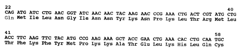
Fig. 1 (A) shows the amino acid (SEQ ID NO:1) and
DNA sequence (SEQ ID NO:2) of PEP (aa22-58 of full
length IL-2), Fig. 1(B) shows a schematic drawing
of IL-2 where helices are shown as cylinders
(McKay, D.B., (1992)), Fig. 1(C) shows a
stereogram of a Ca atom backbone trace of one IL-2
molecule (McKay, D.B., (1992)), Fig. 1(D) shows a
ribbon diagram of a member of the right-handed
cylinder family of predicted IL-2 structure (Cohen
et al., (1986)), wherein the PEP sequence is
highlighted and the disulfide bond is shown in
Figs. 1(B) , 1(C) , and 1(D) ;
Fig. 2 shows a schematic diagram of the chemical
production of a permeability enhancing peptide
(PEP) (A) dimer and (B) monomer;
Fig. 3 shows the results of biodistribution
studies with recombinant human IL-2 (rhIL-2) and
CA 02219961 2008-04-30
9
PEP immunoconjugates in tumor-bearing nude mice,
wherein the results are expressed as either (A) %
injected dose/gram or (B) tumor/organ ratios,
(n=4) ; and
Fig. 4 shows the results of biodistribution
studies with rhIL-2, PEP, PEP monomer, and PEP
dimer immunoconjugates in tumor bearing nude mice,
wherein the results are expressed as (A) %
injected dose/gram and (B) tumor/organ ratios.
DETAILED DESCRIPTION
This invention provides an active IL-2 peptide,
preferably synthetic, and its dimer which have
vasopermeability activity but which are devoid of
cytokine activity. The invention also provides potent
vasoactive immunoconjugates of these peptides with
tumor-specific antibodies. Such conjugates facilitate
the delivery of therapeutic and diagnostic agents in
tumors and other tissues.
Permeability Enhancinct Peptides
The invention provides vasoactive IL-2 peptides,
preferably free of cytokine activity. These novel
peptides include portions of the amino acid sequence of
IL-2, sequences which can also be deduced from the
nucleotide sequence, described by Taniguchi et al.
(Nature 302: 305-310 (1983)). The peptides are preferably
synthetic. The monomeric peptides can also be isolated from
naturally occurring IL-2 by known techniques.
A series of distinct permeability enhancing
peptides (PEP) have been synthesized which, when linked
to an appropriate delivery vehicle, are responsible for
increased vascular permeability in vivo. Moreover, the
unprotected synthetic peptides by themselves are short-
CA 02219961 1998-01-09
lived after intravenous administration and have
negligible effects on vascular permeability relative to
unaltered IL-2. Consequently, the vasoactive peptide
must be joined to an appropriate delivery vehicle to
5 maximize the vasopermeablity effects of the peptides.
Preferably, the peptides, alone or joined to a delivery
vehicle, exhibit negligible cytokine activity in IL-2
bioassays, such as T-cell proliferation and
cytotoxicity assays. Taken together, these
10 characteristics of the PEP provide for a powerful
vasoactive agent when linked to an appropriate delivery
vehicle, but minimize any potential toxic effects on
normal tissues.
The length of the PEP is preferably at least about
22 amino acids in length and most preferably about 37
amino acids in length. Preferred embodiments of the
peptide include amino acids residue numbers 37 to 58,
33 to 58, or 37 to 72 of amino acid sequence SEQ ID NO:
3. These preferred embodiments exhibit about 50% of
the vasopermeability effects of an IL-2 immunoconjugate
when joined to an appropriate delivery vehicle. The
most preferred embodiment of PEP comprises residue
numbers 22 to 58 of SEQ ID NO:3, i.e., the entire amino
acid sequence of SEQ ID NO: 1. This PEP embodiment
exhibits an optimum of about 100% of the
vasopermeability of an IL-2 immunoconjugate, when
joined to an appropriate delivery vehicle.
The complete amino acid sequence of the IL-2
peptide fragment that is the most preferred PEP (SEQ ID
NO: 1), as well as the corresponding DNA sequence (SEQ
ID NO: 2), is shown in Figure 1A. The location of this
fragment in the intact IL-2 molecule is shown
schematically in three diagrams, which have been used
by investigators to represent the IL-2 molecule (see
Figures 1B, 1C, and 1D).
CA 02219961 1998-01-09
11
The permeability effects of the peptides of the
present invention are further optimized when the PEP
comprises a dimer, preferably linked by a disulfide
bond. Consequently, a preferred embodiment of the PEP
includes a cysteine residue and is capable of forming a
disulfide bridge with another PEP molecule. A most
preferred embodiment comprises a PEP dimer, having a
disulfide bridge connecting two cysteine residues.
The PEP molecules acquire their ability to produce
a localized increase in vascular permeability when they
are joined to delivery vehicles, which can direct the
vasoactive peptides to appropriate tumor targets. The
joining of PEP with appropriate delivery vehicles, such
as tumor-specific monoclonal antibodies (MAb), can be
readily accomplished by chemical conjugation means, as
described below. Alternatively, the PEP can be joined
to the tumor-specific MAb using genetic engineering
methods to give a PEP/MAb fusion protein, also
described below. In addition to the PEP, the
conjugates or fusion proteins may include appropriate
linker molecules, e.g. peptides or bifunctional
reagents, which may overcome perturbations of the PEP
or MAb's tertiary structure.
The permeability enhancing properties of the
conjugates can be determined by in vivo experiments,
such as those described in Example 7. Exemplary in
vitro assays for cytokine activity are found in Example
8.
Selection of Delivery Vehicles
An important aspect of the invention comprises the
potency of a vasoactive peptide when linked to a tumor-
specific delivery vehicle. MAbs are ideal delivery
vehicles because they are homogeneous, recognize
specific determinants, and are relatively
CA 02219961 2008-04-30
12
biocompatible. Preferred delivery vehicles include
MAbs of mouse, rabbit, or other mammalian species of
origin. Most preferably, the immunogenicity of non-
hunian MAbs is avoided by the selection of human or
human-mouse chimeric MAbs as delivery vehicles.
Suitable monoclonal antibodies (MAbs) for use in
the invention comprise not only those having a
specificity for antigens unique to tumor cells, but
also those having a shared specificity for antigens of
noi-mal tissues. The essential property of these
monoclonal antibodies is their effectiveness as
carriers, which preferentially concentrate vasoactive
agents at the site of the tumor. Suitable monoclonal
antibodies are those having a specificity to antigens
that are either more abundant or more easily bound in
turnor tissue than in normal tissue.
Some MAbs against tumor or normal cellular
antigens, suitable for use in.the immunoconjugates are
available commercially (e.g., Centocor, Malvern, PA).
Others may be prepared by the well-established
hybridoma procedure of Kohler and Milstein (Nature 256:
495 (1975)), and commercial kits facilitate this
process, e.g.HyBRLi Prep Kit (Bethesda Research Labs,
Bethesda Research Labs, Bethesda, MD).
The selection of hybridoma cell lines producing
suitable MAbs is accomplished by first growing
hybridoma cells for several days, for example, in the
wells of microtiter plates. Cell supernatants are then
tested for the presence of MAb to tumor or cellular
antigens by any convenient immunoassay, for example, an
ELISA. Cells testing positive are then expanded into
larger scale cultures to produce larger quantities of
MAbs. An adequate amount of MAb can then be purified
CA 02219961 2008-04-30
13
from the supernatants, for example, using Protein A
affinity chromotography.
In a preferred embodiment of the invention,
commercially available MAbs specific for lymphoma
cells, e.g., Lym-1 and Lym-2, are used (Techniclone,
Corp., Tustin, CA).
In another preferred embodiment, MAbs specific for
intracellular antigens accessible in degenerating
cells, e.g. TNT-1 and TNT-2 are used (Techniclone,
Corp., Tustin, CA).
In yet another preferred embodiment, MAbs specific
for tumor vessels, e.g. TV-1 (Epstein, A.L, Cancer Res.
55: 2673-2680 (1995).
The MAb of the immunoconjugate may be either
intact whole antibody, the monovalent HL isoform, the
F(ab')2 portion of antibody, or Fab antibody fragments.
Removal of all or part of the Fc portion of the
antibody molecule can facilitate it use by removing
sites or domains which interact with non-tumor
components such as Fc receptors or complement while
leaving the antigen binding sites intact. Antibody
fragments like Fab, HL, and F(ab')2, which have 1/3,
1/2, and 2/3 the weight of whole antibody,
respectively, are better able to diffuse through the
interstitial tissue and into the tumor. However, the
Fab, HL, and F(ab')2 fragments are cleared from the
circulation more rapidly. Fab fragments may be
prepared by digestion of whole antibody with papain, or
digestion of whole antibody with pepsin to give F(ab')2
fragments, followed by digestion of interchain
disulfide bonds to yield univalent fragments.
CA 02219961 1998-01-09
14
In addition, suitable delivery vehicles should
retain their ability to bind with antigen following
chemical conjugation with vasoactive peptides. The
immunoreactivity of MAbs, before and after conjugation
with peptides, can be determined by any suitable
immunoassay, such as the radioimmunoassay described in
Example 6. Preferrably, immunoconjugates having
greater than 75% immunoreactivity, as compared to the
unconjugated antibody, are used in vivo.
Chemical Coniugation Methods
The structural link between the MAb and the
vasoactive peptide, as well as the chemical method by
which they are joined, should be chosen so that the
binding ability of the MAb and the biological activity
of the peptide, when joined in the conjugate, are
minimally compromised. As will be appreciated by those
skilled in the art, there are a number of suitable
chemical conjugation methods, including the following
procedures.
1. Conlugation by the CDI Method
Carbodiimides (CDIs), which are anhydrides of
urea, can produce cross-links between the antibody and
the peptide, regardless of either molecule's
orientation. Conjugants are derived by condensation of
the antibody and peptide under acidic conditions with
CDI. This method provides a rapid and simple means of
conjugation.
2. Conlugation by the SPDP Method
N-succinimidyl 3-(2-pyridyldithio) propionate
(SPDP) is a heterobifunctional reagent which introduces
thiol groups to the terminal amino of proteins, and has
been used in a number of immunoconjugates.
CA 02219961 1998-01-09
3. Conjugation by the SMCC Method
Peptides can also be coupled to antibodies using
the bifunctional reagent, succinimidyl-4-(N-maleimido
methyl) cyclohexane 1-carboxylate (SMCC).
5
4. Conjugation by the NHS Method
N-hydroxisuccinimide (NHS) activates a terminal
COOH group, for example, of a peptide, to form an
active ester derivative that can be covalently coupled
10 to the protein of the monoclonal antibody.
5. Glutaraldehyde
An alternative method for the conjugation of
peptides to proteins uses glutaraldehyde as a reagent
15 for coupling. Nucleophilic groups such as sulfhydryl
and amino groups covalently add to the aldehyde forming
a Schiff base. Excess active glutaraldehyde groups can
be subsequently blocked by addition of glycine, and the
excess peptide and glycine molecules removed by
dialysis.
Genetically Engineered Fusion Proteins
Genetically engineered fusion proteins,
constructed by cloning the gene sequences of antibody
light chains and heavy chains fused to sequences
encoding vasoactive peptides, present an attractive
alternative to the chemical linkage of vasoactive
peptides to MAbs. These constructs can be tailored to
be less immunogenic than MAbs from non-human sources.
Moreover, fusion proteins allow defined molar amounts
of PEP monomer or, alternatively, at least two tandemly
linked PEP sequences, to be attached at specific sites
of the MAb.
As an example, mRNA from hybridoma cells
expressing a monoclonal antibody is isolated. From
this mRNA, cDNA is reverse transcribed and amplified by
CA 02219961 1998-01-09
16
polymerase chain reaction. Specific regions encoding
heavy and light chains of an immunoglobulin, e.g.
variable and/or constant regions, can be amplified by
the selection of appropriate oligonucleotide primers
targeting the desired region(s). The cDNA is
sequenced, mapped by restriction endonucleases, and
cloned into an appropriate transfer vector. At a
minimum, the immunoglobulin sequences encoding an
antigen binding domain, i.e. the variable light chain
and variable heavy chain regions, are contained in the
transfer vector. In addition, a truncated or full
length portion of the constant region encoding the
original or another immunoglobin can be joined in frame
with the variable region, to allow expression of the
joined regions. For example, a preferred embodiment of
the invention encodes a chimeric MAb, comprised of
murine variable regions linked to their corresponding
human constant regions of the heavy and light chains.
An appropriate DNA sequence, encoding at least one
vasoactive peptide, is then ligated proximate to a
region of an immunoglobulin gene encoding the carboxy-
terminus, preferably a constant region, most preferably
the constant region of a heavy chain. The best site
for attachment for each vasoactive peptide may be
different and may be easily determined via experimental
methods. For example, none or various lengths of amino
acid encoding linkers may be inserted between the PEP
and the carboxy-terminus of the immunoglobulin gene. In
addition, two or more tandemly linked PEP sequences can
be joined to the appropriate region(s) of an
immunoglobulin gene. The resulting expression products
can then be tested for biologic activity.
The completed engineered gene for the fusion
protein is inserted into an expression vector, which
can be introduced into eukaryotic or prokaryotic cells
CA 02219961 1998-01-09
17
by gene transfection methods, e.g. electroporation or
the calcium phosphate method. The fusion protein
product can then be expressed in large scale cell
culture and purified.
Use of Vasoactive Peptides
A successful vasoactive immunoconjugate or fusion
protein will maximize the clinical effectiveness of
monoclonal antibody-based diagnosis and therapy.
Clinically, the vasoactive immunoconjugate or fusion
protein is given before or with an intravenously
injected immunodiagnostic, chemotherapeutic, or
immunotherapeutic agent. Induction of a localized
permeability change within the tumor vasculature will
make the tumor more susceptible to penetration and
improve the delivery of drugs, toxins, radioisotopes,
monoclonal antibodies, or conjugates of monoclonal
antibodies with drugs, toxins, or radioisotopes to the
tumor site.
The suitability of tumor specific antibodies,
immunoconjugates, and genetically engineered fusion
proteins for use in vivo is determined by their
biodistribution, cellular localization, selective
binding, and rate of clearance from the tumor host, or
an animal model of the tumor host. Studies to asses
this suitability are conveniently carried out by means
of labeled MAbs. For example, radioiodination of
antibody moieties can be accomplished by the modified
chloramine T method of Example 6. A tumor host is
treated with immunoconjugate, a fusion protein, or left
untreated. After injecting a tumor host with the
labeled MAb, the effectiveness of a vasoactive
conjugate or fusion protein can be evaluated by
appropriate radioimaging, biodistribution, histological
studies, and autoradiographic methods.
CA 02219961 1998-01-09
18
The time required to produce the maximum
vasoactive effect depends on the specific conjugate or
fusion protein chosen. However, an optimal interval
between the time of administering the vasoactive agent
and the therapeutic or diagnostic agent can be
determined experimentally. For example, the ability of
a radiolabelled MAb to concentrate selectively at a
tumor site can be determined by radioimaging.
Posterior gamma scintillation images (100,000 cpm) are
obtained from an anesthetized host on alternate days
after injection of radiolabeled MAb, using a gamma
scintillation camera with a pinhole collimator. The
camera is preferably interfaced with a computer system.
An appropriate 131I standard with the same activity is
counted to quantitate the data.
Further biodistribution studies can be performed
using animal models, wherein the host animal is
sacrificed at an optimal time, as determined by the
imaging studies described above. Blood, major organs
and tumor tissues are then excised, weighed, and
counted'to determine the biodistribution of the MAb.
In addition, tumor tissue can be fixed and embedded,
and tissue sections examined by autoradiography to
determine the location of the bound radiolabeled MAb in
the tumor.
It is anticipated that the minimum time between
the administration of the vasoactive conjugate or
fusion protein and the administration of a diagnostic
or therapeutic agent is at least about 20 minutes, and
the maximum time is about 72 hours.
The dose of vasoactive immunoconjugate or fusion
protein to be given is based on criteria of medical
judgment and experience, both objective and subjective.
However, an adequate measure of an effective dose is
CA 02219961 1998-01-09
19
that amount which improves the clinical efficacy of
therapy, or accuracy of diagnosis, to a statistically
significant degree. Comparisons can be made between
treated and untreated tumor hosts to whom equivalent
doses of the diagnostic or therapeutic agents are
administered. Where a diagnostic or therapeutic agent
is toxic to normal tissue, an effective dose of
vasoactive conjugate or fusion protein is one which
minimizes such toxic effects.
A preferred therapeutic agent is a clinically
useful Mab. In addition, an antineoplastic therapeutic
agent can be a tumoricidal agent, such as a
radioisotope, a chemotherapeutic drug, or a toxin.
Moreover, the MAb can be attached to a tumoricidal
agent, e.g., radioisotope, chemotherapeutic drug, or
toxin.
A diagnostic agent can be used for tumor imaging
and is comprised of a MAb having a specificity for a
tumor, which has a label detectable in vivo.
Preferably, this label comprises a radioactive isotope.
In addition to the detectable label, the tumor imaging
agent can also be attached to a cytotoxic agent, such
as a radioisotope, drug, or toxin.
In another version of the invention, the
vasoactive immunoconjugate or fusion protein is linked
to a tumoricidal agent. Consequently, the therapeutic
method is a simplified procedure comprised of
administering to a.tumor bearing host an effective
amount of a vasoactive conjugate or fusion protein,
which is linked to a chemotherapeutic agent, toxin, or
radioisotope.
Similarly, the vasoactive immunoconjugate or
fusion protein can be linked directly to a detectable
CA 02219961 2008-04-30
label, such as a radioisotope. Consequently, the
diagnostic method can comprise simply administering to
a tumor bearing host the labeled vasoactive
immunoconjugate in an amount sufficient to give a clear
5 tumor image.
The previous versions of the present invention
have many advantages including the ability to increase
vascular permeability at the site of neoplastic or
10 other diseased tissue. Moreover, the previous versions
of the invention provide potent vasoactive agents that
enhance the uptake of therapeutic and diagnostic agents
at a tumor site with a minimum of toxic side effects on
no:rmal tissues.
MkMPLLS
Reagents
All chemicals, such as N-hydroxysuccinimide
(sulfo-NHS), 1-cvclohexy-3-
(morpholinoethyl)carbodiimide metho-p-toluenesulfonate
(CDI), and chloramine T were purchased from Sigma
Chemical Co. (St. Louis, MO). Iodo-beads were purchased
from Pierce (Rockford, IL). Al1 solvents were of
analytical grade and were used as purchased. Iodine-
125 was obtained as sodium iodide in 0.05 N sodium
hydroxide solution (ICN Biomedicals, Irvine, CA).
Radioactive samples were measured using either a 12B2
CompugammaTM counter (LI:B .instruments, Pleasant Hill, CA)
or a CRC-& dose calibrator (Capintec Inc., Pittsburgh,
PA) .
Murine monoclonal antibodies Lym-1 (IgGZa) and TNT-
1(IgG2a) were obtained from Techniclone, Corn. (Tustin,
CA.). Lym-1 is directed against a variant of the HLA-Dr
antigen expressed on the cell surface of human B-
lymphocYtes and malignant lymphomas (Epstein, A.L. , et
a1.., Cancer Res. 47: 830-840 (1987)), whereas TIvT-1
CA 02219961 2008-04-30
21
recognizes an epitope of nucleohistones expressed in
the nucleus of mammalian cells (Epstein, A.L., et al.,
Cancer Res. 48: 5842-5848 (1988)). Protein
concentrations of the antibody preparations were
estimated by optical spectroscopy at 280 nm.
Recombinant human IL-2 (rhIL-2) was obtained from
Hoffman La-Roche (Nutley, NJ) or Chiron (Emeryville,
CA). Human serum albumin (HSA) was obtained from Sigma
Chemical Company.
For in vivo experiments, the Raji Burkitt's
lymphoma cell line and the ME-180 human cervical
carcinoma cell line were used as previously described
(Chen, F.-M., et al., J. Nucl. Med. 31: 1059-1066
(1990)). Both cell lines were grown in RPMI-1640
medium containing 10% fetal calf serum (Hyclone
Laboratories, Logan, UT), penicillin G (100 U/ml), and
streptomycin sulfate (100 gg/ml). For in vitro
cyt.otoxicity studies, the K562 human erythroleukemia
cell line, the Daudi Burkitt's lymphoma cell line, and
the mouse P815 mastocytoma cell line were used. All of
the cell lines were cultured in a 37 C well-humidified
5% COz incubator and were routinely passaged twice
weekly.
E%AMPLE 1
Synthesis of Human IL-2 Peptide Fragments
Peptides were synthesized by the Merrifield method
(Merrifield, B., Science 232: 341-347 (1986)) using a
one-column peptide synthesizer (Model 430A, Applied
Biosystems, Foster City, CA). The protected peptides
were assembled by solid-phase synthesis and cleaved by
trifluoroacetic acid (Fields, C.G., et al., Peptide
Res. 4: 95-101 (1991); King, D.S., et al., Int. J.
Peptide Prot. Res. 36: 255-266 (1990)). The peptides
were then purified by gel filtration on Sephadex' G-10
in 30% acetic acid and lyophilized. A list of the
CA 02219961 2008-04-30
22
different peptide fragments of IL-2 generated by these
procedures is provided in Table 1 (see below).
EXAMPLE 2
Conjugation of Recombinant IL-2 to Tumor-Specific
Monoclonal Antibody
Recombinant IL-2 was radio-iodinated and used in
trace amounts during subsequent coupling reactions to
ascertain the binding of IL-2 to antibody or HSA.
Lyophilized IL-2 was dissolved in sufficient water to
give a final concentration of 2 mg/ml. Fifty l of IL-
2 solution (100 g) , 100 .Ci of carrier free iodine-125
and 5 l of chloramine T (10 mg/ml) in water were added
to 100 l in 0.1 M phosphate buffer, pH 7.4, and the
reaction was allowed to proceed for 1 min at room
temperature. The reaction was quenched with 100 l of
anion exchange resin (AG1=X8; Bio-Rad Laboratories,
Richmond, CA) in PBS. After 1 min the suspension was
wit:hdrawn and filtered in a Spin-XTM centrifuge unit
(Costar, Cambridge, MA) to remove the resin.
The coupling reaction was initiated by the
addition of 500 l of IL-2 (2 mg,/ml) to 500 l of
antibody (10 mg/ml), CDI (14 mg), and sulfo-NHS (8mg)
to give a total volume of 1.2 ml in 0.1 M phosphate
buffer, pH 7_4. The reaction was incubated overnight
at 4 C. After centrifugation, the soluble coupled
antibody was chromatographed on a SephadexTM G-100 column
calibrated with blue dextran. The radioactivity and
antibody peaks co-eluted indicating the IL-2 had
attached to the antibody. From the antibody
concentration and radioactivity, approximately one
molecule of IL-2 was calculated to be bound to each
antibody molecule. These immunoconjugates retained a
minimum of 75% of the antibody binding reactivity as
determined by a live cell binding assay (Epstein et
CA 02219961 1998-01-09
23
al.,(1987); Gaffar, S.A., et al., J. Immunoassay 12: 1-
4 (1991)).
EXAMPLE 3
Conjugation of IL-2 peptide fragments to antibody and
human serum albumin
Portions of the IL-2 peptide fragments, prepared
according to Example 1, were also radio-iodinated prior
to conjugation with antibody or HSA using a slightly
different procedure. Lyophilized peptide fragments
were dissolved in 10% aqueous ethanol to a final
concentration of 1 mg/ml. One hundred l of this
solution was added to a solution of 100 Ci of Na1251 in
0.1 N NaOH neutralized with an equivalent volume in 0.1
M acetic acid. The mixture was stirred vigorously and
two iodo-beads were added. The reaction was allowed to
proceed for 1 hr. After incubation the mixture was
withdrawn into a syringe, and the iodo-beads were
washed twice with 100% aqueous ethanol. Combined wash
liquids were purified on a short Sephadex G-10 column
(eluted with PBS, pH 7.4).
The purity of the radiolabeled fragments was
determined by analytical instant thin layer
chromatography (ITLC). ITLC strips (2 x 20 cm) having
silica gel impregnated fibers (No. 61886, Gelman
Sciences, Ann Arbor, MI), were activated by heating at
110 C for 15 min prior to use, spotted with 1 l of
sample, air dried, cut in half, and counted to
determine fragment bound and unbound radioactivity. In
this system, free iodine migrates with the solvent,
while labeled peptide fragments remain near the origin.
In all cases, greater than 90% of the radioactivity was
associated with the IL-2 peptide fragments. The
different radiolabeled IL-2 fragments were used in
trace amounts in the reaction mixture to ascertain the
CA 02219961 1998-01-09
24
binding of peptide fragments to the antibody, as noted
below.
Coupling reactions were initiated by adding
different peptide fragments to the antibody or HSA,
CDI, and sulfo-NHS in a 1:2:50:50 ratio by weight to
give a total volume of 0.6 ml in 0.1 M phosphate
buffer, pH 7.4. The reactions were incubated overnight
at 4 C. After centrifugation, the soluble coupled
antibody was chromatographed on a G-100 column
calibrated with blue dextran. From the antibody
concentration and radioactivity, approximately one-half
molecule,of IL-2 peptide fragment was calculated to be
bound to each antibody or HSA molecule.
An alternative method used for the conjugation of
peptides to proteins used glutaraldehyde as a reagent
for coupling. Nucleophilic groups such as sulfhydryl
and amino groups covalently add to the aldehyde forming
a Schiff base. Two mg of protein (10 mg/ml in PBS, pH
8.0) were mixed with 2-3 mg peptide (5 mg/ml in HZO) at
room temperature. The pH was maintained at 8.0 with
the addition of dilute NaOH. One hundred l of a 0.02%
solution of fresh glutaraldehyde was added to the
reaction mixture with mixing over 9-10 min, and the
mixture stored overnight at 4 C. The remaining active
glutaraldehyde groups were blocked by addition of 0.2 M
glycine (0.2 ml) for 2 hr. The excess peptide and
glycine molecules were removed by dialysis.
Conjugated peptide fragments were analyzed by fast
protein liquid chromatography (FPLC) performed at room
temperature using a Pharmacia system (Pharmacia,
Piscataway, NJ) equipped with two P-500 solvent pumps,
a MV-8 motor valve injector, a single path UV monitor,
a LLC-500 automated controller, and an REC-482 dual pen
chart recorder. The conjugates were eluted from a
CA 02219961 1998-01-09
superose-12 HR 10/30 pre-packed column (Pharmacia),
using 0.1 M PBS, pH 7.2 as the solvent system, at a
flow rate of 1.0 ml/min. The UV absorbance of the FPLC
eluate was detected at 280 nm. The conjugated
5 antibodies appeared at 650 seconds and the unbound
fragments at 1170 seconds. Immunoconjugates retained a
minimum of 75% of the antibody binding reactivity as
determined by an indirect cell binding assay (Epstein
et al.,(1987); Gaffar et al.(1991)).
EXAMPLE 4
Conjugation of PEP Dimer to antibody
The PEP dimer was prepared by linking the
monovalent peptide through the intrinsic cysteine
(amino acid #58), to form a disulfide bond as shown
diagrammatically in Fig. 2A. The thiol form of PEP was
regenerated by treatment with 10 mM 2-
mercaptoethylamine for 30 min, followed by gel
filtration on a Sephadex G-10 column equilibrated with
0.1 M sodium phosphate, pH 6.8. The peptide was then
incubated for 16 hr at room temperature at pH 9 by the
addition of 5 M NaOH (Figure 2). The desired peptide
dimer was purified from the reaction mixture by gel
filtration on a Sephadex G-25 column equilibrated with
phosphate buffer, pH 7.4. Yields of 90% PEP dimer were
found under those conditions without the formation of
high molecular weight species. The PEP dimer was
coupled to antibody using the conditions described
above and was found to have approximately the same
conjugation yield as the other peptides.
EXAMPLE 5
Conjugation of PEP-Phenylmaleimide Monomer to antibody
1. Synthesis of N-phenylmaleimide
The approach to synthesizing N-phenylmaleimide is
shown schematically in Figure 2B. Maleic anhydride
(1.33 g, 13.6 mmol) was dissolved in toluene (15 ml)
CA 02219961 2008-04-30
26
and aniline (1.3 g, 13.9 mmol) in toluene (20 ml) was
added dropwise over a 20 min period. The reaction
mixture was stirred for 45 min at room temperature and
then cooled in an ice-water bath. The precipitated
product, N-phenylmaleamic acid, was collected by
filtration, washed with hexane and dried overnight (2.1
g y:ield).
Proton (1H)nuclear magnetic resonance (NMR)
ana:lysis of the product was recorded on a Hitachi
Per.kin-Elmer R-24 60 MHz instrument. NMR sample
concentrations were about 10% (w/v) in the indicated
solvent. Chemical shifts (ppm) are reported down field
(6) relative to the internal tetramethylsilane (TMS)
standard. The following results verified that the
product was N-phenylmaleamic acid: 'H NMR (MeZSO-d6, b) ;
10.3 (1H, singlet, OH), 7-7.8 (SH, multiplets, 5 aryl
CH), 6.4 (2H, doublet of doublets, COCH=CHCO).
N-phenylmaleamic acid (2.0 g, 10 mmol) was added
and the solution stirred at 120 C. The brown
precipitate was filtered and evaporated to dryness
under reduced pressure and the residue was dissolved in
diethyl ether. The ether mixture was filtered and the
filtrate was again evaporated to dryness. The residue
obtained was applied to a flash chromatography column
(30 x 200 mm) of KieselgelTM 60, 230-400 mesh (No. 9385,
E. Merck, Darmstadt, Germany). Elution with 500 ml of
ethyl acetate/hexane (1:3) yielded fifty fractions.
Fractions 25-40 were combined to provide pure N-
phenylmaleimide (1.5 g yield): TLC (EtOAc/hexane, 1:3)
Rf 0.45. ~ NMR (CDCL3, 6): 7-7.8 (5H, multiplets, 5
aryl CH); 6.8 (2H, singlet, COCH=CHCO).
Product isolation and identification was conducted
by high performance liquid chromatography (HPLC) using
a Beckman System Gold Instrument (Beckman Instruments
CA 02219961 2008-04-30
27
Inc., Fullerton, CA) equipped with two 110B solvent
pumps, a 210A injector valve, a 166 programmable
absorbance detector, and a 406 analog interface module.
A ZorbaxT GF-250 reversed-phase column (DuPont,
Wilmington, DE) was eluted at a flow rate of 1 ml/min
with 100% acetonitrile. Peak detection was determined
by UV absorbance at 254 nm. The starting material, N-
phe:nylmaleamic acid, appeared at 220 seconds followed
by the desired product at 340 seconds.
2. Reaction of PEP with N-phenylmaleimide and
formation of the immunoconluctate
The conjugation of N-phenylmaleimide to the PEP
was accomplished by the addition of a 2.5-fold molar
excess of N-phenylmaleimide (in 15 l methanol) to PEP
dissolved in 0.1 M citrate buffer, pH 6Ø The
reaction was allowed to proceed for 30 min at 37 C. The
reaction mixture containing the PEP-phenylmaleimide
conjugate was exposed to 15 mM mercaptoethylamine to
reduce any disulfide bonds that might have formed
during the reaction and left to react overnight. The
final reaction conjugate was purified by gel filtration
on a SephadexTOG-10 column which was eluted with 0.01 M
PBS, pH 7.2. As with the dimer, coupling of the PEP-
phenylmaleimide monomer to the antibody was performed
as described above and produced approximately the same
conjugation yield.
EXAMPLE 6
Preparation and Analysis of Monoclonal Antibodies
1. Radioiodinati.on of Antibodies
F(ab')z fragments of Lym-1 and TNT-1 monoclonal
antibodies were radiolabeled with iodine-125 using a
modified chloramine T method. Briefly, the iodination
reaction was initiated by adding chloramine T at a
weight ratio of 10:1 (antibody:chloramine T). The
reaction was quenched by the addition of sodium
CA 02219961 2008-04-30
28
metabisulfite, and the mixture was chromatographed on a
Sephadex.. G-25 gel column that was previously
equilibrated with PBS containing 1% bovine serum
albumin (Sigma). Fractions of 125I-labeled monoclonal
antibodies were collected and diluted with the same
buffer to an appropriate volume for injection.
Radiolabeled antibodies were analyzed using an
analytical ITLC system as described in Example 3. All
preparations revealed the same radiochemical purity
2. Immunoreactivity of Radiolabeled Monoclonal
Antibodies
The immunoreactivity of radiolabeled Lym-1
preparations was monitored by a live cell
radioimmunoassay. Raji cells were washed twice in cold
PBS containing 1 mg/ml bovine serum albumin and 0.02%
sodium azide. Cells (5 x 10⁵) resuspended in 100 l of
wash buffer were pipetted into microtiter wells
(Inlmulon RemovawellTMStrips; Dynatech Labs, Inc.,
Alexandria, VA). The microtiter plates were
pre-treated the previous night with BSA (10 mg/ml) in
PBS with azide in order to prevent the antibody
solutions from binding to the wells. Radiolabeled Lym-
1 or Lym-1 immunoconjugates were then added (100,000
cpm/well) in a volume of 100 l/well and the plates
were incubated for 30 min at room temiDerature with
co:nstant shaking. The plates were then washed 4 times
by spinning at 1,000 rpm for 5 min, and aspirating the
supernatants with a 12-tip micromatic manifold, and
then resuspending the cells in 200 l of wash buffer
using a TitertekTMMultichannel pipet (Flow Labs, McLean,
VA). The wells were then separated mechanically and
counted in a gamma counter to quantitate the amount of
label binding to the cells.
CA 02219961 1998-01-09
29
Approximately 80% of radiolabeled Lym-1 F(ab')2
preparations were found to bind Raji cells by live cell
radioimmunoassay. The radiolabeled TNT-1 F(ab')2 had
an immunoreactivity of ~80% in a paraformaldehyde-
acetone-treated cell assay developed in our laboratory
(Gaffar et al., (1991)).
EXAMPLE 7
In Vivo Vasopermeability Studies
1. Tumor Models and Biodistribution Studies
TNT-1 immunoconjugates were tested in the ME-180
human cervical carcinoma system to demonstrate
targeting of TNT-1 immunoconjugates to intracellular
antigens accessible in permeable (dead) tumor cells.
The ME-180 human cervical carcinoma cell line was
heterotransplanted in the left thigh of 6-week old
female athymic nude mice (Harlan Sprague Dawley, San
Diego, CA) by the subcutaneous injection of a 0.2 ml
inoculum consisting of 107 cells. The tumors were grown
for 3-4 weeks until they grew to approximately 1 cm in
diameter.
Lym-1 immunoconjugates were tested in the Raji
lymphoma model to demonstrate targeting cell-surface
antigens. The Raji lymphoma cell line was used to
produce heterotransplants in 6-week-old female nude
mice by the subcutaneous injection of a 0.2 ml inoculum
consisting of 4 x 10⁷ Raji cells and 4 x 10⁶ human fetal
fibroblast feeder cells in the left thigh. Three days
prior to injection, the mice were irradiated with 400
rads using a cesium irradiator to ensure a high take
rate of the implanted cells. The tumors were grown for
14-18 days until they grew to approximately 1 cm in
diameter.
To test the relative effects of the
immunoconjugates on the biodistribution and tumor
CA 02219961 1998-01-09
uptake of Lym-1 or TNT-i in tumor-bearing mice,
separate groups of 4-5 mice were given intravenous
injections of 30 g of antibody alone or antibody
conjugate. At 2.5 hr after injection, each group
5 received 50 Ci of 125I-labeled Lym-1 or TNT-1 F(ab')2
fragment as tracer.
All animals were sacrificed 72 hr later, by sodium
pentobarbital overdose, for biodistribution analysis.
10 Various organs, blood, and tumor were removed, weighed,
and samples were counted in a gamma counter. For each
mouse, data were expressed as tumor:organ ratio (cpm
per gram tumor/cpm per gram organ) and percent injected
dose/gram (aID/g). From these data, the mean and the
15 standard deviation were calculated for each group.
2. Identification of vasoactive IL-2 peptide
fragments
Based on the primary, secondary and tertiary
20 structures of IL-2, a series of distinct peptides were
synthesized in order to identify the sequences
responsible for increased vascular permeability. The
peptides and their sequences are listed in Table 1.
Each peptide and rhIL-2, as well as their respective
25 immunoconjugates with MAb Lym-1, were assayed for their
ability to induce tumor vascular permeability and
enhanced antibody uptake in Raji tumor-bearing nude
mice.
30 TABLE 1. Vasopermeability Activity of Interleukin-2
Synthetic Peptide Fragments and
Immunoconjugates
Fragment/ Amino Acid Sequence Vasopermeability
3 5 Immunoconjugatel (YLym-1/IL-2)
3A 44-58 n.t.2
Lym-1/3A 0
CA 02219961 1998-01-09
31
B1 37-58 n.t
Lym-1/B1 50
3B 33-58 n.t.
Lym-1/3B 50
3C 22-58 0
Lym-1/3C 100
E6 22-38 n.t
Lym-1/E6 0
A3 37-72 n.t.
Lym-1/A3 50
4A 105-133 n.t.
Lym-1/4A 0
4B 87-133 n.t
Lym-1/4B 0
IL-2 1-133 75
Lym-1/IL-2 100
1 30-40 pM of peptide were added per assay
2 not tested
Control studies used intact IL-2 and the Lym-1/IL-
2 immunoconjugate to establish markedly enhanced levels
of Lym-1 uptake in Raji tumor bearing nude mice for
comparison. As noted previously (LeBerthon et al.
(1991)), enhanced permeability can be obtained despite
the fact that chemically conjugated MAb/IL-2 does not
demonstrate cytokine activity. As shown in Table 1, a
vasoconjugate derived from one synthetic peptide,
designated 3C, produced approximately 100% of the
vasopermeability effects of Lym-1/IL-2 chemical
conjugate. Three other vasoconjugates, composed of
synthetic peptides 3B, Bl, and A3, which contained
smaller fragments of 3C, produced approximately half
the vasopermeability effects of Lym-1/IL-2 in these
assays.
As expected, intravenous administration of the
CA 02219961 1998-01-09
32
unprotected and short-lived unconjugated synthetic
fragments by themselves had no effect of Lym-1 uptake
in tumor-bearing nude mice. Hence, conjugation of
peptides to another macromolecule, such as an antibody,
is required to demonstrate the biologic activity of the
synthetic peptides. By comparison, native IL-2 had 75%
vasopermeability in the in vivo model.
From the data presented in Table 1, it appears
that the entire sequence of amino acids 22-58 of SEQ ID
NO:3 produces optimal vasopermeability. However,
conjugates composed of amino acids 37-58, 33-58, and
37-72 of SEQ ID NO:3 retain 50% of the activity,
whereas fragment E6, consisting of amino acids 22-38 of
SEQ ID NO:3, has no activity.
3. In vivo analysis of PEP immunoconlugates
MAb alone, MAb/IL-2, or MAb/PEP immunoconjugates
were used to pre-treat tumor-bearing nude mice in two
tumor models in order to demonstrate increased tumor
uptake of radiolabeled MAb 2.5 hours after pre-
treatment. TNT-1 immunoconjugates were used in the ME-
180 human cervical carcinoma system to demonstrate
targeting to intracellular antigens accessible in
permeable (dead) tumor cells. In complementary
studies, Lym-1 immunoconjugates were used in the Raji
lymphoma model to demonstrate targeting cell-surface
antigens.
As shown in Figure 3A, TNT-1 pre-treatment gave
1.28% of the injected dose in the tumor and TNT-1/IL-2
and TNT-1/PEP pre-treatments led to 4.5 and 4.4 percent
injected dose/gram, respectively. Equally as
impressive, pre-treatment with Lym-1 alone led to only
1.4% of the injected dose of radiolabeled Lym-1
accumulating in the tumor, while Lym-l/IL-2 and Lym-
1/PEP gave 5.7 and 5.6 percent injected dose/gram,
CA 02219961 1998-01-09
33
respectively (Figure 4A). In both systems, there was
an approximate four-fold increase in radiolabeled
antibody within the tumor.
In addition to these findings, use of IL-2 or PEP
immunoconjugates increased the specific targeting of
the radiolabeled antibodies as shown by the higher
tumor/organ ratios (Figures 4A and 4B).
These results indicate that PEP is equivalent to
rhIL-2 after conjugation to two different monoclonal
antibodies for the enhancement of antibody uptake in
tumor. Unlike IL-2, however, unconjugated PEP, which
has a molecular weight of 3,700 Daltons, showed no
vasopermeability activity after intravenous
administration in the mouse (Table 1), presumably
because of its rapid degradation and clearance from the
circulation.
4. in vivo evaluation of PEP monomer and dimer
immunoconjuaates
The presence of the terminal cysteine (amino acid
#58) suggests that dimerization of the synthetic
peptide might be occurring during the conjugation
procedures. In order to assess whether dimerization
affected the vasopermeability effects of PEP, monomer
and dimer forms of PEP were produced before conjugation
as described in Example 5 and summarized in Figure 2.
Vasoconjugates constructed with these chemically-
generated fragments were therefore composed of only
monomer or dimer forms of PEP for comparative purposes.
When used as a pre-treatment in tumor-bearing nude
mice, biodistribution analysis demonstrated that the
vasoconjugate consisting of the dimer had an
approximately two-fold enhancement of antibody uptake
in tumor compared to the vasoconjugate constructed with
CA 02219961 1998-01-09
34
the PEP monomer (Figure 4). In addition, the
vasoconjugate constructed with the PEP dimer gave
approximately the same enhancement in antibody uptake
as the MAb/IL-2 conjugate, indicating that dimerization
was important in the generation of optimal
vasopermeability at the tumor site in this model
system.
EXAMPLE 8
Cytokine Studies
1. IL-2 Bioassays (Proliferation assay)
The growth of an IL-2 dependent indicator cell
line, CTLL-2, was used to compare the biologic activity
of PEP, PEP conjugates, and positive control human
recombinant IL-2. Samples of PEP, PEP conjugates, or
IL-2 standards (100 l/well) were serially diluted 3-
fold from an initial concentration of 8.1 pM
(recombinant IL-2) in sterile 96-well flat bottom
microtiter plates. CTLL cells (4 x 10⁵) in a volume of
50 l were added to each well. Plates were incubated
for 18 hr in 5% CO 2 at 37 C, then pulsed with 0.5 Ci of
3H-thymidine for 6 hr (25 l of a 1:50 dilution of 1.0
mCi in media; Amersham, Arlington Hts. IL) prior to
harvesting wells onto glass fiber filter paper and
liquid scintillation counting in glass minivials.
While recombinant IL-2 was highly active as a positive
control, none of the PEP-containing preparations were
found to support the proliferation of the T cell line.
2. Cytotoxicity Assays
The ability of PEP and PEP conjugates to induce
LAK cell killing was tested by 51Cr release
microcytoxicity assays in 96-well microtiter plates as
previously described (Katsanis, E., et al., Blood 78:
1286-1291 (1991). Two populations of effector cells
were used, human peripheral blood mononuclear cells
(PBMC) or murine splenocytes. The effector cells were
CA 02219961 2008-04-30
isolated by Fico11TM density gradient centrifugation and
activated for 4 days in vitro in media containing 13.7
pM or 80 pM PEP, or 13.7 pM antibody/PEP or HSA/PEP
conjugates at a density of 0.5 x 10⁶ cells/ml. Freshly
5 isolated effectors in media without human recombinant
IL-2 were used as controls. Human cells were cultured
in RPMI-1640 with 2 mM L-glutamine, 100 U/ml
penicillin, 100 g/mi streptomycin, and 10% fetal calf
serum. Murine cells were grown in the same culture
10 medium as above, but were supplemented with 10 mM non-
essential amino acids, 100 mM sodium pyruvate, and 25
mM 2-mercaptoethanol (Sigma).
Three different tumor target cell lines were
15 tested. PBMC effectors were tested against two
malignant tumor target cell lines, K562 (NK sensitive)
and. Daudi (NK resistant). For assessment of the
killing potential of T cells, activated murine
splenocytes were tested against the P815 mastocytoma
20 cell line in a tumor directed antibody-dependent
cellular cytotoxicity assay (reverse ADCC)(Anderson,
P.M., et al., j. Immunol. 142: 1383-1394 (1989)).
Addition of 10 ng/ml of 145-2C11 anti-murine CD3
antibody (Boehringer Mannheim, Indianapolis, IN) to
25 plates containing the Fc receptor positive P815 cell
liries results in markedly augmented killing by
activated T cells.
Cytotoxicity assays used 500 51Cr-labeled tumor
30 targets per well in V-bottom microtiter plates and
effector:target ratios of 30:1, 10:1, and 3.3:1
achieved by 3-fold serial dilution of the first row
prior to the addition of radiolabelled targets. Plates
were centrifuged 5 min at 500 rpm to ensure cell
35 contact, incubated 4 hr at 37 C, and then centrifuged
again at 1,000 rpm. One hundred microliters of
supernatant was harvested into glass scintillation
CA 02219961 2008-04-30
36
vials prior to liquid scintillation counting.
None of the PEP or PEP conjugate preparations
induced LAK cell killing of target cell lines in any of
the cytoxicity assays described above. By comparison,
recombinant human IL-2, which served as a positive
control, was highly active.
EXAMPLE 9
Recombinantly Engineered Vasoactive Immunoconjugate
Construction of a PEP/MAb fusion protein
expression vector can be carried out using standard
molecular cloning techniques. A transfer vector for a
human-mouse chimeric monoclonal antibody, can be
constructed and used as a parent vector. The transfer
vector will carry cDNA sequences for a chimeric human-
mouse heavy chain under the control of a first promoter
and a chimeric human-mouse light chain under the
cor.Ltrol of a second promoter. An example of such a
transfer vector is the baculovirus vector, pBVchLYM-1,
of Hu et al. (Hum. Antibod. Hybridomas 6(1): 57-67
(1995),
Nucleotide sequences encoding the PEP, i.e. a cDNA
subtantially homologous to SEQ ID NO: 2, will be
inserted into an appropriate restriction enzyme site
near the 3' end of the heavy chain gene. The resulting
expression vector will encode a chimeric light chain as
well as a fusion protein consisting of the chimeric
heavy chain with PEP attached at the carboxy-terminus.
The expression vector will be tranfected into a
su:'_table cell line and the light chain and heavy chain
fusion proteins will be co-expressed in cell cultures.
The heavy and light chains of the chimeric PEP/MAb
fusion protein will self assemble within the
transfected cells and can be subsequently purified from
the cell culture by protein A affinity chromatography.
CA 02219961 1998-01-09
37
EXAMPLE 10
Clinical Use and Application
PEP immunoconjugates or fusion proteins can be
used to enhance the delivery of therapeutic or tumor
imaging agents. The mechanism of action of the PEP-
containing molecules is to increase vascular
permeability at the tumor site. In the animal model,
described in Example 7, administration of PEP
immunoconjugates 2.5 hours before the administration of
radioiodinated MAbs produced markedly enhanced uptake
of the radioactive tracer in tumors. Accordingly, the
PEP immunoconjugate or fusion protein will generally be
administered to the tumor host 1-3 hours before the
subsequent dose of therapeutic or tumor imaging agent.
Although the present invention has been described
in considerable detail with reference to certain
preferred versions thereof, other versions are
possible. For example, the PEP may be joined to a
delivery vehicle which includes a toxin. Therefore,
the spirit and scope of the appended claims should not
be limited to the description of the preferred versions
contained herein.