Note: Descriptions are shown in the official language in which they were submitted.
CA 02220388 1997-11-06
WO 96/38809 PCTlUS96/07916
- 1 -
METHOD .POND APPARATUS FOR ASSESSING SLIDE AND
SPECIMEN PREPARATION QUALITY
This invention relates to a method and apparatus
for assessing slide and specimen preparation quality,
and more particularly to an automated assessor for a
population of biological specimens fixed and stained
on slides.
BACKGROUND OF THE INVENTION
Detection of disease processes for which a
specimen is taken may not be possible if specimen
preparations such as fixation or staining are improper
or inconsisi~ent. As a result, there is a need to
provide biological specimens having at least a minimum
quality level to ensure a higher efficacy of disease
screening. A method for assessment of specimen
preparation ensures that specimens are prepared using
the technic~~ue and material that provides for a
selected level of q~.ality to allow detection of
disease processes of interest.
Furthermore, any such assessment may further
determine whether prepared specimens are suitable for
computer examination. The assessment may be adjusted
to fit requirements for a selected automated system.
Moreover, the assessment may include information about
the specimen. preparation and physical characteristics
requiring improvement.
Specime=n preparations affect specimen
characteristics including morphological detail of
cells. Morphologic detail of the cells in biological
specimens must remain intact and be made visible for
visual or computer examinations of biological
specimens to be effective. Cell fixation immobilizes
cellular structure. Staining makes the fixed cellular
structures visible. Improper fixing of samples allows
cell morphology to change and degenerate. Improper
CA 02220388 2002-O1-10
77501-5
- 2 -
staining may obscure ce7_lular detail or may not make
cellular structure vis:i~~le. The assessment should determine
whether specimen prepa_r~~t;ion is conducted in such a manner
as to provide good fixation and staining of cells for visual
examination or analysis by automated devices. See, for
example, the introduction to "Diagnostic Cytopathology of
the Uterine Cervix", pp. 1-9, by S. Patten, and "Sensitivity
and Specificity of Rou~~ine Diagnostic Cytology", by S.
Patten in "The Automation of Uterine Cancer Cytology", pp.
406-415, edited by Wied, Bahr, and Bartels, published by
Tutorials of Cytology, c='hicago Illinois, 1976. Currently,
specimen preparation i~~ evaluated periodically by human
visual review.
As described in the above references, specimen
collection or sampling is another characteristic which has a
strong impact on diagnostic viability of a specimen. If the
sample was taken in the wrong anatomical location or sampled
with poor technique, true proper spectrum of cell types may
not be present. The a~~sessment should determine whether
specimen collection provides a good s<~mple of cells for
examination.
Phy;~ica7.. characteristics of slides are important
for automated examinat:i.or. of bioloc~ica=L specimens . Physical
characteristics such as t=hi.ckness of a slide, alignment of a
coverslip or marks_ng of a slide rnu;~t be within predetermined
parameters fo:r effective imaging and computer examination.
These qualities include the quality and sufficiency of the
sample obtained. The present invention provides, for the
first time, a practical. cbject.ive method and apparatus for
measuring these qualities.
It :LS therefore a motivation of the invention to
provide a method and apparatus to provide objective measures
CA 02220388 2002-O1-10
77501-5
- 3 -
of specimen staining a:nd fixation, specimen collection
quality, and slide physi.cal_ characteristics.
SUMMARY OF THE INVENTION
The invention provides a method and apparatus for
the assessment of s.lids~ characteristics and specimen
preparation quality. ':Che method and apparatus of the
invention comprises an image analysis system that further
comprises an image gatlue~ring system having a camera, a
motion controller, an :i_llumination system, and an image
transfer interface. T:ne image gatlzeri.ng system is
constructed fir gatherir:.g image data of a specimen mounted
on a slide. The image gathering system is coupled to a data
processing system to tr-amsfer image data from the image
gathering system i:o the data processing system. The data
processing system imple~rr~ents a multip:Le step process. A
first step comprises s:i.ide physica_L characteristic
assessment. A second st=ep comprises :specimen collection
quality assessment. A t:r<ird step comprises slide handling
quality assessment. A fourth step comprises specimen
preparation quality assessment. A fifth step comprises
specimen analysis accuracy assessment.
In accordance with the present invention, there is
provided a method for ~.valuating a slide set comprising the
steps of: a) usirug an automated slide scanning system for
evaluating physical characteristics of the slide; and b)
determining whether a slide may be successfully scanned :by a
predetermined automated biological specimen analyzer based
on the step of evaluat~.ng physical characteristics of the
slide.
In <accordance: with the present invention, there is
further provided a method for testing a slide set for slide
CA 02220388 2002-O1-10
77501-5
- 3 a --
and specimen preparati~~n quality comprising the steps of:
a) evaluating slide and specimen preparation quality for a
slide in the slide set t:o produce an evaluation, wherein the
step of evaluating slic~f: and specimen quality further
comprises evaluating handling quality of the slide; and b)
determining the slide and specimen preparation quality of
the slide set based on t=he evaluation.
In accordance with the present invention, there is
further provided a method for testing a slide set for slide
and specimen :preparati~:~r. quality comprising the steps of:
a) evaluating slide and. specimen preparation quality for a
slide in the slide set t;o produce an evaluation of specimen
material quality, wherein the step of evaluating specimen
material quality further camprises thc~ step of checking
whether a distribution cf_ viewable specimen material on the
slide meets predetermined criteria; and b) determining the
slide and spe~~imen preparation quality of the slide set
based on the evaluation.
In accordance with the present invention, there is
further provided a method for testing a slide set for slide
and specimen preparation quality, and wherein the slide is
viewed by an automated system having <~n imager, and wherein
the imager is capable c7f focusing on <~ specimen, the method
comprising the steps of: a) evaluating slide and specimen
preparation quality for a slide in the slide set to produce
an evaluation of ~~pecinuen material quality, and wherein the
step of evaluating spec:i.men material quality further
comprises the step of checking whether the automated system
has successfully focused on more than a predetermined number
of regions of the spec~.men; and b) det=ermining the slide and
specimen preparation qua.l.ity of the s7_ide set based on the
evaluation.
CA 02220388 2002-O1-10
77501-5
- 3 b ---
In accordance with the present invention, there is
further provided a method for testing a slide set for slide
and specimen preparation quality comprising the steps of:
a) evaluating slide and specimen preparation quality for' a
slide in the slide set to produce an evaluation of specimen
material quality, wherein the step of evaluating specimen
material quality furthez~ comprises the step of checking
whether a specimen is ti1_ted more ~-vha:n a predetermined
degree; and b) determixning the slide and specimen
preparation quality of the slide set cased on the
evaluation.
In accordancE:e with the present invention, there is
further provided a metrnod for testing a slide set for slide
and specimen preparation quality comp:r:ising the steps of:
a) evaluating slide anc::i specimen prep<~ration quality for a
slide in the slide set to produce an evaluation of specimen
material quality, whereir:, the step of evaluating specimen
material quality further- comprises the=_ step of checking
whether there are fewer than a predetermined number of
fields ranked after a low power mac~ni~=ication scan; and b)
determining the slide and specimen preparation quality of
the slide set. based on th.e evaluation.
In accordance with the present invention, there is
further provi.c~ed a metlw_od for testing a slide set for slide
and specimen preparation quality comprising the steps of:
a) evaluating slide and specimen preparation quality for a
slide in the slide set t.o produce an evaluation of specimen
material quality, wherein the step of evaluating specimen
material quality further comprises the step of checking
whether there are fewer than a predetermined number of
points for a high power: magnification focus surface model to
be created; and b) determining the slide and specimen
CA 02220388 2002-O1-10
77501-5
- 3c -
preparation quality of t:.he slide set based on the
evaluation.
In accordance with the present invention, there is
further provided a method for testing a slide set for slide
and specimen preparation quality comprising the steps of:
a) evaluating slide and specimen preparation quality for' a
slide in the slide set to produce an evaluation of specimen
material quality where're the step of evaluating specimen.
material quality further comprises the step of checking
whether a high power focus surface is more variable than a
predetermined amount; <~r._d b) determining the slide and
specimen preparation quality of the slide set based on the
evaluation.
In .accordance with the present invention, there is
further provided a method for testing <~ slide set for slide
and specimen preparation quality comprising the steps of:
a) evaluating slide anc~ specimen preparation quality for a
slide in the .slide set t:o produce an evaluation of specimen
material quality where-r~ the step of evaluating specimen
material quality further comprises the step of checking
whether there are fewer than a predetE=_rmined number of
focused fields in a hir~h power scan; and b) determining the
slide and specimen preparation quality of the slide set
based on the E=_valuation of the slide.
In <accordance with the present invention, there is
further provided a metr~od for testvng a slide set for slide
and specimen preparation quality comprising the steps of:
a) evaluating slide ancz specimen prep<~ration quality for a
slide in the ;glide set to produce an evaluation of specimen
material quality wherein the step of evaluating specimen
material qual=ity further comprises the step of checking
whether there are fewer than a predetermined number of
CA 02220388 2002-O1-10
77501-5
- 3 d --
reference cells; and b) determining the slide and specimen
preparation quality of the slide set based cn the
evaluation.
In accordance with the present invention, there is
further provided a method f_or testing a slide set for slide
and specimen preparation quality comprising the steps of:
a) evaluating slide and ~>pecimen preparation quality for a
slide in the slide set to produce an ~svaluation by testing
for preparation quality failures wherein the step of testing
for preparation quality failures fort:-per comprises the step
of checking for more b:.ri:~ble area o:z the slide than a
predetermined amount; b) determining she slide and specimen
preparation qwalit:y of t=he slide sE=t based on the
evaluation.
In accordance with the press=nt invention, there is
further provided a met;'uod for testing a slide set for slide
and specimen preparation quality comps:ising the steps of:
a) evaluating slide anca specimen preparation quality for a
slide in the ;Aide set to produce an evaluation by testing
for preparation quality failures where=in the step of testing
for preparation quality failures further comprises the step
of checking staining quality; and b) determining the slide
and specimen preparation quality of the slide set based on
the evaluation.
In <~ccordancE; with the present invention, there is
further provided an apparatus for ana_Lyzing slide and
specimen preparation qi.rality for a population of slides
comprising: a) an image gathering means for obtaining image
data, wherein the .image. data is repre:~entative of the
population of slides; );:>) a means for calculating at least
one slide quality measurement connected to receive the image
data, having a slide quality data output and further
CA 02220388 2002-O1-10
77501-5
- 3 a --
comprising a means for testing slide physical
characteristics connected to receive the image data and
providing a slide physical characteristics data output
connected to the slide quality data output; and c) a data
processing system connected to receive and integrate the
slide quality data output: wherein the data processing system
provides a population suitability data output.
In accordance with the present invention, there is
further provided an app:~ratus for analyzing slide and
specimen preparation qu~~lity for a population of slides
comprising: a) an image gathering means for obtaining
image data, wherein the image data is representative of the
population of slides; b; a means for calculating at least
one slide quality measuo~ement connected t.o receive the image
data, having a slide quality data outputs further compri:>ing
a means for testing system accuracy connected to receive the
image data and providing a system accuracy data output
connected to the slide duality data output; and c) a data
processing system connected to receive and integrate the
slide quality data output wherein the dam processing sy:~tem
provides a pc>pulation suitability data. output.
In accordance with the present invention, there is
further provided a method for testing a slide set for slide
and specimen preparation quality comprising the steps oi=:
2~~ a) evaluating slide and specimen preparation quality for a
slide in the slide set.?~o produce an evaluation of specimen
material quality by checking whether there is less than a
predetermined percentage of fields fc>cused on an at least
one initial t:ry; and b) determining the slide and specimen
preparation quality of the slide set based on the
evaluation.
CA 02220388 2002-O1-10
77501-5
- 3f
In accordance with the present invention, there is
further provided a met:hc>d for testing a slide set for slide
and specimen preparation quality comprising the steps of:
a) evaluating sli:~e and specimen preparation quality for a
slide in the slide set t:o produce an evaluation of specimen
material quality by checking whether there is more than a
predetermined percentage of fields never focused; and b)
determining the slide ,:end specimen pheparation quality of
the slide set based on the evaluation.
Other objects, features and advantages of the
present invention will become apparent t.o those skilled in
the art through the desr_:~ription of the preferred embodiment,
claims and drawings he.rc~>in wherein like numerals refer to
like elements.
BRIEF DESCRIPTION OF THE DRAWINGS
To illustrate this invention, a preferred
embodiment will be descz~ibed herein with reference to the
accompanying drawings.
Figures 1A, 1E3 and 1C show one embodiment of the
invention.
Figure 2 shows a flow chart of the method for
CA 02220388 1997-11-06
WO 96/38809 PCT/US96/07916
- 4 -
assessing slide and specimen preparation quality of
the invention.
Figure 3 is a flow chart of a method for
assessing slide and specimen preparation quality.
Figure 4 shows a simplified schematic block
diagram of an apparatus of the preferred embodiment.
Figure 5A shows a schematic of a coverslip and
slide containing a specimen to be analyzed.
Figure 5B is a diagram of a single low
ZO magnification field of view.
Figure 6 is a high level flow diagram of a
process by which the best focus positions on a
specimen are determined.
Figure 7 is a flow diagram of a process by which
images from different focal depths are gathered and
processed in an example of the preferred embodiment.
Figures 8 and 9 comprise a flow diagram of the
processing of an initial focus scan, which uses a
method referred to as a gradient focus score, and
determines a starting point for the application of the
pattern recognition focusing method.
Figure 10 is an example plot of a gradient focus
score across a set of focal depths, a filtered version
of the same, and the computed derivative of the
filtered version, where the plots are used to
illustrate a method by which peaks are found in the
gradient focus score.
Figure 11 shows paths of pattern recognition
focus scans, referred to as cellular focus scans, over
the surface of a specimen.
Figures 12A, 12B, 12C and 12D illustrate four
simple binary morphological operations.
Figure 13 is a data flow diagram of the cellular
focus score morphological process.
CA 02220388 2002-09-06
77501-5
_ 5 _
DETAILED DESCRIPTION OF THE PREFERRED EMBODIMENT
In a presently preferred embodiment of the
invention, the system disclosed herein :is used in a system
for analyzing cervical pap smears, suah as that shown and
disclosed in United Stages Patent 5,78'?,188 i~;sued July 28,
1998, entitled "Method For Identifying Normal Biomedical
Specimens", by Alan C. Nelson, et al., TJnited States Patent
5,528,703 issued June 18, 1996, entitled "Method For
Identifying Objects Using Data Process:i.ng Techniques", by S.
James Lee, et al., U.S. Pat. No. 5,310,700, issued 5/24/1994
entitled "Method And Apparatus For Rapidly Processing Data
Sequences", by Richard S. Johnston, et~ al., United States
Patent 5,361,140 :issued November 1, 19!:34 entitled "Method
and Apparatus for Dynamic Correction ai: Microscopic Image
Signals" by Jon W. Hayenga, et al.; and United. States Patent
5, 912, 699 Hayenga, et al.. issued June 1.5, 1.999 entitled
"Method and Apparatus far Rapid Capture of Focused
Microscopic Images" to Hayenga, et al.
The present. invention is also related to
biological and cytological systems as described in the
following patents which are assigned t<:> the same assignee as
the present invention, i.ncludin<~ United States Patent
5,715,326 issued Feb. 03, 1998, to Ortj;~n et al., entitled
"CYTOLOGICAL SYSTEM ILLUMINATION INTEGRITY CHECKING
APPARATUS AND METHOD", LJni.ted States Patent No. 5,581,631,
issued Dec. 03, 1996 to Ortyn et al., entitled "CYTOLOGICAL
SYSTEM IMAGE COLLECTION INTEGRITY C~HECh::I:NC~ APPARATUS" ,
United States Patent No. 5,557,097, is~;ued Sep. 17, 1996, to
Ortyn et al., entitled "CY'T'OLOGICAL SY~~TEM AUTOFOCUS
INTEGRITY CHECKING APPARATUS", United ~:,t;ates Patent No.
5,499,097, issued Mar. 1.2, 1996, to Ort.yn et al., entitled
"AUTOMATED CYTOLOGY SYSTEM POSI'T'ION INTEGRITY CHECKING
CA 02220388 2002-09-06
77501-5
_ 6 .._
METHOD AND APPARATUS", United States Pater:~t No. 5,875,258,
issued February 23, 1999, to Ortyn et ..~1., ent:itled
"BIOLOGICAL SPECIMEN ANALYSIS SYSTEM PROCESSING INTEGRITY
CHECKING APPARATUS"
The present invention is also related to
biological and cytological systems as described in the
following patents which are assigned to the same assignee as
the present invention, including Uniter_i State; Patent
5,757,954 to Kuan issued May 26, 1998 entitled, "Field
Prioritization-Apparatus and Method" , T.7ni.ted 8;tates Patent
5,978,498 issued November 2, 1999 to Wilhelm eat al.,
entitled "Apparatus for Aut.ornated. Ident:ifi.cati.on of Cell
Groupings on a Biological Specimen", Llrl:ited States Patent
5,987,158 issued November 16, 1999 to Meyer et. al. entitled
"Apparatus for Automated Identification of Thick Cell
Groupings on a Biological Specimen", tln:ited States Patent
5, 828, 776 to Lee, et al . :i.ssued Oct.obez: 27, 1998 entitled
"Apparatus for Identification and Intec~.ration of Multiple
Cell Patterns", United States Patent 5,62'7,908 to Lee, et
al. issued May 6, 1997 ent.:itled ".A Method for Cytological
System Dynamic Normalization", United ~t~ates Patent
5,638,459 to Rosenlof, et al., issued ~:~une 10, 1997 entitled
"Method and Apparatus for Detecting a Micx°oscope Slide
Coverslip", United States Patent 5,566,249 to Rosenlof, et
al. issued October 15, 1996 entitled '"Apparatu.s for
Detecting Bubbles in Cove.rsli~ Adhesivce", United States
Patent 5,933,519 to Lee, et al. issued August 3, 1999,
entitled "Cytological Slide Scoring Apparatus", United
States Patent 5,692,066 to Lee, et al. issued November 25,
1997 entitled "Method and Apparatus fo:x- Image Plane
Modulation Pattern Recogn~_tion", Unitec:l States Patent
5,978,497 to Lee, et al. issued November_ 2, 1999 entitled
"Apparatus for the Identification of Free-Lying
CA 02220388 2002-09-06
77501-5
Cells", United States Patent. 5,740,269 to Oh, et al. issued
April 14, 1998 entitled "A Method and Apparatus for Robust
Biological Specimen Classification', Z~Tn:ited States Patent
5,715,327 to Wilhelm, et al. issued Fel.~ruary ?~, 1998
entitled "Method and Apparatus for Detection of Unsuitable
Conditions for Automated Cytology Scor~_ng~' .
Now refer to Figures 1A, 1B ~~nd 1C which show a
schematic diagram of one embodiment of the apparatus of the
invention for assessing slide and specimen preparation
CA 02220388 2002-O1-10
77501-5
_ g _.
quality 500. The appar~~tus of the invention comprises an
imaging system 502, a mot=ion control system 504, an image
processing system 536, ~~ central processing system 540, and
a workstation. 542. The imaging system 5G2 is comprised of
an illuminatcr 508, imac~_ing optics 510, a CCD camera 512, an
illumination sensor 51.4 and an image capture and focus
system 516. The image capture and focus system 516 provides
video timing data to the CCD cameras 512, the CCD cameras
512 provide images com;pi-is:ing scan lines to the image
capture and focus systerz 5:16. An illumination sensor
intensity is provided to the image capture and focus system
516 where an illumination sensor 514 receives the sample of
the image from the optics 510. In some embodiments optics
510 may comprise color filters. In one embodiment of the
invention, the optics -tn_~v further comprise an automated
microscope 511. 'The il:l_~zminator 508 provides illumination
of a slide. The image capture and focus system 516 provides
data to a VME bus 538. The VME bus distributes the data to
an image processing system 536. The image processing system
536 is comprised of fial.d-of-view processors 568. The
images are sent along the image bus 564 from the image
capture and focus system 516. A central. processor 540
controls the operation of the invention through the VME bus
538. In one embodiment the host CPU 562 comprises a
MOTOROLA* 68030 CPU. 'rl~ie motion controller 504 is comprised
of a tray handler 518, ..~ microscope stage controller 520, a
microscope tray control:l_er 522, and a calibration slide 524.
The motor drivers 526 ~~o~~ition the slide under the optics.
A bar code reader 528 rt~ads a barcode located on the slide
524. A touch sensor 530 determines whether a slide is under
the microscope objectives, and a door interlock 532 prevents
operation in case the doors are open. Motion controller 534
*Trade-mark
CA 02220388 2002-O1-10
77501-5
_ ga ._
controls the motor drivers 526 in response to the central
processor 54C. An Ethernet communication system 560
communicates to a workstation 542 to provide control of the
system. A hard disk 54~t is controlled by processor 550. In
one embodiment, processor 550 may comprise a workstation. A
tape drive 546 is connect=ed to the processor 550 as well- as
a modem 548, a monitor 552, a keyboard 554, and a mouse
pointing device 556. A ~?rinter 558 is connected to the
Ethernet 560.
During operat_~on, the central computer 540,
running a real time operating system, controls the
microscope 511 and the processor to acquire and digitize
images from the microscope 511. The computer 540 also
controls the microscope 511 stage to position the specimen
under the microscope objective, and from one to fifteen
field of view (FOV) processors 568 which receive images
under control of the computer 540.
It is to be understood that the various proce:~ses
described herein may be :implemented in software suitable for
running on a digital processor. The software may be
embedded, for example, un the central processor 540.
Refer now to Figure 2 which shows a process flow
diagram of the method for assessing slide and specimen
preparation quality of t=he invention. A technician gathers
a set of laboratory slices with representative normal and
abnormal slides in step :LO. In the
CA 02220388 1997-11-06
WO 96138809 PCTlCTS96/07916
- 9 -
preferred embodiment, the assessor acquires 400
slides. The slide set comprises the following slides:
200 within normal limit slides,
150 low grade SIL slides, and
S 50 high grade SIL slides.
Low grade squamous intraephithelial lesions (SIL)
and high grade SIL are low grade and high grade
squamous intraephithelial lesions of the uterine
cervix.
The slides in each category may advantageously be
less than one' year old and should be randomly selected
from a laboratory's single slide case archive. In one
preferred embodiment, all of the slides may preferably
have glass coverslips.
An automated system, such as, for example, is
described in the referenced patents, processes the
slide set to obtain data for assessing slide and
specimen preparation duality in step 20. In one
preferred embodiment, the automated system may
comprise the AutoPap~ 300, available from NeoPath,
Inc, located in Bellevue, WA. The automated system
processes and obtains data from the acquired slides.
In step:a 30-70, the automated system performs a
series of teats on the data obtained in step 20. In
step 30, tree automated system performs a Slide
Physical Characteristics Test to evaluate the physical
characteristics of Pap Smear slides to determine if
they may be successful:Ly scanned by a predetermined
automated biological specimen analyzer, such as the
AutoPap~ 300 System. The Slide Physical
Characteristics Test evaluates the physical
characteristics of the slides acquired from the
laboratory. These physical characteristics may
include, for example, the characteristics shown in
Table 1.
CA 02220388 1997-11-06
WO 96/38509 PCTIUS96/07916
- 10 -
Table 1
Slide too thick
Unable map coverslip
to
surface
Coverslip edges not detected
Coverslip length not 40, 50,
or 60 mm
Coverslip width not with
limits
Coverslip corners not square
Coverslipped
area too
small
Coverslip skewed on slide
Unable focus on specimen
to
Coverslip and specimen too
thin
Coverslip and specimen too
thick
The Apparatus and the Specimen
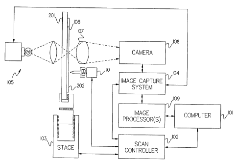
Referring to Figure 4, a simplified schematic
block diagram of one example of the apparatus of the
invention is shown for greater ease in explaining the
method and apparatus of the invention. The apparatus
shown comprises a central computer 101, a real time
scan controller system 102, which coordinates the
motion of the motorized stage 103 of the microscope
with the image capture system 104, a stroboscopic
illumination system 105, a low-power microscope
objective 107, an electronic camera 108 of the CCD
type, one or more dedicated image processing systems
109, and a touch sensor 110. The stroboscopic
illumination system 105 focuses a brief flash of light
on the specimen 106. The specimen 106 is mounted on
a glass slide 201 and protected under a transparent
coverslip 202.
CA 02220388 1997-11-06
WO 96138809 PCTlUS96In7936
- 11 -
The computer 101 may be advantageously programmed
to guide the steps of the focusing procedure as
described in detail below. In Figure 4, the arrows
between the various components generally represent the
flow of information between the parts of the
apparatus.
Figure 5A schematically shows a more detailed
view of a sl:Lde 201 on which a typical specimen 106 is
mounted, them covered with a transparent coverslip
202. A typical slide 201 may be eighty millimeters
long by twenty seven millimeters wide by one
millimeter thick. A typical coverslip 202 may be
sixty millimeters long by twenty four millimeters wide
by 0.13 mil:Limeters thick. The best focus on the
specimen 106 varies from point to point, due both to
warpage in the slide-coverslip combination, and to the
intrinsically three-dimensional nature of the specimen
itself.
A grid 203 is shown superimposed over the slide
201 in Figur<~ 5B. The grid 203 may not be visible in
the physical embodiment of the invention, but is used
herein for illustrative purposes. Grid 203 is not
shown to sca7.e. The grid 203 illustrates the division
of the slide: into low magnification fields of view
such as 210, shown in more detail in Figure 5B. Each
of the low magnification fields of view 210 is divided
into twenty five high magnification fields of view
211, for example. In one embodiment of the invention,
a captured, digitized image of a low magnification
field of view contains 512 x 512 pixels, and
represents a specimen area of about 1.4 mm x 1.4 mm.
Finding the t:overslip
Referring to Figure 4 while now also referring to
Figure 6, focusing on a specimen begins with the
central computer 101 issuing instructions to the scan
CA 02220388 1997-11-06
WO 96/38809 PCT/US96/07916
- 12 -
controller 102 to move the stage 103 to a predefined
central location at process step 401. The central
location is chosen with respect to the approximately
known physical location of the specimen 106 in such a
way that even a small coverslip, if properly placed
over the specimen, must cover a substantial region
around the central location.
At step 402, the central computer 101 instructs
the scan controller 102 to move the stage 103 in the
axis perpendicular to the slide 201, so that the
specimen 106 approaches the touch sensor 110. This
motion continues until either the touch sensor 110
records contact with the coverslip 202 over the
specimen 106 at step 403, or the end of travel is
reached, step 404. Reaching the end of travel at step
404 indicates that either no slide, or a slide which
is too thin for the apparatus, is present, in which
case the automatic processing of the slide in question
halts at step 405.
When the touch sensor 110 indicates contact with
the coverslip over the specimen 106 at step 403, the
scan controller 102 reports the stage location at
which the touch occurred to the central computer 101,
which stores it at step 406. This location indicates
the stage location of the top of the coverslip at the
central point, and is used as a starting point for
focusing. If the touch sensor indicates contact with
the coverslip at a point higher than a predetermined
height, the slide is too thick. In which case the
automatic processing of the slide in question halts.
In particular, the location of the touch sensor 110 is
calibrated to be a known distance from the focal
plane of the objective lens 107 by using targets
designed for this purpose. At step 411, this
calibration is used to move the stage 103 to a
CA 02220388 1997-11-06
WO 96138809 PCT/L1S96/07936
- 13 -
position such that the focal plane of the objective
107 lies just below the top of the coverslip 202 at
the central touch location.
Before focusing can continue, however, the
location of i~he coverslip must be better determined.
Toward this end, at step 407, four more touches,
substantially similar to the first one described above
at steps 402 through 404, are performed at separate
locations on. the slide within a minimum central
coverslip area. The location of the coverslip at each
of these touches is also recorded. At step 408, a
least squares plane is constructed from the five touch
locations, and the tilt of the plane is compared with
an allowed maximum. If the tilt exceeds the maximum,
or if any of the four touches failed to record a
location, processing of the slide is halted at step
405. An excessively tilted coverslip at step 408
usually indicates that the slide is improperly loaded
in the apparatus.
At this point, since the approximate position of
the coverslip is known, a more detailed search for its
edges is undertaken at step 409. At step 410, if the
coverslip edges were not found, or if they indicated
an inappropriate size or shape, processing of the
slide is once again halted at step 405.
At step 411, the stage is returned to the center
touch location, at such a height that the objective
107 focal plane is just beneath the touched surface of
the coversli~> 202. At step 412, focusing of the
specimen 106 ,proceeds, with the central computer 101
instructing the scan controller 102 to coordinate the
stage 103 motion with the image capture system 104 in
order to perform an initial focus scan, starting from
the position where the objective 107 focuses an image
from just beneath the surface of the coverslip 202 at
CA 02220388 1997-11-06
WO 96/38809 PCT/LTS96/07916
- 14 -
the central touch location onto the CCD camera 108.
The Initial Focus Scan
At this point we describe the initial focus scan
in detail, referring in turn to Figures 5A, 5B, 6, 7,
8, 9. 10 and 11, while also referring to Figure 4.
The purpose of a focus scan is to acquire and
process images from different focal planes in a
specimen, in order to find the best focal plane. Any
focus scan, in this example of the preferred
embodiment, is performed as follows. Referring
jointly to Figures 4 and 7, the central computer 101
passes to the scan controller 102 a list of stage
locations at which images are to be collected, at step
1501. The stage locations are chosen to cause the
objective 107 to focus at different planes in the
specimen 106.
The scan controller 102 computes the timing of
the motion of the stage, and constructs a table,
giving successive positions of the stage when images
are to be collected, and the time when the stage will
be at each location. The table entries are computed
and passed to the image capture system 104 at step
1502.
Once the image capture system 104 has received
the table, at step 1503, the scan controller 102
initiates the motion of the stage 103. The motion of
the stage is monitored by encoders at step 1504 to
ensure accuracy. Any incorrect stage locations will
be reported to the computer 101, which may reset the
stage and restart processing.
At each time listed in the table, the image
capture system 104 signals the stroboscopic
illuminator 105 to flash at step 1505. The
illuminator 105 focuses a brief flash of light on the
specimen 106 at the specified location at step 1506.
CA 02220388 1997-11-06
W O 96138809 PCT/CTS96/079I6
- 15 -
The illuminator system monitors the flashes of the
strobe with a light sensor at step 1507. Any missing
flashes, or :Flashes of incorrect intensity, will be
reported to the computer 101, which may halt
processing of the slide.
At step 1508, the objective 107 focuses an image
from the illuminated field of view of the specimen
106 onto the camera 108. At step 1509, the image
capture system 104 collects a digital representation
of the image thus acquired from the camera 108. In
one example, the digits:l representation of the image
consists of 512 rows by 512 columns of pixels, each of
which is assigned a gray level from zero, representing
the darkest level, to 255, representing the
brightest. If necessary, the image may be sent in
analog form from the camera 108 to the image capture
system 104, then passed through an analog to digital
converter to create the digital representation.
The image capture system 104 sends each digital
image it acquires to the image processors) 109 at
step 1510. The dedicated image processors) 109
perform a pre-programmed sequence of morphological,
computational., and logical operations on each image
sent from the. image capture system 104 to derive one
or more measures of focus quality. These measures are
computed and sent to the computer 101 at step 1511.
Once the computer 101 receives the measures from every
image in the list originally sent to the scan
controller 102 at step 1501, it processes the list of
measures in order to determine the optimum focus
location at sctep 1512.
All the while images are being captured and
processed, the stage 103 continues to move the
' specimen 106 in accordance with the instructions from
the scan controller 102, from step 1503 onward, until
CA 02220388 1997-11-06
WO 96/38809 PCT/ITS96/07916
- 16 -
the list of images to be collected is exhausted.
The initial focus scan starts, as noted above,
from a position where the objective 107 is focused on
an image plane just beneath the surface of the
coverslip at the central touch location. It proceeds
further beneath the coverslip, collecting one image
for each depth of focus of the objective 107, until it
is past the depth corresponding to the maximum
coverslip optical thickness. The maximum coverslip
optical thickness may be a predetermined allowable
thickness depending upon the particular apparatus
employed.
The initial focus scan is used to identify a
starting point, called the seed point, for focusing
the system on the specimen. Since it is not important
whether or not this starting point is derived from
cells in the specimen, or just dust or other matter on
the surface of the slide, morphological pattern
recognition is not used for the initial focus scan.
Instead, a simpler intensity gradient focus
quality measure is computed as follows. Refer to
Figure 8, which shows the process flow diagram for the
image processor when computing the gradient focus
score. To begin with, at step 601, a histogram is
computed of the gray levels of the image. This
histogram is used to calculate a measure of the
illumination brightness, or light level, present in
the image. In particular, the light level may be
defined as the highest intensity level at which 0.1
or more of the pixels of the image record a still
higher intensity.
At step 602, the horizontal and vertical
gradients in light intensity are computed at each of
the pixels in the digitized image. For the vertical
gradient, the computation is performed at each pixel
CA 02220388 1997-11-06
WO 96/38809 PCTlUS96/079I6
- 17 -
by subtracting the intensity value of the pixel
immediately below from that of the pixel immediately
above. The horizontal gradient is computed in an
analogous way. These gradients are compared to a
threshold in order to reduce the effect of image noise
on the measure of image sharpness. Gradients below
the predetermined threshold value are treated as zero .
Those skilled in the art, having the benefit of this
disclosure, will understand that the threshold value
may be derived empirically, or from the noise
characteristics of the specific imaging apparatus
used.
In order to be able to distinguish the best focus
locations of different regions within the field of
view, the image processor divides the field of view
into a five by five grid, like the one shown in
Figure 5B, .at step 603. Subsequent processing
computes twenty five separate focus measures, one for
each of the twenty five regions in the grid.
At step 604, fifty histograms are computed, two
for each of the twenty five grid regions in the image.
The two hisstograms are computed on the horizontal
and vertical gradient images, respectively.
Even a focusing procedure which does not perform
pattern recognition must take some account of the size
of the objector to be focused, in order to acquire
information from the appropriate range of spatial
frequency. Because the focusing system described here
is designed t=o work at low magnification on small
objects, the intensity gradient algorithm takes size
into account by using on7_y the fifty highest gradients
in each of the twenty five regions at step 605. The
rest of the gradient histogram is ignored.
At step 606, the sc~,iares of these fifty gradients
are summed, and divided by the light level, in order
CA 02220388 1997-11-06
WO 96/38809 PCT/US96/07916
- 18 -
to produce twenty five focus scores for each image in
the focus scan. The light level is used to normalize
the scores in order to reduce their dependence on
illumination from quadratic to linear. This is useful
because the algorithm may be used on images in a
context where the illumination may be, for some
reason, obscured.
Once the image processing system 109 has computed
the twenty five gradient focus scores for each image
in the initial focus scan, it passes these scores,
along with the matching focus positions, back to the
central computer 101, as described above and shown as
step 1511 in Figure 7.
The task of the central computer 101 in step 1512
of Figure 7 is to look for peaks in the focus score as
a function of focus position in each of the twenty
five regions, ignore spurious fluctuations due to
noise, and make an initial approximation of the best
focal plane. It accomplishes this as shown in Figure
9.
First, at step 620, the scores for each region
are filtered across focus position in order to reduce
noise in the focus scores. For this purpose, a
Gaussian kernel with a half width at half maximum
equal to the depth of field of the objective is used.
Second, at step 621, the derivative of the focus
score with respect to position is computed for each
region and position by subtracting the filtered focus
score at each position and region from the succeeding
position's filtered focus score for the same region.
Third, at step 622, peaks in the focus score in
all regions are identified by looking for patterns
across position of two positive derivatives followed
by two negative derivatives, and checking to make sure
that the focus score at the peak is above a
CA 02220388 2002-O1-10
77501-5
pre-defined minimum, to avoid finding spurious peaks.
The precise location of the peak is found by linear
interpolation of the gradient to the zero crossing.
Figure 10 illustrates the process of finding the
peaks by plotting an example of the original focus
scores 701, the Gaussian-filtered focus scores 702,
and the differences of the filtered scores 703, versus
focus position. The scores plotted in Figure 10
represent the values found from a single region. The
interpolated zero of the derivative at 704 represents
the calculated position of the peak. Note the
positive derivatives before the peak, and the negative
derivatives after the peak.
Fourth, in step o23, the sharpness of each peak
is measured by dividing the magnitude of the second
derivative of the filtered focus scores at the peak by
the magnitude of the peak. The sharpness provides an
indication of how definite a preference for the given
focus position the peak indicates.
Fifth, in step 6:'.4 , all of the peaks found in
all regions are divided into two classes: those which
are one minimum cover:~lip optical thickness or more
below the highest peak found, and those which are not.
They are divided in order to separate' any peaks which
may be coming from dust on top of the coverslip from
peaks coming from the specimen proper.
Sixth, at step 625, in each region, the peak
with the highest focu:a score in each class is kept,
while any or_her peaks un the same region and class are
ignored. As a result, there are at most twenty five
peaks in each class to consider.
Seventh, at step 626, a weighted average of the
position of the peaks in each class is taken to
represent the best focus position for the full field
35~ of view in each class. The peak positions are
CA 02220388 1997-11-06
WO 96/38809 PCTlUS96/07916
- 20 -
weighted by the relative peak sharpness calculated in
step 623 to derive the weighted average. If any peak
has a sharpness which is more than a factor of four
less than the sharpest peak in the class, it is
dropped from the averaging as being too soft a peak at
this step. This leaves at most two possible focus
positions. Note that it is possible that all the
peaks are in the upper class, in which case there is
only one focus position at this stage.
Eighth, to avoid the possibility of focusing on
the top of the coverslip, if there are two focus
positions, the class which is lower (further beneath
the coverslip) is chosen as representing best focus on
the specimen at step 627.
Ninth and finally, at step 628, if there were no
valid peaks found at step 622, the scan fails to find
a best focus position at step 630. Otherwise, the
focus position chosen at step 627 is stored by the
computer 101 at step 629. This completes the
discussion of the initial focus scan.
Multiple Tries of Initial Scan
Referring back to Figure 6, the result of the
initial focus scan at step 412 is thus either a
starting focus position, or a failure to find a peak.
In nearly all cases, when a specimen-bearing slide
which meets the physical requirements for coverslip
thickness and placement is used, the initial focus
scan described above is successful. This is because
it requires very little material to focus on, and the
scan is undertaken in the center of the slide, where
there is likely to be some specimen.
However, if a failure is found at step 413,
additional attempts are made to find a seed point for
focusing. In particular, if fewer than a set number
of attempts have been made at step 414, a new location
CA 02220388 1997-11-06
WO 96!38809 PCTlUS96/07936
- 21
is selected at step 415 on a field of view adjacent
to
the one at which a focus scan was just attempted, and
processing returns to attempt another initial scan at
step 412. The succeeding attempts may occur in a
r
spiral pattern around the original touch point, so as
to continue selecting new fields of view while
remaining as close as possible to the central touch
location. Only if all of the set number of attempts
have been unsuccessful at step 414 does processing of
the slide cease at step 405.
Once an initial focus position is found at step
412, the pattern recognition, or cellular, focus
scans begin from this point, referred to as the seed
point to cre<~te a map of the focus surface.
Figure 11 illustrates the path of the cellular
focus scans across the surface of the specimen, where
the location of the seed point is marked with an "X".
The squares 801 indicate the fields of view scanned,
while the arrows 802 show the path the stage follows.
The purpose of following the path indicated is to
come as close as possible to achieving a
representative sample from the slide, while minimizing
the time taken to scan. The stage used takes no more
time to move simultaneously in two dimensions than to
move in just one, so the diagonal moves illustrated
maximize the speed of motion.
Referring also back to Figure 6, at step 416,
the first scans occur to the right of , and adj acent
to, the seed point in Figure 11. The zig-zag pattern
illustrated i.n Figure 11 turns around, as for example
at 804, each time scanning approaches one of the edges
of the covers;lip. The entire pattern must come to an
end before t:he far right end of the coverslip in
Figure 11. Scanning then resumes at steps 422 and
416, again starting adjacent to the seed point, to the
CA 02220388 1997-11-06
WO 96/38809 PCT/US96/07916
- 22 -
left of the seed point, at the scan marked with a
circle in Figure 11. Note that the last reversal of
scanning 803 drawn in Figure 11 takes five steps,
rather than the three steps taken by 804 and every
other reversal. This illustrates the fact that, under
conditions to be described below, the number of steps
in a reversal is increased from three to five in order
to speed processing of the specimen.
The first two cellular focus scans are centered
about the focal plane defined by the seed point.
Cellular focus scans are much more shallow than the
gradient scans described above, consisting of the
acquisition and processing of only four images, again
separated by roughly the depth of focus of the
objective lens. This makes the cellular scans much
faster. Finding the best focus position from a
cellular scan is necessarily a simpler operation than
from a gradient scan, because there are only four
points to work with. More burden is placed on the
processing of the. image to weed out signal from noise,
and in particular, to recognize and focus principally
on the nuclei of well-separated cells. Note that
cells in clumps often provide less useful information,
if their nuclei cannot be clearly distinguished.
Cellular Morphology
The processing of an image in a cellular focus
scan is comprised of a combination of simple
morphological operations. Figures 12A-12D illustrate
four simple binary morphological operations. Figure
12A illustrates an erosion with a three by three
block, while Figure 12B demonstrates a dilation with
the same block. Figure 12C shows an erosion with a
five by five wire frame, and Figure 12D illustrates a
dilation with the same wire frame.
A morphological operation, such as an erosion or
CA 02220388 1997-11-06
WO 96!38809 PCTlUS96/079I6
- 23 -
dilation, involves two entities. The first entity is
the image which is operated on, and the second entity
is a structuring element with which the operation is
performed. The structuring element may be pictured as
a grid of pixels, whose values are either "on" or
"off", and which possesses a unique center pixel. The
center pixels of the structuring elements in Figures
12A-12D are marked with X's.
A morphological operation may be envisions as
placing the center of the structuring element, in
turn, over each pixel in the original image. A
binary operai=ion operates on images whose pixels are
either "on" or "off". The two simplest operations are
binary erosion and dilation. Binary erosion turns
"off" all pixels in the image which, when the
structuring element is centered on them, have at
least one "o:f f " pixel of the image aligned with an
"on" pixel o:~ the element. All other pixels in the
image are set to "on". Dilation turns "on" in the
image all pixels which, when the structuring element
is centered on them, have at least one "on" pixel of
the image aligned with an "on" structuring element
pixel. All other pixels are set to "off".
Binary erosion and dilation are readily
generalized to grayscale erosion and dilation.
Grayscale erosion replaces each pixel's value in the
image by the minimum of the values of those pixels
which correspond to "on" pixels in the element.
Grayscale dilation rep7_aces each pixel's value with
the maximum of the values of those pixels which
correspond to "on" pixels in the element. Erosion and
dilation are combined to make compound operations. In
particular, a dilation followed by an erosion with the
same element is referred to as a closing, while an
erosion followed by a dilation is an opening.
CA 02220388 1997-11-06
WO 96/38809 PCT/US96/07916
- 24 -
Figure 13 is a data flow diagram of the cellular
focus score morphological process. Each image from
each cellular focus scan, after being stored in the
camera 108 and digitized by the image capture system
104, is processed through this set of operations by
the image processors) 109. As shown in Figure 10,
the process has four main branches.
On the first branch, a histogram 1011 is taken of
the grayscale values of the image 1001, and three
grayscale values 1012, called the white, cyto, and
dark levels, are computed from the histogram. The
white value is the same as the light level described
in the discussion of the gradient focus score above.
The cyto level is defined as 95 0 of the white value.
As the name implies, regions of the image with gray
levels below this level probably represent areas with
at least some cytoplasm. The dark level is defined as
1/15 of the white value, plus a quantity representing
a noise floor. Regions of the image with gray levels
below the dark value represent either thick clumps of
specimen, or artifacts of other material. Such
regions are excluded from consideration in focusing.
On the second branch of the cellular morphology
process, each pixel in the original image 1001 is
tested at step 1021 to see if its gray level exceeds
the cyto threshold. If it does, the corresponding
pixel in a binary image is set to zero; if not, the
binary pixel is set to one. The binary image thus
produced passes through a morphological opening 1022
by a five by five box, followed by an opening 1023 by
a 43 by 43 box.
The opening 1022 by the five by five box is
designed to reject regions which are too small to
actually represent cells,~while the opening 1023 by '
the 43 by 43 box detects regions which are so large
CA 02220388 1997-11-06
W O 96/38809 PCT'lUS96/07916
- 25 -
that they must represent groups of cells stuck
together, rather than cells with gaps between them.
In accord 'with the requirements of focusing on
free-lying cells, the results of the two openings are
L
exclusive-or'd together at step 1024 to generate a
cytoplasmic binary image, which is then dilated 1025
by a five-by--five box, to pick up any nuclei which may
be on the edges of their cells.
The third branch of the cellular focus score
algorithm checks each pixel of the original image 1001
at step 103:1 to find out if it has a gray level
greater than the dark level defined above. If it
does, the corresponding pixel of a binary image is set
to one; if not, to zero. The resulting dark binary
image is eroded 1032 by a 43 by 43 box to prevent
focusing on t:he edges of thick clumps of specimen, or
on regions laced with artifacts. The dark binary
image is then and'd 1033 with the cytoplasmic binary
image, to produce a binary image representing the
allowed regions for nuclei to be recognized.
The foux-th and final branch of the algorithm is
designed to detect nuclei. It begins with a grayscale
closing 1041 of the original image 1001 by a five by
five brick. This closing will usually efface the
nuclei of interest from the image. The original
image 1001 i:~ then subtracted at step 1042 from the
result of the closing to produce a texture-enhanced
inverted image, in which the nuclei appear as
prominent bright spots.
In order to develop a binary image which
identifies the nuclei, the result of the closing is
divided by eight at step 1043 , then tested against the
texture-enhanced image at step 1044. If the gray
level of the texture-enhanced image exceeds that of
this measure of the local light level, the pixel in
CA 02220388 1997-11-06
WO 96/38809 PCT/US96/07916
- 26 -
question is flagged as potentially part of a nucleus
at step 1044 . This binary image is and' d 1045 with
the allowed regions binary image, to generate a binary
mask of possible nuclear pixels.
The binary mask image thus produced at step 1045
has the defect that few restrictions on nuclear size
or shape have been placed on it. The next steps are
designed to rectify this limitation. First, an
opening 1046 by a two by two box is applied to
eliminate solitary pixels or single-width strands of
pixels. Second, a dilation 1047 of the resulting mask
by a seven by seven hollow frame of pixels is inverted
1048, then and'd 1049 with the mask to get rid of
those parts of prospective nuclei which will not fit
inside the seven by seven frame. This requires that
the prospective nuclei be roughly elliptical in shape .
Third and finally, the resulting binary image is
dilated 1050 with a three-by-three box, in order to
include the edges of the nuclei in the mask. The
result of step 1050 is the final mask image, which
identifies nuclei meeting all the requirements for
focusing.
Even after the nuclei have been identified, it
remains necessary to measure the sharpness with which
they are focused. In order to do this, the
texture-enhanced image produced at step 1042 is eroded
1051 by a three-by-three brick, and the resulting
image subtracted 1052 from the texture-enhanced image
itself, to produce an enhanced gradient image. At
step 1053, this enhanced gradient image is then set to
zero wherever the final mask image from step 1050
contains a zero, and left unaltered where the final
mask image contains a one.
Finally, a histogram 1054 is taken of the gray
levels of the resulting image, in order to add up a
CA 02220388 1997-11-06
WO 96/38809 PCT/US961079I6
- 27 -
measure of the quantity of nuclear matter found, and
of course, t:he sharpness of the focus on the nuclei.
Two measures are computed from the histogram 1054 at
step 1055 as follows. First, any level of the
enhanced gradient image below 5 a of the white level
is ignored a:> due to non-nuclear material. Then, any
pixel above this level is counted toward the sum of
the total number of acceptable nuclear pixels, and the
nuclear sharpness measure is computed as the mean
to square enhanced gradient level of those pixels which
are above 5 a of the white level. The sum of
acceptable nuclear pixels is a measure of the amount
of useful specimen found, while the nuclear sharpness
measure is divided by the white level to reduce the
dependence on light level, then used as the cellular
focus score.
Cellular Focus Processing
The central computer 101 thus receives four focus
scores and four pixel counts from the image
processors) 109 for each cellular focus scan
performed. 'The computer processes these measures.
The processing occurs as described below.
First,.t:he computer determines which focus score
is the highesi~ . If the h ighest focus score is the one
furthest from the cover:alip, the result of the focus
scan is an indication that it is necessary to move
further from the coverslip to seek a better focus
plane. If not, the nuclear pixel count of the image
with the highest focus score is checked to see if it
exceeds 0.1 0 of the image. If not, there is
apparently not enough to focus on in this field of
view, and the computer determines to continue scanning
in the same plane.
If the nuclear pixel count is greater than 0.1
0 of the image, and the image with the highest focus
CA 02220388 2002-O1-10
77501-5
28 -
score is the one close:~t to the caverslip, the result
of the focus scan is <3n indication that it might be
necessary to move closer to the coverslip to seek a
better focus plane. Sufficient data is required
'e before moving toward the coverslip in order to prevent
moving to attempted focus on material on the
coverslip over very spa rse slides.
If the image with the highest focus score is
neither at the top nor at the bottom of the scan, and
the image with the highest focus score has a nuclear
pixel count exceeding 0.1 0 of the image, then the
differences in focus scores can be linearly
interpolated to locate the peak. The interpolation is
made to the zero crossing in the derivative, analogous
1~~ to the interpolated zero crossing shown as 704 in
Figure 10. This indicates a successful.findinc~ of best
focus, and its location is recorded. Finally, the
indication for further focusing is to center about the
best focus plane recorded.
Refer again to Figure 6 to follow the usage of
cellular focus scans in the focus scanning of the
specimen. As noted above, the first two cellular
focus scans in step 415 are centered about the plane
defined by the seed point. At step 417, the result of
2~~ the first of these scares is processed by the computer
101. After deriving ~~he result, the computer 101
requests the next cellular scan at step 418.
If the result of the focus scan just processed
indicated that focus should be lowered, the
requested scan 418 is centered one focus step lower
than the scan just processed. If the result indicated
that not enough data was available, the requested
scan 418 is centered about the same plane as the scan
just processed. If r_he result indicated that focus
3~~ should be brought higher, the plane of the last
CA 02220388 1997-11-06
WO 96/38809 PCTlLTS96/079I6
- 29 -
successful focus scan is tested to see if the current
focus position is less than half a minimum coverslip
optical thickness above it. If so, the requested scan
418 is cente~__~ed one focus step higher. If not, then,
' 5 to prevent attempting focus on the top of the
coverslip, the requested scan 418 is centered about
the same plane as the scan just processed.
Referring again to Figure 6, at step 419 the
computer tests the position of the focus scan just
requested to see if it is at the far right or left end
of the coverslip, in accord with the scanning pattern
shown in Figure 11. If it is not, the computer
returns to step 417 to compute the result of the scan
just completed, then request a new scan again at step
418. Note that the focal height of each cellular scan
is based on the result. of the scan two before it.
This allows the scan controller 102, image capture
system 104, image processors) 109, and computer 101
to continuou:~ly process focus scans in parallel as
fast as the stage can move, with no lag time spent
waiting for the results of a computation.
When an end of the coverslip is reached at step
419, there are two focus scan results still to be
processed at step 420. Then, at step 421, the
computer checks to see if focus scanning has already
proceeded in both directions from the seed point as
shown in Figure 11. If it has, the cellular focus
scanning of the specimen is complete at step 423. If
not, the machine returns to the seed point at step
422, then begins scanning in the opposite direction
back at step 416. The first two focus scans in both
directions are centered about the plane of the seed
point.
A minimum number of successful cellular focus
scans is needed to accurately focus on a specimen.
CA 02220388 2002-O1-10
77501-5
- 30 -
In one example embodiment, the minimum number is 24
scans. If this number is not reached after all scans
as shown in Figure 11 are completed, the specimen must
be rejected for automatic processing. Pap smears
rejected for this reason are usually unsatisfactory
because of insufficient squamous cellularity.
Additionally, it is possible to have successful scans
only in one region of the slide. If successful focus
scans do not cover all areas of the slide, the slide
is rejected as having specimen distributed in too
small an area.
Once a model of the focus surface of the specimen
is available, it may be used as a guide to scan the
entire specimen under the coverslip at low power
magnification. It may also be used as a starting
point for the high powex- magnification focusing of the
specimen. If the focua surface is too tilted, high
power focusing may not x~e possible for the entire high
power field of view. If the focus surface is too
tilted slide processing ceases. If the focus surface
is too variable, focus pans may have provided
inaccurate focus information. If the focus map is too
variable; slide processing ceases as "unable to focus
on specimen".
The successful focus scan data may also be used,
along with the height of the coverslip given by the
touch sensor, to estimate the optical thickness of the
coverslip over the speca.men. If the coverslip optical
thickness is too large or too small then an
unacceptably large sphearical aberration is produced
when the specimen is viewed through a high resolution
objective lens. 8s a result the specimen may be
unsuitable for high power microscopic examination. If
the specimen to coverslip top measure, optical
thickness, .is greater than a first predetermined
CA 02220388 1997-11-06
WO 96138809 PCT1US961079I6
- 31 -
distance or 7.ess than a second predetermined distance
the processing of the slide ceases.
In the preferred embodiment, the focus surface
model is u:>ed to guide low power magnification
scanning of t=he specimen. Areas of the specimen are
analyzed for potential abnormality. Each slide area
field of view is ranked for likelihood of containing
abnormality. If too few fields of view are ranked,
the slide may not have been scanned properly, so
l0 processing ce=ases for the slide. During low power
magnification. scan, bubble areas are identified. If
too much bubble area exists, slide processing ceases.
In one embodiment, after the low power
magnification scan, a focus surface model is created
for the high power magnification scan. Since depth of
field is reduced at high power magnification, a more
accurate focus surface is required. Focus pans are
done in much the same manner as for the low power
magnification focus surface, except that the pans are
done at high power magnification. If the high power
magnification surface is too variable or if too few
focus pans are successful, processing of the slide
ceases.
A high power magnification scan is executed after
the high power magnification focus surface model has
been created. During 'the high power magnification
scan, regions of the slide identified as potentially
containing abnormality during low power scan are
imaged and analyzed at high power magnification. The
high power ma~~nification focus surface serves as the
. initial estimate of focus position for high power
imaging. If the position given by the high power
magnification focus surface leads to imagery which
does not meet predetermined focus criterion,
additional image acquisition attempts are made at the
CA 02220388 2002-O1-10
77501-5
same slide region at different focus positions. If
after a predetermined :number of acquisition attempts,
the region remains outside of the predetermined focus
criterion, the slide region is abandoned. If too few
slide regions are focuased adequately during the high
power magnification scan, slide processing ceases. If
during high power mae:nification scar, fewer than a
predetermined number of slide regions are adequately focused
on first try, or too many slide regions are not focused
1Ci adequately, the high power magnif ication focus surface
model may have been inaccurate.
Image quality is checked to ensure good imagery.
Image saturation, pixels with Values of 0 or z55, are
counted for each image. The numbers of images with
lf~ saturation are counted. If too many images are
saturated during e~.ther the low or high power
magnification scan the optics may not be adequate for
reliable specimen viewing or imaging. Additionally,
if dirt obscures the optical path, imaging artifacts
2C~ such as striping. may he detectable on some systems.
If striping is detected in more than a predetermined
number of images, the slide maybe too dirty to allow
accurate specimen viewing or imaging.
During evaluation, the aur_ornated system
2°_i discontinues processing for slides that fall outside
of an acceptable range for any of the preselected
criteria. The automated system may count a proportion
of slides failing processing. In one preferred
embodiment, the slide set is considered to pass if the
3c) proportion of slides failing processing is less than
6%; otherwise the slide set fails.
In step 40, the automated system performs a
Specimen Collection C~uality Test to evaluate the
quality and sufficiency of the specimen material
3'~ sampled on the slide. Specimen collection quality is
CA 02220388 1997-11-06
WO 96138809 PCTltJS96Ia79I6
- 33
highly dependent upon a clinic's sampling tools and
techniques for specimen collection. In the preferred
embodiment, the Specimen Collection Quality Test may
comprise two tests. Tables 2 and 3 list qualities for
which the slide set may be tested. Slides failing
these tests ~~omprise the specimen collection quality
failures. '.C'able 2 tabulates slide set-up related
failures. 'Table 3 tabulates failures related to
process suitability failures. Process suitability
failures may include, for example, slides for which
process results cannot be expected to be reliable, for
example, when the process detects too few reference
cells. The proportion of slides failing processing
for these reasons is measured. In the preferred
embodiment, _Lf the proportion of slides that failed
the first teat is less than 70, the slide set is
considered to pass the first test; otherwise, the
slide set fails.
In the preferred embodiment, the second specimen
quality test measures and ranks the reference cell
ratio for all normal slides. The reference cell ratio
is the number' of detected reference cells ( that is,
free-lying intermediate cells) on a slide divided by
the number of all objects detected on the slide. In
one preferred embodiment, if 85% of the normal slides
have a reference cell ratio greater than 0.015, then
the slide set is considered to pass the test;
otherwise, the slide set fails.
The slide set is required to pass both specimen
quality tests to pass the Specimen Collection Quality
Test.
CA 02220388 1997-11-06
WO 96/38809 PCT/US96/07916
- 34 -
Table 2
Lack of material in center
Too few points for low-power
f ocus map
Specimen distributed in small
area
Unable to focus on specimen
Specimen tilt
Too few fields ranked in low-
power scan
Too few points for high-power
focus map
High-power focus surface too
variable
Too few focused fields in high-
power scan
Table 3
Insufficient reference cells
Image quality not within limits,
percentage of fields focused on
first try.
Image quality not within limits,
percentage of fields never
focused .
The automated system performs a Slide Handling
Quality Test in step 50. The Slide Handling Quality
Test determines if slide handling practices may need
to be modified to facilitate effective processing on
a selected automated system, such as the AutoPap° 300
System. The test evaluates the quality of slide
barcoding, cleaning, and loading practices at a
preselected clinical site. Tables 4 and 5 list tests
for slide handling quality failures. Table 4
tabulates slide set-up related failures. Table 5
tabulates failures related to image processing methods
CA 02220388 1997-11-06
WO 96/38809 PCT/US96/07916
- 35 -
suitability failures. The system measures the
proportion of slides failing these tests. In the
preferred embodiment, i_f the proportion of slides that
failed is less than 50, the slide set is considered to
pass the slide handling quality test; otherwise, the
slide set fails.
Table 4
Slide barcode not read
Slide tilted
Tables 5
Image quality not within limits,
excessive striping.
Image quality not within limits,
high power magnification image
saturation (small amounts)
Image quality not within limits,
high power magnification image
saturation (large amounts)
Image quality not within limits,
low power magnification
image
saturation.
The automated system performs a Preparation
Quality Test in step 60. The Preparation Quality Test
evaluates the result of laboratory fixation, staining,
and coverslipping processes to see if the presentation
of cells is within an acceptable range. In the
preferred embodiment, five tests comprise preparation
quality test - to pass the full test, the slide set
must pass a1:1 tests. Referring to Tables 6 and 7,
slides which :fail processing for the tabulated reasons
comprise the preparation quality failures.
Cytoplasm stain density is' measured. If the
cytoplasm stain measure outside of predetermined
limits, the automated device may not accurately score
the slide. T'he slide is called unsuitable for image
CA 02220388 1997-11-06
WO 96/38809 PCT/LTS96/07916
- 36 -
processing methods.
Also the contrast between reference cell nuclei
and cytoplasm. If this contrast measure is outside of
predetermined limits the slide is called unsuitable
for image processing methods of the device.
The proportion of slides failing processing for
these reasons is measured. Table 6 tabulates slide
set-up related failures. Table 7 tabulates failures
related to image processing methods suitability
failures. In the preferred embodiment, if the
proportion of slides that failed the first test is
less than 50, the slide set passes the first test;
otherwise, the slide set fails.
Table 6
Too many bubbles
Too few fields ranked in low-
power scan
Table 7
Stain average not within limits
Cytoplasm Staining not within
limits
Staining detail not within limits
Nuclear/Cytoplasm contrast not
within limits
Insufficient reference cells
Image quality not within limits,
high power magnification image
saturation (large amounts)
Image quality not within limits,
low power magnification image
saturation.
The second preparation quality test measures the
nuclear stain density of the reference cells detected
on the slide. Measurements are stored in a "mean
stain" bin. The mean optical density for each
CA 02220388 1997-11-06
WO 96138809 PCTlUS96/079I6
- 37 -
detected intermediate cell nucleus is calculated.
Data for all the detected intermediate cell nuclei on
- the slide is accumulated in a 10-bin histogram. The
average staining score for the normal slides is
calculated. In the preferred embodiment, if the
average staining score is greater than 4.2 and less
than 6.4, the slide set passes the test; otherwise,
the slide set fails.
The third preparation quality test counts the
l0 number of poi=entially abnormal cell nuclei detected on
a slide (stage 3 abnormals). The 80th percentile of
the normal slides which contain endocervical component
cells is calculated. In the preferred embodiment, if
the 80th percentile is greater than 3, the slide set
passes the test; othervaise, the slide set fails.
The fourth preparation quality test measures the
80th percentile of the QC score of the normal slides
which contain endocervical component cells. In the
preferred embodiment, if the 80th percentile is
greater than. 0.15 and less than 0.6, the slide set
passes the test; otherwise, the slide set fails.
The fifth preparation quality test measures the
median of reference cell nuclear texture (nuclear blur
average) for the normal slides which contain
endocervical component cells. In the preferred
embodiment, if the median is greater than 5.65, the
slide set passes the test; otherwise, the slide set
fails .
In step 70, the automated system performs a
Classification Test. The Classification Test
evaluates whether the customer slide and cell
presentation are within the training range of the
AutoPap~ 300 System to enable an effective
interpretation by the system. The test evaluates the
accuracy of :slide classifications.
CA 02220388 2002-O1-10
77501-5
- 38 -
The system accuracy test evaluates sensitivity to
abnormal specimen morphology. The 80th percentile of
the QC score of the normal slides is calculated. In
the preferred embodiment., if more than 70% of the low
grade slides and 80% of the high grade slides have QC
scores above the 80th percentile for normal slides,
the slide set passes the test; otherwise, the slide
set fails.
In step 80, the automated system then integrates
the results from the beasts in steps 30-'70. In the
preferred embodiment, the slide set must pass each
test or the slide set i;s considered to fail. If the
slide set passes, the test result integration
states that the slide set is acceptable in step 90.
If the slide set fails, the test result integration
makes recommendations for adjustment of the larioratory
or clinic process in step 100.
Now referring to Figure 3, Figure 3 shows a more
detailed flow chart of the method for assessing slide
and specimen preparation quality of the invention. In
one embodiment of the invention slides are collected
at step 103. At process step 104 the collected slides
are cleaned and a barccde is affixed to the slides.
At process step 106 the slides are processed in
accordance with the various quality control methods
described herein. Processing includes process steps
108 through process step 126 as shown in Figure 3 and
as described with reference to the tables hereinbelow.
At process step 108 a percentage of slides is
determined as failing quality control processing for
physical characteristics. At process step 118 slides
are determined to be unacceptable as failing quality
control processing.for physical characteristics if
more than 6% of the slides failed this test. At
process step 110 a percentage of slides is determined
CA 02220388 1997-11-06
WO 96138809 PCT/US96/07936
39 -
as failing quality control processing for specimen
collection characteristics. At process step 120
slides are determined to be unacceptable as failing
quality control processing for specimen collection
' S characteristics if more than 70 of the slides failed
this test. At process step 112 a percentage of slides
is determined as failing quality control processing
for slide handling quality characteristics. At
process step 122 slides are determined to be
unacceptable as failing quality control processing for
slide handling quality characteristics if more than 5 0
of the slides failed this test. At process step 114
a percentages of slides is determined as failing
quality control processing for specimen preparation
characteristics. At process step 124 slides are
determined to be unacceptable as failing quality
control processing for specimen quality
characteristics if more than 50 of the slides failed
this test. At proce;~s step 116 a percentage of
abnormal slides is determined as scoring higher than
the 80th percentile of normal specimens. At process
step 126 slides are determined to be not acceptable if
fewer than 700 of the low grade slides or fewer than
800 of the high grade slides have scores higher than
the 80th percentile of normal specimens.
The invention has been described herein in
considerable detail in order to comply with the Patent
Statutes and to provide those skilled in the art with
the information needed to apply the novel principles
and to construct and use such specialized components
as are required. However, it is to be understood that
the invention can be carried out by specifically
different equipment and devices, and that various
modification:, both as to the equipment details and
operating px-ocedures, can be accomplished without
CA 02220388 1997-11-06
WO 96/38809 PCT/US96/07916
- 40 -
departing from the scope of the invention itself.
What is claimed is: