Note: Descriptions are shown in the official language in which they were submitted.
CA 02220667 1997-11-10
-1-
ENDOSCOPE WITH IMPROVED
FLEXiBLE INSERTION TUBE
Background
Field of the Invention
The present invention relates generally to flexible endoscopes; specifically
to an
improvement in the design of their flexible insertion tubes.
State of the Prior Art
A typical endoscope comprises an elongated tube suitable for introduction into
a
human or animal body. A lens at a distal tip of the endoscope forms an image
of an internal
area of the body. Means such as fiber optic cables or video transmission
transmit the image
along the length of the endoscope to a point outside of the body where it can
be viewed by a
surgeon or other user of the endoscope. Of course, endoscopes are not limited
to medical
uses, and are also useful in machine diagnostics and repair among other uses.
Regardless of its
intended use, a flexible distal portion allows the endoscope to negotiate non-
linear passages.
In addition to the image transmission means, the distal portion of the
endoscope
typically contains one or more tubular passages for passing air, liquid, or
instruments. Flexible
endoscopes usually also contain one or more wires for controlling movement of
the tip of the
endoscope. A flexible sheath surrounds the flexible portion of the endoscope
to protect it
from its environment and to protect a patient's body or other environment from
the internal
parts of the endoscope.
To prevent noscomical infection, endoscopes are typically washed and then
either
sterilized or subjected to high level disinfection after each use. For added
convenience during
these procedures, many endoscopes are provided with an entirely water tight
structure to
protect the internal components of the endoscope from washing, disinfection
and sterilization
CA 02220667 1997-11-10
-2-
agents. In a flexible endoscope, the flexible elastomeric sheath surrounding
the flexible
portion of the endoscope forms an integral part of this water tight structure.
Delicate medical instruments, such as flexible endoscopes and the like, are
notoriously
difficult to sterilize and disinfect due to the complexity of their structure
and design.
Elastomeric parts on flexible endoscopes cannot survive the intense heat of
steam sterilization
typically used in the hospital and clinical environment. Typically, these
instruments are now
dipped into baths of liquid sterilants or high level disinfectants, with some
of the liquid being
forced through the long lumens within the endoscopes. Such processes have
limitations. For
instance, the high toxicity of many of the preferred liquid sterilants or
disinfectants classifies
them as hazardous waste after the procedure and makes them dangerous to work
with. Also,
liquid does not penetrate small crevices within an instrument as well as
gaseous phase
sterilants such as high pressure steam and gaseous chemical sterilants.
= Gaseous sterilization with strong oxidizing agents such as hydrogen peroxide
is a well
established method for sterilizing delicate instruments such as flexible
endoscopes. Ethylene
oxide (EtO) gas is one such sterilant. However, it must be handled carefully
as it is extremely
toxic and mutagenic. One particularly effective gaseous technology is hydrogen
peroxide gas
plasma sterilization such as that provided by the STERRAD Systems of Advanced
Sterilization Products, a division of Johnson & Johnson Medical, Inc. In this
type of system,
instruments are placed into a sealed chamber and exposed to an atmosphere
containing
hydrogen peroxide in the gaseous phase. The chamber is placed under a vacuum
prior to
admitting the hydrogen peroxide to encourage the hydrogen peroxide vapor to
reach all areas
of the instrument. Once the vapor has reached all surfaces on the instruments
in the chamber,
an electromagnetic field is applied to the chamber driving the hydrogen
peroxide into the
plasma phase of matter. This enhances the sterilizing effect of the hydrogen
peroxide.
Further, when the field is released, the free radicals in the plasma recombine
to form water and
oxygen, thereby leaving no harmful residuals.
CA 02220667 2006-07-04
-3-
However, when flexible endoscopes have been subjected to this type of process,
many
experienced rapid degradation of their elastomeric outer sheath. This was
curious as it was
not thought that the hydrogen peroxide would affect such parts. Even more
perplexing was
the apparent random nature of the problem. Many theories were propounded,
including some
s unknown interaction between the hydrogen peroxide, the plasma state and the
elastomers. It
was discovered that the degradation stems not from the action of the oxidizer
on the
elastomer, but from the action of the oxidizer on lubricating substances
within the interior
space within the insertion tube which in turn form compounds which degrade the
elastomers.
Certain lubricants found in endoscopes and other instruments breakdown in the
oxidizing
lo environment of the hydrogen peroxide vapor to form acids which can damage
the elastomeric
parts of delicate rriedical instruments. The lubricants are members of the
class of metal
dichalcogenides, such as molybdenum disulfide.
The oxidative chemical sterilant vapor reaches interior space primarily
through two
15 avenues. First, vapor may enter the space through a pressure relief port in
the insertion tube.
A significant area of unused space occupies the interior of the insertion tube
of most flexible
endoscopes. Of course, this space is filled with gas, typically air. As the
pressure is reduced
during a sterilization procedure, the gas trapped inside of the endoscope
exerts tremendous
pressure against the elastomeric sheath. If this pressure is not released, the
sheath could
2o rupture. Many endoscopes are provided with a sealable port leading into the
interior of the
.. õ
endoscope. During sterilization in a reduced pressure environment the port may
be opened to
allow the interior of the endoscope to communicate with the sterilization
atmosphere and thus
relieve the excess pressure within the endoscope. The port is also used to
check for leaks in
the endoscope, especially in the sheath, through the controlled application of
gas pressure to
25 the endoscope's interior while it is submerged in water. U.S. Patent No.
5,364,880 and its
foreign equivalents including EPO Patent No. 0 744 153 disclose a two-way
check valve and
filter mechanism to relieve the pressure differential across the outer sheath
without admitting
hydrogen peroxide or other gaseous sterilants into the inner space of the
insertion tube.
CA 02220667 2006-07-04
-4-
Second, polyurethane is a preferred elastomer for forming the outer sheath due
to its
biocompatibility, surface lubricity softness and to the ease with which it can
be manufactured.
However, oxidative sterilant vapors such as hydrogen peroxide can permeate
through a sheath
formed of polyurethane to enter the interior space of the endoscope. Once
within the interior
space it interacts with lubricants such as molybdenum disulfide to form acidic
products which
attack the polyurethane sheath. This action also degrades the effectiveness of
the lubricant,
but the sheath generally fails before the lubricant is exhausted in this
manner. Further, the
chemical sterilant vapor may cause other damage within the insertion tube and
it may not
dissipate during the normal course of the sterilization cycle, leaving
residual chemical sterilant
within the interior space after the sterilization procedure is completed, thus
extending the
chemical reaction between sterilants gas and molybdenum disulfide.
U.S. Patent No. 4,753,222 discloses an endoscope sheath with a two layer
construction.
However, it does not disclose that the inner layer may be made to prevent
passage of hydrogen peroxide
or other sterilants into the interior space of the insertion tube.
Summary of the Invention
The present invention overcomes these limitations and others in the prior art
by
providing a vapor barrier between the outer sheath and the interior space of
the insertion tube,
thereby preventing hydrogen peroxide or other sterilants from entering the
insertion tube
interior space and interacting with the lubricants and mechanisms therein.
In a flexible endoscope having a flexible insertion tube comprising a tubular,
biocompatible elastomeric outer covering thereabout which encloses an interior
space, the
improvement according to the present invention comprises a vapor barrier
between the outer
covering and the interior space. Thus, vapor passing through the outer
covering from an
atmosphere thereabout is prevented from entering the interior space by the
vapor barrier.
CA 02220667 2006-07-04
=
-5-
Further, the vapor is thus prevented from interacting with substances within
the interior space
to produce agents harmful to the elastomeric covering.
Preferably, the vapor barrier is formed of a material impervious to the vapor
phase of
sterilizing substances such as hydrogen peroxide, ethylene dioxide, chlorine,
and chlorine
dioxide. Suitable materials include, polyolefins, fluorinated polyolefins,
polyvinyidene
chloride, polyvinyldene fluoride, fluro-chloro polymers or non-porous polymer
coating
materials such as Paralyne;1a product of Union Carbide. The elastomeric outer
covering is
preferably formed of polyurethane.
The vapor barrier may be formed in any number of ways, but it is preferably
formed
either by coating a polymer onto an inner surface of the elastomeric outer
covering, or by co-
extruding the material_ of the vapor barrier with the outer covering.
A method according to the present invention for protecting a biocompatible
elastomeric coating on a flexible endoscope insertion tube from the effects of
exposing the
insertion tube to.a sterilizing chemical vapor, the method comprises the
following steps. An
interior space of the insertion tube is enclosed with a tubular, biocompatible
elastomeric outer
covering. A vapor barrier is placed between the outer covering and the
interior space, thereby
2o preventing any of the chemical vapor which passes through the outer
covering from entering
the interior space. The chemical vapor is thus prevented from interacting with
substances
within the interior space to produce agents harmful to the elastomeric
covering.
Brief Descriation of the Drawings
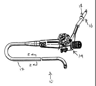
FIG. 1 is a perspective view of a flexible endoscope according to the present
invention;
FIG. 2 is a sectional view of an insertion portion of the endoscope taken
along line 2-
2 of FIG. 1; and
CA 02220667 1997-11-10
-6-
FIG. 3 is a partial sectional view of the insertion portion of FIG. 2, showing
the
construction of the sheath.
Detailed Description
FIGS. 1 and 2 depict an endoscope 10 according to the present invention which
comprises in gross, a flexible insertion tube 12, a handpiece 14 and an
accessory connector 16
with a valved port 181eading to an interior space 20 of the insertion tube 12.
The port 18 thus
allows pressure communication between the interior space 20 and atmosphere.
As best seen in FIG. 2, the interior space 20 carries a pair of fiber optic
bundles 22 and
24, one for carrying light for illumination and the other for carrying the
image to be viewed.
Some other type of flexible endoscopes use a CCD chip (charge couple device)
for electronic
image transmission. A lumen 26 and a steering mechanism 28 also pass through
the interior
space 20. Each fiber optic bundle 22 and 24 comprises a large number of
individual optical
fibers 30 and a sheath 32 surrounding the fibers 30. Generally, the sheath 32
is formed of
silicone. Molybdenum disulfide lubricates the fiber optic bundles 22 and 24 to
reduce friction
between the individual optical fibers 30 as they slide against each other
during the
maneuvering o~xhe insertion tube 12. Molybdenum disulfide is generally also
dispersed
throughout the interior space 20 of the insertion tube 12 to also lubricate
the steering
mechanism 28, the lumen 26 and any other components as they slide against each
other. Other
flexible endoscope, such as gastrointestinal scopes and colonoscopes, may have
more internal
channels to facilitate passage of air, water and the like.
Turning to FIG. 3, the insertion tube 12 comprises a metal inner spiral layer
34 for
rigidity, over which lies a braided metal layer 36 and an outer sheath 38. The
sheath
comprises two layers; an outer layer 40 which ultimately contacts the body of
the patient and
an inner vapor barrier layer 42. Since the insertion tube 12 is inserted
inside the body, the
material choice is critical. The material of the outer layer 40 must be
flexible and
biocompatible. Polyurethanes are commonly used for such application. They
offer a good
CA 02220667 2006-07-04
k~k#
-7-
combination of lubricity, flexibility, strength, durability, and stability, as
well as
biocompatibility. However, due to the vapor permeability of polyurethane, in a
typical
endoscope with a single layer sheath formed of polyurethane, vastly greater
amounts of
hydrogen peroxide or other sterilant may, in some instances, enter the
interior of the interior
space by diffusion through the sheath than would enter through the pressure
equalization port.
In the present endoscope 10, the inner vapor barrier layer 42 prevents the
sterilant
from passing into the interior space 20. It may be formed of any substance
which will be
flexible, not interact with, absorb or allow permeation of vapor based
sterilants such as
io hydrogen peroxide, chlorine dioxide, ethylene dioxide and the like.
Preferred materials
include polyolefins, fluorinated polyolefins, polyvinyldene chloride,
polyvinyldene fluoride, or
fluro-chioro polymers. The inner layer 42 may be formed by any known method,
however, it
is preferred to either co-extrude the inner layer 42 with the outer layer 40
or to deposit the
inner layer 42 onto the outer layer 40 as a film coating. In the latter
instance a non-porous
polymer such as Paralynela product of Union Carbide) may be employed or, a
separate
internal layer between the multilayer sheath (38,36 & 34) and the inner
channels ('including
metal braiding) made of the above mentioned polymers, and the internal
components of the
endoscopes can.be installed.
In addition to hydrogen peroxide gas plasma, there are other sterilization
methods
which employ oxidization processes or strong oxidizers for sterilization. Some
other oxidizing
sterilants and methods include ozone (03), chlorine dioxide (C1O2), EtO,
hydrogen peroxide
vapor without plasma, and peracetic acid. These oxidizing sterilants are
expected to react
similarly with molybdenum disulfide and cause material degradation. The two-
layer
construction of the sheath 38 will protect the endoscope 10 against attack by
any of these
agents.
In a sterilization procedure, the endoscope 10 will typically be washed to
free it of
organic matter and then placed into a sealed chamber (not shown). The pressure
in the
CA 02220667 1997-11-10
-8-
chamber will be reduced and then a vapor based sterilizing agent such as
hydrogen peroxide
will be introduced into the chamber. The sterilant may penetrate and sterilize
the outer layer
40, but the inner layer 42 will block its passage into the interior space 20.
At the time the
sterilant is introduced, the port 18 will be closed thereby preventing access
to the interior
space 20 through this avenue. As the interior space 20 is otherwise sealed,
there remains no
avenue for the sterilant to pass into the interior space and interact with the
mechanisms or
lubricants therein. If using a plasma type process, a plasma is then created
which enhances the
sterilization process and leaves behind no harmful residuals.
While the invention has been particularly described in connection with
specific embodiments thereof, it is to be understood that this is by way of
illustration and not of
limitation, and that the scope of the appended claims should be construed as
broadly as the
prior art will permit. For instance, a particular construction of an endoscope
10 and insertion
tube 12 have been illustrated. However, the invention is not limited thereto;
there are many
variations of such designs known to those of skill in the art, and one of
ordinary skill in the art
would understand that the present teaching of the two-layer sheath 38 could be
applied to any
common design of endoscope and insertion tube. The endoscope 10 illustrated
herein is
suitable for a sigmoidoscopy, however, it will be appreciated that other types
of endoscopes
are capable of incorporating the present invention. Also, polymer materials
are disclosed as
preferred for the inner layer 42, but other materials such as metals, ceramics
and others which
can meet the requirements set forth herein may be substituted therefor. While
the invention is
most useful in vapor phase sterilization, the construction disclosed herein
would also prevent
liquids which may penetrate a polyurethane outer sheath from entering the
interior space.
