Note: Descriptions are shown in the official language in which they were submitted.
CA 02220770 1997-11-12
WO 97/35636 PCTlCTS97/04301
1
DETECTION OF PRESSURE WAVES TRANSMITTED THROUGH
CATHETERILEAD BODY
FIELD OF THE INVENTION
The present invention generally relates to implantable medical devices having
a catheter or lead in direct or indirect contact with a body organ, muscle
group or
limb capable of mechanical movement and more particularly to a method and
apparatus for detecting pressure waves of mechanical and/or acoustic origin
caused
by movement of the body organ and propagated by the catheter or lead body to
the
implanted medical device.
BACKGROUND OF THE INVENTION
Certain organs of the body mechanically expand and contract on a regular
basis, most notably the diaphragm and lungs during breathing and the heart as
it
beats. Muscle groups and body limbs also mechanically expand and contract and
move under the control of the central nervous system. When a dysfunction
occurs
IS in these body systems, examinations are conducted to determine the nature
of the
dysfunction and a variety of therapies are prescribed to restore the function.
Such
examinations include monitoring and the delivery of therapies including drugs
andlor electrical stimulation to restore the affected function.
With respect to the heart, the cells of the chambers of the heart relax and
contract in an organized and relatively rhythmic cycle due to gradual
polarization
followed by rapid and organized depolarization of the cardiac cells. The
depolarization is accompanied by a relatively forceful contraction of the
chamber in
a manner described extensively in the literature. Cycles of this type exhibit
characteristic electrical signal waveforms of the polarization-depolarization-
re-
polarization wavefronts referred to as the PQRST electrogram complex. The
accompanying relaxation of the heart muscle draws blood into the chambers and
the
forceful mechanical contraction ejects blood through the valves. The resulting
blood pressure waves are audible through the adjacent tissue of the thoracic
cavity
as a characteristic "lub-dub" sound. In a similar manner, the relaxation and
CA 02220770 1997-11-12
WO 97/35636 PCT/US97l04301
2
contraction of the diaphragm causes air to be drawn in and ejected from the
lungs
with a characteristic motion and sound.
For a wide variety of reasons, the detection, display and/or measurement of
the electrical signals and the acoustic waves or sounds of these heart and
lung cycles
have long been of medical interest. In addition, the mechanical motion of the
lungs
and surrounding thoracic cavity have been the subject of measurement using
impedance plethysmography.
Of course, cardiac sounds and the sounds of respiration are commonly
listened to by trained medical personnel employing use of passive or active
stethoscopes manually positioned over the patient's chest during a medical
examination. In this manner, congestion in the Lungs, if present, or the
characteristic lub-dub sounds of the sequential depolarization of the atria
and
ventricles of the heart may be listened to. Together with other symptoms, a
number
of illnesses of the heart and lungs may be diagnosed and treatment prescribed.
Efforts have been underway for many years to develop implantable sensors
for temporary or chronic use in a body organ or vessel for a variety of uses,
and
particularly for measuring aspects of the cardiac and breathing cycles.
Catheters
and leads are used in conjunction with a wide variety of cardiac medical
devices,
including implanted and external pacemakers, cardioverter/defibrillators, drug
dispensers, cardiac monitors, cardiac assist devices, implanted
cardiomyoplasty
stimulators, muscle and nerve stimulators, and the like. One or more catheter
or
lead is attached at the proximal end thereof to a device port or terminal and
the
distal end segment thereof is introduced into direct contact with the heart
muscle or
extended within the atrial and/or ventricular heart chamber in contact with
blood
and in indirect contact with the heart muscle. A condition of the heart or of
the
blood is sensed and/or a therapy is delivered through the lead or catheter.
For example, a simple cardiac catheter for measuring blood pressure changes
includes an elongated catheter body extending between a proximal connector end
and a distal end having one or more end openings or a balloon adapted to be
introduced into a heart chamber. Drugs or agents may be dispensed through the
CA 02220770 1997-11-12
WO 97135636 PCT/US97104301
3
catheter lumen to a desired location. Blood pressure fluctuations in the
column of
fluid in the lumen may be measured as long as the distal end opening remains
open
or the balloon remains capable of flexing with pressure changes. However,
unless
anticoagulants are continuously dispensed through the catheter lumen, the
distal end
of the catheter becomes encased in fibrosis interfering with balloon motion
and/or
closing the end openings within a relatively short time. For this and other
reasons,
such simple blood pressure measuring catheters cannot be left in place
chronically
or implanted permanently in association with an implanted medical device.
Catheters have also been proposed including sensors incorporated into the
IO catheter distal tip for measuring various blood parameters. In these cases,
the
catheter body incorporates electrical conductors and proximal end connector
terminals in order to power such sensors and to convey electrical signals from
the
sensors to an implanted or external medical device,.
A lead is a form of a catheter having one or more lead conductors extending
between a proximal connector terminals) and distal exposed electrodes) from
which electrical stimulation may be delivered or electrical signals of the
body may
be detected. A pacing lead includes one or more pace/sense electrodes and
associated lead conductors. A cardioversion/ defibrillation lead includes one
or
more cardioversion/defibrillation~electrodes and associated lead conductors,
and
may also include one or more pace/sense electrodes and associated Iead
conductors.
In the context of cardiac pacemakers and/or cardioverter/defibrillators
comprising implantable pulse generators (IPGs) and leads of this type, a
variety of
indwelling sensors have been proposed in combination with the leads for
sensing
parameters including blood pressure, the rate of change of blood pressure,
blood gas
concentrations, blood pH, and blood temperature or for sensing the mechanical
motion of the heart. The sensed signals as well as the P-wave and/or R-wave of
the
heart sensed through the pace/sense electrodes in contact with the patient's
atrium
and/or ventricle, respectively, are employed for a variety of reasons.
In pacemakers, such indwelling sensors have been proposed for use in
algorithms for adjusting the pacing rate to meet the demand for cardiac output
as
CA 02220770 2004-03-29
66742-647
g
related to a characteristic of the sensed parameter that varies with exercise.
Other
rate-responsive pacemakers have been widely commercialized employing an IPG
mounted piezoelectric crystal sensor responsive to the level of patient
activity,
referred to as an "activity" sensor. The use of an activity sensor and
indwelling
sensor or two or more indwelling sensors in combination and the processing of
the
sensor signals to derive a pacing rate control signal are disclosed, for
example, in
commonly assigned U.S. Patent No. 5,188,078.
Still other rate-responsive pacemakers have been commercialized employing
impedance variations with respiration as measured between spaced thoracic
electrodes as disclosed, for example, in EPO Patent Nos. 0 089 014 and 0 151
689.
The thoracic electrodes may be spaced apart and
away from the heart or may include a pacing lead electrode. In the '689
patent, a
pacing rate control signal is developed from the variation in the measured
impedance as a,function of pulmonary minute ventilation.
1S The use of a variety of sensors in pacemakers is also proposed for
detecting
capture of the heart by a pacing pulse, i.e., detecting the evoked
depolarization in
response to preceding pacing pulse. In order to conserve battery power and
prolong
the life of implanted pacemakers, it is desirable to minimize the energy of
the pacing
pulse tb provide a minimal "safety margin" of the pulse energy over the
threshold
energy su~cient to capture the heart. The detection of the evoked electrical
signal
is difficult because the pacemaker sense amplifier is "blinded" by the
residual
"polarization" energy of the pacing pulse at the sensing electrode for a time
period.
While the detection of an evoked ventricular depolarization R-wave
superimposed
on the decaying residual polarization wave may be possible under carefully
controlled circumstances, the detection of the much lower amplitude evoked P-
wave
has not proven possible. Therefore, the alternate use of indwelling sensors of
the
types described above to detect the change in blood temperature, gas
concentration;
or pressure accompanying the evoked response has been proposed as described in
detail in commonly assigned U.S. Patent Nos. 5,320,643, 5,331,996 and
5,342,406.
CA 02220770 1997-11-12
WO 97135636 PCT/US97/04301
In a further approach, a system disclosed in U.S. Patent No. 4,114,628
suggests the use of a mechanical heart motion sensor in contact with the heart
to
detect capture or LOC. The disclosed sensor in the ' 628 patent is a moving
core,
coiled wire inductor transducer mounted within an endocardial or epicardial
lead
5 coupled to the IPG.
In the context of automatic implantable cardioverter/defibrilIators,
fibrillation is typically detected by the continuous detection and analysis of
features
of the P-waves or R-waves, including high rate, rate regularity, sudden onset
of
high rate, etc. When the atria or ventricles of the heart are in fibrillation,
the
associated P-waves or R-waves become chaotic, and the heart chamber is unable
to
vigorously contract in the normal manner. The confirmation of ventricular
fibrillation by detecting the absence of blood pressure is proposed in U. S.
Reissue
Patent No. Re 27,652. The confirmation of ventricular fibrillation by
detecting the
absence of heart motion characteristic of normal contractions along with a R-
wave
high rate has been proposed in U.S. Patent Nos. 3,815,611 and in the above-
referenced ' 628 patent. The use of the indwelling blood pressure sensors,
blood
gas concentration sensors or temperature sensors to confirm fibrillation by
detecting
a change in the measured parameter in conjunction with the high rate P-wave or
R-
wave is also suggested in the prior art.
Implantable cardiac monitors, e.g. the MEDTRONIC~ implantable
hemodynamic monitor employ pacing leads attached to the patient's heart for
simply
monitoring and storing EGM data and an indwelling pressure sensor for
developing
an absolute pressure signal from the heart. A reference pressure sensor is
incorporated into the connector block assembly for detecting ambient
atmospheric
pressure that is subtracted from the pressure sensor signal to provide the
absolute
pressure sensor. Patient's suffering from congestive heart failure are
candidates for
such implantable hemodynamic monitors. Such patient's suffer from episodes of
cardiac insufficiency accompanied by labored breathing that is not presently
monitored.
CA 02220770 1997-11-12
WO 97135636 PCT/LTS97l0430I
6
The detection of heart sounds rather than the R-wave peak or a pressure
sensor signal has also been suggested anonymously in RESEARCH DISCLOSURE
No. 37I50, entitled "Use of Heart Valve Sounds as Input to Cardiac Assist
Devices"
(March, 1995) for use in controlling operations of pulsatile cardiac assist
devices.
The Listed cardiac assist devices include intra-atrial blood pumps (IABPs),
cardiomyoplasty/cardiac assist devices (of the type described in commonly
assigned
U.S. Patent No. 5,069,680, for example), aortomyoplasty and ventricular assist
devices (VADs). The heart sounds are picked up by a microphone, amplified,
bandpass filtered and compared to a "signature" sound pattern to derive a
control
signal timed to the second or "dub" heart sound, which is related to the
dicrotic
notch of the aortic pressure wave. No specific structure for accomplishing
this is
disclosed. In U.S. Patent No. 4,763,646 to Lekholm, a heart sound detector is
also proposed to be mounted in one or more pacing leads arranged in or about
the
heart or to be mounted in the LPG case for acoustically sensing heart sounds
transmitted through a fluid filled lumen. The use of a pressure sensor,
microphone
or accelerometer is proposed for the heart sound detector.
Despite the prodigious effort expended in developing such indwelling
sensors, few have been found to be useful or acceptable for chronic use.
Typically,
the foreign object body reaction encapsulates the sensor and isolates it from
the
parameter to be detected and measured. In addition, the active sensors require
additional Lead conductors and are electrically inefficient. Consequently,
such
sensors are complex in design, difficult to manufacture, relatively expensive,
and
short Lived.
For example, a great deal of effort has been expended in developing
indwelling absolute pressure and pressure rate of change sensors as noted
above for
measuring these parameters in a heart vessel or chamber. Many designs of such
chronically or permanently implantable pressure sensors have been placed in
limited
clinical use. Piezoelectric crystal or piezoresistive pressure transducers
mounted at
or near the distal tips of pacing leads, for pacing applications, or catheters
for
monitoring applications, are described in U.S. Patent Nos. 4,023,562,
4,407,296,
CA 02220770 2004-03-29
66742-647
7
4,432,372, 4,485,813, 4,858,615, 4,967,755, and 5,324,326, and in PCT
Publication No. WO 94113200, for example. These sensors and an.improved sensor
and operating system are described in detail in commonly
assigned U.S. Patent No. 5,564,434 issued Oct. 15, 1996,
for IMPLANTABLE CAPACITIVE ABSOLUTE PRESSURE AND TEMPERATURE
SENSOR and U.S. Patent No. 5,535,752 issued July 16, 1996,
fog IMPLANTABLE CAPACITIVE ABSOLUTE PRESSURE AND TEMPERATURE
MONITOR SYSTEM.
Other semiconductor sensors employ CMOS IC technology in the fabrication
of pressure responsive silicon diaphragm bearing capacitive plates that are
spaced
from stationary plates. The change in capacitance due to pressure waves acting
on
the diaphragm is measured, typically through a bridge circuit, as disclosed,
for
example, in the article "A Design of Capacitive Pressure Transducer" by Ko et
al.,
in IEEE Proc..Symp. Biosensors, 1984, p.32. Again, fabrication for lung term
implantation and stability is complicated and unproven.
This concentrated focus on the development of accurate, efficient, reliable
and permanently implantable, indwelling pressure sensors on lead bodies has
taken
place in recognition that the detection of pressure waves, particularly in the
cardiac
' and pleural context, has a great number of applications as described above.
However, the indwelling pressure sensor approach requires the implantation of
the
sensor into the heart chamber, where certain sensor types may become fibrosed
and
lose the ability to respond to mechanical heart motion or blood pressure
changes
associated with the contraction of the heart chamber or in association with
the lungs
or diaphragm where fibrosis may also render it ineffective.
In virtually all approaches, it is necessary to rely on additional components
and circuitry which consume more energy, require additional lead conductors,
increase the bulk and cost of the system, increase the cost of the
implantation, and
raise reliability issues. The additional components and circuitry are
increased
further in dual chamber pacemakers. Very few of the numerous approaches of the
CA 02220770 2004-03-29
66742-647
8
prior art have been attempted in an implantable pacemaker
system and fewer yet have been proven clinically useful and
commercially successful.
Therefore, despite the considerable effort that
has been expended in designing such sensors, a need exists
for a body implantable, durable, long-lived and low power
sensor for accurately sensing pressure waves and related
parameters in the body over many years of implantation.
SUMMARY OF THE INVENTION
Accordingly, embodiments of the present invention
provide a practical and durable substitute for an indwelling
sensor particularly for measuring pressure waves at a site
of interest in a patient's body.
Further embodiments of the present invention
employ the lead or catheter body to convey pressure waves
from the site of interest remotely to a low power pressure
wave transducer in contact therewith.
In one aspect, the invention provides a system for
detecting pressure waves emanating from a site in the
patient's body through a catheter extending to the site for
use in diagnosing a body function or timing a medical device
operation comprising: a catheter having an elongated
catheter body extending between a proximal connector end and
a distal end adapted to be placed at the site such that a
pressure wave imparts movement to said catheter body and is
transmitted through said catheter body in response to a body
function; a connector assembly for attachment with said
proximal connector end; a pressure wave detection transducer
mounted in said connector assembly in operative relation
with said proximal connector end for detecting said pressure
wave and providing a pressure wave signal representative
' CA 02220770 2004-03-29
66742-647
8a
thereof; and means for processing the pressure wave signal
for use in an operation of the medical device.
The pressure wave signal is preferably amplified,
bandpass filtered to the pressure wave of interest and used
to trigger a device operation. The system may also include
a reference transducer having the same pressure wave
response characteristics as the pressure wave transducer but
isolated from the proximal connector end for providing a
reference signal including common mode pressure
CA 02220770 1997-11-12
WO 97/35636 PCT/U897/04301
9
wave noise that both transducers are simultaneously subjected to. The
reference
signal is also amplified, bandpass filtered to the pressure wave of interest
and
subtracted from the pressure wave signal to eliminate the common mode pressure
wave noise component. The resulting signal is employed to trigger the desired
device action.
The pressure wave transducer and the reference transducer (if present) are
preferably a piezoelectric crystal transducer, a pressure sensor or an
accelerometer
in direct or indirect mechanical contact with the proximal connector end of
the
catheter. Preferably, the pressure wave and reference transducers are of the
same
type and have the same characteristics and specifications. Any transducer can
be
used, including resonant micro beams, piezoresistive diaphragms etc., so long
as
they are of sufficient sensitivity and responsive to the appropriate frequency
passage.
The pressure wave transducer is preferably encapsulated within the
connector assembly in a position making contact with the lead body through a
connector element. The reference transducer, when present, is preferably also
encapsulated from the body within the connector assembly away from direct or
indirect coupling with the lead connector end and aligned in parallel with the
pressure wave transducer. The reference transducer may alternatively located
elsewhere in the implantable medical device.
Preferably, the catheter is a electrically conducting lead extending into
direct
or indirect contact with the patient's heart, organ, or muscle of interest,
and the
system comprises an implanted medical device of the types described above
coupled
to the proximal connector end of the lead. In the context of a cardiac monitor
or
IPG, cardiac pressure waves and respiration pressure waves are both
transmitted
proximally through the lead body to the pressure wave transducer. The cardiac
pressure waves are caused by the change in blood pressure or direct mechanical
motion of the heart, e.g. the opening and closing of a heart valve, against
the distal
end segment of the lead. The cardiac pressure wave therefore reflects a heart
sound
- and/or mechanical shock originating in the heart. The respiration pressure
waves
CA 02220770 1997-11-12
WO 97/35636 PC"T/US97/04301
are caused by mechanical motion of the Iungs transmitted to the heart and
thoracic
cavity during the respiratory cycle. Either or both pressure wave signals may
be
derived in parallel to detect particular characteristics of the cardiac and/or
respiratory cycle for monitoring the cardiac and/or respiratory cycles or to
provide
5 timing signals) for controlling the device operations described above. It is
believed
that any of the commonly used leads for pacemakers can perform satisfactorily
for
this invention.
The present invention advantageously provides the ability to reliably provide
timing signals for a wide variety of applications described above without
having to
10 solve the problems associated with directly exposing a sensor, particularly
a
pressure or motion sensor, to the hostile environment of the body and without
having to provide additional conductors in the Iead body.
The present invention may be of use with catheters, but is of particular
advantage in use with leads of the types described above. In particular,
pacing Ieads
have been implanted for many years in a large number of patient's hearts in
the
context of pacemaker and pacemaker-cardioverter-defibrillator IPG's. Such
IPG's
may be replaced with IPG's constructed in accordance with the present
invention
having the improved capabilities and features thereof without having to
replace the
Ieads with sensor bearing leads, resulting in a considerable savings and while
providing improved patient treatment.
Moreover, the invention may also be used advantageously in conjunction
with indwelling leads or catheters coupled to external medical devices.
BRIEF DESCRIPTION OF THE DRAWINGS
Other objects, advantages and features of the present invention will be
readily appreciated as the same becomes better understood by reference to the
following detailed description when considered in connection with the
accompanying drawings, in which like reference numerals designate like parts
throughout the figures thereof and wherein:
CA 02220770 1997-11-12
WO 97/35636 PCT/US97/0430I
11
Figure 1 is a schematic illustration of an IPG or monitor implanted in a
patient's chest and an endocardial lead transvenously introduced into the
heart and
traversing the patient's chest;
Figure 2 is a side cross-section view of a lead connector assembly taken
along lines 2-2 of Figure 1 within which at least a piezoelectric crystal
pressure
wave transducer and a reference transducer are incorporated in relation to the
lead
proximal connector end in accordance with a first embodiment of the invention;
Figure 3 is an end cross-section view taken along lines 3 - 3 of the connector
assembly of Figure 2;
Figure 4 is a side cross-section view of a lead connector assembly also taken
along lines 3 - 3 of Figure 1 within which at least a piezoelectric pressure
wave
transducer and a reference transducer are incorporated in relation to the lead
proximal connector end in accordance with a second embodiment of the
invention;
Figure 5 is a side cross-section view of a lead connector assembly also taken
along lines 3 - 3 of Figure 1 within which an accelerometer pressure wave
transducer is incorporated in in-line indirect mechanical contact the lead
proximal
connector end in accordance with a third embodiment of the invention;
Figure 6 is a block diagram of a signal processing system for processing the
pressure wave and reference signals from the pressure wave transducers and
reference transducers;
Figure 7 is a waveform diagram depicting the cardiac cycle pressure wave
detected by the pressure wave transducer in relation to the preceding
intrinsic
PQRST complex;
Figure 8 is a waveform diagram depicting the respiration cycle pressure
wave detected by the pressure wave transducer in relation to a series of
intrinsic
PQRST complexes; and
Figure 9 is a waveform diagram depicting the cardiac cycle pressure wave
detected by the pressure wave transducer in relation to a preceding pace
pulse.
J
CA 02220770 2004-03-29
66742-647
1z
DETAILED DESCRIPTION OF THE PREFERRED EMBODIMENTS
The preferred embodiment of the invention is illustrated in the context of a
connector block for an implantable patient monitor, e.g. the MEDTRONIC~
implantable hemodynamic monitor, or an TPG, e.g. an implantable pacemaker IPG
of the type described in detail in the above-referenced ' 078 and ' 406
patents or an
implantable pacemaker-cardioverter-defibrillator IPG of the type described in
commonly assigned U.S. Patent No. 5,312,441. In such
monitors and IPGs, the connector assembly is molded as
a separate piece part and attached to the hermetically sealed case or can for
the
power source and electronic components in a manner shown, for example, in
commonly assigned U.S. Patent No. 5,070,605.
Figure 1 is a schematic illustration of such an IPG or monitor 10 implanted
in a patient's chest 12 and an endocardial lead 18 (or leads) transvenously
introduced into the heart 14 and traversing the patient's chest 12. The IPG or
monitor 10 includes the connector assembly 20 and the case or can 22 enclosing
the
power supply and circuitry. As the heart 14 contracts and expands, it creates
pressure waves which are transmitted into the distal end segment 16 of lead 18
and
are conducted proximally to the relatively still IPG or monitor 10. Similarly,
as the
lungs 24, 26 expand and contract the pleural cavity and chest with the
respiration
cycle controlled by the diaphragm 28, the chest movement creates pressure
waves
that to the elongated lead 18 impart movement and are
conducted proximally to the relatively still TPG or monitor 10.
Since the lead distal end segment 16 is typically firmly attached to the heart
14 (and may in fact be alternatively attached to the epicardium) so that good
electrical contact is maintained, the cardiac contraction pressure wave may
constitute a reaction to a physical shock, i.e. a rapid mechanical movement,
imparted to the distal end segment of the relatively forceful contraction of
the heart.
The transmitted cardiac contraction pressure wave may comprise the.mechanical_
movement itself effecting an acoustic or ringing response of the lead body and
may
CA 02220770 1997-11-12
WO 97!35636 PCT/US97J04301
13
include a component of the actual cardiac contraction sound, and we define it
as
such.
We have discovered that the cardiac contraction pressure wave may be
transmitted through the solid lead body and readily detected at the proximal
connector end of the lead I8 by a sensor in direct or indirect mechanical
contact
with the lead body. Similarly, we have discovered that the respiratory
pressure
wave, which is more gradual and prinnarily attributable to mechanical motion
of the
lead body may also be readily detected at the proximal connector end of the
lead 18.
This discovery allows the replacement of sensors in the distal tip segment of
a lead
or catheter, which suffer the deficiencies detailed above, with sensors
associated
with the lead's proximal connector end, preferably in the connector block
assembly
of the implanted device. The principles of the invention illustrated in the
following
preferred embodiments are also applicable to detection of pressure waves
originating
in other organs, muscles, muscle groups or limbs conveyed through catheters or
IS leads to the pressure wave sensor in the medical device.
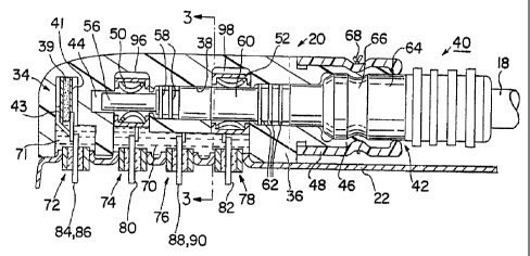
Figures 2 and 3 depict the lead connector module or assembly 20 coupled
with a proximal connector end 40 of a lead 18 and the incorporation of a
pressure
wave transducer 32 and a reference transducer 34 in accordance with a first
embodiment of the invention. Although a specific connector block and lead type
are
illustrated in the figures, it will be understood that the invention may be
practiced
with any lead configuration having in-line or bifurcated lead proximal
connector
ends and connector assembly configurations for such lead connector ends.
In this first embodiment, the transducers 32 and 34 are each formed of a
piezoelectric crystal of the type employed as an activity sensor in
commercially
available MEDTRONIC~ THERA~ DR IPGs for rate-responsive pacing in the
DDDR mode and other modes. In Figure 1, such an activity sensor 30 is depicted
adhered within the can 22. Such transducers 32, 34 are formed of a rectangular
piezoelectric crystal of about 0.250 x 0.125 x 0.022 inches which is reduced
in size
from the activity sensor. The major opposed surfaces of the piezoelectric
crystal 33
are coated with thin film electrodes 35 and 37, and the major opposed surfaces
of
CA 02220770 2004-03-29
66742-647
14
the piezoelectric crystal 39 are coated with thin film electrodes 41 and 43
that are
electrically attached to sensor lead wires as described below. The resulting
capacitive transducer provides an electrical output signal on the sensor lead
wires
that varies in amplitude in response to minute deff~efions of the
piezoelectric crystal
-' in response to the acoustically or mechanically conducted cardiac and
respiratory
pressure waves.
It should be noted that the orientation of the reference transducer 34 should
be in a parallel plane with plane of the pressure wave transducer 32, rather
than in a
transverse plane as depicted for convenience of illustration in the Figures 2
and 3.
The parallel orientation provides a more exact response of both transducers to
common mode noise originating elsewhere in the body, for example.
The connector assembly 20 shown in Figures 2 and 3 is similar to that
described and shown in Figures 4-6 of the above. ' 605 patent. In
particular, the connector 20 is formed of a connector housing 36 of uncolored,
transparent epoxy molded to form an elongated, lead connector end bore 38 open
at
the tubular end 42 and terminating in a pin receptacle chamber 44. The
connector
housing 36 also encloses the transducers 32, 34, feedthrough terminal pins
identified
below and in-line lead retainers 50 and 52 described below. A flexible sleeve
48 fits
over tubular end extension 46.
The bore 38 is shaped to receive the proximal connector end 40 of in=line,
bipolar lead I8. The lead 18 is typically constructed of coaxially arranged
and
electrically insulated coiled wire conductors extending the length of an outer
insulating sheath and forming the lead body surrounding a lumen 54 but may be
constructed without a lumen. The proximal connector end 40 conforms to the IS-
1
standard for bipolar in-Iine lead connectors and includes a proximal connector
pin
56 coupled to the inner coiled wire conductor and sized to fit within the pin
engaging, deflectable beam, cylindrical lead retainer 50. An insulating sheath
overlies the junction of the connector pin 56 and the inner coiled wire
conductor and
is formed with annular moisture sealing ribs 58 that engage the walls of the
bore 38.
CA 02220770 2004-03-29
66742-647
A connector ring 60 is coupled to the outer coiled wire conductor (not
shown) and sized to fit within the pin engaging, deflectable beam, lead
retainer 52.
An insulating sheath overlies the junction of the connector ring 60 and the
outer
coiled wire conductor and is formed with further annular moisture sealing ribs
62
5 thatengage the walls of the bore 38.
The lead connector end 40 is enlarged to a diameter 64 distally to the
connector ring 60 and has an annular groove 66 in diameter 64 shaped to be
retained
in a necked down annular portion of the tubular end extension 46. The
attachment
of the lead connector end 40 in the bore 18 may be secured by a suture ring
68.
10 The secure electrical connection of the connector pin 56 with the
electrically
conductive lead retainer 50 and the connector ring 60 with the electrically
conductive lead retainer 52 is described in detail in the above ' b05
patent.
A series .of electrical feedthroughs 72, 74, 76, 78 are mounted to extend
15 through the mating surface of the can 22 and into cavities 70 or 71
(preferably
minimized into channels) sealed with medical grade silicone rubber adhesive or
the
like when the connector assembly 20 is attached to the can 22. Lead
feedthrough
pins 80 and 82 extend through the lead feedthroughs 74 and 78, respectively
and are
electrically connected to the lead retainers 50 and 52, respectively, by short
wire
conductors. Reference feedthrough pins 84 and 86 extend through double pin,
reference feedthrough ?2 and are electrically connected with the thin film
electrodes
41 and 43, respectively, of the reference transducer 34 by short transducer
wire
conductors. Similarly, pressure wave feedthrough pins 88 and 90 extend through
double pin, pressure wave feedthrough 76 and are electrically connected with
the
thin film electrodes 35 and 37, respectively, of pressure wave transducer 32
by
short transducer wire conductors. Double pin transducer feedthroughs 72 and 76
may be employed because of the extremely low voltage and current signals
generated by the pressure and reference wave transducers 32 and 34.
The connector assembly may be fabricated in one way by positioning the
pressure and reference wave transducers 32, 34 and attached wires within
opening
CA 02220770 1997-11-12
WO 97/35636 PCTlUS97/04301
16
92 of cavity 70 and within cavity 71, respectively, and positioning the lead
retainers
50 and 52 and attaching wires in the depicted enlarged open portions 96 and 98
of
bore 38. The inserted components can then be fixed and sealed from the
environment in those positions with silicone rubber adhesive while leaving the
ends
of the wires exposed for attachment to feedthrough pins. The backfilling of
the gap
between the pressure wave transducer 32 and the outer surface of the retainer
52
with silicone adhesive ensures that a direct mechanical contact is made with
the lead
retainer 52 and indirect contact is made with the lead body. Care must be
taken to
avoid entraining air bubbles in the backfilled silicone rubber adhesive
insulating
layer between the lead retainer 52 side wall and the adjacent conductive thin
film
electrode 35.
Alternatively as shown in Figure 3, the pressure wave transducer 32 is
carefully spaced from the Iead retainer 52 by an electrical insulating layer
35 to
prevent it from contacting the thin film electrode 35 while ensuring indirect
contact
through the lead retainer 52 to the lead body. In practice, the insulating
layer 35
may be a more rigid plastic adhesive for adhering the lead retainer 52 and
pressure
wave transducer 32 (and associated sensor and retainer leads) together as a
sub-
assembly that is inserted into the open portion 98 before it is backfilled.
A further alternative approach providing direct contact of the lead retainer
52
with the piezoelectric crystal 33 can be practiced if the two electrodes are
deposited
on the side where electrode 37 is depicted. Intimate direct contact between
the
pressure wave transducer 32 and the lead retainer 52 can also be achieved by a
thin
layer of adhesive at the contact Line.
In any case, the connector housing 36 may be formed with welding access
ports through which a welding probe may be introduced to weld the conductor
wire
ends to the feedthrough pins as exemplified by welding ports 87 and 89 shown
in
Figure 3. In this final assembly process, the connector assembly 20 is secured
to
the mating surface of can 22, and the conductor wire ends are welded to the
feedthrough pins through the welding access ports. Then, the interior spaces
70, 71
CA 02220770 1997-11-12
WO 97/35636 PCT/US97/04301
17
(or channels) and the access ports are backfilled with medical grade silicone
rubber
adhesive.
The resulting connector assembly 20 of the first embodiment therefore
includes a pressure wave transducer 32 that makes direct mechanical contact
with
the lead 18 and an reference transducer that is isolated from the lead 18 but
subjected to common mode noise sources at the location of the monitor or IPG.
For
example, such common mode noise sources may include pressure waves induced by
body or limb movement, speech, coughing, snoring, footfalls and extraneous
ambient noise.
Turning to Figure 4, it depicts and alternative arrangement of the locations
of the piezoelectric crystal pressure wave transducer 32 and reference
transducer 34.
This orientation allows the direct conduction of mechanical pressure wave
energy in
the pressure wave conveyed up the Lead lumen 54 to deflect the piezoelectric
crystal
33. The pressure wave transducer 32 is in direct axial alignment with the lead
connector pin and mechanically coupled to it by a flexible spacer. e.g. a leaf
spring
100. The leaf spring 100 is maintained in the end of the bore chamber 44 so
that
mechanical contact with the lead connector pin 56 may be maintained given lead
and
connector fit tolerances. As shown, the chamber 44 is extended to the thin
film
electrode 35, and the non-conductive leaf spring 100 fits in that space. A
conductive leaf spring 100 may be used in a monitor or if the thin film
electrode 35
is insulated or if the electrode 33 is located alongside electrode 35. All
other
aspects of the fabrication of the connector assembly 20 of Figure 4 are
similar to
those described above.
The reference transducer 34 is located in a cavity 45 in molded housing 36
that is separated from the lead bore 38 by an internal wall of molded housing
36.
Channels are also formed in the molded housing 36 to direct the transducer
conductors to the reference feedthrough pins 84, 86. After the reference
transducer
34 is positioned in the cavity 45, it is backfilled with silicone rubber
adhesive.
In these embodiments of Figures 1 - 4, the placements of the reference
transducer 34 and the related conductors and feedthrough 72 are arbitrarily
depicted.
CA 02220770 1997-11-12
WO 97/35636 PCT/LTS97/04301
I8
They may be situated in the connector housing 36 at any convenient location
that
provides isolation from the pressure wave conducted up the lead 18. The
preferred
location and orientation of the reference transducer 34 and its related
components is
in a parallel plane to the plane of the pressure wave transducer 32. In an
alternative
embodiment, it is possible to eliminate the reference transducer 34 and
associated
components and employ the signals provided by the activity sensor 30 as
reference
signals for eliminating common mode noise.
The pressure wave transducer 32 may also be placed at any convenient angle
to either of the lead retainers 50 and 52. Moreover, although a single channel
monitor or IPG IO is depicted for the sake of simplicity, it will be
understood that
the same approaches may be taken to provide a second pressure wave transducer
in
relation to a second lead for a dual chamber monitor or IPG of the types
incorporated above.
In addition, although piezoelectric crystal transducers of the type described
are preferred due to their low cost, reliability, low current drain and
responsiveness
to pressure waves of the type described, piezoelectric crystal moving beam
accelerometers may also be used. Other solid state, micro-miniaturized IC
accelerometers and transducers may be substituted for the piezoelectric
crystal
transducers, including miniature IC beam accelerometers and capacitive
accelerometers.
Turning now to Figure 5, it depicts a further embodiment of the invention
employing a micro-miniaturized, accelerometer 102 mounted in alignment with
the
lead connector pin 56 and in indirect contact therewith through a leaf spring
100.
Such accelerometers are typically mounted on a diaphragm, and motion of the
diaphragm effects motion of the moving element of the accelerometer.
The accelerometer I02 is inserted into the chamber 44 through an access port
47 in molded housing 36 that is backfilled with silicone rubber adhesive. The
accelerometer leads I04, 106 are routed to pressure wave feedthrough pins 88,
90 of
the pressure wave feedthrough 76. A reference accelerometer isolated from the
pressure wave sensing accelerometer may also be provided in the embodiment of
CA 02220770 1997-11-12
WO 97/35636 PCT1US97/04301
19
Figure 5 in the same manner as the reference transducer 34 of Figures 2-4. All
other aspects of the fabrication of the connector assembly 20 of Figure 5 and
its
attachment to the can 22 are similar to those described above.
Although the embodiments depicted in Figures 1-5 and described above
employ leads in relation to monitoring the cardiac cycle and/or the
respiratory cycle,
it will be understood that implantable monitors and lPGs are used in a wide
variety
of contexts. A number of other organ and muscle stimulators have been
developed
to effect the contraction of a stimulated organ, muscle, muscle group or limb
and
monitors have been developed to detect the intrinsic contractions or motions
thereof.
I0 It will also be understood that the present invention may be implemented in
catheters used chronically with implantable drug dispensers for monitoring the
cardiac cycle, the respiratory cycle or any other motion of an organ, limb or
muscle
group related to the operation of the drug dispenser.
Turning now to Figure 6, it is a block diagram of a signal processing system
IS for processing the pressure wave and reference signals developed by the
pressure
wave transducer 32 and the reference transducer 34 (if present) in the above
described IPG or implantable monitor embodiments or in any alternative medical
device embodiment using a catheter or lead as described above. The pressure
wave
and reference signals are first amplified in amplifiers 110 and 112,
respectively.
20 The amplified pressure wave and reference signals are then bandpass
filtered in
bandpass filters 114 and 116, respectively. Then, the amplified and filtered
reference signal is subtracted from the amplified and filtered pressure wave
signal in
differential amplifier 118. The resulting signal is applied to the signal
processor
120 of the operating system I22 of interest. The operating system 122 may be
25 microcomputer based IPG, implantable monitor or any other medical equipment
that
the Lead or catheter is coupled to.
In the context of an implantable monitor or an IPG, an additional sense
amplifier 124 is coupled to the lead feedthrough pins 80 and 82 for providing
a
electrical sense signal to the operating system in a manner well known in the
art. In
CA 02220770 1997-11-12
WO 97/35636 PCTlUS97104301
2a
cardiac medical device applications, the electrical sense signal may be the
electrogram of the patient's heart detected through the lead 18.
The bandpass filter characteristics are tailored to pass the range of
amplified
signal frequencies of interest and to reject frequency components in the
signals that
are outside that range. For example, the piezoelectric transducers as
described
above are sensitive to heart sound or motion frequencies of interest as well
as to
footfalls when the patient is ambulatory, muscle artifacts or myopotentials
associated
with limb movements and exercise, and may be responsive to speech and exterior
environmental noise. These may constitute "noise" that are first filtered out
to the
IO extent possible and then subtracted in differential amplifier I18 to derive
the signal
of interest. Moreover, increased signal-to-noise performance may be obtained
by
gating (time windowing) the output signal of differential amplifer 118 inside
signal
processor 120 to a particular time of interest in any given cycle.
In accordance with the present invention, parallel signal processing channels
for deriving multiple signals responsive to each of these sources may be
provided in
the same system. For example, the atrial and ventricular contractions of the
heart,
the respiratory cycle and the patient activity level related to the patient's
ambulatory
rate may alI be derived form the pressure wave signal and the reference signal
{if
present) in parallel signal processing circuits of Figure 6.
In either case, the frequency range of the bandpass filters for each such
channel is selected for the signal to be derived. In sensing cardiac sounds
and
motion components of the pressure wave, the frequency range of interest is
believed
to be between about 0.5-7.0 Hz in the atrium and in the ventricle but may be a
different range depending on the waveform characteristic to be measured. To
sense
patient activity related to ambulatory movement, i.e., footfalls. the
frequency range
of interest representing footfalls is between about 0.5-15 Hz. To detect the
amplitude and frequency of the respiratory cycle, the frequency range of
interest is
about 0.05-0.8 Hz.
Figure 7 is a two second waveform diagram depicting the cardiac cycle
pressure wave detected by the pressure wave transducer in relation to the
preceding
CA 02220770 1997-11-12
WO 97/35636 PCTIUS97/0430I
21
intrinsic PQRST complex as detected from the pressure wave transmitted through
a
conventional pacing Lead implanted in the ventricle of a healthy dog. In this
experiment, a wide bandpass filter was employed, and only the pressure wave
transducer of the embodiment of Figures 2 and 3 was used. Figure 9 is an
idealized
waveform diagram depicting the cardiac cycle pressure wave that would be
detected
by the pressure wave transducer in relation to a cardiac depolarization evoked
by a
preceding pace pulse.
A lag between the peaks of the PQRST complex and the peaks of the double
pulses is observed that is greater than the lag observed between the PQRST
peaks
and the peaks of the lub-dub sound waves observed using conventional chest
electrodes and sound transducers as illustrated in the above-cited RESEARCH
DISCLOSURE No. 37150. The peaks of Figure 7 may represent the pressure ,
waveform of the ventricles in forcefully contracting and expelling blood and
then
relaxing and filling with blood that takes place in closer timed relation to
the
IS PQRST complex. A clear correlation between the double signal peaks of the
pressure wave and the PQRST complex is observed. This correlation is effective
with either an intrinsic depolarization or an evoked depolarization of the
heart and in
both the atrial and ventricular heart chambers.
Figure 8 is a 20 second waveform diagram depicting the respiration cycle
pressure wave detected by the pressure wave transducer in relation to a series
of
PQRST complexes in the same dog experiment. The respiration cycle is much
longer than the cardiac cycle. Pulmonary minute ventilation may be determined
from the amplitude of the peaks of the respiratory cycle and the interval
between
peaks in a manner well known in the art.
While there has been shown what are considered to be the preferred
embodiments of the invention, it will be manifest that many changes and
modifications may be made therein without departing from the essential spirit
of the
invention. It is intended, therefore, in the following claims to cover alI
such
changes and modifications as may fall within the true scope of the invention.