Note: Descriptions are shown in the official language in which they were submitted.
CA 02220949 1997-08-13
WO 96/25098 . PCT/US95/06425
REPEAT FIXATION FOR FRAMELESS STEREOTACTIC PROCEDURE
Background of the Invention
The present invention relates to device, system and
method for stereotactic medical procedures. More
specifically, it provides for-repeated accurate
positioning (fixation) of a patient or part of a patient
for carrying out medical procedures which are done at
different times.
Various medical procedures involve repeated
treatments at different times. For example, application
of radiation is sometimes used for treating brain tumors
or other conditions. Although a single application of
radiation may sometimes be used, under many circumstances
there are sound medical reasons to use repeated
application of radiation at different times.
The treatment of a radiation therapy patient can be
broken down into four stages. These are (1) diagnostic
evaluation, (2) treatment planning, (3) simulation and
(4) treatment. Our repeat fixation device is applicable
to the latter three phases of the treatment process. In
the first stage of diagnostic evaluation the physician
decides which tissues are at risk of disease and should
be targeted. The patient may undergo many diagnostic
tests including angiography, computerized tomography (CT)
' 25 and magnetic resonance (MR) imaging. After the physician
is satisfied that they have identified the tissues at
-1-
CA 02220949 1997-08-13
WO 96/25098 PCT/US95/06425
risk, the patient then undergoes a process known as
treatment simulation. This process involves obtaining a
set of images such as plane films, digital images, CT,
MRI, and ultrasound images. These radiographs allow the
physician to select a specific path for each radiation
beam which only includes the tissues at risk and, excludes
normal tissues. Because the tissues the physician has
targeted are often radiographically transparent the
physician routinely relies upon radiographic landmarks to
infer the proper beam alignment. These same landmarks
are subsequently imaged on similar radiographs taken with
the therapeutic x-ray beam prior to administering the
radiation treatment. These pretreatment radiographs,
which are known as therapy portal films, allow the
physician to judge the appropriate alignment of the
treatment beam and the patients anatomy. The frequency
at which these portal films are repeated is dependent
upon the complexity of the patient setup and the
proximity of the beam to critical structures (such as a
patient's optic nerve).
A routine course of radiation therapy may span
anywhere from 10 to 64 fractions over a period of two to
six weeks. The number of treatments dependent upon the
specifics of the particular disease. For each fraction
- 2 - '
CA 02220949 2002-O1-10
the patient must be repositioned at the teletherapy unit
and aligned relati~re to the radiation beam.
There exists a clinical situation in which the
target tissues cannot be adequately localized by their
proximity to radiographically opaque structures as
required by the above simulation procedure.
Arteriovenous malformations, acoustic neurinomas and
other small intracranial targets are examples of such
clinical entities. To enable the identification, and
subsequent treatment of such targets, a new and very
powerful technique known as radiotherapy has been
developed. (Radiosurgery is usually considered to be a
single fraction radiotherapy treatment, meaning a single
treatment, although it may also be more broadly
interpreted. Multiple radiotherapy treatments are often
called high precision radiotherapy or fractioned
stereotactic radiot.herapy.) This technique allows small
intracranial targets to be identified and treated to a
very high degree of precision.
The radiosurgical technique uses stereotactic
principles for targeting, localization and treatment.
The procedure begins with a stereotactic reference system
being fixed to the patient's skull. This reference
system remains fixed relative to all intracranial points
throughout the entire radiosurgical procedure. All
- 3 -
CA 02220949 2002-O1-10
3
diagnostic exams, such as angiography, 'CT and MR scanning
include a set of fiducial markers which allow all points
within the image to be localized relative to the
stereotactic reference frame.
Once the target tissues have been identified the
path of radiation t>eams can be mathematically computed.
The computer algorithms, which support this procedure,
allow the clinicians to evaluate the amount of dose which
would be deposited within the patient if the simulated
beams were actually x-ray beams were applied along the
proposed paths. In an attempt to arrive at a treatment
plan which adequately confines the radiation dose to the
target tissues while limiting the dose to all normal
tissues the beams of radiation are modified, eliminated
or new beams added to the plan. Once a plan with an
acceptable dose distribution has been arrived at the
information on beam trajectory is transferred to the
radiotherapy treatment unit. A single fraction of
radiation is then given to the patient and the
stereotactic frame is removed. The entire length of the
procedure, from frame applicati0I1 through treatments
usually spans 6 to 8 hours.
The present inventors' prior U.S. patents listed
below, assigned to the assignee of the present
application disclo~,e
- 4 -
CA 02220949 1997-08-13
WO 96!25098 PCT/US95/06425
techniques for providing stereotactic radiosurgery with a
high degree of precision:
U.S. Patent Issue Date Title
5,027,818 July 2, 1991 DOSIMETRIC TECHNIQUE
v 5 - FOR STEREOTACTIC
RADIOSURGERY
5,189,687 February 23, 1993 APPARATUS FOR STEREO-
TACTIC RADIOSURGERY
The techniques of the inventors' above patents allow
the patient to be precisely positioned relative to
radiation beams of stereotactic radiosurgery to within
0.2 mm plus or minus 0.1 mm. Although this works very
well for single fraction therapy, there exist clinical
settings where fractionating the total dose, i.e.
dividing the dose into many small fractions, would yield
additional therapeutic advantage. In the radiotherapy
procedure, once the reference frame has been removed from
the patient the relationship between intracranial target
points and the reference system is lost. Because the
above procedure would require the reference frame to
remain fixed to the patient's skull through the entire
course of treatment, which may last several weeks, this
approach is considered inappropriate for fractionated
therapy. Alternately, each fractional treatment would
r
require a laborious and time-consuming procedure to re-
- 5 -
CA 02220949 1997-08-13
WO 96/25098 ~ PCT/US9S/06425
determine patient position for second and subsequent
treatments.
There exist several different techniques for non- ~
invasive repeat fixation. These methods can be broken
down into three basic categories. These are bite plate
systems, contour realignment systems and mask systems.
All of these systems have design flaws which can lead to
unacceptable, and undetectable, positional errors.
The mask techniques have been used in radiation
therapy for over three decades. In these system a custom
mask, which snugly fits either the face or the entire
head, is fabricated. For high precision radiotherapy the
mask is then attached to a stereotactic reference frame,
similar to the frame used for any stereotactic procedure.
Prior to each diagnostic exam the patient is placed into
the mask/frame system and normal stereotactic fiducial
systems are used for image registration.
Mask immobilization and repositioning systems have
been used extensively in radiation therapy. From
multiple reports in the literature mask systems appear to
have a repeat fixation tolerance no better than 3 to 5
mm. It is our opinion that this level of accuracy is
unacceptable for fractionated radiotherapy.
Bite plate systems have also been used in
radiotherapy for several decades. This technique
-
CA 02220949 1997-08-13
WO 96/25098 ' PCT/L1S95/06425
requires the fabrication of a customized bite plate. The
plate fits snugly onto the patient's teeth. As with the
~ mask/frame systems, the bite plate is fixed to a
stereotactic reference frame which then accepts the
routine set of fiducial markers for both plane film
radiography, CT and MR scanning. The primary
disadvantage of this system is that the bite plate is
used for both localization and patient fixation. The
bite plate not only provides the reference for
l0 stereotactic localization, but it also is the mechanism
which is used to move the patient into position. Moving
the patient by use of the bite plate produces torque on
the bite plate-teeth interface. An analysis of this
approach reveals that very small movements in the bite
plate position, relative to the patient's teeth, can
result in large translations and rotations of the
intracranial targets. Since no method of alignment
verification has ever been developed, these errors go
undetected.
An alternate system for patient positioning uses the
patient's own anatomical contours as the stereotactic
reference system. In this approach a CT or MR scan is
taken and a three dimensional reconstruction of the
patient's surface is obtained. These contours act as the
' 25 reference system for stereotactic localization.
_ 7 _
CA 02220949 1997-08-13
WO 96/25098 PCT/US95/06425
The usual diagnostic exams are carried out and the
treatment is then planned using the same stereotactic
principles used in routine radiotherapy. The target is
identified and the patient's surface contour coordinates
are measured relative to the isocenter. The patient is
placed at the teletherapy treatment unit: and the surface
contours are again obtained through the use of surface
digitization. A set of algorithms then calculate the
translations as well rotations required to reposition the
patient's target over the teletherapy units isocenter.
The accuracy of such systems under clinical test
conditions have been shown to be approximately two to
three mm.
When performing fractionated radiotherapy, accuracy
in applying the radiation is very important. Some tumors
or other conditions require that the radiation be
concentrated in relatively small volumes. Misalignment
of the radiation beam may cause an insufficient amount of
radiation to be applied to the tumor or other target.
Further, such misalignment may increase the likelihood
and/or degree of damage to healthy tissue adjacent the
tumor or other target.
Fractionated radiotherapy may be imprecise if the
tumor or other target cannot be localized with a
sufficient degree of accuracy. However, this need for '
_ 8 _
CA 02220949 2002-O1-10
proper localization-~s the same need wh~i~h one has when
performing single dose radiotherapy and this need is
addressed by the present inventors' incorporated by
reference patents. The additional factor in fractionated
radiotherapy is the need to easily and accurately repeat
a position of the patient. If the position of the
patient was accurate relative to the first treatment, the
repositioning should normally cause the patient to assume
the exact same position (relative to the treatment
mechanism) for the second and subsequent treatments.
However, if the second or other subsequent treatment is
performed with the patient only slightly moved from the
first treatment position, this will introduce
inaccuracies. The repeat fixation techniques discussed
above have the indicated disadvantages.
More generally, the need for repeat fixation of a
patient or portion of a patient exists outside of
radiotherapy. In the general case, one wishes to perform
a first medical procedure on a patient with a precise
localization of portions of the patient, and, at some
later time, perform a second medical procedure on the
patient with a precise localization of portions of the
patient. One can rE_peat laborious and time-consuming
localization steps for the second medical procedure, but
this increases medical costs and complexity. As used
_ g _
CA 02220949 2002-O1-10
herein, a medical px:~ocedure is a procedure for diagnostic
and/or remedial purposes.
Surr~mary of the Invention
The present invention provides a new and improved
method and system fcr repeat fixat:ian in performing medical
procedures. More specifically, the present invention
provides for highly precise non-invasive repeat fixat:ion.
Further, the present invention provides repeat fixation in
which a locator is mechanically independent from any
structures used for positioning the patient. That is, any
structure used to position the patient does not move the
locator except by way of the patient. The present invention
also provides repeat f_:ixation for stereotactic radiotherapy.
Further, the present invention provides repeat fixation
which allows relative=Ly fast relocalization of a patient
after an initial loca=_:ization. The px-esent invention also
provides repeat fixat:_on which minimizes or avoids the
disadvantages o.f prior techniques discussed above.
- 10 -
CA 02220949 1997-08-13
WO 96/25098 PCTlUS95/0642~
The above and other features of the present
invention which will be more readily understood when the
following detailed description is considered in
conjunction with the accompanying drawings are realized
by a medical method including the steps, not necessarily
in order, of: positioning a patient for a first medical
procedure: and attaching a mechanically free locator to
a patient, the locator having at least 3 LEDs (light
emitting diode) thereon and being in registry with a
portion of the patient. As used herein, a mechanically
free locator is one which is used for localization
without being rigidly fixed to a structure other than
possibly a portion of a patient. LEDs are used a first
time to get precise positioning information relative to
at least part of the patient.
A first medical procedure is performed on the
patient. After the first medical procedure, the locator
is removed from the patient. At a later time, the
locator is re-attached to the patient, the locator again
being in registry with the portion of the patient and
having an identical orientation relative to the portion
of the patient as when the locator was previously
attached. After the re-attaching step, the LEDs are used
a second time to get precise positioning information
relative to the at least part of the patient. After the
- 11 -
CA 02220949 1997-08-13
WO 96!25098 PCT/US95/06425
re-attaching step, a second medical procedure is
performed on the patient.
Preferably, the attaching and re-attaching of the
locator is non-invasive. As used herein, non-invasive
shall mean that no holes need to be created in a patient
and no patient tissue needs to be removed in ordex to
attach and re-attach the locator.
More specifically, the locator is a bite plate with
an external portion connected thereto, and the LEDs are
on the external portion. The attaching includes using a
mold of dental impression material to bring the bite
plate in registry with teeth of the patient, and wherein
the re-attaching uses the mold to bring the bite plate in
registry with teeth of the patient with an identical
orientation relative to the teeth as when the bite plate
was previously attached.
In one technique of the invention, the first medical
procedure is an imaging of at least a portion of the
patient and the second medical procedure is a remedial
procedure treating at least one problem precisely
localized in the first medical procedure. The second
medical procedure may use a probe inserted in the patient
for treatment of the patient or the second medical
procedure includes radiotherapy.
- 12 -
CA 02220949 1997-08-13
WO 96/25098 PCT/US95/06425
In another aspect of the invention, both the first
and second medical procedures include radiotherapy.
The using of the LEDs the first and second times
utilizes a sensing subsystem for sensing the positions of
the LEDs. Before performing the second medical
procedure, the patient is positioned using a positioner
independent of the locator to secure at least the portion
of the patient in a desired position.
The present invention may alternately be described
as a system for medical procedures, the system including
a locator attachable to a patient, having at least 3 LEDs
thereon, and having a registration portion for
registration with a portion of a patient's body. The
registration portion allows removal of the locator from
the patient and re-attachment to the patient with an
identical orientation relative to the portion of the
patient as when the locator was previously attached. The
locator is mechanically free such that a patient is
positionable without applying forces to the locator
during patient positioning. The system has a positioner
independent of the locator and operable to secure at
least the portion of the patient in a desired position.
A sensing subsystem is operable for sensing the positions
of the LEDs when the patient is in the desired position.
The locator is non-invasive. The locator is more
- 13 -
CA 02220949 1997-08-13
WO 96!25098 PCT/US95l06425
specifically a bite plate with an external portion
connected thereto, and the LEDs are on the external
portion, and the bite plate has dental impression -
material for fabrication of a mold to bring the bite
plate in registry with teeth of the patient, and the mold
is operable to bring the bite plate in registry with
teeth of the patient with an identical orientation
relative to the teeth as when the bite plate was
previously attached.
The system further includes a radiotherapy apparatus
for applying radiation treatment to a patient, the
positioner and sensing subsystem allowing proper
positioning of the patient for applying radiation
treatment. The system further includes an imaging
subsystem for imaging the patient.
The present invention may further be described as a
medical method comprising the steps, not necessarily in
order, of: positioning a patient for a first medical
procedure; attaching a locator to a patient, the locator
having at least 3 fiducial markers thereon and being in
registry with a portion of the patient; using fiducial
markers a first time to get precise positioning
information relative to at least part of the patient: and
performing a first medical procedure on the patient.
After the first medical procedure, the locator is removed
- 14 - '
CA 02220949 1997-08-13
R'O 96/25098 PCT/US9510642~
from the patient. At a later time after the removal of
the locator, the locator is re-attached to the patient,
the locator again being in registry with the portion of
the patient and having an identical orientation relative
to the portion of the patient as when the locator was
previously attached. After the re-attaching step,
fiducial markers are used a second time to get precise
positioning information relative to at least part of the
patient. After the re-attaching step, a second medical
procedure is performed on the patient. The locator is a
bite plate with an external portion connected thereto,
and the fiducial markers are on the external portion, and
wherein the attaching includes using a mold of dental
impression material to bring the bite plate in registry
with teeth of the patient. The re-attaching uses the
mold to bring the bite plate in registry with teeth of
the patient with an identical orientation relative to the
teeth as when the bite plate was previously attached.
In one technique, the first medical procedure is an
2o imaging of at least a portion of the patient and the
second medical procedure is a remedial procedure treating
at least one problem precisely localized in the first
medical procedure. The second medical procedure uses a
probe inserted in the patient for treatment of the
- 15 -
CA 02220949 1997-08-13
WO 96/25098 PCT/US95/06a2~
patient or the second medical procedure includes
radiotherapy.
In another technique of the invention, both the
first and second medical procedures include radiotherapy.
In a specific aspect of the invention, the fiducial
markers used the first time are objects other than LEDs
and the fiducial markers used the second time are LEDs
put on the external portion at locations of the objects.
In an alternate specific aspect of the invention,
the fiducial markers used the first and second times are
LEDs and the using of the fiducial markers the first and
second times utilizes a sensing subsystem for sensing the
positions of the LEDs. Before performing each of the
first and second medical procedures, the patient is
positioned using a positioner independent of the locator
to secure at least the portion of the patient in a
desired position.
The present invention may alternately be described
as a system for medical procedures, the system including
a locator attachable to a patient, having at least 3
fiducial markers thereon, and having a registration
portion for registration witha portion of a patient's
body, the registration portion allowing removal of the
locator from the patient and re-attachment to the patient
with an identical orientation relative to the portion of
- 16 -
CA 02220949 1997-08-13
WO 96125098 ' PCT/US95/06425
the patient as when the locator was previously attached,
the locator being mechanically free such that a patient
is positionable without applying forces to the locator
during patient positioning. A positioner is independent
of the locator and operable to secure at least the
portion of the patient in a desired position. A,sensing
subsystem senses the positions of the fiducial markers
when the patient is in the desired position. In a
specific aspect of the invention, the locator is a bite
plate with an external portion connected thereto, the
fiducial markers are on the external portion, and the
bite plate has a mold to bring the bite plate in registry
with teeth of the patient, and the mold is operable to
bring the bite plate in registry with teeth of the
patient with an identical orientation relative to the
teeth as when the bite plate was previously attached.
The system may further include a radiotherapy
apparatus for applying radiation treatment to a patient,
the positioner and sensing subsystem allowing proper
positioning of the patient for applying radiation
treatment. A radiotherapy apparatus configured for
radiosurgery, a standard linear accelerator, a
radiosurgery apparatus as described in the above prior
patents, and any other device for applying therapeutic
f
- 17 -
CA 02220949 1997-08-13
WO 96!25098 PCT/US95/06425
radiation would be considered a radiotherapy apparatus as
the term is used herein.
The fiducial markers includes three LEDs which
uniquely define a plane.
Brief Descrit~tion of the Drawings
The above and other features of the present
invention will be more readily understood when the
following detailed description is considered in
conjunction with the accompanying drawings wherein like
characters represent like parts throughout the several
views and in which:
FIG. 1 is a simplified diagram of the system of the
present invention;
FIG. 2 is an enlarged side view of a patient's head
with portions of the present invention attached thereto;
FIG. 3 is a detailed view of locator according to
the present invention; and
FIG. 4 is an exploded view of the locator of FIG. 3.
Detailed Description
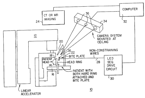
The system 10 of the present invention is shown in
FIG. 1 as having a linear accelerator 12 for performing
stereotactic radiotherapy on a patient's head 14 which is
18
CA 02220949 1997-08-13
WO 96/25098 PCTlUS95/06425
on a surgical table 16 (shown only partially) and secured
thereto by way of a head ring 18. The details of the
accelerator 12 and table 16 are not a necessary part of
the present invention and need not be discussed.
Moreover, these would be constructed and operable in the
manner discussed with respect to the structures and
techniques of the above incorporated by reference U.S.
Patents of the inventors, this allowing the precision
application of radiotherapy to the patient.
As an alternative or additionally to the accelerator
12, a probe 20 (constructed in known fashion) for
stereotactic surgery may be mounted to anchor 22 secured
to the table 16 as shown or to a wall or other structure
such as a linear accelerator, CT, MR, or any other
reference required (not shown). The probe 20, which is a
scalpel, laser, or other surgical apparatus, may
alternately have LEDs thereon for sensing the exact
position and direction (orientation) of the probe in
space using known techniques such that the probe need not
be attached to anything.) A further alternative or
additional feature may be an imaging system such as
computerized tomography (CT) or magnetic resonance (MR)
system 24. One or more of the accelerator 12, probe 20,
and imaging system 24 are used to perform medical
" 25 procedures on the patient.
- 19 -
CA 02220949 1997-08-13
WO 96/25098 PCT/US95/06425
The present invention provides for repeated fixation
of a locator in registry with (i.e., uniquely positioned
relative to) a portion of a patient. Fefore discussing
details of how this is accomplished, it will generally be
noted that the locator is used to provide a frame of
reference for performing a first medical procedure and
the locator is then removed. The locator is then re-
attached to the patient such that a second medical
procedure could be performed. The medical procedures may
be any diagnostic and/or treatment procedures. However,
the discussion which follows will emphasize use of the
technique for fractionated stereotactic radiotherapy.
The present system uses a bite plate 26 connected
by non-constraining (i.e., they are loose and do not
significantly pull on the bite plate) wires 28 to an LED
sequential drive circuit 30. (In lieu of the wires, a
wireless arrangement, not shown, could be used to strobe
the LEDs or a drive circuit could be on the bite plate
itself.) Circuit 30 is also connected to a computer 32.
The computer is connected to the imaging system 24 and a
camera system 34. The camera system 34, which serves as
a sensing subsystem, may be of a known type having
several cameras 36 as part thereof in order to locate the
bite plate 26 by way of several LEDs (not shown in FIG.
1) thereon. The camera system 34 and technique for '
- 20 - '
CA 02220949 2002-O1-10
strobing the LEDs (sequentially-lighting them one at a
time) may be that disclosed in U.S. Patent 5,198,877,
issued to Schulz r~r~ March 30, 1993, assigned on its face
to PixSys, Inc.
Such a camera system is commercially available from
PixSys, Inc.
With reference now to FIG. ~, the patient's head 14
is restrained and can be positioned by use of a head ring
18, which ring would then be fixed in place using
techniques discussed in the present inventors'
incorporated by reference patents. The head ring 18 may
be of any type used or developed to constrain the head.
The bite plate 26 is a type of locator and has at
least three LEDs 38 (only two visible in FIG. 2) thereon.
The three LEDs are not in a line and therefore uniquely
define a plane. Most advantageously, the bite plate 26
is mechanically free such that a patient is positionable
without applying forces to the locator during patient
positioning. The bite plate 26 i.s more specifically
independent any structures (such as ring 18) used for
positioning the patient (such structures being called
positioning structures). That is, any structure used to
position the patient does not move the bite plate 26
except by way of the patient. In that fashion, no forces
or torques are applied to the bite plate 26 which might
- 21 -
CA 02220949 2002-O1-10
cause it to slightl}' change its position relative to the
patient.
With reference to FIGS. 3 and 4, the bite plate 26
has a plastic mouth portion 39 having tooth imprints 40
(only a few shown fcr ease of illustration) previously
formed of dental mold material on mouth portion 38 in
known fashion. A mount plate 42 is integral with or
fixed to mouth portion 39. Three holes 44 are disposed
within the mount plate 42 and allow it to removably
l0 receive a marker plate 46 having three posts 48 mating to
the holes 44. The marker plate 46, which is planar and
parallel to the likewise planar mount plate 42, can be
constructed of transparent plastic and have LEDs 38
disposed therein (as shown) or mounted on a surface
thereof.
An alternate marker plate 50, shown in FIG. 4 only,
may be shaped the same as marker plate 46 and have three
posts 52 (only one visible) for securing it to the mount
plate 42 by way of holes 44. Instead of using LEDs as
fiducial markers, marker plate 50 has three markers 54
which may be radiopaque markers for angiography or CT
scanning or which may be magnetic resonance markers for
MR scanning. Only two of the markers 54 are visible in
FIG. 4, but it will be appreciated that their placement
- 22
CA 02220949 1997-08-13
R'O 96!25098 PCTlUS95/06425
and positioning would preferably be the same as shown for
LEDs 38 in FIG. 3.
_ Considering now all of the FIGS., the operation of
the invention for fractionated stereotactic radiotherapy
will be discussed.
Prior to the patient undergoing either angiography,
CT scanning, or MR scanning, the mold corresponding to
tooth imprints 40 is made by placing mouth portion 39
with dental impression material against the teeth of the
1o patient. Known techniques allow such a mold to be made
in about 10 minutes. The mouth portion 39 would then be
permanently fixed by adhesive or otherwise to the mount
plate 42 (assuming mount plate 42 was not integral with
mouth portion 39). The mount plate 42 may be about 3 cm
by 6 cm and would have the three holes 44 therein.
A temporary adhesive may then be used to fix marker
plate 50 to the mount plate 42 by having posts 52
inserted in the corresponding holes 44. The imaging
system 24 images the brain of the patient and senses the
position of the at least three markers 54. Three
dimensional positions are determined within 0.2 mm
throughout the region of interest. Although FIG. 1 has
shown the imaging system 24 at the same location as the
accelerator 12, it will be appreciated that they could be
at separate locations. Instead of using the markers 54
- 23 -
CA 02220949 1997-08-13
WO 96/25098 ' PCT/US95/0642s
sensed by imaging system 24, one could alternately use
the LEDs 38 on the marker plate 46 during the initial
imaging and the computer 32 could combine position data -
relative to the LEDs 38 with the imaging data from imager
24.
During the imaging, the head clamp ring 18 would not
necessarily be used, but some patient restraint would
normally be used just to remind the patient to hold still
for the approximate 30 seconds for complete imaging.
After the diagnostic images have been obtained, a
routine stereotactic radiosurgical planning session is
conducted. After an acceptable plan has been arrived at
the isocenter, or isocenters, of the plan are identified
relative to the bite plate markers. This then creates a
link between the external reference system, the markers,
and the intracranial target.
The patient is then brought into the treatment area.
They are positioned and immobilized through the use of
comfortable head clamps. At this point the markers used
in the diagnostic procedures can be localized through the
use of a high precision digitizing probe (not shown).
Instead of using a digitizing probe (not shown) to
locate specific marker points on the marker plate such as
plate 50, the marker plate 50 could be separated from
mount plate 42 and the marker plate 46 attached to mount
- 24 -
CA 02220949 1997-08-13
WO 96125098 PCT/US95/06425
plate 42 before mouth portion 39 is placed back in the
patient with his or her teeth in registry with the
- imprints 40. Using marker plate 46, the infrared LEDs 38
are strobed and the camera system 34 identifies the exact
position of the plate 46 with respect to six degrees of
freedom. In other words, the use of at least three LEDs
not in a line allows a precise determination of the
position of plate 46 relative to x, y, and z axes and
rotation about x, y, and z axes (hence six degrees of
freedom).
Since the positions of the markers relative to the
intracranial target (such as a brain tumor) are known, it
will be known what the positions of the markers should be
in order for the target to be at the isocenter of the
accelerator 12. Camera system 34 provides the current
position of the markers to the computer 32. Comparing
the current positions of the markers with the proper
positions, the computer 32 computes the appropriate 3
dimensional translations and 3 axis rotations which are
required to move the patient to the proper position. For
each subsequent treatment after the first radiation
treatment, the patient is again placed at the approximate
treatment position, the positions of the fiducial markers
are determined and the required movements are computed
and performed.
- 25 -
CA 02220949 1997-08-13
WO 96/25098 PCTlUS9510642~
Most importantly, the repositioning of the patient
to the proper position for treatment does not use the
bite plate 26. Instead such repositioning of the patient
would use the head clamp ring 18. Therefore, and since
the bite plate 26 is not connected to the positioning
structure, such repositioning does not ~~ut forces or
torques on the bite plate 26. Thus, the position locator
(bite plate) avoids the misalignments or errors which
would otherwise to introduced by having a locator plate
fixed to a structure used to reposition the patient.
In order to test the above system, both the known
surface contour method as well as the present technique
has been implemented in anatomical phantoms. To test out
the accuracy and precision of the technique the phantom,
a Styrofoam manikin, was fitted with a rigid stereotactic
frame. The phantom was then scanned and localized and
placed into the correct treatment position. The
anatomical contours and the bite plate markers were
localized. The phantom then underwent a series of
precise moves which included both individual translations
and rotations and combined moves. These moves were
carried out to within 0.1 mm and 0.2 degrees. After each
move the contours and bite plate positions were again
obtained. The inverse move, the move required to
reposition the phantom back to isocenter was then
- 26 -
CA 02220949 1997-08-13
R'O 96/25098 PCT/US95/06425
computed. The results of the experiment showed that the
contour method was able to reposition the phantom to
.. within 2 mm of the initial position. The bite plate
system was able to accomplish this move to within 0.3 mm.
The above increase in precision is nearly an order
of magnitude. More importantly the dose gradient
routinely obtained in radiotherapy results in a decrease
in dose from the 90% intensity to the 50% intensity in
approximately 2 mm. This means that tissues at the edge
l0 of the targeted volume have a high probability of
receiving a subclinical dose for any given fraction. The
increased accuracy obtainable with the bite plate system
substantially reduces the probability of positional
targeting errors.
Although the locator is a bite plate in the
preferred embodiment, the present invention broadly
contemplates other locators which can be place in
registry with a portion of a patient.
Advantageously, the bite plate used herein is a non-
invasive locator and avoids the discomfort associated
with techniques requiring one to put one or more holes in
a patient or otherwise remove tissue from a patient.
However, the present invention also has applicability to
invasive locators which are mechanically independent of
- 27 -
CA 02220949 1997-08-13
WO 96/25098 PCT/US95106425
any patient positioning structure (i.e., members used to
change or adjust patient position).
Although the present description h<~s assumed the use
of three markers such as LEDs 38 or markers 54, more than
three could be used and may help provide more accurate
positioning information. For example, a fourth LED not
in the plane defined by LEDs 38 could provide useful
additional information.
Various computer programs may be used to provide the
relationship between intracranial or other target points
and the markers or LEDs. Likewise, various computer
programs may be used to compute the appropriate 3
dimensional translations and 3 axis rotations which are
required to move the patient to the proper position.
The discussion has so far assumed that one would
want to adjust the patient position after re-attachment
of the bite plate so that the patient position for a
second medical procedure (either diagnostic or remedial)
is identical to the initial patient position. However,
the present invention also contemplates that the second
position could be stabilized offset from the first
patient position. In that case, the second medical
procedure could use a transformation so that treatment by
the probe 20 or imaging by imager 24 could be adjusted to
take into account differences between the first patient
- 28 -
CA 02220949 1997-08-13
R'O 96/25098 PCT/US95/06425
position and the second patient position. Because the
accelerator 12 movement relative to the patient is
normally limited to arcs about two transverse axes, it
would be more difficult to adjust for offset between the
first patient position and the second patient position,
although a radiation head with a greater degree of
freedom of movement could allow one to use such a
transformation. Using such a transformation technique
would allow one to secure the patient position without
requiring that the patient position is identical to what
it was for the previous treatment. Under such
circumstances, a positioner which simply stabilizes the
patient position would be sufficient even if the
positioner did not provide the ability to move or re-
position the patient by way of it. Moreover, if the
medical procedure was sufficiently fast, one might be
able to avoid use of even a simply position-stabilizing
positioner.
Although not shown, one could also have a set of
LEDs on the radiation emitting head, collimator, or other
part of the linear accelerator 12 and/or the head
support. By proper placement of the LEDs to detect any
misalignments of the type discussed in the inventors'
incorporated by reference patents, the various
misalignment correcting mechanisms of those patents would
- 29 -
CA 02220949 1997-08-13
WO 96/25098 PCT/US95/06425
not be required. Instead of correcting for misalignments
using those mechanisms, use of such LED:a on part of the
linear accelerator 12 and/or the head support would allow
the system to not only compute the translation/rotation
of the patient relative to the nominal isocenter of the
linear accelerator, but would allow the system to compute
the actual isocenter. Thus, the patient could be moved
to proper position relative to the actual isocenter.
This compensates for any offset between the nominal
isocenter (isocenter absent the misalignments) and the
actual isocenter.
Although specific constructions have been presented
herein, it is to be understood that these are for
illustrative purposes only. Various modifications and
adaptations will be apparent to those of sJtill in the
art. In view of possible modifications, it will be
appreciated that the scope of the present invention
should be determined by reference to the claims appended
hereto.
- 30 -