Note: Descriptions are shown in the official language in which they were submitted.
CA 02221446 2005-03-09
OPTICAL SENSOR INCLUDING INFORMATION ELEMENT
BacktZround of the Invention
Field of the Invention
The present invention relates generally to more effective calibration and use
of light-emitting
diodes. More particularly, the present invention relates to an apparatus and
method of calibrating and
using light-emitting diodes in a sensor for use with an oximeter system.
Description of the Related Art
Light-emitting diodes (LEDs) are used in many applications. In certain
applications, Imowledge
of the particular wavelength of operation of the LED is required to obtain
accurate measurements. One
such application is noninvasive oximeters conventionally used to monitor
arterial oxygen saturation.
In conventional oximetry procedures to determine arterial oxygen saturation,
light energy is
transmitted from LEDs, each having a respective wavelength, through human
tissue carrying blood.
Generally, the LEDs are part of a sensor attached to an oximeter system. In
common usage, the sensor
is attached to a finger or an earlobe. The light energy, which is attenuated
by the blood, is detected with
a photodetector and analyzed to determine the oxygen saturation. Additional
constituents and
characteristics of the blood, such as the saturation of carboxyhemoglobin and
scattering can be
monitored by utilizing additional LEDs with additional wavelengths.
U.S. Patent No. 4,653,498 to New, Jr., et al., discloses a pulse oximeter that
utilizes two LEDs
to provide incident light energy of two different, but carefully selected,
wavelengths.
In conventional oximeters, the wavelength of each LED in a sensor must be
precisely known in
order to calculate accurately the oxygen saturation. However, the sensors are
detachable from the
oximeter system to allow for replacement or disinfection.
When a sensor is replaced, the LEDs of the new sensor may have a slightly
different wavelength
for the predetermined LED drive current due to manufacturing tolerances.
Accordingly, conventional
oximeters provide for indicating to the oxiineter the particular wavelength of
the LEDs for a given
sensor. In one lmown system, a resistor is used to code each transmission
LEDs. The resistor is selected
to have a value indicative of the wavelength of the LED. The oximeter reads
the resistor value on the
sensor and utilizes the value of the resistor to determine the actual
wavelength of the LEDs. This
calibration procedure is described in U.S. Patent No. 4,621,643, assigned to
Nellcor, Inc. Such a prior art
sensor is depicted in FIGURE 1.
Summary of the Invention
In conventional oximeters which provide an indication of the operational
wavelength of each
LED for each sensor, the oximeter systems are programmed to perform the
desired calculations for
various wavelengths. This complicates the design of the oximeter system, and
therefore, adds expense to
CA 02221446 2005-03-09
2
the oximeter system. Accordingly, it would be advantageous to provide sensors
which exhibit the same
wavelength characteristics from sensor to sensor.
In addition, conventional sensors require an additional LED for each
additional wavelength
desired. For replaceable sensors, each LED can add significant total
additional cost because of the large
number of sensors that are used in hospitals and the like. Therefore, it would
be desirable to provide a
sensor which provides more than one wavelength from a single LED.
Many LEDs are observed to exhibit a wavelength shift in response to a change
in drive current,
drive voltage, temperature, or other tuning parameters such as light directed
on the LED. The present
invention involves an improved method and apparatus to calibrate LEDs by
utilizing this wavelength
shift. In addition, the present invention involves utilizing the wavelength
shift to allow a single LED to
provide more than one operating wavelength. The addition of a wavelength
provides the ability to
monitor additional parameters in a medium under test without adding an LED. In
oximetry, this allows
monitoring of additional constituents in the blood without adding additional
LEDs to the oximeter
sensor.
The present invention also involves an application of the wavelength shift i:n
LEDs to obtain
physiological data regarding the oxygen saturation of blood without knowing
the precise operational
wavelength of an LED in the sensor.
In general, various aspects of the invention are provided, as follows:
An oximeter sensor comprising:
a first light emitting device configured to generate light at a first lrnown
wavelength and which
is active at or above a first voltage level and inactive below said first
voltage level;
an information element electrically connected in parallel with said first
light emitting device;
and
a detector responsive to light which originated from said first light emitting
device to generate
an output signal.
A medical sensor comprising:
a first light emitting element associated with said medical sensor and
configured to generate
light of a selected wavelength, said first light emitting element in
communication with a first signal line
and adapted to receive a drive signal on said first signal line;
an information element, said information element also in communication with
said first signal
line and configured to provide information on said first signal line; and
a detector associated with said medical sensor and responsive to light which
originated from
said first light emitting element to provide data on a second signal line.
An information system for a physiological monitor comprising:
a physiological monitor having a first signal line on which the physiological
monitor provides a
drive signal and on which the physiological monitor obtains information data;
and
an information element in communication with said first signal line, said
information element
CA 02221446 2005-03-09
2a
configured to provide said information data on said first signal line.
A medical monitor comprising:
a sensor comprising:
a first signal line;
a light emitting element configured to generate light of a selected wavelength
in response to a
drive current on said first signal line; and
an information element in cornmunication with said first signal line to
provide infonnation on
said first signal line; and
a detector responsive to light which originated from said light emitting
element; and
a processor in communication with said first signal line and in eommunication
with said
detector, said processor responsive to said information on said first signal
line from said information
element and providing said drive current for said light emitting element via
said first signal line.
A sensor used in an oximeter system for monitoring oxygen level in blood of a
patient, the
sensor comprising:
at least one light emitting diode configured to transmit light energy through
human tissue
carrying the blood, wherein the blood attenuates the light energy;
a photodetector configured to detect the attenuated light energy; and
an information element configured to indicate a characteristic of the patient,
wherein the infonnation element is electrically coupled in parallel with the
at least one light
emitting diode.
A medical probe for non-invasive inonitoring of a constituent in blood, said
medical probe
comprising:
a light emitter configured to transmit light of a selected wavelength, wherein
said light is
attenuated after traveling through a medium with blood flow;
a detector configured to receive said attenuated light; and
an information element electrically coupled to said light emitter and
configured to indicate a
patient type.
A medical probe for non-invasive monitoring of a constituent in blood, said
medical probe
comprising:
a light emitter configured to transmit light of a selected wavelength, wherein
said light is
attenuated after traveling through a medium with blood flow and the selected
wavelength of the light
emitter changes to monitor a different constituent;
a detector configured to receive said attenuated light; and
an information element electrically coupled to said light emitter and
configured to indicate a
patient type.
CA 02221446 2005-03-09
2b
A probe for medical monitoring of a patient, the probe comprising:
a light emitting diode configured to receive a drive signal and to generate
light energy for
transmission through a fleshy medium of the patient;
a photodetector configured to receive the liglit energy attenuated by the
transmission through the
fleshy medium and to generate an output signal corresponding to intensity of
the attenuated light energy;
and
an indicator configured to communicate a characteristic of the patient,
wherein the indicator is
electrically coupled in parallel with the light emitting diode.
A probe for medical lnonitoring of a patient, the probe comprising:
a light emitting diode configured to receive a drive signal and to generate
liglit energy for
transmission through a fleshy medium of the patient;
a photodetector configured to receive the light energy attenuated by the
transmission through the
fleshy medium and to generate an output signal corresponding to intensity of
the attenuated light energy;
and
an indicator configured to communicate a characteristic of the patient,
wherein the indicator is
electrically coupled in parallel with the light emitting diode, and the drive
signal operates at a relatively
high frequency, and the indicator communicates at a relatively low frequency.
One aspect of the present invention provides a tuned light transmission
network for transmitting
light energy at a preselected wavelength. The network has a current source
configured to provide a
preselected source current witlz a light emitting diode coupled to the current
source. The light emitting
diode is of the type that exhibits a shift in wavelength with a shift in a
selected tuning parameter.
Advantageously, the tuning parameter is drive current or drive voltage. A
tuning resistor connected in
parallel with the light emitting diode has a value selected to draw at least a
first portion of the
preselected source current such that a second portion of the preselected
source current passes through
the light emitting diode. The second portion of the preselected source current
is selected to cause
the light emitting diode to generate light energy of a preselected wavelength.
In the present embodiment, the tuned light transmission network also comprises
a detector
responsive to light energy from the light emitting diode to generate an output
signal indicative of the
intensity of the light energy.
Another aspect of the present invention involves a method for precalibrating a
light generating
sensor. The method involves a number of steps. A first level of current
passing through a light source
as required to operate the light source at a preselected wavelength is
detennined. A second level of
current is then defined. The second level of current is higher than the first
level of current. The second
level of current forms a drive current. A resistor is then selected which when
coupled in parallel with the
light source fonms a tuned light source network. The resistor is selected such
that when it is connected
in parallel with the light source, it draws a sufficient amount of the drive
current such that the first level
CA 02221446 2005-03-09
2c
of current passes through the light source.
Another aspect of the present invention is a method of providing two
wavelengths from a single
light emitting diode. A light emitting diode is selected of the type that
exhibits a wavelength shift with a
change in drive current through the light emitting diode for a range of drive
currents. A source of
electrical energy is coupled to the light emitting diode to provide the drive
cun ents. The light emitting
diode is driven with a first level of drive current within the range of drive
current to cause the light
emitting diode to become active and operate at a first wavelength in response
to the first level of drive
currents. The light emitting diode is then driven with a second level
CA 02221446 1997-11-18
WO 96/41138 PCTIUS96/08631
3-
of drive current within the range of drive current and different from the
first level of drive current to cause the light
emitting diode to become active and operate at a second wavelength in response
to the second level of drive current.
In an embodiment where the light emitting diode is configured to transmit
light energy to a medium under
test, the method comprises further steps. While the light emitting diode is
operating at the first wavelength, light
is transmitted as a first light energy at the first wavelength through the
medium under test. The first wavelength
is chosen for a first predetermined attenuation characteristic of the light
energy as it propagates through the medium
under test. The attenuated light energy is measured from the light emitting
diode with a photodetector. In addition,
while the light emitting diode is operating at the second wavelength, light
energy is transmitted at the second
wavelength through the medium under test. The second wavelength is chosen for
a second predetermined
attenuation characteristic of the light energy as it propagates through the
medium under test. The attenuated light
energy is measured at the second wavelength from the light emitting diode.
In one advantageous embodiment, the method is used to determine the oxygen
saturation of blood, and the
medium'under test comprises a portion of the human body having flowing blood.
In this embodiment, the method
further involves coupling the source of energy to a second light emitting
diode which operates at a third wavelength
distinct from the first and the second wavelengths. Further, the change in
wavelength between the first and second
wavelengths has a preselected value. Third light energy is transmitted at the
third wavelength through the medium
under test, and the third light energy is measured after propagation through
the medium under test. Based upon the
measurements, the oxygen saturation of the blood is determined.
In one embodiment, parameters in addition to oxygen saturation may also be
determined relating to the
medium under test when the first wavelength has a known value, and the change
in wavelength between the first
and the second wavelengths has a preselected value. In this embodiment, value
of the second wavelength is
determined, and another parameter is calculated relating to the blood. In one
embodiment, the another parameter
is the saturation of carboxyhemoglobin. Alternatively, another parameter is
scattering. Yet another parameter is
Methhemoglobin.
Advantageously, using the apparatus described above for tuning, the first
light emitting diode is adjusted
with an adjusting resistor such that the change in wavelength for an
incremental change in current matches a
preselected wavelength change. Preferably, adjusting involves placing the
adjusting resistor in parallel with the first
light emitting diode, and selecting the value of the adjusting resistor to
cause the first light emitting diode to exhibit
the preselected change for the incremental change in current.
Yet a further aspect of the present invention provides an oximeter sensor
having a first light emitting device
configured to generate a light at a first known wavelength with a resistor in
parallel with the first light emitting
device. Preferably, the light emitting device comprises a light emitting
diode. In one embodiment, the resistor
comprises an encoding resistor having a value indicative of the first known
wavelength value. The value of the
encoding resistor is sufficiently high such that the encoding resistor draws
effectively insignificant current during
active operation of the first light emitting device.
CA 02221446 1997-11-18
WO 96/41138 PCTIUS96/08631
-4-
In another embodiment, the resistor comprises a security resistor having a
value indicative that the oximeter
sensor is of a predetermined type. In addition, the value of the security
resistor is sufficiently high such that the
security resistor draws effectively insignificant current during active
operation of the first light emitting device.
Still a further aspect of the present invention involves a method of tuning a
light emitting diode to operate
at a preselected wavelength within a range of wavelengths. the method involves
selecting a light emitting diode that
exhibits a wavelength shift in response to a change in drive current within a
range of drive current and driving the =
light emitting diode with a first drive current. The wavelength of the light
emitting diode during operation at the
first drive current is measured, and, if the light emitting diode is not
operating at the preselected wavelength, the
drive current is adjusted within the range of drive current to a second drive
current such that the light emitting diode
operates at the preselected wavelength.
Another aspect of the present invention involves a sensor configured to
transmit and detect light. The
sensor has at least one light emitting element, the light emitting element
having an emission with a centroid
transmission wavelength. The sensor further has first and second
photodetectors, the emission of the light emitting
element being within the response of the first and second photodetectors. A
light directing member is configured
to direct light from the at least one light emitting element to the first and
second photodetectors. A filter positioned
between the second photodetector and the at least one light emitting element
has a transition band selected to
encompass the centroid transmission wavelength.
In one embodiment, the sensor comprises an oximeter sensor, and the at least
one light emitting element
comprises first and second light emitting diodes. Advantageously, the first
light emitting diode has a centroid
wavelength in the red range and the second light emitting diode has a centroid
wavelength in the infrared range.
Advantageously, the filter has a transition band which encompasses the
centroid wavelength of the first light emitting
diode.
In one advantageous embodiment, the light directing member comprises an
integrating optical sphere having
the first and second photodetectors positioned about the sphere so as to
receive substantially equivalent portions
of light from the at least one light emitting element.
In another embodiment, light directing member comprises a beam splitting
member positioned to substantially
equally divide light from the at least one light emitting member and to direct
substantially equal portions of the light
to the first and the second photodetectors.
Still another aspect of the present invention involves a method of determining
the centroid wavelength of
a light emitting element. The method involves providing a set of a plurality
of predetermined ratios, each of the
plurality of predetermined ratios corresponding to an associated centroid
wavelength. Light is transmitted from the =
light emitting element to a first light detecting element to obtain a first
intensity, and light is transmitted from the
light emitting element through a filter which attenuates the light to a second
light detecting element to obtain a
second intensity. A ratio of the second intensity to the first intensity is
then calculated. The ratio is compared to
the set of predetermined ratios to reference the centroid wavelength of the
light emitting element.
In one embodiment, the first and second light detecting elements comprise the
same light detecting element.
CA 02221446 1997-11-18
WO 96/41138 PCT/US96/08631
-5-
Brief Description of the Drawinas
FIGURE 1 represents a calibrated prior art oximeter probe;
FIGURE 2 depicts a representational graph illustrating the relationship
between the extinction coefficients
of three constituents of blood with respect to the transmission wavelength of
light transmitted through the blood;
FIGURES 3A and 3B depict exemplary LED characteristics;
FIGURE 4A depicts a representation of a tuned oximeter sensor according to one
aspect of the present
invention;
FIGURE 4B depicts an oximeter system with a digit for monitoring;
FIGURES 5A and 5B depict a representational diagram of one embodiment of a
resistor for use in
accordance with the present invention;
FIGURE 6 depicts the averaging effect in the wavelength of two simultaneously
active LEDs with close
transmission wavelengths;
FIGURE 7 depicts an embodiment of an oximeter sensor according to another
aspect of the present
invention; and
FIGURES 8 and 8A depict exemplary embodiments of improved calibrated oximeter
sensors;
FIGURE 9A and 9B depict alternative embodiments sensors in accordance with of
one aspect of the present
invention relating to detecting the wavelength of light emitting diodes;
FIGURES 10A, 10B, 10C, and 10D depict graphs relating to the wavelength
detection aspect of the present
invention; and
FIGURES 11 and 11A depict graphs of filter response curves for various filters
in accordance with the
wavelength detection aspect of the present invention.
FIGURES 12 - 15 depict four different probe configurations for use with the
present invention.
Detailed Description of the Preferred Embodiment
The present invention has applicability to the use of medical probes and LEDs
in general. However, an
understanding is facilitated with the following description of the application
of the principles of the present invention
to oximetry.
The advantages of noninvasive techniques in monitoring the arterial oxygen (or
other constituents) saturation
of a patient are well-known. In oximetry, light of a known wavelength is
transmitted through a medium (e.g., a
human digit such as a finger) under test. The light energy is partially
absorbed and scattered by the constituents
that make up the medium as the light propagates through the medium. The
absorption and scattering of the light
energy by any given constituent depends upon the wavelength of the light
passing through the constituent, as well
as several other parameters. The absorption by a constituent is characterized
with what is known as the extinction
coefficient.
FIGURE 2 represents an exemplary graph 100 of the relationship between the
extinction coefficient of three
possible constituents of blood with respect to the wavelength of light.
Specifically, a first curve 102 illustrates the
relationship between the extinction coefficient of oxyhemoglobin (oxygenated
hemoglobin) with respect to the
CA 02221446 1997-11-18
WO 96/41138 PCT/US96/08631
6-
transmission wavelength; a second curve 104 illustrates the relationship
between the extinction coefficient of reduced
hemoglobin with respect to the transmission wavelength; and a third curve 106
illustrates the relationship between
the extinction coefficient of carboxyhemoglobin (hemoglobin containing carbon
monoxide) with respect to the
transmission wavelength. This relationship is well understood in the art. One
wavelength is required for
each separate constituent in the medium. The wavelengths used for oximetry are
chosen to maximize sensitivity of
the measurement (i.e., oxygen saturation, etc.). These principles are well
understood in the art. =
The amplitude of the energy incident on a homogeneous media having at least
one constituent under test
is approximately related to the amplitude of the energy transmitted through
the media as follows:
N
- ~. di~/ci (1)
'O e f=1
where lo is the energy incident on the medium, I is the attenuated signal, d;
is the thickness of the i,h constituent
through which light energy passes, s; is the extinction (or absorption)
coefficient of the i, constituent through which
the light energy passes (the optical path length of the i,h constituent), and
c; is the concentration of the i,h
constituent in thickness d;. As well-understood in the art, this basic
relationship is utilized to obtain oxygen
saturation using conventional oximetry techniques.
It should be understood that the above equation is simplified for discussion
purposes. Other factors such
as multiple scattering also contribute to the resulting attenuation of the
light energy. Multiple scattering is discussed
in a paper by Joseph M. Schmitt entitled, "Simpte Photon Diffusion Analysis of
the Effects of Multiple Scattering
on Pulse Oximetry," IEEE Transactions on Biomedical Ennineerina, vol. 38, no.
12, Dec. 1991.
However, for further discussion purposes, the simplified equation (1) will be
utilized. In procedures based
on oximetry technology, the accuracy of the physiological measurement is
impacted by the accuracy of the
wavelength of the transmission LEDs because, as depicted in FIGURE 2, the
extinction coefficient is dependent upon
the wavelength of
the transmission LED. In order to obtain oxygen saturation, two LEDs, one in
the red wavelength range and one in
the infrared wavelength range, are typically utilized in order to obtain the
saturation measurement for a patient.
Further, as set forth in Equation (1), the extinction coefficient is a
critical variable in the equation. Accordingly, it
is important that the oximeter be provided with information as to the specific
wavelength of the transmission LEDs =
for the sensor. However, the wavelength of different LEDs, although
manufactured for a specified wavelength, varies
for the same drive current from LED to LED due to manufacturing tolerances.
Wavelenath Tuned LEDs _
One aspect of the present invention provides an apparatus and method for
tuning each LED in a sensor such
that the operating wavelengths for LEDs do not vary significantly from sensor
to sensor. The tuning is performed
CA 02221446 1997-11-18
WO 96/41138 PCTIUS96/08631
7-
by utilizing the wavelength shift exhibited in many LEDs in response to a
change in drive current. FIGURES 3A and
3B illustrate this wavelength shift principle in two graphs. The graph 110 of
FIGURE 3A depicts (with a curve 112)
current in the vertical axis versus voltage in the horizontal axis for a
typical LED. The graph 110 of FIGURE 3A is
well-understood in the art. In the area referenced between the axis indicated
A and B, just beyond the shoulder of
the curve 112, the wavelength of certain LEDs shifts in a substantially linear
fashion in response to a corresponding
change in drive current or voltage. The amount of wavelength shift per
incremental change in drive current typically
differs for each LED (designed for the same wavelength), just as the operating
wavelength for LEDs (designed for
a specific wavelength) varies for the same drive current from LED to LED.
FIGURE 3B depicts an exemplary graph 120 of the wavelength of an LED in
response to the drive current
in the area of the shoulder depicted in FIGURE 3A. This graph
depicts in a curve 122 an exemplary wavelength shift for an LED in the red
range in response to drive current
changes. The slope of the curve 122 depicted in FIGURE 3B varies from LED to
LED, as does the wavelength range.
However, for conventional LEDs used in blood oximetry, an incremental shift in
drive current through the LEDs causes
some incremental shift in the wavelength.. Because this relationship is
substantially linear in the area just beyond
the shoulder of the curve 112 depicted in FIGURE 3A, in one preferred
embodiment, the shift is obtained in the area
beyond the shoulder. The graph of FIGURE 3B is not meant to represent all
LEDs, but merely to represent one
possible wavelength shift corresponding to a particular change in drive
current.
Accordingly, one way to obtain a selected wavelength is to drive the LEDs with
the current necessary to
obtain the wavelength. However, such embodiment would require an oximeter
design which varies the LED drive
current for each sensor.
In one advantageous embodiment, in order to avoid the added complexity of
oximeter system design, a
resistor is placed in parallel with an LED in order to adjust the drive
current through the LED to a level which will
result in a selected wavelength. In such embodiment, the oximeter system is
designed to operate at the selected
wavelength for each LED in the sensor. And, the oximeter need only provide a
fixed drive current. Accordingly, in
one embodiment, the design of the oximeter is simpler in that it need not take
into account variations of wavelength
from sensor to sensor. The oximeter can simply be designed to operate at the
selected wavelengths and have a
fixed drive current.
Each LED sensor manufactured for the oximeter is tuned, using the wavelength
shift, such that the LEDs
in the sensor generate light at the selected wavelengths for the oximeter.
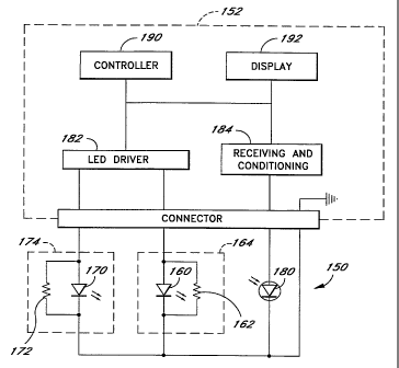
FIGURE 4 depicts one embodiment of a
tuned sensor 150, connected to an exemplary oximeter system 152, according to
the LED tuning aspect of the
present invention.
The sensor 150 is illustrated with a first light source 160 and a second light
source 170, typically LEDs.
A first tuning resistor 162 connected in parallel with the first LED 160 forms
a first tuned LED network 164.
Similarly, a second tuning resistor 172 is connected in parallel with the
second LED 170 to form a second tuned
LED network 174. The sensor 150 further comprises a photodetector 180. A power
source in the oximeter system,
such as an LED driver 182, is coupled to the tuned LED networks 164, 174 in
order to provide a predetermined drive
CA 02221446 1997-11-18
WO 96/41138 PCTIUS96/08631
-8-
current at the input of the tuned LED networks 164, 174. Advantageously, the
LED driver 182 provides current to
only one of the tuned LED networks 164, 174 at any given time. The
photodetector 180 is coupled to receiving
and conditioning circuitry 184 in the oximeter system 152. In operation, the
photodetector receives the attenuated
light energy and responds with an output signal representing the intensity of
the alternative light energy. The
oximeter system 152 further comprises a controller 190 with supporting
resources and a display 192. The oximeter
system receives the signals obtained from the sensor 150 and analyzes the
signals to determine information regarding =
the medium through which the light energy has been transmitted. It should be
understood that the oximeter system
is depicted in simplified form for discussion purposes. Oximeter systems are
well known in the art. One possible
oximeter system comprises the oximeter system disclosed in International
Publication No. WO 96112435 published
on 2 May 1996. Other oximeter systems are well known and can be designed to
operate at the selected
wavelengths.
As depicted in FIGURE 4B, for oximetry, a typical medium may include a finger
200 or an earlobe, as well-
known in the art. Media such as the finger and earlobe typically comprise a
number of constituents such as skin,
tissue, muscle, arterial blood and venous blood (having several constituents
each), and fat. Each constituent absorbs
and scatters light energy of a particular wavelength differently due to
different extinction coefficients. In general
operation, the first LED 162 emits incident light in response to the drive
current from the LED driver 182. The light
propagates through the medium under test. As the transmitted light propagates
through the medium, it is partially
absorbed by the medium. The attenuated light emerging from the medium is
received by the photodetector 180.
The photodetector 180 produces an electrical signal indicative of the
intensity of the attenuated light energy incident
on the photodetector 180. This signal is provided to the oximeter system 152,
which analyzes the signal to
determine the characteristics of a selected constituent of the medium through
which the light energy has passed.
The tuning is now explained with reference to the first LED 160. The tuning is
also applicable to the
second LED 172. As explained above, in response to a particular drive current,
different LEDs respond with different
wavelengths, even though the LEDs were manufactured to generate the same
wavelength. Tuning the first LED 160
in accordance with the present invention invoives determining the amount of
current required to operate the first LED
160 at the selected wavelength and adjusting the current through the first LED
160 in order to obtain the selected
wavelength.
For instance, typical operational values for red LEDs used in oximetry range
between 645 nm and 670 nm.
For a particular embodiment of an oximeter, the oximeter may be designed to
operate with a selected wavelength
within that range, for example, 670 nm. However, the LEDs manufactured to
produce the selected wavelength of =
670 nm involve manufacturing tolerances typically in the range of 2-10 nm for
the same drive current. However,
for a typical LED used in oximetry, the drive current can be varied in order
to obtain the desired output wavelength
for the LED. For instance, as illustrated in FIGURE 3B, the represented LED
has an operating wavelength of 660
nm for the typical 50 mA drive current. If the drive current is increased to
approximately 85 mA, the operating
wavelength becomes the selected wavelength of the present example (670 nm).
The present invention takes
CA 02221446 1997-11-18
WO 96/41138 PCT/US96/08631
-9-
advantage of the observed wavelength shift in response to a drive current
change to tune each LED to obtain the
selected wavelength, such as 670 nm.
For purposes of discussion, the first LED 160 is defined to exhibit the
wavelength characteristic depicted
in FIGURE 3B. To tune the first LED 160, the drive current from the LED driver
182 is assumed to be preset or
fixed. In the present embodiment, the drive current is preferably somewhat
larger than the drive current necessary
to drive the first LED 160 alone (e.g., 100 mA or more). This is because the
first tuning resistor 162 carries some
of the fixed drive current from the LED driver 182. The first tuning resistor
162 is selected to draw an appropriate
amount of the fixed drive current to adjust the amount of current flowing
through the first LED 160 to result in the
selected output wavelength. In the present example, the resistor is chosen to
carry approximately 15 mA (of the
100 mA from the LED driver 182) in order to reduce the current through the
first LED 160 to approximately 85 mA
to obtain the 670 nm selected wavelength. Accordingly, each LED can be driven
with the same fixed drive current
from the LED driver 182, yet the current through any particular LED differs in
accordance with the value of the
associated tuning resistor. In this manner, the LED driver 182 can be designed
to provide the same fixed drive
current for every sensor connected to the oximeter. The oximeter system 152 is
thus designed to make its calculation
based on the assumption that the corresponding wavelengths remain constant
from sensor to sensor.
One particular advantageous method of selecting the tuning resistor involves
the use of a semiconductor
substrate resistor, such as the resistor 210 depicted in FIGURE 5A and 5B. The
resistor 210 depicted in FIGURE
5A comprises a semiconductor substrate 212, a resistive coating pad 214, and
connective conductors 216, 218.
In one embodiment a tunable LED 220 (i.e., an LED that exhibits wavelength
shift with drive current change) is
' 20 connected in parallel with the semiconductor substrate resistor 210. The
fixed (preset) drive current is then applied
with a current source 222 to the network formed by the substrate resistor 210
and the tunable LED 220. The
operating wavelength of the tunable LED 220 is measured. Preferably, the
initial substrate resistor has less
resistance than will be necessary to obtain the desired output wavelength. A
laser is used to scribe the resistive
pad 214, as depicted by the line 224 in FIGURE 5B. The scribe line 224
effectively removes a portion of the
resistive pad 214, and thereby increases the resistance of the remaining
resistive pad 214, as well known in the
art. Using the laser, the increase in resistance can be controlled very
precisely. The resistive pad 214 can be laser
trimmed until the current through the tunable LED 220 causes the tunable LED
220 to generate the selected
operating wavelength. The resulting resistor/LED pair forms a tuned LED
network. This tuning method is
advantageous because of the precision and the resulting low-cost of the tuned
LED.
Other methods of selecting the first tuning resistor 162, such as calculating
the wavelength shift for a given
current change for the first LED 160, and then selecting the appropriate
resistor to cause the correct amount of
current to flow through the LED to obtain the selected operating wavelength,
can also be used. Similarly, a
potentiometer could be used. Preferably, each LED for each sensor is tuned in
a similar manner such that the
operating wavelength is a selected operating wavelength for the sensor. For
instance, a two wavelength oximeter
operating may have selected wavelengths for the two LEDs of 670 nm and 905 nm.
For each.sensor, a first LED
is tuned for the 670 nm selected wavelength, and a second LED is tuned for the
905 nm selected wavelength.
CA 02221446 1997-11-18
WO 96/41138 PCT/US96/08631
-10-
In sum, the tuning aspect of the present invention involves using the
principle of wavelength shift in an LED
to tune each LED to obtain a respective selected operating wavelength.
It should be understood that for some LEDs, the manufacturing tolerance may be
too far from the respective
selected wavelength to enable the use of the shift in wavelength to properly
tune the LED; or the wavelength shift
may be insufficient to obtain the selected wavelength. In one embodiment, such
LEDs would not be utilized, and
would be considered out of tolerance. Alternatively, if the obtainable
wavelength shift is not sufficient to allow for =
proper tuning, it is also possible to use two LEDs having wavelengths very
near each other and near the selected
wavelength. One LED has a wavelength below the selected wavelength, and one
LED has a wavelength above the
selected wavelength. As the graph of FIGURE 6 illustrates, when two LEDs are
both active and placed adjacent one
another, the light from the two LEDs combines to form a combined wavelength
which is the average wavelength of
the two LEDs. The combined wavelength has a broader wavelength range, but has
a known average. Preferably,
to fine tune the average wavelength, the wavelength shift of one or both of
the two LEDs can be utilized using
tuning resistors as described above such that the average wavelength is the
selected wavelength. Accordingly, two
LEDs (preferably tuned in accordance with the present invention as a pair) can
be used to obtain the selected
wavelength for operation in a given oximeter.
As another alternative, if sufficient wavelength shift is not available to
allow for tuning all LEDs to the
selected wavelengths, a few selected wavelengths could be used. For instance,
for determining oxygen saturation,
the selected red wavelengths could be 660 nm, 670 nm and 680 nm. The selected
infrared wavelengths could be
900 nm, 920 nm, and 940 nm, independent of the red wavelengths. Each sensor
would be tuned using the tuning
resistors described above such that the red and infrared LEDs operate at one
of the selected red and infrared
wavelengths, respectively. An indicator would then be provided an the sensor,
or the connector attached to the
sensor, to allow the oximeter to determine which of the selected wavelengths
is present on the sensor attached to
the oximeter. Alternatively, a wavelength detection device could be provided
with the oximeter system to determine
which of the selected wavelengths is present in a sensor attached to the
oximeter system. Although this
embodiment requires some means for the oximeter to determine which of the
selected wavelengths is present on the
attached sensor, the selected wavelengths are precise from sensor to sensor.
Two-Wave(enath LED
Another aspect of the present invention involves using the principle of
wavelength shift in an LED for a
given change in current in order to use a single LED to provide two operating
wavelengths. This is advantageous
in making physiological measurements, such as blood oximetry measurements,
because for each additional wavelength
added, the saturation of an additional constituent in the blood can be
measured. For instance, with a two-
wavelength
oximeter, only the ratio of one of two constituents to the total of the two
constituents (e.g., oxygen
saturation) can be accurately monitored. If oxygen saturation is monitored
with two wavelengths, other constituents
which are significantly present in the blood affect the measurement of oxygen
saturation.
If an additional constituent present in the blood has a significant effect
upon the oxygen saturation reading
for a particular patient, the failure to detect the constituent can be
detrimental to the patient. An example of a
CA 02221446 1997-11-18
WO 96/41138 PCT/US96/08631
-11-
constituent which, when present in the blood, will significantly impact the
oxygen saturation reading provided by a
two-wavelength oximeter is carbon monoxide. This is because the extinction
coefficient magnitude for
carboxyhemoglobin (depicted in the curve 106 of Figure 2) approaches the
extinction coefficient of oxyhemoglobin
(depicted in the curve 102 of FIGURE 2) for light energy in the range of 660
nm. Therefore, carboxyhemoglobin may
be detected as oxyhemoglobin. This leads to a false indication of the oxygen
saturation (i.e., overestimation) in the
blood using a two-wavelength oximeter. In this manner, the attending physician
may fail to detect the lack of
oxygen, and the increase of carbon monoxide in a patient. If an additional
transmission wavelength is provided on
the sensor, the oximeter can monitor another constituent, such as
carboxyhemoglobin.
In accordance with the present invention, the principle of wavelength shift in
an LED is utilized in order to
drive one LED with two appropriate drive current levels to provide two
distinct wavelengths. In its simplest form,
this is accomplished by first driving an LED (which exhibits wavelength shift
with drive current change) with a first
known drive current to a first known wavelength, and then driving the same LED
with a second known current to
a second known wavelength.
FIGURE 7 depicts one advantageous embodiment of a sensor 250 for blood
oximetry measurements coupled
to an oximeter system 252 designed in accordance with this aspect of the
present invention. The sensor 250
comprises a first LED 254 and a second LED 256. For blood oximetry the first
LED 254 preferably operates in the
red wavelength range and the second LED 256 preferably operates in the
infrared wavelength range. The sensor
250 further comprises a photodetector 258. The photodetector 258 is coupled to
receiving and conditioning circuitry
262. The oximeter system is under the control of a controller 264 and has a
display 266. As well-understood in
the art, an LED driver 260 sequentially drives the LEDs 254, 256 with a
predetermined drive current. The
photodetector 258 detects the light energy, attenuated by the medium under
test. The oximeter 252 receives and
analyzes the signal from the photodetector 258 to determine information
regarding the medium through which the
light energy has been transmitted. As with the embodiment of FIGURE 4, the
oximeter system 252 is depicted in
simplified form. Appropriate oximeter systems include the system disclosed in
International Publication No. WO
96112435, published on May 2, 1996. Other monitors well understood in the art
also exist. The oximeter system
252 is modified in accordance with the present invention to drive the shifting
LED as described below.
In the present example for blood oximetry, the first LED 254 is the shifting
LED and is used to provide two
wavelengths. In order to accurately provide two wavelengths, the wavelength
shift principle is utilized. According
to one embodiment, LEDs are evaluated at the time a sensor is manufactured,
and an indicator is provided on the
sensor which can be read by the oximeter system 252 to indicate the drive
current change necessary in order to
effectuate a desired shift in wavelength. Indicators may comprise a resistor
on the sensor or sensor connector, a
memory on the sensor or sensor connector, or a similar device. Alternatively,
the indicator can provide a indication
to the oximeter of the amount of wavelength shift which is obtained due to a
preset drive current change. Another
alternative is to provide a wavelength detector 268 for the oximeter, which
allows the oximeter system 252 to
detect the transmission wavelength of an active LED. Wavelength detectors,
such as a monochrometer, are well
known in the art. However, conventional monochrometers are expensive and
bulky. This description sets forth a
CA 02221446 1997-11-18
WO 96/41138 PCT/US96/08631
-12-
more practical approach to detecting wavelength below. In this embodiment, the
LED driver 260 changes the drive
current until the desired wavelength is obtained, utilizing the wavelength
detector 268 to monitor the wavelength.
In one preferred embodiment allowing for a simpler oximeter design, in order
to accurately provide two
wavelengths with a single LED such as the first LED 254, a network 270 of a
slope adjusting resistor 272 and the
first LED 254 is slope adjusted such that a preselected change in drive
current (AI) entering the first slope adjusted
network, causes a preselected shift in wavelength (AA) in the first LED 254.
In other words, as depicted in FIGURE
3B, each LED exhibits an inherent slope of the curve 122. However, the slope
of this curve often differs from LED
to LED, even for LEDs rated for a particular wavelength. In order for an
oximeter to be designed for simplicity in
obtaining a repeatable preselected wavelength shift, it is advantageous to
have the preselected wavelength shift (AA)
for each first LED in different sensors correspond to the same preselected
drive current change (AI). Accordingly,
it is desirous that the first LED (for the present example) on different
probes respond with the same preselected
change in wavelength for the same change in drive current provided by the LED
driver 260. In other words, it is
advantageous that the slope of the curve 100 depicted in FIGURE 3B be the same
for each corresponding LED
network, since it is not typically the same for each individual LED. In this
manner, the oximeter is designed to drive
the LEDs with two drive current levels, where the two drive current levels are
preselected and remain constant from
sensor to sensor.
Just as the first tuning resistor 162 tunes the first LED 160 to a particular
selected wavelength for a
selected drive current, a slope adjusting resistor, such as the slope
adjusting resistor 272, can be used to alter the
slope of the curve 122 exhibited for the particular corresponding LED network
(e.g., the first slope adjusted LED
network 270). In most instances, the slope adjusting resistor 272, if used to
alter the slope, cannot also be used
to tune the precise wavelength of the first LED 254. However, other methods
and procedures to indicate to the
oximeter what the particular wavelength of operation of the first LED for a
given drive current can be utilized. For
instance, an indicator (such as a resistor or low cost memory device) can be
provided with the sensor 250 which
can be read by the oximeter 252, which indicator provides the initial
operating wavelength of the slope adjusted LED
network 270.
Slope adjustment can be accomplished in the same manner as described above
with respect to the
semiconductor substrate resistor 210. However, the substrate resistor
functions as the slope adjusting resistor rather
than a wavelength tuning resistor (i.e., the substrate resistor is adjusted to
cause a preselected change in wavelength
for a preselected change in drive current for the LEDlresistor network). In
other words, for the first LED 254, the
substrate resistor 210 depicted in FIGURE 5A and 5B is coupled to the first
LED 254 to form the slope adjusting
resistor 272. A laser is used to trim the resistor until the preselected
change in drive current for the network 270
results in the preselected change in wavelength for the first LED 254.
It should be noted that if LEDs are available that exhibit the same wavelength
shift with respect to the
same change in drive current, the first slope adjusting resistor 272 is
unnecessary.
For determining oxygen saturation, the second LED 256 operates at a fixed
infrared wavelength (e.g., 905
nm). Preferably, if the infrared LEDs exhibit manufacturing tolerances, the
infrared LEDs can be tuned using a tuning
CA 02221446 1997-11-18
WO 96/41138 PCT/US96/08631
-13-
resistor 274, in the same manner as the tuning resistor 162 of FIGURE 4, to
operate at the selected infrared
wavelength. With a tuned second (infrared) LED 256 and a slope adjusted first
LED 254 (configured to provide two
wavelengths), measurements at three wavelengths can be taken using the sensor
250.
In use, the sensor 250 of FIGURE 7 is first driven with an initial drive
current to cause the first LED 254
to generate light energy of a first wavelength (e.g., 660 nm). The attenuated
signal at this first wavelength is
detected by the photodetector 258 and received by the oximeter 252. Next, the
first slope adjusted LED 254 is
driven with a new drive current varied by the preselected change in drive
current to cause the preselected
wavelength shift to obtain a second wavelength (e.g., 675). As long as the
initial wavelength is provided to the
oximeter system 252, and the slope (change in wavelength due to change in
current) of the first LED network 270
is properly adjusted to match the preselected slope, the second wavelength
will also be a known quantity. A third
measurement is taken by driving the second LED 256 and receiving the
attenuated signal with the photodetector 258.
Measurements are stored in the oximeter system 252. Based upon the three
measurements taken, the arterial
saturation of two constituents of blood may be determined (e.g., oxyhemoglobin
and carboxyhemoglobin), thus
providing more precise information regarding the physiological makeup of the
blood of a patient under test.
In an oximeter system where monitoring of carbon monoxide and oxygen is
desired, the first wavelength
may be 660 nm, the second wavelength may be 675 nm or 680 nm and the third
wavelength will be an infrared
wavelength such as 900 nm or 905 nm. With these three wavelengths provided by
two LEDs, the saturation of both
oxyhemoglobin and carboxyhemoglobin in blood can be determined. The use of two
LEDs to perform measurements
at three wavelengths reduces the cost of the sensor, which is particularly
advantageous if the sensor is a disposable
or replaceable sensor.
In addition to the uses described above, it should also be noted that the
wavelength shift principal described
above could be used to obtain an additional wavelength with one LED.
Measurements Without Precise WavelenOth Information
A further aspect of the present invention involves an apparatus and method of
measuring the saturation
of a selected constituent in a medium under test (e.g., oxyhemoglobin in
blood) without knowing the precise
operational wavelength af one LED. According to this aspect of the present
invention, if the wavelength shift for
an LED is known for a known change in drive current, the operational
wavelength for the LED need not be known
if other information is also available, as further explained below.
As explained above, obtaining a known wavelength shift for a selected change
in current can be
accomplished by adjusting presently existing LEDs, such that the LEDs react to
a preselected change in drive current
(AI) with a preselected change in wavelength (AA). Alternatively, if LEDs are
available having a repeatable (from
LED to LED) change in wavelength for a selected change in current, those LEDs
can be used without adjustment.
An understanding of this aspect of the present invention is explained with
reference to arterial oxygen saturation
determination using two-wavelength oximeters.
As explained above, FIGURE 2 depicts a graph illustrating the relationship
between the typical extinction
coefficient for three constituents of blood with respect to the transmission
wavelength of light transmitted through
CA 02221446 1997-11-18
WO 96/41138 PCT/US96/08631
-14-
the blood. For purposes of determining oxygen saturation, the first curve 102
and second curve 104 are of interest.
As illustrated by the first curve 102, the extinction coefficient of
oxyhemoglobin for light transmitted
between approximately 665 nm (indicated as A, on the graph) and 690 nm
(indicated as A2 on the graph) is
substantially constant (more apparent when the Y-axis of FIGURE 2 is not a log
scale axis). When light within that
same range (i.e., A, aZ) is transmitted through reduced hemoglobin (the second
curve 104), the extinction coefficient
of the reduced hemoglobin exhibits a substantially linear relationship as a
function of transmission wavelength. These
known properties of blood constituents are utilized in the apparatus and
method of the present invention to obtain
information regarding the oxygen saturation (or other constituent saturation)
of the blood without knowing the
particular wavelength of one of two LEDs.
Assuming that incident light is represented by the letter Ia and the
attenuated signal is represented by I,
the attenuated signal is represented by Equation (1) above. In other words,
for the LED sensor 250 of FIGURE 7,
the attenuated signal I is received by the photodetector 258 and is a function
of the ambient transmission, as set
forth in Equation (1).
Where light of wavelength A is transmitted through tissue with blood
containing two forms of hemoglobin
(oxyhemoglobin and reduced hemoglobin), Equation (1) can be expanded for these
two constituents of blood:
n
s
(e h, ~~~ (~p -alE1zCi) !e ds2,tCL\ (2)
where:
d is the thickness of the medium.
s,,, is the absorption coefficient of reduced hemoglobin at wavelength.l,
s,,, is the absorption coefficient of oxyhemoglobin at wavelength A,
c, is the concentration of reduced hemoglobin,
cZ is the concentration of oxyhemoglobin,
s; is the absorption coefficient of the j" layer of attenuating material (not
including oxyhemoglobin
and reduced hemoglobin),
di is the thickness of the j'h layer of attenuation material (not including
oxyhemoglobin and reduced
hemoglobin), and
c; is the concentration of the j'h layer of attenuating material (not
including oxyhemoglobin and
reduced hemoglobin).
Equation (2) can be further expressed as follows:
CA 02221446 1997-11-18
WO 96/41138 PCT/US96/08631
-15-
S = In i = -d (E1.,C1 + E21 C2) (3)
IBL
where:
n
-E Ejdjcl
lBL = /o( e ' ' ) = baseline
s is a value obtained by measuring I with the photodetector and calculating
the ratio of I to I, after
taking the natural log.
For determining oxygen saturation, where the light is transmitted at a first
red wavelength A,, Equation (3)
is expressed as follows:
S, = In T" A, = -d (s,a c, + e2A,c2) (4)
l J
Where light is transmitted at an infrared wavelength.l,A, Equation (3) is
expressed as follows:
SIR= In i I a,R= -d (c1C1 + g2;~'RC2 (5)
IBL
When the wavelength A, and the wavelength A,R are both known, the oxygen
saturation can be determined,
as well-understood in the art. This is briefly illustrated with the following
derivation:
LET N1= S1 and N2 = SIR (6)
d d
Equations (4) and (5) become:
CA 02221446 1997-11-18
WO 96/41138 PCT/US96/08631
-16-
N1 = C2E21, + C1E1;L
N2 = C2E2X IR + V1 E1 ;L IR (8)
In matrix notation, Equations (7) and (8) become:
A= E211 E1x, X C2 B_ Ni
E 2.1,R E 1XR Ci N2
A X=B E2;L' E 1;L ' (2)=(Z)E 1X2
Or. (c2)( = E2a., E1;Lj -1 (N1
C1 E 21,R E 1'X,R N2
(E1a,R Ni -E1a.,N2)
Hence: (C2 - (:2,X,E1,X,R-E1;.,E21,) (10)
Ci (- E 2X, N1 + E 2;L, N2)
(E2;L1 E 1 ;L/R-E 1 ;L1 E2.X/)10
As well understood in the art, oxygen saturation is defined as the following
ratio:
CA 02221446 1997-11-18
WO 96/41138 PCT/US96/08631
-17-
oxygen: SAT= C2 ~ 1= C2 + C'
C2+C, SAT C2
Or. 1 =1 + c'
SAT C2
(-E211 Ni +E2a., N2)
Hence: Cj
C2 (Ey;L,RN1 -E1x,N2)
(E27L, E1;./R-E1 X 1 E2X/)
-
Substituting. N1= s' and N2 = SIR
d d
and multiplying the numerator and denominator by -1:
(s1 S,R
and Simplifying. C' = E d d
2X1
C2 _ E S1 + E S/R
1 a,R d 1 a., d
CA 02221446 1997-11-18
WO 96/41138 PCTIUS96/08631
-18-
Multiplying numerator and denominator by d:
C1 = E (S1- S/R) (12)
~1
C2 2 (-E1,ti,RS1 +E1'XS/R)
Substituting Equation (12) into Equation (11) above:
1 - E (S1 - S/R) + 1
SA T ~- E 1;L,RS1 + E 111 S/R)
1 = ~E211S1-~2a.j S2-E1a./,S1 +E1a.j S2)
SimP/ifYin9'= SAT -E S S
1~IR 1 +E1~1 2)
AND FINALLY:
SAT= (E1,XIRS1 +~1;L'S2) (13)
(-E2~iS1 +E2X1'>2 +E1~.,R'S1 -E1~,iS2) Wh
e n
the
wavelength A, and the a,R are both known, the extinction coefficients, 61,1
Eu,, &,,,, and s2A,R for the
corresponding constituents at A, and A. are also known. As explained above, S,
and S,R can be obtained by
measuring I and lo and taking the natural log of this ratio at the various
wavelengths during operation. Accordingly,
all of the variables in the saturation equation are known or obtainable
through measurement.
However, if the wavelengths for the transmission LEDs are not specifically
known, the extinction
coefficients c will not be known. In accordance with one aspect of the present
invention, the oxygen saturation
can be computed without knowing the precise wavelength of one of the LEDs. For
purposes of discussion herein,
CA 02221446 1997-11-18
WO 96/41138 PCT/US96/08631
-19-
the LED in the red range is chosen for illustration of this aspect of the
present invention. In accordance with the
present invention, and as explained above, the red LED can be adjusted to
exhibit a preselected wavelength shift,
even though the precise wavelength may not be known. Accordingly, the red LED
can be driven with two different
drive currents to obtain two different wavelengths, the shift between which is
preselected and known. However,
as explained above, the precise wavelength may be unknown without some
indication of at least the starting
wavelength. In accordance with the present invention, as long as the
preselected wavelength shift is known, the
starting wavelength need not be known.
In an application where the extinction coefficients vary with respect to
shifts in wavelength on the order
of 1- 3 nm, it would be possible to determine the wavelength without prior
information regarding the wavelength
or the wavelength shift. This would be accomplished by calculating the desired
measurement (e.g., oxygen saturation)
at several (e.g., two or more) different LED drive currents and using the
change in the measurement in connection
with an empirically generated data set (i.e., curves) of measurements with
respect to wavelengths to determine the
wavelength of the LED.
If the preselected wavelength shift is utilized, the oximeter system can make
measurements at three
wavelengths aõ aZ and .1, Thus, a third equation in addition to Equations (3)
and (4) is obtained.
Where the light is transmitted at a second red wavelength A2, Equation (3) is
expressed as follows:
S2 = In ' ) 1 12= -d (P- 1112C1 + E2a,2C2) 4)
'BL
As depicted in FIGURE 2, within the range of 650 nm - 700 nm, the extinction
coefficient does not
significantly change. More particularly, within the range of A, - A2 - 665 mm -
690 mm,
(16)
E2,X2 G5 E211
Furthermore within the same range,
g1~2 - (~g1~1 - Qgy~ (16)
CA 02221446 1997-11-18
WO 96/41138 PCT/US96/08631
-20-
As, is known for a known wavelength shift within the described range, because
the change in the extinction
coefficient AE, is substantially linear.
Substituting Equations (14) and (15) into Equation (4), (5), and (14) results
in the following equations:
S1 - -d (c1c1 + E212C2) (17)
SIR -d(E 1 ;LIRC1 + E2,XIRC2) (18)
S2 = -d((E111-'& E1) C1 + E2,X2C2) (19)
As explained above, Sõ S2, and S,R are calculated by measuring I and IBL.
Accordingly, Sõ SZ, and S,A, are
known values. The extinction coefficients s, and Sz for the infrared
wavelength LED are assumed to be known
because in the infrared wavelength of interest (e.g., 850 mn - 920 nm) and
more particularly 890 nm - 910 nm),
the extinction coefficient is substantially constant for both curves 102 and
104. In another embodiment, the
accuracy would be improved slightly by tuning the LED. The extinction
coefficients for oxyhemoglobin at A, and A2
are also known, as long as the wavelength is in the range where the extinction
coefficient remains constant. In the
present example, this range is defined as 665 nm to 690 nm. Furthermore,
because the change in the absorption
coefficient (As,) for reduced hemoglobin is known for a known wavelength shift
between A, - A2 - 665 nm - 690
nm, Os, is also a known quantity because s, is linear with A. The total
thickness of the medium, d, generally is
unknown for most applications. However, for the determination of oxygen
saturation, as illustrated above, the
thickness (d) cancels because saturation is a ratio.
Accordingly, for the determination of oxygen saturation, Equations (17), (18),
and (19) provide three
equations with three unknowns (e,,,,, c, and c2). Algebraic techniques
following those of Equations (6) to (13) may
be applied to solve the three equations to obtain the oxygen saturation ratio
of c2/(c,+cZ). Accordingly, it is not
necessary to know the precise operating wavelength of the first LED 254, as
long as the operating wavelength for
the first LED 254 is in a known range where a preselected change in drive
current causes a preselected change in
the wavelength, and where the extinction coefficient of one constituent is
constant and the extinction coefficient
of the second constituent is substantially linear such that the change in the
extinction coefficient for a preselected
change in wavelength is also known.
Accordingly, this aspect of the present invention permits the user to obtain
physiological data without
knowing the precise operational frequency of an LED.
CA 02221446 1997-11-18
WO 96/41138 PCT/US96/08631
-21-
Improved Calibration of LED Sensor
An additional aspect of the present invention involves an improved calibration
technique for an oximeter
sensor where a resistor is utilized to code the LED rather than tune the LED.
As depicted in the prior art calibrated
oximeter probe of FIGURE 1, an encoding resistor 300 utilizes a separate
electrical connection lead and connects to
a common ground lead 304. With the ever increasing use of replaceable or
disposable sensors, any reduction in the
complexity of the replaceable sensor can result in a significant cost savings
over time. In accordance with present
invention, the characteristics of an LED as depicted in FIGURE 3A can be
utilized to provide a more cost effective
coded or calibrated oximeter probe where the coding or calibration is provided
using a coding resistor.
In accordance with this aspect of the present invention, one of the LED
electrical connections can also be
used for the coding resistor. FIGURE 8 depicts a schematic diagram of an
exemplary oximeter sensor where a coding
resistor 332 can be read using one of the LED electrical connections rather
than a separate electrical connection.
A sensor 310 comprises a first LED 312, a second LED 314 and a photodetector
316. The first LED 312 has a
first corresponding electrical connection 318; the second LED 314 has a second
corresponding electrical connection
320; and the photodetector 316 has a corresponding electrical connection 322.
Each of the LEDs 312, 314 and
the photodetector 316 are connected at their outputs to a common ground
electrical connection 330. In the present
embodiment, the coding resistor 332 is coupled in parallel with the first LED
312 or the second LED 314. In this
embodiment, the coding resistor 332 is not provided to tune the first LED 312
or to slope adjust the first LED
network, but is provided as an indicator which can be read by an attached
oximeter system 340. The resistor can
be used to indicate the operating wavelength of the first and second LEDs 312,
314, or more advantageousiy, to
indicate the type of probe. In other words, the value of the coding resistor
332 can be selected to indicate that
the probe is an adult probe, a pediatric probe, a neonatal probe, a disposable
probe or a reusable probe. In one
preferred embodiment, coding resistors could be provided across each of the
LEDs 312, 314 to allow additional
information about the probe to be coded without added leads. However, any
resistor or impedance device could be
used without it being used in parallel with the LEDs to encode the change in
wavelength or other information for
the LEDs.
For instance, the coding resistor could be utilized for security purposes. In
other words, the value of the
coding resistor, and the placement across the LED 312 could be used to ensure
that the probe is configured properly
for the oximeter. For instance, the coding resistor could be utilized to
indicate that the probe is from an authorized
supplier such as a"Masimo" standard probe, "Patient Monitoring Company 1"
probe, "Patient Monitoring Company
2" probe, etc.
In addition, it should be noted that the resistor need not be a passive
element. Coding information could
also be provided through an active circuit such as a transistor network,
memory chip, or other identification device,
for instance Dallas Semiconductor DS 1990 or DS 2401 or other automatic
identification chip.
In order to read the coding resistor 332, the oximeter system 340 drives the
first LED 312lcoding resistor
332 combination at a level that is low enough that the LED draws effectively
insignificant current because of the
CA 02221446 1997-11-18
WO 96/41138 PCT/US96/08631
-22-
exponential relationship between I and V, as illustrated in the graph of
FIGURE 3A. As well understood in the art,
the LED becomes active in the area of the shoulder, designated with the A axis
indicator. Below the voltage level
at A, the LED is effectively inactive and draws effectively insignificant
current. In other words, the current through
the first LED 312 is negligible. Significantly all of the current through the
first electrical connection 318 flows
through the coding resistor 332.
The current which flows through the coding resistor for the voltage applied is
measured by the oximeter
system by measuring the current through the first electrical connection 318.
In turn, the oximeter system 340
determines the value of the coding resistor 332 which is preselected to
indicate the type of probe, the operating
wavelength or other parameters about the probe. In essence, by reducing the
drive voltage across the first electrical
connection 318 and ground to a low level that does not activate the first LED
312, the first LED 312 is effectively
removed from the electrical circuit. In the present embodiment, it has been
found that for conventional LEDs in the
red and IR range, 0.5V is a particularly advantageous voltage. At 0.5V,
current through the LED is generally less
than 1,uA (an insignificant amount).
Preferably, the coding resistor 332 is chosen to be of a sufficiently high
vaiue that when the current supply
to the first electrical connection 318 rises to a level sufficient to drive
the first LED 312, the coding resistor 332
is effectively removed from the electrical circuit because of its high
resistance as compared to the resistance of the
first LED 312 at active operating currents.
Accordingly, a coding resistor can be used in connection with an oximeter LED
sensor without the addition
of an electrical connector dedicated to the coding resistor. This reduces the
cost of the sensor in accordance with
the present invention.
In one advantageous embodiment, the oximeter can monitor the coding resistor
continuously by providing
a.5V coding resistor reading signal at a frequency different from the LED
drive current. For instance, if the LED
drive current is turned on and off at a frequency of 625 Hz, the .5V coding
resistor reading voltage can be provided
at a frequency much lower than 625 Hz, such that the 625 Hz signal can be
easily filtered with a low pass filter
with a cutoff significantly below 625 Hz, but with a pass band which allows
the .5V signal to pass. This would
allow the oximeter to continuously monitor the coding resistor 332 in case of
a change in the sensor by the system
operator.
This particularly advantageous embodiment of using the coding resistor 332 can
also be utilized with a
conventional back-to-back configuration for the red and infrared LEDs, as is
typical in oximeters. Such a
configuration is depicted in FIGURE 8A. FIGURE 8A is similar to FIGURE 8,
except that the first LED 312 and the
second LED 314 are connected in a back-to-back configuration such that the
first electrical connection 318 is
required and the voltage can be alternated from positive to negative to draw
current through either the second LED
314 or the first LED 312. This eliminates the need for an electrical
connection to the oximeter probe, thereby
further reducing the cost of the probe. In the back-to-back configuration of
FIGURE 8A, if the second LED 314 is
a red LED with a knee of approximately 2.OV and that the second LED 312 is an
infrared (IR) LED with a knee of
approximately 1.5V, a positive voltage is advantageously applied to the first
electrical connection 318 at
CA 02221446 1997-11-18
WO 96/41138 PCT/US96/08631
-23-
approximately 0.5V in order to measure the coding resistor 332. Because the
knee for the red LED is 2.OV, very
little (less than 1,uA) current will flow through the red LED and essentially
no current will flow through the infrared
LED 312 (because the infrared LED 312 is reverse biased). In such a scenario,
the current which passes through
the network of the first LED 312, the second LED 314, and the coding resistor
332 is approximately equal to the
current through the coding resistor 332. The resistance of the coding resistor
332 is then easily determined via
Ohms Law by dividing the voltage applied to the network by the current which
flows through the network. Care
must be taken to insure that the element (active or passive) does not create
electromagnetic noise which could lead
to reduced system signal to noise ratio.
Wavelenath Detection
As briefly discussed above, in certain circumstances, it is useful directly to
obtain information regarding the
wavelength of an LED connected to an oximeter. As illustrated in FIGURE 7, a
wavelength detector 268 can be
provided. However, a wavelength detector requires some configuration
operations to be performed by the operator.
In a hospital environment, it is advantageous to simplify the use of the
oximeter. Accordingly, in another
embodiment, each LED sensor is configured with a wavelength detection
configuration. FIGURE 9A and 9B depict
diagrams of possible embodiments of LED sensors configured with filters. These
sensor configurations can be used
to obtain the wavelength of the LED for the sensor.
As depicted in FIGURE 9A, a sensor 400 comprises a transmission LED network
402, a first photodetector
404, a second photodetector 406, a diffuser 407, a beam splitter 408, an
optical filter 410 and an optional optical
filter 471. The transmission LED network 402, the first photodetector 404 and
the second photodetector 406 all
couple to an oximeter system 412. A third photodetector 413 is also depicted
in dotted line to illustrate the
photodetector for the oximetry measurement. This third photodetector 413 is
not discussed in the following
discussion which relates to the calibration portion of the oximeter probe 400.
The transmission LED network 402
preferably comprises at least two LEDs, one in the red wavelength range (e.g.,
660 nm) and one in the infrared
wavelength range (e.g., 905 nm). Determining the wavelength of one of the LEDs
in the LED network 402 using the
configuration of the sensor 400 depicted in FIGURE 9A is described below.
As seen in FIGURE 9A, the LED network 402 transmits light 414 which first
passes through the diffuser
407. The diffuser 407 is provided advantageously in the preferred embodiment
in order to remove polarization of
the light because the beam splitter 408 is sensitive to polarized light, and
most LEDs transmit some percentage of
polarized light. The light then passes to the beam splitter 408 where it is
divided. The beam splitter 408 is
preferably coated with a material which is partially reflective to light of
the wavelength of the LEDs of interest in
the LED network 402. Advantageously, the beam splitter 408 reflects
approximately one-half of the light 414 and
directs it to the first photodetector 404. The remainder of the light passes
through the beam splitter 408 and
through the filter 410 and is received by the second photodetector 406. The
oximeter system 412 receives the
intensity reading from the first and second photodetectors404, 406 and
utilizes the relative intensities from the first
and second photodetectors 404, 406 to determine the centroid of the emission
wavelength for the LEDs 402, as
further explained below.
CA 02221446 1997-11-18
WO 96/41138 PCTIUS96/08631
-24-
As is well understood in the art, obtaining a beam splitter to precisely
divide the light by 50 percent would
be costly to construct. However, it is not necessary to obtain a 50 percent
split of the light because imprecision
can be accommodated with calibration. In an embodiment where no second filter
411 is provided, the system can
be calibrated by activating the infrared LED. This is possible because the
first filter 410 is transparent to the
infrared wavelength, and thus, each photodetector 404, 406 senses the same
signal. In such an embodiment, the
intensity outputs from the first and second photodetectors 404, 406 can be
compared and equalized through
calibration constants during run-time. This compensates for imprecision in the
photodetectors, beam splitter 408 and
diffuser 407.
In an embodiment where the infrared is not used to calibrate, the
photodetectors 404, 406, the beam
splitter 408 and the diffuser 407 can be calibrated prior to delivery with a
passive or active coding element 415
for each device. It should be understood that the box 415 represents one or
more coding elements. It should also
be understood that a single coding element could be used for all of the
optical devices within the box 515.
Preferably, the elements provided for calibration (those within the box in
dotted lines labelled 515) in this embodiment
are positioned in a reusable portion of the probe such that the increased
expense is not too significant.
The filter 410 may also have imprecision due to temperature sensitivity and
imprecision of manufacturing
process. Therefore, in order to calibrate for imprecision with respect to the
filter 410 (preferably a shot glass) due
to shift in temperature, a temperature detector 405 is provided in a preferred
embodiment. Because temperature
sensitivity in shot glass filters are well known, by detecting the
temperature, the shift in filter characteristics can
also be determined. With respect to the imprecision in manufacturing, a
passive or active coding element 415 can
be provided on the probe to provide information about the variation from a
selected (ideal) filter characteristic
(transition band for filter).
Another preferred embodiment utilizing a filter configuration is depicted in
FIGURE 9B. FIGURE 9B depicts
a sensor having a transmission LED network 420, a diffuser 421, a first
photodetector 422, and a second
photodetector 424. As in FIGURE 9A, a third photodetector 431 is depicted
representing the photodetector used
for oximetry measurements. The first and second photodetectors 422, 424 are
positioned at the interior periphery
of an integrating optical sphere 426, or the like. As can be seen in FIGURE
913, the integrating optical sphere 426
has an aperture 428 through which light 429 from the LED network 420 is
directed for monitoring and for
wavelength determination. The light which enters the aperture is reflected
about the interior of the optical sphere
426, without significant absorption. Advantageously, the interior of the
integrating optical sphere is reflective to
the wavelengths of the light from the LED network 420. In addition, the
interior of the integrating optical sphere
426 scatters the light. Advantageously, the first and second photodetectors
422, 424 are spaced laterally across
the integrating optical sphere, with the aperture 428 positioned equidistance
between the first and second
photodetectors422, 424. In this manner, each of the first and second
photodetectors422, 424 receive substantially
the same amount of light originating from the LED network 420.
As with the embodiment of FIGURE 9A, the second photodetector 424 has an
associated low pass optical
filter 430, through which the light incident on the second photodetector 424
passes prior to reaching the second
CA 02221446 1997-11-18
WO 96/41138 PCT/US96/08631
-25-
photodetector 424. Accordingly, like the embodiment of FIGURE 9A, the second
photodetector 424 in FIGURE 9B
receives light attenuated by the filter 430, and the first photodetector 422
receives light unattenuated by the filter
430.
As with the embodiment of Figure 9A, as is well understood in the art,
obtaining an integrating optical
sphere precisely integrate the light would be costly to construct. However,
again, it is not necessary to obtain a
perfect integrating sphere because imprecision in the sphere (as well as in
other elements) can be accommodated
with calibration. For instance, the system of Figure 9B can be calibrated by
activating the infrared LED if no infrared
filter (corresponding to the filter 411 in Figure 9A) is used. This is
possible because the filter 430 is transparent
to the infrared wavelength, and thus, each photodetector 422, 424 senses
unfiltered signal (which ideally would be
the same). In such an embodiment, the intensity outputs from the first and
second photodetectors 422, 424 can
be compared and equalized through calibration constants during run-time. This
compensates for imprecision in the
photodetectors, optical sphere, and diffuser.
As with the embodiment of Figure 9A, if the infrared is not used to calibrate,
the photodetectors422, 424,
the optical sphere 426, and the diffuser 421 can be calibrated prior to
delivery with passive or active coding
element(s) 432 for each device.
As with the embodiment of Figure 9A, the filter 430 may have imprecision due
to temperature sensitivity
and imprecision due to manufacturing. Therefore, in order to calibrate for
imprecision with respect to the filter 430
(preferably a shot glass) due to shift in temperature and manufacturing
tolerances, a temperature detector 425 is
provided in a preferred embodiment, as with the embodiment of Figure 9A. With
respect to the imprecision in
manufacturing, a passive or active coding element 432 can be provided on the
probe to provide information about
the variation from a selected (ideal) filter characteristic (transition band
for filter).
It should also be understood, that in one embodiment, a single memory element
or other passive or active
element (415, 432) could be provided with enough identification capability to
provide characteristic information for
each of the diffuser, the photodetectors, filters, and the beam splitter (or
optical sphere). For instance, a memory
device or transistor network could be provided with several bits of
information for device.
In the present embodiment, with red (e.g., 640-680 nm) and infrared (e.g., 900-
940 nm) LEDs in the LED
networks 402, 420 of FIGURES 9A and 913, the wavelength of the red LED is the
most critical for blood oximetry.
Accordingly, accurate determination of the centroid operating wavelength of
the red LED in the LED networks 402,
420 is desired. In this case, the filters 410, 430 advantageously are selected
to partially attenuate light in the red
wavelength range, and pass light in the infrared range unattenuated.
The principle by which the sensors of FIGURE 9A and 9B can be used to identify
the wavelength of the
LEDs for those sensors is now described. As well understood in the art, LEDs
for use in blood oximetry and the
like have an emission characteristic similar to the emission curve depicted
with the curve 440 of FIGURE 10A. As
depicted in FIGURE 10A, the ideal LED has a centroid wavelength at ao (e.g.,
660 nm). However, as well
understood, the actual centroid wavelength for a batch of LEDs with a target
centroid wavelength of Ao differs due
to manufacturing tolerances. For instance, the emission curve may be shifted
to the right as in the dotted emission
CA 02221446 1997-11-18
WO 96/41138 PCT/US96/08631
-26-
curve 440A depicted in FIGURE 10A. The actual centroid wavelength is
significant in accurate oximetry
measurements.
The filters 410, 430 preferably have a response as depicted by the curve 450
in FIGURE 10B. With a filter
chosen with the middle of its transition band selected at the target centroid
wavelength,.lo, the filter transition band
advantageously extends from a lower anticipated wavelength a, to an upper
anticipated wavelength A2. The range
(GI, - A2) preferably encompasses the anticipated variance in wavelengths for
LEDs due to manufacturing tolerances.
In other words, the manufacturing tolerance range for LEDs manufactured to
have a target wavelength of Ao, should
not extend beyond the upper or lower bounds of the filter transition band.
For LEDs having a centroid wavelength in the area of the transition band of
the filter, a ratio of the overall
intensity detected from a sensor LED without filtering to the intensity of the
same sensor LED detected with filtering
provides useful information, as further explained.
FIGURE 10C is illustrative of the ratio for an LED having a wavelength just
above than the target
wavelength.lo. The LED emission without filtering is represented by the LED
emission curve 440A. The emission
with filtering is depicted by the filtered emission curve 441. The filtered
emission curve 441 represents the filter
response multiplied by the LED emission without filtering as well understood
for filtered emission. The significant
ratio is the ratio of the area under the filtered LED emission curve 441
(illustrated with cross hatching) to the area
of under the unfiltered LED emission curve 440A. It will be understood that
this ratio will vary from 0 - 1, for LEDs
with a centroid in the range a, - a2, and assuming the same filter response.
This ratio of the two areas can be determined from the ratio of intensities
received from the photodetectors
404, 406 or 422, 424 as follows: Let the normalized intensity of the
unfiltered light k(A) and the intensity of the
filtered light, If(A) be represented by the following equations.
2
/LM = 1 2 (30)
1 +(~ -/~oL)
1 2
W,) = 2
-(;. -F
1+e ~od25 The energy of the unfiltered light as received by the photodetector
404, 422 can be expressed as the
integral over the range of wavelengths of the LED emission as follows:
CA 02221446 1997-11-18
WO 96/41138 PCT/US96/08631
-27-
X2/L(~)' 'X) dX (31)
E(a'2y X 1)(no ff/terj - fil
where IJA) is the LED emission vs. wavelength (A) and P(A) is the photodiode
response vs. wavelength (A).
For simplicity, where the photodiode response is "1" (P(A) = 1) in the range
of interest (A, - A2) (in other
words, the light emitted from the LED falls within the range of the LED), the
signal of the first photodetector 404,
422 (no filter) will be as follows:
E(X 2y X 1)(no filter) - fxX2/L(X)d~, (32)
1
Similarly, the energy of the light received by the second photodetector406,
424 which has passed through
the filter 410, 430 can be expressed as follows:
E(a'2y X 1)(with filter) fF(A)I(A)o'A (33)
If all LEDs for a batch of sensors have the same peak emission and bandwidth
in the area of interest (A, -
A2), and can be represented by the same equation (30) except for a
multiplicative constant Io, then a normalized
ratio of the energies can be defined as follows:
E ~, ~, ~ ~ X2F(~,) /L(~.) d~,
( 21 1) (with filtel) x 1
E(norm) - E(X 2, a,1) (no ~2
filtel) /o f~1 /L(i1,)da,
_l .1 a 12F(~") /L(~') d(~') fx 12F(~') IL(~") d~" (3a)
E(norm)(X) - ~ -
lo (',12IL(X) dX constant
CA 02221446 1997-11-18
WO 96/41138 PCT/US96/08631
-28-
The generalized ratio of equation (34) is a ratio of the entire area of the
LED emission attenuated by
filtering (designated with cross-hatching in FIGURE 10C) to the area under the
entire LED emission curve.
The function Eõa,,, is single valued and monotonic in the area (.1, -A2) and
depends only on the centroid
wavelength shift of the LED with respect to the center of the transition band,
aD, of the filter.
Accordingly, for a filter with a center of the transition band at .io, the
ratio of the energy detected by
second photodetector (filter present) to the energy detected by the first
photodetector (filter not present) in the
wavelength range (.1, - A2), will be a value between 0 and 1. The precise
ratio depends upon the centroid
wavelength for the LED under test. As can be seen from FIGURE 10C, as the
centroid wavelength increases toward
.tZ, the ratio approaches "1", and as the centroid wavelength approaches aõ
the ratio approaches "0". This
relationship is depicted in FIGURE 10D for A , - - 610nm and A2 =-- 710nm.
In use, a ratio can be calculated to correspond to each possible LED
wavelength in the range (A, - a2).
For instance, a test batch of LEDs representing the range of wavelengths (.1, -
A2) can be used to obtain
corresponding ratios of the intensity of filtered light to unfiltered light.
An accurate wavelength detection device,
such as a monochrometer, can be used to measure the centroid wavelength for
each tested LED. The centroid
wavelength can be stored for each tested LED in association with the measured
ratio for each tested LED. This
leads to a normalized photodiode response, which can be referenced to obtain
the wavelength of an LED having an
unknown wavelength in the wavelength range (A, - A 2).
In other words, for any LED having a centroid wavelength in the range (A , -
A2), with a sensor as depicted
in FIGURE 9A and 9B, the wavelength of the LED for the sensor can be
determined by taking the ratio of the
intensities of the second and first photodetectors, and using the ratio to
reference the normalized photodiode response
to find the wavelength. In the present embodiment, this is accomplished with a
look-up table stored in a memory
for the oximeter system. The look-up table stores the ratio values
corresponding to associated waveiength values.
Accordingly, with the sensor embodiments of FIGURES 9A and 913, the oximeter
simply continually initiates
measurements for calibration purposes. The oximeter, using the method
described above, calculates the ratio between
the two intensities (filtered and unfiltered) and obtains the respective
wavelength for the sensor. This is for testing
purposes. Accordingly, the LEDs or shot glass purchased advantageously should
produce a ration less than 1 and
greater than 0, otherwise the LED wavelength will be undeterminable. In case
the ratio equals 1 or zero, the system
should either not operate or use a calibration equation that is closest to the
extreme (e.g., for ratio = 0, assume
wavelength is 630 nm and for a ratio s 1, assume wavelength is 670nm in the
present embodiment).
As mentioned above, knowledge about the precise wavelength of the red LED in
an oximeter probe is
generally more critical than knowledge of the precise wavelength of the
infrared LED. Accordingly, the filters of the
sensors of FIGURES 9A and 9B are chosen with the center of their transition
band, Ao, in the red wavelength range.
As seen from the filter response curve of FIGURE 10B, if the center of the
transition band is in the red range, the
infrared light will not be attenuated by the filter.
Examples of preferable filter responses are depicted in FIGURE 11. FIGURE 11
depicts the response curve
for three filters, adequate for the present invention, depending upon the
expected wavelengths. A first filter has the
CA 02221446 1997-11-18
WO 96/41138 PCT/US96/08631
-29-
center of its transition band at 645 nm, a second filter has the center of its
transition band at 665 nm and a third
filter has the center of its transition band at 695 nm. Other filters are also
appropriate depending upon the target
centroid wavelength.
However, it should be understood that the principle explained above could also
be used for the infrared LED,
if the filters are chosen with the center of their transition band at.io
selected at the anticipated or target infrared
wavelength (e.g., 905 nm). In addition, the second filter 411 (FIGURE 9A) can
be provided as a filter, with the
center of its transition band selected at the anticipated or target infrared
wavelength in order to calibrate the
infrared LED as well. In other words, the second filter 411 would pass red
wavelengths (would be transparent to
the red LED light) and would have its transition band centered around 900 or
905nm. Such a filter is depicted in
FIGURE 11A.
The wavelength detection described above could also be implemented with a
sensor having only one
photodetector, and a removable filter. The operator would initiate an
intensity measurement as prompted by the
oximeter without the filter. Then, the operator would place the filter in the
light path between the LED and the
photodetector, and initiate a second reading. The ratio of the second reading
to the first reading provides the ratio
Ino,,,,, which is used to reference the operating wavelength.
Probe Examales
FIGURES 12 - 14 illustrate three different of probes used in medical
monitoring of patients.
FIGURE 12 depicts a wrap-around type probe 500 with an associated connector
502 coupled to a cable
504 which couples to an oximeter system (not shown in FIGURE 12). FIGURE 12A
depicts the bottom of the
connector 502. FIGURE 12B depicts a bottom view of the wrap-around probe of
FIGURE 12, and FIGURE 12C
depicts a side view of the wrap-around probe of FIGURE 12. The wrap around
probe 500 has an LED emitter 506,
a photodetector 508 at the end of a cavity 509, a flexible circuit 510, and
friction electrical connection fingers 512.
The probe 500 also has a connection port 519. In one embodiment, where the
probe would be used for the
calibratable probe of FIGURES 9A, the wrap-around probe would also have a
light tunnel 514 (FIGURE 12B) to
channel some of the light from the emitter 506 to the connector 502. In such
an embodiment, all of the probe
calibration elements marked in the dashed line 515, 515A in Figures 9A and 9B
are positioned in a cavity 516
(FIGURE 12A) which receives the light channeled through the light tunnel 514
and coupled to the connector 502 via
an aperture 518 at the end of the light tunnel 514. As seen in FIGURE 12A,
electrical friction connectors 520 on
the connector are configured to couple with the electrical connectors 512 of
the wrap-around probe 500. The
flexible circuit connects the emitters 506 and the detector 508 to the
connection fingers 512.
In use, the wrap-around probe is placed on the digit of a patient, and the
photodetector 508 is positioned
opposite the emitter 506 so as to receive light from the emitter 506
attenuated by transmission through a fleshy
medium.
FIGURE 13 depicts another embodiment of a wrap-around probe 530 for medical
monitoring of infants. The
probe has a first flexible portion 532 configured to be wrapped about the
digit of a neonate. attached to the first
flexible portion 532 is a second flexible member carrying emitters 534 (LEDs)
and photodetector 536. In one
CA 02221446 1997-11-18
WO 96/41138 PCT/US96/08631
-30-
embodiment where the calibration probe of FIGURE 9A is implemented with the
probe of FIGURE 13, a fiber optic
538 is provided to carry part of the light from the emitter 534 to the
connector port 540 of the probe 530. In this
manner, the same connector 502 having a photodetector can be utilized with the
infant style probe of FIGURE 13.
Alternatively, a light channel or tunnel could be used instead of the fiber
optic to carry a portion of the light from
the emitter 534 to the connector port 540. The same connector 542 is used for
the neonatal probe 530.
Accordingly, as with the embodiment of FIGURE 12, all of the calibration
elements within the dotted box 515, 515A
of FIGURES 9A and 9B are positioned within the connector 502.
FIGURE 14 depicts yet another probe for use in medical monitoring. The probe
of FIGURE 14 comprises
a clip-on probe 550 which couples via a cable 552 to a connector port 554
which is the same as the connector port
540 of FIGURE 13 and the connector port 519 of FiGURE 12. The clip-on probe
carries emitters 556 and a
photodetector 558. With this embodiment, some light from the emitters 556
enters a fiber optic 560 which channels
light to the connector port 554 as in the embodiment of FIGURE 13. Again, the
probe calibrations elements within
the same connector 502 are preferably contained within the connector 502 which
is advantageously the same as
the connector for the embodiments of FIGURES 12 and 13.
FIGURES 15-15D depict yet another embodiment of a wrap-around probe 600
comprising a flexible wrap
portion 602 with an associated connector 604 coupled to a cable 506 which
couples to an oximeter system (not
shown in FIGURE 15). FIGURE 15 depicts a perspective view of the entire probe
600. FIGURE 15A depicts the
underside of the connector 604. FIGURE 15C depicts a top view of the wrap
portion 602 and FIGURE 15D depicts
a bottom view of the wrap portion 602. The connector 604 has two portions: an
emitter portion 610 and a
connection portion 612. The emitter portion 610 advantageously contains the
emitters (such as LEDs) for the
selected wavelengths. This emitter portion 610 can be reused for a period of
time, preferably weeks to months,
thereby allowing for further reduced cost of the wrap-around portion 602 which
is disposable after each use. In
other words, emitters need not be provided for each wrap portion 602. Yet, the
emitter portion 610 is removably
coupled to the connection portion 612 of the connector 604, allowing the
connection portion 612 to be reusable for
a much longer period of time.
In this embodiment, the wrap portion 602 is flexible and disposable after each
use with a very low cost.
The wrap portion has a flexible layer 626 made from polymer or other flexible
materials and has a connector port
614 on the flexible layer 626. The connector port 614 has electrical finger
friction connectors 616 which are
adapted to couple to electrical finger friction connectors 620 (FIGURE 15A) on
the bottom of the connection portion
612 of the connector 604. The electrical finger friction connectors 616 for
the wrap portion 602 couple to a
flexible circuit 618 which connects to a detector 622 which is shielded (not
shown) for the detector 622. Two of
the connections couple to the detector 622 and the third is for the shield
which is preferably a conventional Faraday
shield to protect the detector from electromagnetic interference and the like.
The wrap around probe 600 has an aperture 624 that provides a window for the
transmission of light
energy from the emitters in the emitter portion 610. The emitters are
positioned to transmit light through an
aperture 628 (FIGURE 15A) in the emitter portion 610 which is configured to
match with the aperture 624 in the
CA 02221446 1997-11-18
WO 96/41138 PCTIUS96/08631
-31-
wrap portion 602 when the connector 604 is positioned in the connection port
614. Thus, the light transmits from
the emitters in the emitter portion 610 through the aperture 628 in the
emitter portion 610 and through the aperture
624 in the wrap portion 602 when the connector 604 is inserted into the
connector port 614 and the emitters are
activated.
In use, the wrap portion 602 is wrapped around a digit of the patient (e.g., a
finger) and the detector 622
is positioned to receive light transmitted through the aperture 624 and
through at least a portion of the digit. For
instance, the wrap portion 602 can be wrapped around a finger in a manner that
the detector 622 is opposite the
aperture 624 from which light energy is transmitted.
In one embodiment, the probe 600 is used for the calibratable probe of FIGURES
9A and 9B. In this
embodiment, the connection portion 612 has the elements in the dotted boxes
515 and 515A of FIGURES 9A and
9B positioned in the connection portion 612. In this manner, the calibration
elements are reusable, yet work with
the LEDS in the emitter portion 610 to form a calibratable embodiment. In such
an embodiment, the emitters are
positioned in the emitter portion 610 such that the majority of the light
energy transmits through the aperture 628
and that some light energy transmits to a light aperture 620 in the end of the
connection portion 612 (Figure 15B)=
The connection portion 612 contains the calibration elements depicted in the
boxes 515 and 515A (FIGURES 9A AND
9B) housed in the connection portion 612.
Figure 15B depicts an end view of the connection portion 612 depicting the
light channel 620 and two
electrical connector 613A, 613B which provide connections for LEDs (red and
infrared connected back-to-back in the
present embodiment) in the emitter portion.
It will be understood that the apparatus and method of the present invention
may be employed in any
circumstance where a measurement of transmitted or reflected energy is
required, including but not limited to
measurements taken on a finger, an earlobe, or a lip. Thus, there are numerous
other embodiments which will be
obvious to one skilled in the art. Furthermore, the apparatus and method of
the present invention may be employed
for any LED application that is wavelength sensitive. The present invention
may thus be embodied in other specific
forms without departing from its spirit or essential characteristics. The
described embodiments are to be considered
in all respects only as illustrative and not restrictive. The scope of the
invention is, therefore, indicated by the
following appended claims. All changes which come within the meaning and range
of equivalency of these claims
are to be embraced within their scope.