Note: Descriptions are shown in the official language in which they were submitted.
CA 02221793 1998-O1-27
Technical field
The present invention relates to a novel antibacterial protein, designated
gloverin, representing a new class of antibacterial insect proteins which can
be
isolated from Lepidoptera, more specifically the pupa of Hyalophora giant silk
moths.
Furthermore, the invention relates to pharmaceutical compositions comprising
gloverin as a medicament and use thereof in a method against bacterial
infection.
Background of the invention
Infection of pupae of lepidopterans with live non-pathogenic bacteria induces
the synthesis of a variety of antibacterial polypeptides secreted into the
hemolymph. Previous studies have identified three main classes of
antibacterial proteins namely lysozyme, cecropins and attacins [1 ]. Lysozyme
[2,3] attacks the cell wall of gram-positive bacteria. The small (4-5 kDa),
cationic cecropins [3,4] display a strong bactericidal effect against a
variety of
gram-positive and gram-negative bacteria. The attacins [5,6,7] (20 kDa) exist
in
two forms; one basic (p1=9) and one neutral p1=7) and the antibacterial effect
is
directed only against gram-negative bacteria.
Several forms of these antibacterial proteins have been found in various
insect
species. Peptides related to cecropins can be found not only in insects but
also
in vertebrates [8]. The same is true for the ubiquitous lysozymes. A protein
related to the attacins, sarcotoxin IIA, has been found in the dipteran
Sarcophaga [9].
Another class of antibacterial proteins from insects is the insect defensins
[10].
They are characterised by an amino acid sequence of 38 to 43 amino acids
containing six cysteines, forming three disulphide bridges. Different variants
of
insect defensins have been found in several insect species. Other related
insect proteins are the diptericins with a molecular mass of 8.6 kDa that are
effective against gram-negative bacteria [11 ] and the hemolins that belong to
CA 02221793 1998-O1-27
2
the immunoglobulin superfamily and are suggested to play a role in the
regulation of cell adhesion during the cellular response to bacterial
infections
(12,13]
In addition to the antibacterial proteins from insects, there is also a number
of
antibacterial proteins isolated from mammalians e.g. the
bactericidallpermeability increasing protein (BPI} [14,15] and the defensins
[16]. The mammalian defensins differ structurally from insect defensins,
although they have similar size and charge.
Summary of the invention
The present invention provides a novel antibacterial protein, called gloverin.
Gloverin is a basic (with a p1 of about 9) protein with a molecular weight of
about 14 kD containing a large number of glycine residues but no cystein.
Gloverin displays no strong sequence similarity to other known proteins.
Gloverin inhibits the growth of gram-negative bacteria, such as Escherichia
coli.
The minimal concentration required for inhibition of bacterial growth is 1-
3~.M,
which is less than 5% of the concentration of gloverin in the hemolymph of
infected pupae. The synthesis of vital outer membrane proteins and,
consequently, the permeability of the outer membrane are affected, indicating
that the activity of gloverin is directed to the outer membrane of gram-
negative
bacteria.
Preferably, the novel antibacterial protein, gloverin, according to the
invention
is isolated from Hyalophora moths. Alternatively, gloverin is produced by
genetic engineering or by chemical synthesis.
Also, the invention relates to pharmaceutical compositions comprising gloverin
or pharmaceutically active fragments thereof and use of gloverin or fragments
thereof as a medicament against bacterial infection. Furthermore, the
invention
relates to a method of treating bacterial infection comprising administration
of
gloverin or fragments thereof.
CA 02221793 2001-12-14
63786-116
3
Detailed description of the invention
MATERIALS AND METHODS
Isolation of protein. Diapausing pupae of Hyalophora gloveri were injected
with 105 live Enterobacter cloacae f312. After 7 days the hemolymph was
collected as previously described [3). Gloverin was purified from freshly
collected or frozen hemolymph by the following procedure: 50 ml hemolymph
was diluted five times with ice-cold distilled water and centrifuged for 10
min at
17000xg, 4 °C. Saturated ammonium sulphate (SAS) solution was added to
the
supernatant to give 25 % SAS final concentration. After 30 min at room
temperature the precipitate was collected by centrifugation for 10 min at
17000xg, 4°C. The precipitate was dissolved in 10 ml distilled water
and
desalted on a Sephac~eX G-25, PD-10 column (Pharmacia, Sweden)
equilibrated with the starting buffer used in the subsequent ion-exchange
chromatography step. This was performed on a DBE-sepharose*cL-~B
column (3 x 6 cm)(Pharmacia, Sweden) equilibrated with 20 mM
diaminopropane, adjusted to pH 10.1 with hydrochloric acid, at room
temperature. Proteins were eluted using a gradient of 1 M sodium chloride in
starting buffer. A subsequent gel filtration step was pertormed on a sugerde~*-
75 column (1x30cm) (Pharmacia, Sweden) equilibrated with 0.1 M
ammoniumbicarbonate.
Approximately 1.5 mg of purified gloverin were recovered from 50 ml of
hemolymph collected from 50 pupae. The purity of the isolated protein was
ascertained by sodium dodecyl sulphate -polyacrylamide gel electrophoresis
(SDS-PAGE) and mass spectrometry as described below.
Electrophoresis. SDS-PAGE was performed in 12.5% (wlv) slab gels by the
method of Laemmli [17) but with a 4.5% stacking gels containing 9% glycerol.
Isoelectric focusing was performed using a Phast* system (Pharmacia,
Sweden) following the manufacturers standard protocols.
* Trade-mark
CA 02221793 2001-12-14
63786-116
4
Automated amino acid sequence analysis [18J was performed using an ABI
477A (Applied Biosystems) protein sequencer with an on-line ABI 120A PTH
analyser following standard protocols.
Gloverin was cleaved using cyanogen bromide, ,Glu-C, Lys-C or Arg-C
endoproteinase (Boehringer Mannheim). Following cleavage with cyanogen
bromide or Glu-C endoproteinase the digest was separated on a ~perde~c* - 7 s
gel filtration column in 0.1 M ammoniu~nbicarbonate. When cleav~rl viritl~ Lys-
C
or Arg-C endoproteinase the digest was separated by RP-HPLC on a ~rc~~ee*
C-18, 5 m column, 2.1x30 mm, eluted with a gradient of 0 - 70% acetonitrile in
water containing 0.1 % trifluoroacetic acid during 60 min with a flow rate of
0.3
mllmin.
Chromatography was carried out using an FPLC system (Pharmacia, Sweden).
The effluent was monitored at 214 nm. All fractions collected were analysed by
mass spectrometry
Sequence comparison. The databases Swiss protein (release 27.0), and PIR
protein (release 35.0) were searched by the program FASTA [19) using the
Genetic computer group software [20].
Amino acid analysis. Amino acid analyses were performed by the ion
exchange ninhydrin method.
Mass spectrometry. Plasma desorption mass spectra for cleavage products
during sequence work were obtained using a BIOION 20 mass spectrometer
(Applied Biosystems).
Circular dichroism. Circular dichroism (CD) measurements were performed
on a Jasco*#1A spectropolariometer. d-10 Camphor-sulphonic acid was used
for calibration with D a taken as +2.37 at 290 nm. All spectra were recorded
at
25°C using a 0.1 cm cell. Protein concentrations used were 0.1 mg/ml
for
estimating the optimum concentration of hexafluoro-iso-propanol and 0.3 mglml
for recording the complete spectra, respectively.
* Trade-mark
CA 02221793 2001-12-14
63786-116
Protein concentrations were determined spectrophotometrically at 280 nm
using the absorptive value of 18 350 M-1 cm-1. The mean residue ellipticity
expressed in deg.cm/dmol was calculated at every nm and is given as the
average of two analyses. The mean residue weight used was 106.4 glmol.
NMR-analyses. 1 D 1 H-NMR analyses were performed on a Varian*400 MHz
FT NMR spectrometer.
Ultracentrifugation. Equilibrium and sedimentation experiments were
performed using an Optima XL-A (Beckman Inc.) analytical ultracentrifuge.
Bacterial strains. D21f2 [21] is a rfa mutant of the E. coli K-12 strain D21
[22],
with a deep rough, heptose-less lipopolysaccharide (LPS) (= chemotype Re).
The gram-positive strain used was Bacillus megaterium Bm 11 [23].
The term "deep rough" used herein means that the LPS chain is shortened.
Antibacterial assay. The antibacterial activity of purified gloverin was
assayed
by recording the growth of liquid cultures in microtiter plates
(NUNC,Denmark),
200ullwell. GIoVerin was added to LB medium at 5 - 10 ~.M final concentration
and this mixture was inoculated with 5x1 Os cells in mid-log phase. The
cultures
were incubated at 37°C on a rotary shaker and growth was recorded every
20
min by monitoring the absorbance at 560 nm with a Titertek ~o~tis~an*
spectrophotometer.
In some experiments samples were withdrawn from the growing cultures at
different times and spread on LB agar plates to determine the correlation
between number of viable cells and absorbance.
Radioactive labelling of bacterial proteins. Cells were grown as described
above for the antibacterial assay except that LB was substituted with M9
minimal medium supplemented with 0.4% (w/v) glucose and amino acids,
except methionine. L-[35S] methionine (>37 TBq/mmol; Amersham,UK) was
added to the cultures after 2 h, to a final concentration of 25~Ci/ml.
Labelling
* Trade-mark
CA 02221793 2001-12-14
63786-116
6
was continued for 10 min and then stopped by the addition of trichloroacetic
acid to a final concentration of 10%(w/v). The labelled and precipitated cells
were analysed on SDS-PAGE and the dried gels were overlaid with ~s~dak' x-
omat AR-film and exposed for two days at room temperature.
The invention will now be described below with reference to the accompanying
drawings, in which:
Fig. 1 represents SDS PAGE analysis of SDS-precipitated hemolymph and
purified proteins.
Lanes: (1 ) Non-immune hemolymph (2) immune hemoplymph at day 7
(3) purified gloverin (4) purifed basic attacin (5) purified neutral attacin
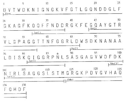
Fig. 2 The amino acid sequence of gloverin.
The peptides used for sequencing are underlined to show overlaps. The
peptide obtained by cleavage with cyanogenbromide is designated "CNBr.1."
The peptides obtained by digestion with endoproteinase Glu-C, Arg-C and Lys-
C are designated "Glu-C.1-2.", "Arg-C.1. and "Lys-C.1-3.", respectively. A
potential glycosylation site is indicated by *.
Fig. 3 Laser desorption mass spectrum for gloverin.
Fig.4 Circular dichroism spectrum for gloverin.
Gloverin dissolved in 10 mM phosphate pH 6.4 (1 ) and with the addition of
20%(v/v) hexafluoro-iso-propanol (2).
Fig. 5 Effect of gloverin on the growth of D21f2.
Gloverin was added at time zero at a concentration of 5mM (1) or 10mM (2).
The control (C) represents growth in the absence of gloverin. Panel A shows
the optical density of the growing cultures. Panel B shows the number of
viable
cells in samples withdrawn from the cultures at times indicated.
* Trade-mark
CA 02221793 2001-12-14
63786-116
7
Fig. 6 Effect of Triton X-100 and.lysozyme on D21f2 treated with gloverin.
Triton X-100 (final concentration; 1 %(w/v) (panel A); or chicken lysozyme
(final
concentration; 200mglml) (panel B); was added at time indicated by arrow to
cultures grown in the absence (1 ) or presence (3) of gloverin (5 ~,M). Curves
(2) represent growth in the presence of gloverin (5 pM) only, and curves (C)
are controls without any additions.
Fig. 7 Effect of lipopolysaccharide (LPS) and magnesium on the growth-
inhibiting activity of gloverin on D21f2.
In panel A curve (1) represents growth with the addition of 50 ~M of LPS. .
Curve (2) and (3) represent growth with the addition of 10 ~.M of gloverin
and°
50 ~~M or 30 ~M of LPS, respectively. Curve (4) represents growth with the
addition of 10 uM of gloverin solely and curve (C) represents the growth of
D21f2 without any additives. In panel B curve (1) represents growth of D21f2
in
the presence of 10 pM of gloverin and 40 mM MgCl2. Curve (2) shows growth in
10uM gloverin only and (C) represents the growth of D21f2 without any
additives.
The addition of 40 mM MgCl2 to the control culture has no effect (not shown).
Fig. 8 Autoradiogram of SDS-PAGE showing the effect of gloverin on
synthesis of the outer membrane proteins Omp F/C and Omp A.in 35S-
methionine labelled D21f2 cells.
Lanes: (1 ) Control, untreated bacteria; (2) Bacteria incubated with gloverin
(10~M) for 2 h
RESULTS
Isolation of protein. Ion-exchange chromatography of ammonium sulphate
precipitated immune hemolymph resulted in two large peaks as determined at
280 nm. Analysis by SDS-PAGE showed that the first eluted of these consisted
of gloverin and the basic form of attacin, while the second peak contained the
neutral form of attacin and some additional proteins (data not shown). In
order
to further separate gloverin from attacin the gloverin containing peak from
the
* Trade-mark
CA 02221793 1998-O1-27
8
ion-exchanger was applied on a Superdex 75 column which yielded gloverin
free of attacin.
The SDS-PAGE analysis (Fig. 1 ) of purified proteins and the hemolymph from
immunised and non-immunised pupae demonstrates that gloverin is induced by
infection. Isoelectric focusing showed that the purified gloverin has an
isoelectric point of 8.5 (data not shown).
Sequence analysis. The amino acid sequence of the above described gloverin
is shown in Fig. 2, which also includes the sequence of the different cleavage
fragments used. One digestion with Glu-C endoprotease was by accident
performed without sufficient buffering causing the enzyme to cleave after both
glutamic acid and aspartic acid. This produced the peptide from amino acid 98
to 113 and gave an overlapping sequence in the region of amino acid number
100. Comparison of the sequence of gloverin with other sequences in current
data banks revealed no proteins with strong sequence similarities.
With knowledge of the amino acid sequence it is possible to produce gloverin
by chemical synthesis. The invention relates to gloverin and gloverin-like
sequences. The main criterion is that the specific gloverin-activity is
retained in
the protein/fragment.
It is realized by the skilled man in the art that the DNA sequence encoding
gloverin can be obtained from the above information. Thus, the invention also
encompasses DNA sequences encoding gloverin and gloverin-like proteins.
Furthermore, the invention relates to such proteins produced by conventional
genetic engineering.
Amino acid analysis. The result of the amino acid analysis of gloverin is
presented in Table 1 and is compared with the composition deduced from the
sequence. Included is also the amino acid composition for the corresponding
protein isolated from Hyalophora cecropia.
CA 02221793 2001-12-14
63786-116
9
Table 1.
Amino acid composition for gloverin
Amino acid Amino acid
Amino acid composition composition fof
fof
COmpOSltlon fof gioverfa #rom g~overi~ ~rcm
g3ower~n from Hyaloph6ra gloveriHyalop~hora cecropia
Hyakaphc~a gloveriaccording to according to a.a-
a.a-
Amino acidacco~a~ng to ec~enceanalysis analysis
Ala 10 9,8 10,3
Arg 6 5, 8 5,1
Asn 9
Asp 13 20, 5 20,1
Cys 0 0 0,8
Gln 7
Glu 1 7,3 7,7
Gly 24 22,5 21,7
His 2 2,1 2,0
Ile 3 3,2 3,5
Leu 8 8,p 7,g
Lys 9 7,9 8,7
Met 1 1,1 1,2
Phe 10 9,7 9,4
Pro 3 3,3 3,8
Ser 7 7,2 . 7,4
Thr 7 6,8 7,3
Trp 3 - _
Tyr 1 1,1 1,7
Val 6 5,8 6,2 '
Mass spectrometry. Laser desorption mass spectra of gloverin (Fig. 3)
indicated a molecular mass of 13786 Da, which is in good agreement with the
value of 13785 Da as calculated from the amino acid sequence. There is no
CA 02221793 1998-O1-27
indication that gloverin is glycosylated, although there is a potential
glycosylation site at asparagine 87.
Conformational studies. The CD-spectrum of gloverin in 10 mM phosphate,
pH 6.4 , can be interpreted as a reflection of a mainly random-coil structure
(Fig. 4). To estimate the possible structure present in a more hydrophobic,
membrane-like environment, CD-spectra were recorded in different
concentrations of hexafluoro-iso-propanol. In a hydrophobic environment the
spectrum changes to reflect a conformation having large amounts (approx.
50%) of alpha-helix structure {Fig. 4). The degree of assumed alpha-helix
reaches a maximum at a concentration of 20 % of hexafluoro-iso-propanol.
The result from the NMR-analysis confirms the conformational change that was
indicated by the CD experiments (data not shown).
From the ultracentrifugation sedimentation experiments of gloverin in 10 mM
phosphate, pH 6.4, the following parameters were calculated: sedimentation
coefficient (S°20(w )) = 1.4 S, diffusion coefficient (D) = 8.95 x10⁻⁷
cm2s-1 and
a friction ratio {f/f0) = 1.5. These values are in accordance with the
expected
values to be obtained for a protein of estimated molecular weight of 13.8 kDa
and present in an extended conformation.
The equilibrium experiments gave a molecular weight of 13.8 kDa showing that
the protein exists as a monomer in water solution.
Antibacterial activity.
The growth of E. coli K-12 is inhibited. Addition of gloverin to growing
cultures
of sensitive E. coli caused a decrease in the growth rate. This effect was
noticeable after 1 h. After 2 - 3 h, growth was completely inhibited (Fig. 5)
and
prolonged exposure to gloverin resulted in a decrease in cell density. The
remaining cells were still viable since the cultures recovered and continued
to
grow when incubated over night (data not shown).
Included in Fig. 5 is also a viable count experiment showing the correlation
between cell density and the number of viable cells. No inhibitory effect of
gloverin on the gram-positive cell Bacillus megaterum could be observed, using
CA 02221793 1998-O1-27
11
concentrations of up to 100 mM of gloverin (not shown). The growth-inhibiting
effect of gloverin is not significantly affected by heating the protein to
100°C for
min (data not shown).
The permeability of the outer membrane increases. Addition of the non-ionic
detergent Triton X-100 to a culture of E. coli D21f2 grown for 2.2 h in the
presence of gloverin resulted in a drastic drop in absorbance, in contrast to
the
much smaller effect of Triton X-100 on untreated control cultures (Fig. 6).
The
sensitivity to lysozyme was also icreased by gloverin-treatment (Fig. 6).
These
results suggest that gloverin affects the integrity of the outer membrane,
allowing entry of substances that are normally excluded by this permeability
barrier, such as conventional antibiotics. Combined therapy with gloverin and
conventional antibiotics) will lower the dose normally required for the
antibiotic(s).
Mg2+ inhibits the activity of gioverin. The effect of gloverin on the growth
of
D21f2 was inhibited in the presence of 40 mM Mg2+ (Fig. 7B). This result is in
accordance with the role of magnesium in stabilising the outer membrane.
Binding to free LPS inhibits activity. Pre-incubation of gloverin with soluble
LPS (Rd)(Sigma) for 30 min at 37°C prior to addition of the mixture to
a growing
culture of D21f2 cells blocks the antibacterial effect of gloverin (Fig. 7A).
The
inhibitory effect of LPS is concentration-dependent.
The synthesis of outer membrane proteins is affected. SDS-PAGE analysis of
the protein content of gloverin-treated and radioactively labelled D21f2 cells
showed that there was no general effect on protein synthesis. However,
gloverin caused a specific inhibition of the synthesis of the outer membrane
proteins Omp F, Omp C and Omp A. Some additional, unidentified proteins
were also affected (Fig. 8).
DISCUSSION
A novel, antibacterial protein isolated from the immune hemolymph of
Hyalophora gloveri pupae, is described in functional and structural terms.
CA 02221793 1998-O1-27
12
The studied gloverin has a molecular mass of 13785 Da and consists of 130
amino acids without any cysteines but with a high content (18.5%) of glycine.
Ultracentrifugation and circular dichroism show that gloverin exists as a
monomeric random coil in water solution, while, according to circular
dichroism,
an alpha helix structure can be induced by the addition of hexafluoro-iso-
propanol. The direct measurement of molecular weight by mass spectrometry
compared to the mass deduced from the amino acid sequence indicates that
the gloverin is not subject to post-translational modifications, e.g.
glycosylation.
A protein corresponding to gloverin was isolated from the closely related
lepidopteran Hyalophora cecropia and the sequence for the 38 N-terminal
amino acids was found to be identical. Also the amino acid analysis for the
two
proteins gave similar results. Comparison of the gloverin sequence with those
of other proteins found in the data base disclosed no structural similarity to
known proteins. Thus, we conclude that gloverin represents a novel class of
antibacterial proteins.
The antibacterial effect of gloverin seems to be directed towards certain gram-
negative bacteria. The sensitivity of E. coli K-12 increases with decreasing
length of the polysaccharide chain of the lipopolysaccharide (LPS). Strain
D21 f2 used in the experiments is an LPS mutant with an Re-type of LPS, and
the most sensitive strain. The parent strain D21 (LPS Ra) is about 10 times
less
sensitive. The fact that gloverin renders these bacteria sensitive to the
detergent Triton X-100 and to lysozyme, - compounds that are normally
inactive against these cells due to their inability to penetrate the outer
membrane - indicates that gloverin has an effect directed against the cell
envelope. This effect could be almost completely inhibited by Mg2+ that is
known to have an important role in stabilisation of the outer membrane of gram-
negative bacteria. The observed increase in permeability is accompanied by a
decrease in outer membrane proteins, an effect which further indicates that
the
outer membrane is the target for gloverin.
CA 02221793 1998-O1-27
13
The observation that the sensitivity of the cell to gloverin increases with
decreasing length of the polysaccharide chain of LPS, in combination with the
fact that the effect of gloverin on growth is inhibited by pre-incubation with
LPS
in solution, indicates that binding to LPS is important for the action of
gloverin.
The polysaccharide chains of LPS may hinder gloverin simply by steric
interactions. The shorter the chain, the easier it is for gloverin to get
access to
the inner parts of the LPS-layer. Possible binding sites for the basic
gloverin
might be provided by the lipid A part of LPS andlor the phosphate groups
present both on lipid A and on the 2-keto-3-deoxyoctonic acid {KDO).
The activity of gloverin resembles in many respects (permeability, Omp
synthesis, inhibition by free LPS) that of attacin (6,7)
A comparison of gloverin and mammalian BPI, shows that the effect of BPI is
also inversely dependant of the length of the LPS polysaccharide chains [14].
Addition of magnesium ions also inhibits the effect of BPI. However, in
contrast
to gloverin and attacin, BPI does not seem to have the same profound effect on
the synthesis of outer membrane proteins [15]. Gloverin retains its
antibacterial
properties after boiling which shows that the activity is not due to any
catalytic
effect. This is also true for the attacins, cecropins and BPI.
The novel antibacterial proteins of the invention enable new antimicrobial
therapy with new antimicrobial agents, i.e. gloverins. Gloverin can be
combined
with other conventional antimicrobial agents, such as penicillins, to enhance
the antimicrobial effect. Furthermore, they provide useful tools for studies
of
the regulation of assembly and synthesis of the bacterial outer membrane.
CA 02221793 1998-O1-27
14
REFERENCES
1. Boman H.G., Faye I., Gudmundsson G.H., Lee J-Y & Lindholm D.A. (1991 )
Cell-free immunity in Cecropia. A model system for antibacterial proteins,
Eur.
J. Biochem. 201, 23-31.
2. Powning R.F. & Davidson W.J. (1976) Studies on insect bacteriolytic
enzymes-II. Some physical and enzymatic properties of lysozyme from
hemolymph of Galleria mellonella, Comp. Biochem. Physiol. 55, 221-228.
3. Hultmark D., Steiner H., Rasmuson T., & Boman H.G. (1980) Insect
immunity: purification and properties of three inducible bactericidal proteins
from hemolymph of immunized pupae of Hyalophora cecropia, Eur. J. Biochem.
106, 7-16.
4. Steiner H., Hultmark D., Engstrom A., Bennich H. & Boman H.G. (1981)
Sequence and specificity of two antibacterial proteins involved in insect
immunity, Nature 292, 246-248.
5. Hultmark D., Engstrom A., Andersson K., Steiner H., Bennich H. & Boman
H.G. (1983) Insect immunity. Attacins, a family of antibacterial proteins from
Hyalophora cecropia, EMBO J. 4, 571-576.
6. Engstrom P., Carlsson A., Engstrom E., Tao Z-J. & Bennich H. (1984) The
antibacterial effect of attacins from the silk moth Hyalophora cecropia is
directed against the outer membrane of Escherichia coli, EMBO J. 3, 3347-
3351.
7. Carlsson A., Engstrom P., Palva E.T. & Bennich H. (1991 ) Attacin, an
antibacterial protein from Hyalophora cecropia, inhibits synthesis of outer
membrane proteins in Escherichia coli by interfering with omp gene
transcription, Infect. Immun. 59, 3040-3045.
8. Lee, J.-Y., Boman, A., Sun, C., Andersson, M., Jornvall, H., Mutt, V. &
Boman, H. G. (1989) Antibacterial peptides from pig intestine: isolation of a
mammalian cecropin, Proc. Natl. Aced. Sci. USA 86, 9159-9162.
9. Ando K. & Natori S. (1988) Inhibitory effect of sarcotoxin IIA, an
antibacterial protein of Sarcophaga peregrine, on growth of Escherichia coli,
J.
Biochem. 103, 735-739.
CA 02221793 1998-O1-27
10. Hoffman J.A. & Hetru C. (1992) Insect defensins: inducible antibacterial
peptides, Immunol. Today. 13, 411-415.
11. Keppi E. Pugsley A.P. Lambert J. Wicker C. Dimarcq J-L., Hoffman J.A. &
Hoffman D. (1989) Mode of action of diptericin A, a bactericidal peptide
induced in the hemolymph of Phormia terranovae larvae, Arch. Insect.
Biochem. Physiol. 10, 229-239.
12. Sun S-C., Lindstrom I., Boman H.G., Faye I. & Schmidt 0. (1990) Hemolin:
An insect immune protein belonging to the immunoglobulin superfamily,
Science. 250, 1729-1732.
13. Ladendorff N.E. & Kanost M.R. (1991 ) Bacteria-induced protein P4
(hemolin) from Manduca sexta: A member of the immunoglobulin superfamily
which can inhibit hemocyte aggregation, Arch. Insect Biochem. Physiol. 18,
285-300.
14. Elsbach P. & Weiss J. (1993) Immunobiol. 187, 417-429.
15. Elsbach P. & Weiss J. (1986) Phagocytic cells: Oxygen-independent
antimicrobial systems, in Inflammation: Basic principles and clinical
correlates
(Gallin J.I., Goldstein I.M. & Snyderman R., eds) pp. 445-470, Raven Press
Ltd,
New York.
16. Lehrer R.I., Lichtenstein A.K & Ganz T. (1993) Defensins: Antimicrobial
and
cytotoxic peptides of mammalian cells, Annu. Rev. Immunol. 11, 105-128.
17. Laemmli UK. (1970) Cleavage of structural proteins during the assembly of
the head of bacteriophage T4, Nature 277, 680-685.
18. Edman P. & Begg G. (1967) A protein sequenator, Eur. J. Biochem. 1, 80-
91.
19. Pearson W.P. & Lipman D. J. (1988) Improved tools for biological
sequence comparison, Proc. Natl. Acad. Sci. USA 85, 2444-2448.
20. Devereaux J., Haeberli P. & Smithies O. (1984) A comprehensive set of
sequence analysis programs for the VAX, Nucleic Acid Res. 12, 387-395.
21. Roman H.G. & Monner D.A. (1975) Characterization of lipopolysaccharides
from Escherichia coli K-12 mutants, J. Bact. 121, 455-464.
22. Boman H.G. Eriksson-Grennberg K.G., Normark S. & Matsson E. (1968)
Resistance of Escherichia coli to penicillins. IV. Genetic study of mutants
CA 02221793 1998-O1-27
16
resistant to D, I-ampicillin concentrations of 100 ug/ml, Genet. Res
(Cambridge) 12, 169-185.
23. Rasmuson T. & Boman H.G. (1977) in Developmental Immunology
Solomon J.B & Horton J.D., eds) pp. 83-90, ElsevierlNorth-Holland Biomedical
Press, Amsterdam.
CA 02221793 1998-04-08
- 16a -
SEQUENCE LISTING
( 1 ) GENERAL INFORMATION
(i) APPLICANT: BENNICH, HANS
AXEN, ANDREAS
CARLSSON, ANETTE
ENGSTROM, AKE
(ii) TITLE OF INVENTION: NOVEL ANTIBACTERIAL PROTEIN
(iii) NUMBER OF SEQUENCES: 1
(iv) CORRESPONDENCE ADDRESS:
(A) ADDRESSEE: SMART & BIGGAR
(B) STREET: P.O. BOX 2999, STATION D
(C) CITY: OTTAWA
(D) STATE: ONTARIO
(E) COUNTRY: CANADA
(F) ZIP: K1P 5Y6
(v) COMPUTER READABLE FORM:
(A) MEDIUM TYPE: Floppy disk
(B) COMPUTER: IBM PC compatible
(C) OPERATING SYSTEM: PC-DOS/MS-DOS
(D) SOFTWARE: PatentIn Release #1.0, Version #1.30
(vi) CURRENT APPLICATION DATA:
(A) APPLICATION NUMBER: CA 2,221,793
(B) FILING DATE: 27-JAN-1998
(C) CLASSIFICATION:
(vii) PRIOR APPLICATION DATA:
(A) APPLICATION NUMBER: SE 9700244-8
(B) FILING DATE: 28-JAN-1997
(viii) ATTORNEY/AGENT INFORMATION:
(A) NAME: SMART & BIGGAR, ,
(C) REFERENCE/DOCKET NUMBER: 63786-116
(ix) TELECOMMUNICATION INFORMATION:
(A) TELEPHONE: (613)-232 2486
(B) TELEFAX: (613)-232 8440
(2) INFORMATION FOR SEQ ID NO:1:
(i) SEQUENCE CHARACTERISTICS:
(A) LENGTH: 130 amino acids
(B) TYPE: amino acid
(C) STRANDEDNESS:
(D) TOPOLOGY: linear
(ii) MOLECULE TYPE: protein
63786-116
CA 02221793 1998-04-08
- 16b -
(iii) HYPOTHETICAL: NO
(xi) SEQUENCE DESCRIPTION: SEQ ID NO:1:
Asp Val Thr Trp Asp Lys Asn Ile Gly Asn Gly Lys Val Phe Gly Thr
1 5 10 15
Leu Gly Gln Asn Asp Asp Gly Leu Phe Gly Lys Ala Gly Phe Lys Gln
20 25 30
Gln Phe Phe Asn Asp Asp Arg Gly Lys Phe Glu Gly Gln Ala Tyr Gly
35 40 45
Thr Arg Val Leu Gly Pro Ala Gly Gly Thr Thr Asn Phe Gly Gly Arg
50 55 60
Leu Asp Trp Ser Asp Lys Asn Ala Asn Ala Ala Leu Asp Ile Ser Lys
65 70 75 80
Gln Ile Gly Gly Arg Pro Asn Leu Ser Ala Ser Gly Ala Gly Val Trp
85 90 95
Asp Phe Asp Lys Asn Thr Arg Leu Ser Ala Gly Gly Ser Leu Ser Thr
100 105 110
Met Gly Arg Gly Lys Pro Asp Val Gly Val His Ala Gln Phe Gln His
115 120 125
Asp Phe
130
63786-116