Note: Descriptions are shown in the official language in which they were submitted.
CA 02222~0 1997-11-27
W096'105i~ PCT/U',6'0r679
DESCRIPTION
Nucleic Acid Trans~orters For Delivery of
Nucleic Acids Into A Cell
Field of the Invention
The invention was partially supported by a grant from
the United States government under grant number US-PH5 POl
HL50422 awarded by the National Institute of Health. The
U.S. government may have rights in the invention.
Backqround of the Invention
This invention relates to gene therapy using a
transporter system for delivering nucleic acid into a
cell.
Recombinant retroviral vectors have been used for
delivery of genes to cells of living animals. Morgan et
al., Annu. Rev. Biochem., 62:191-217 (1993). Retroviral
vectors permanently integrate the transferred gene into
the host chromosomal DNA. In addition to retroviruses,
other virus have been used for gene delivery. Adeno-
viruses have been developed as a means for gene transfer
into epithelial derived tissues. Stratford-Perricaudet et
al., Hum. Gene. Ther., 1:241-256 (1990); Gilardi et al.,
FEBS, 267:60-62 (1990); Rosenfeld et al., Science,
20 252:4341-4346 (1991); Morgan et al., Annu. Rev. Biochem.,
62:191-217 (1993). Recombinant adenoviral vectors have
the advantage over retroviruses of being able to transduce
nonproliferating cells, as well as an ability to produce
purified high titer virus.
In addition to viral-mediated gene delivery, a more
recent means for DNA delivery has been receptor-mediated
endocytosis. Endocytosis is the process by which
eucaryotic cells continually ingest segments of the plasma
membrane in the form of small endocytotic vesicles.
Alberts et al., Mol. Biol. Cell, Garland Publishing Co.,
New York, 1983. Extracellular fluid and material
CA 02222~0 l997-ll-27
WOA"~33~ PCT~S96/05679
dissolved in it becomes trapped in the vesicle and is
ingested into the cell. Id. This process of bulk fluid-
phase endocytosis can be visualized and quantified using
a tracer such as enzyme peroxidase introduced into the
extracellular fluid. Id. The rate of constitutive
endocytosis varies from cell type to cell type.
Endocytotic vesicles form in a variety of sizes and
shapes and are usually enlarged by fusing with each other
and/or with other intracellular vesicles. Stryer, Bioch.,
Freeman and Co., New York (1988). In most cells the great
majority of endocytotic vesicles ultimately fuse with
small vesicles called primary lysosomes to form secondary
lysosomes which are specialized sites of intra-cellular
digestion. Id. The lysosomes are acidic and contain a
wide variety of degradative enzymes to digest the
macromolecular contents of the vesicles. Silverstein et
al., Annu. Rev. Biochem., 46:669-722 (1977); Simianescu et
al., J. Cell Biol., 64:586-607 (1975).
Many of the endocytotic vesicles are clathrin-coated
and are formed by invaglnation of coated regions of the
plasma membrane called coated pits. Coated pits and
vesicles provide a specialized pathway for taking up
specific macromolecules from the extracellular fluid.
This process is called receptor-mediated endocytosis.
Goldstein et al., Nature, 279:679-685 (1979); Pearse et
al., Annu. Rev. Biochem., 50:85-101 (1981); Postan et al.,
Annu. Rev. Physiol., 43:239-250 (1981). The
macromolecules that bind to specific cell surface
receptors are internalized via coated pits. Goldstein,
supra. Receptor-mediated endocytosis is a selective
mechanism enabling cells to ingest large amounts of
specific ligands without taking in correspondingly large
amounts of extracellular fluid. Goldstein, supra.
One such macromolecule is low density lipoprotein
("LDL"). Numerous studies have been performed involving
LDL and the receptor-mediated endocytotic pathway. In
addition to LDL, many other cell surface receptors have
CA 02222~0 l997-ll-27
W096/40958 PCT~S96/05679
been discovered to be associated with coated pits and
receptor-mediated endocytosis. Pastan et al., Annu. Rev.
Physiol., 43:239-250 (1981). For example, studies have
analyzed the hormone insulin binding to cell surface
receptors and entering the cell via coated pits. Stryer
et al., Biochemistry, Freeman & Co., New York (1988);
Alberts et al., Molecular Biology of the Cell, Garland
Publishing, New York (1983). In addition, it has been
determined that some cell surface receptors associate with
coated pits only after ligand binding. Pastan, supra.
Taking advantage of receptor-mediated endocytosis, the
asialoglycoprotein receptor has been used in targeting DNA
to HepG2 cells in vitro and liver cells in vivo. Wu et
al., J. Biol Chem., 262:4429-4432 (1987); Wu et al.,
Bio., 27:887-892 (1988); Wu et al., J. Biol. Chem.,
263:14620-14624 (1988); Wu et al., J. Biol. Chem.,
264:16985-16987 (1989); Wu et al., J. Biol. Chem.,
266:14338-14342 (1991). These studies used
asialoorosomucoid covalently linked to polylysine with
water soluble carbodiimide, 1-ethyl-3-(3-
dimethylaminopropyl)-carbodiimide or with 3'(2'pyridyl-
dithio)propionic acid n-hydroxysuccinimide ester.
Polylysine in the studies above bound DNA through ionic
interaction. The DNA was ingested by endocytosis.
Other studies have utilized transferrin and the
transferrin receptor for delivery of DNA to cells in
vitro. Wagner et al., P.N.A.S., 87:3410-3414 (1990).
These studies modified transferrin by covalently coupling
transferrin to polylysine. Id. The polylysine interacted
ionically with DNA. Delivery of DNA occurred to cells
through the transferrin receptor. Such analyses were
performed in vitro. Id. Cotten et al., P.N.A.S.,
87:4033-4037 (1990); Zenk et al., P.N.A.S., 87:3655-3659
( 19 9 0 ) .
In addition to DNA, other macromolecules can also be
delivered by receptor-ligand systems. Leamon et al.,
P.N.A.S., 88:5572-5576 (1991); Leamon et al., J. Biol.
CA 02222~0 l997-ll-27
WO~ C~ PCT/U~G/0'~79
Chem., 267:24966-24971 (1992). In particular these
studies have involved the folate receptor, an anchored
glycosylphosphatidyl protein, which is excluded from
coated pits and cycles in and out of the cells by
caveolae. Anderson et al., Science, 252:410-411 (1992).
This uptake mechanism has been called potocytosis. Id.
Folate conjugated enzymes have been delivered into cells
through this receptor system and retained activity for at
least six hours. Leamon et al., P.N.A.S., 88:5572-5576
(1991). Folate receptors have limited tissue distribution
and are overexpressed in several malignant cell lines
derived from many tissues. Weitman et:al., Cancer Res.,
52:3396-3401 (1992); Weitman et al., Cancer Res., 52:6708-
6711 (1992); Campbell, Cancer Res., 51:5329-5338 (1991);
Coney, Cancer Res., 51. 6125-6123 (1991). Other studies
have also used biotin or ~olate conjugated to proteins by
biotinylation for protein delivery to the cell. Low et
al., U.S. Patent 5,108,921.
Once DNA or macromolecules are targeted to a cell for
delivery, the DNA or macromolecule must be released from
the endosome to function as a therapeutic agent. I~ not,
the delivery o~ DNA and macromolecule will be hindered by
lysosomal degradation. Studies have analyzed the
endosomal/lysosomal degradation process. It has been
determined that organisms which are internalized via
receptor-mediated endocytosis or receptor:ligand systems,
like viruses and other microorganisms, escape lysosomal
degradation in order to function. The entry mechanism of
some viruses have been studied extensively. For some
viruses outer membrane proteins have been demonstrated to
be important for endosomal escape. Marsh et al., Adv.
Virus Res., 36:107-151 (1989). Other studies have focused
on methods to prevent lysosomal degradation. These
studies have used substances which pertubate
endosomal/lysosomal function. Mellmann et al., Ann. Rev.
Biochem., 55:663-700 (1986). These substances have only
been used in vitro.
CA 02222~0 l997-ll-27
WO 9f l l~SJv PCT/U', 'C 'Ç79
In addition, studies show that the entire virus-shell
is necessary for efficient endosomal lysis. Marsh et al.,
Adv. Virus Res., 36: 107-151 (1989) . Studies have also
demonstrated that adenovirus will enhance transferrin-
5 polylysine mediated gene delivery. Curiel, P.N.A.S.,
88: 8850-8854 (1991) . These studies improved gene
expression in vitro by using a replication defective
adenovirus incorporated into DNA complexes. The effect of
the adenovirus is to lyse the endosome before the contents
can either be routed to the lysosome or recycled to the
cell surface. To reduce virally induced cell death,
adenovirus has been coupled enzymatically to polylysine
through the ~-NH2 of lysine and the ~-carboxyl of glutamic
acid. Wagner et al., P.N.A.S., 89:6099-03 (1992) .
15 Chemical coupling of polylysine with the acidic residues
of adenovirus also accomplishes the same objective.
In addition to adenoviruses, peptide sequences from
other viruses, such as influenza, have been used to
achieve endosome rupture. Wagner et al., P . N. A . S .,
20 89: 7934-7938 (1992) . A lytic peptide from influenza
hemagglutinin has been used to augment gene transfer by
transferrin-polylysine-DNA complexes. Id. This virus-
like genetic transfer vehicle has been shown to be
functional in vitro but 100-fold less effective than
25 adenovirus, based on the delivery and expression of the
luciferase reporter construct. Id.
Other viruses have also been used for lysis purposes,
like human immunodeficiency virus ("HIV"). U.S. Patent
5,149,782 (Chang et al., issued September 22, 1992) .
30 Peptide segments from HIV have been suggested to be useful
as membrane blending agents to deliver nucleic acids. Id.
These peptides are fusogenic and allow the associated
nucleic acid or molecular conjugate to be inserted into
the cellular plasma membrane. Id. These peptides are 10-
35 30 amino acids in length and are hydrophobic. The fusionproteins used contain repetitious Phe-X-Gly sequences,
where X is a nonpolar amino acid residue. Id.
CA 02222~0 l997-ll-27
W096/~ PCT~S96/05679
A number of bacteria are also internalized via
receptor-mediated endocytosis and are liberated from the
endosome by production of toxins. These toxins lyse the
endosomal membrane. Moulder, Microbiol. Rev., 49:298-337
(1985). Listeria monocytogenes produce a membranolytic
toxin called listeriolysin. Cossart et al., Mol. Biol.
Med., 6:463-474 (1989); Tilney et al., J. Cell Bio.,
109:1597-1608 (1989). Studies have shown that no other
cofactors are needed ~or endosomal escape of Listeria
monocytogenes. Bielecki et al., Nature, 345:175-176
( 19 9 0 ) .
The listeriolysin toxin ~orms pores in membranes which
contain cholesterol. These pores are large enough for
macromolecules like immunoglobulins to pass. Ahnert-
Hilger et al., Mol. Cell Biol., 31:63-90 (1989); Geoffroy
et al., J. Bacteriol., 172:7301-7305 (1990).
In addition, numerous studies have analyzed the role
of polyamines in the intracellular processes involving
nucleic acids. In particular, studies show that
polyamines enhance both transcription and translation, and
are involved in maintaining tRNA structure and activity.
Tabor, et al., Annu. Rev. Biochem., 171:15-42 (1970);
Cohen, Nature, 274:209-210 (1978). Furthermore,
polyamines have been shown to condense nucleic acids which
may be utilized in the cell for packaging processes.
Gosule, et al., J. Mol. Biol., 121:311-326 (1978);
Chattoraj, et al., J. Mol. Biol., 121:327-337 (1978);
Riemer, et al., Biopolymers, 17:785-794 (1970).
Additional studies have found polyamines are active in
ribosome stabilization and in the packaging o~ DNA into
phage heads. Stevens, et al., Ann. N.Y. Acad. Sci.,
171:827-837 (1970); Wilson, et al., Biochemistry, 18:2192-
2196 (1979).
The above studies have investigated the electrostatic
component in the interaction of the polyamines, such as
large poly-L-lysine molecules, with nucleic acids. The
average chain length of these poly-L-lysines ranged from
CA 02222~0 1997-11-27
W096/~095~ PCT~S96/05679
50-200. Use of the poly-L-lysines, however, have been
shown to be toxic to cells in nM concentrations thereby
limiting their applicability. Such studies have also been
performed with spermine, spermidine and putrescine.
Braunlin, et al., Biopolymers, 21:1301-1314 (1982).
Summary of the Invention
Applicant has determined that it is useful to
construct nucleic acid transporter systems for enhanced
delivery of nucleic acid into the cell. These particular
transporter systems enhance delivery of nucleic acid into
the cell by using synthetic lysis and nucleic acid binding
molecules. In particular, the specific lysis agents are
useful in disrupting the endosome thereby allowing the
nucleic acid to avoid lysosomal degradation. The specific
binding molecules are useful in delivering to the cell
stabilized and condensed nucleic acid. In addition, these
specific binding molecules are useful in delivering
stabilized and condensed nucleic acid into the nucleus of
the cell. These transporters can be used to treat
diseases by enhancing delivery of specific nucleic acid to
the appropriately targeted cells. These transporters can
also be used to create trans~ormed cells, as well as
transgenic animals for assessing human disease in an
animal model.
The present invention takes advantage of lysis agents
to avoid the problems of endosomaI/lysosomal degradation
in the delivery of nucleic acid to a cell. In particular,
the present invention ~eatures use of a nucleic acid
transporter system with nucleic acid binding complexes
that includes a specific lysis agent capable of releasing
nucleic acid into the cellular interior from the endosome.
The nucleic acid can be efficiently released without
endosomal/lysosomal degradation. Once released into the
cellular interior, the binding complexes help target the
nucleic acid to the nucleus.
CA 02222~0 1997-11-27
WOg~ sv PCT~S96/05679
The present invention also takes advantage of DNA
binding molecules in order to increase DNA stability and
DNA delivery to cells. In particular, the present
invention features use of nucleic acid transporters with
nucleic acid noncovalently bound to peptides capable of
condensing the nucleic acid. These binding molecules
provide smaller, or condensed, and more stable nucleic
acid particles for delivery, thereby enhancing the
transfection rates of the nucleic acid into the cell and
into the nucleus.
By taking advantage of the characteristics of both the
lysis agents and binding molecules, the present invention
enhances delivery of nucleic acid by the nucleic acid
transporter system. These components can be used alone,
together or with other components of the nucleic acid
transporter described below and disclosed in PCT
publication WO 93/18759, Woo et al., entitled "A DNA
Transporter System and Method of Use," the whole of which
(including drawings) is hereby incorporated by reference.
The transporter system, together with the lysis and
binding molecule, enhances the delivery of nucleic acid to
specific cells by enhancing the release of stable,
condensed nucleic acid from the endosome into the cellular
interior.
In addition to the nucleic acid binding molecule and
the nucleic acid binding complex containing the lysis
agent, the present invention also features various nucleic
acid binding complexes which contain a surface ligand and
a nuclear ligand as well. The surface ligands are capable
of binding to a cell surface receptor and entering a cell
through cytosis (e.g., endocytosis, potocytosis, pinocy-
tosis). By using surface ligands specific to certain
cells, nucleic acid can be delivered using the nucleic
acid transporter systems directly to the desired tissue.
The nuclear ligands are capable of recognizing and trans-
porting nucleic acid through the nuclear membrane to the
nucleus of a cell. Such nuclear ligands help enhance the
CA 02222~0 l997-ll-27
W09~0~58 PCT~S96/05679
binding molecules' ability to target nucleic acid to the
nucleus.
The abilities of the above transporters to deliver
nucleic acid to specific cells and to the nucleus also
allows transgenic animal models to be used for the
dissection of molecular carcinogenesis and disease,
assessing potential chemical and physical carcinogens and
tumor promoters, exploring model therapeutic avenues as
well as livestock agricultural purposes. Furthermore, the
above nucleic acid transporter system advantages allow
methods for administration and treatment of various
diseases. In addition, the above nucleic acid transporter
systems can be used to transform cells to produce
particular proteins, polypeptides, and/or RNA. Likewise,
the above nucleic acid transporter systems can be used in
vi tro with tissue culture cells. In vi tro uses allow the
role o~ various nucleic acids to be studied by targeting
specific expression into specifically targeted tissue
culture cells.
A first aspect of the present invention features a
nucleic acid transporter system for delivering nucleic
acid into a cell. The nucleic acid transporter includes
a nucleic acid binding complex containing a binding
molecule noncovalently bound to nucleic acid and
associated with a lysis agent. In addition, the
transporter can also include an additional binding
molecule noncovalently bound to the nucleic acid. The
nucleic acid binding complex and/or the additional binding
molecule may be noncovalently bound to the nucleic acid at
the same time, i.e., simultaneously, and in various
proportions. Furthermore, the lysis agent can be
associated with the respective binding molecule by a
spacer.
The term "lysis agent" as used herein refers to a
molecule, compound, protein or peptide which is capable of
breaking down an endosomal membrane and freeing the
contents into the cytoplasm of the cell. The lysis agent
CA 02222~0 l997-ll-27
WO9G/1~358 PCT/U'9G~G79
can work by: (1) a membrane fusion mechanism, i . e .,
fusogenic, whereby the lysis agent associates or fuses
with the cell membrane to allow the endosomal contents to
leak into the cytoplasm; (2) a membrane destabilization
mechanism whereby the lysis agent disrupts the structural
organization of the cell membrane thereby causing leakage
through the endosome into the cytoplasmi or (3) other
known or unknown mechanisms which cause endosomal lysis.
This term includes, but is not limited to, synthetic com-
pounds such as the JTS-1 peptide, viruses, lytic peptides,
or derivatives thereof. The term "lytic peptide" refers
to a chemical grouping which penetrates a membrane such
that the structural organization and integrity of the
membrane is lost. As a result of the presence of the
lysis agent, the membrane undergoes lysis, fusion or both.
In the present invention, a preferred lysis agent is
the JTS-1 peptide or derivatives thereof. The amino acid
sequence of JTS-1 lytic peptide is GLFE~TT,T~TTT~.~LW~TT.T.T~.
One skilled in the art will readily appreciate and
understand that such nomenclature is the standard notation
accepted in the art for designating amino acids. The JTS-
1 lytic peptide and derivatives are designed as an ~-
helix, which contains a sequence of amino acids such that
the side ch~ n~ are distributed to yield a peptide with
hydrophobic and hydrophilic sides. Such ~-helixes are
termed amphipathic or amphiphilic. The hydrophobic side
contains highly apolar amino acid side chains, both
neutral and non-neutral. The hydrophilic side contains an
extensive number of glutamic acids but could also contain
aspartic acid, as well as polar or basic amino acids. The
JTS-1 peptide would include any derivatives or
modifications of the backbone thereof. The lytic peptide
undergoes secondary structure changes at acidic pH
resulting in the formation of oligomeric aggregates which
possess selective lytic properties.
In general, parameters that are important for
amphiphilic peptide lysis activity include the following.
CA 02222~0 1997-11-27
W096/40358 PCT~S96/05679
First, Hydrophobicity: The peptide must have a high
enough hydrophobicity of the hydrophobic face to interact
with and penetrate phospholipid-cholesterol membranes,
i. e., lipid binding per se is not sufficient. Red cell
hemolysis assays give better indications of which peptides
will have useful activity. Second, Peptide aqqreqation:
The ability to aggregate plays an important role in lysis
and transfection. Third, pH sensitivity: The amphiphilic
peptide must be pH sensitive. Lysis activity can be
controlled by the introduction of lysine, arginine and
histidine residues into the hydrophilic face of JTS-1.
Fourth, Lipid membrane interaction: The peptide must have
a hydrophobic carboxyl terminal to permit interaction with
lipid membranes, e . g., tyrosine substitution for
tryptophan greatly reduces activity. Finally, PePtide
chain lenqth: The length must be greater than twelve
residues in order to get stable helix formation and lipid
membrane penetration and rupture.
The term "derivative" as used herein refers to a
peptide or compound produced or modified from another
peptide or compound of a similar structure. This could be
produced in one or more steps. The term "modified" or
"modification" as used herein refers to a change in the
structure of the compound or molecule. However, the
activity of the derivative, modified compound or molecule
is retained, enhanced, increased or similar to the
activity of the parent compound or molecule. This would
include the change of one amino acid in the sequence of
the peptide or the introduction of one or more non-
naturally occurring amino acids or other compounds. Thisincludes a change in a chemical body, a change in a
hydrogen placement, or any type of chemical variation. In
addition, "analog" as used herein refers to a compound
that resembles another structure, e.g., JTS-l, K8, KN~ or
spermine (discussed below). Analog is not necessarily an
isomer. The above are only examples and are nonlimiting.
CA 02222~0 l997-ll-27
W0~6/~0958 PCT~S96/05679
For example, the JTS-1 peptide can be modified to
change the L in position 2 to an F so as to have the
structure GFFE~T~T~T~T~T~ Lw~T~T~T~ Such a change can
increase the hydrophobicity of the peptide. Furthermore,
increasing the length of the peptide would also be a
modification, i.e., GLFE~T.T~TT~.~WELLLGLFEA. Such a change
can enhance the efficiency of the JTS-1 peptide. A change
in the S at position 12 to a K to modify JTS-1 to
GLFE~T ~T ~FT ~T ~KLW~T ~T ~T ~~ C an shift the pH optimum for lysis
and enhance proteolysis. The above are only examples and
meant to be nonlimiting.
Other useful lysis agents include, but are not limited
to, peptides of the Othromyxoviridae, Alphaviridae and
Arenaviridae. Lysis agents also can include Pep24, Pep25,
Pep26, (see PCT publication WO 93/18759, hereby
incorporated by reference, including drawings), any
appropriate bacteria toxin, bacteria, adenovirus, para-
influenza virus, herpes virus, retrovirus, hepatitis
virus, or any appropriate lytic peptide or protein from a
virus or bacteria. This includes use of any subfragments
of the above which will provide endosomal escape activity.
Particular bacterial toxins may include cytolytic toxins
or active fragments from alveolysin, bifermentolysin,
botulinolysin, capriciolysin, cereolysin O, chauveolysin,
histolyticolysin O, ivanolysin, laterosporolysin,
oedematolysin O, listeriolysin O, perfringolysin O,
pneumolysin, sealigerolysin, septicolysin O,
sordellilysin, streptolysin O, tetanolysin or
thuringolysin O.
In addition to JTS-1 in another preferred embodiment
the lysis agent can be a replication deficient virus. As
used herein, the term "replication deficient" refers to a
virus lacking one or more of the necessary elements for
replication. In one preferred embodiment, the lysis agent
can also be the adenovirus of the structure F, Pep24,
Pep25, or Pep26. In still another embodiment, bacteria
toxins, listeriolysin or perfringolysin can be used. All
CA 02222~0 l997-ll-27
WO~409S8 PCT~S96/05679
o~ the above are disclosed in PCT publication WO 93/18759,
which is hereby incorporated by reference, including
drawings. The above are only examples and are
nonlimiting.
Lysis agents as used herein are pH sensitive. The pH
optimum is determined by the sequence and the content of
acidic and basic amino acid side ch~n~. A~ter
cointernalization o~ the nucleic acid complex containing
the lysis agent throughout the same coated pit on the
plasma membrane o~ the cell, the decrease in pH that
occurs immediately a~ter endosome formation causes
spontaneous lysis o~ the endosome. The nucleic acid is
then released into the cytoplasm. The above is a
nonlimiting example.
The term ~binding molecule~ as used herein re~ers to
a molecule, compound, protein or peptide which is capable
of stabilizing and condensing nucleic acid. This will
include, but is not limited to, components which are
capable o~ stabilizing and condensing nucleic acid by
electrostatic binding, hydrophobic binding, hydrogen
binding, intercalation or forming helical structures with
the nucleic acid, including interaction in the major
and/or minor grove o~ DNA. The term binding molecule can
also be re~erred herein as condensing agent. The binding
molecule is capable o~ noncovalently binding to nucleic
acid. One skilled ln the art will readily appreciate the
meaning of noncovalent. The binding molecule is also
capable o~ associating with a sur~ace ligand, a nuclear
ligand, and/or a lysis agent.
The term "associated with" as used herein refers to
binding, attaching, connecting or linking molecules
through covalent means or noncovalent means. One skilled
in the art will readily appreciate the me~ning o~ covalent
and noncovalent. "Associated with" includes, but is not
limited to, a binding molecule associated with a sur~ace
ligand, nuclear ligand and/or a lysis agent. In addition,
CA 02222~0 l997-ll-27
W09-~C35~ PCT~S96/05679
it includes the association of a spacer (discussed below)
with the above components.
In the present invention, a preferred binding molecule
is the peptide K8. The amino acid sequence of K8 is
YKAKKKKKKKK~K . In another preferred embodiment, the
binding molecule is any peptide with the formula YKAKNWK,
where N can be between 1-40. This formula or amino acid
structure can be referred to as "KN". This would include
use of any subfragments of the above which provide nucleic
acid stability and condensing characteristics.
Furthermore, this would include any derivatives, analogs
or modifications of K8 or the general YKAKNWK structure KN
The above peptides can include lysine or arginine
residues for electrostatic binding to nucleic acid. These
positively charged amino acids help hold the nucleic acid
intact. The binding molecule can also contain tyrosine
which is useful in determining peptide concentration and
iodination for tracking purposes in vitro and in vivo.
Tryptophan also increases the stability of interaction
with the nucleic acid through intercalation. In addition,
binding of the peptide to DNA quenches tryptophan
fluorescence and allows the kinetics and thermodynamics of
complex formation to be determined. The binding molecule
can also contain helix forming residues such as
tryptophan, alanine, leucine or glutamine. These can act
as spacers which allow the cationic residues to adopt an
optimal configuration for interaction with the nucleic
acid in a helical manner, resulting in a more stable
complex. Furthermore, the binding molecule can also
include a-stabilized cyclic version of K8 or the general
YKAKNWK structure KN Such a cyclic version can be formed
by introducing a lactam or disulfide bridge. Likewise,
dimers of K8 or KN can also be used as a binding molecule.
In general, parameters that are important for binding
molecules include the following. First, the peptide must
contain sufficient lysine or arginine residues to permit
ionic interaction with the DNA. Second, the peptide must
CA 02222~0 1997-11-27
WO ~-'10~ ~ PCT/l~' ,CI~._679
have sufficient length to form a stable helix, eleven or
twelve residues, and condense the DNA to small particles,
e.g., K4 forms larger particles than K8. Third, the
peptide helix that forms upon interaction with DNA can be
5 stabilized by leucine zipper formation which gives a
condensing agent less susceptible to ionic strength.
Finally, the lysine or arginine sequence of the condensing
peptide serves as an additional ~unction as a nuclear
localization sequence.
The binding molecule can also include, but is not
limited to, spermine, spermine derivative, spermidine,
histones, polylysine, polyamines and cationic peptides.
As with K8 or KN/ this includes, but is not limited to,
analogs, modifications or derivatives of the above
15 compounds.
Spermine derivatives include compounds D, IV, VII,
XXI, XXXI I I, XXXVI, LIV, LVI, LXXXII, LXXXIV and CX as
described in PCT publication WO 93/18759, hereby
incorporated by reference, including drawings. When used
2 O with the nucleic acid transporter system, the binding
molecules, such as K8, KN or spermine, whether associated
with a surface ligand, nuclear ligand, lysis agent, or
separate therefrom, can be different or similar binding
molecules and bound at the same time, i. e., simultaneously
25 and in various proportions. In a preferred embodiment the
binding molecule is a spermine derivatlve D, as shown in
the above referenced publication.
K8, KN~ and spermine have advantages over poly-L-lysine
as used for the binding molecule. For example, the
3 0 binding properties of K8, KN~ or spermine have advantages
over the binding properties of poly-L-lysine. First, the
intranuclear K8~ KN~ or spermine concentration is
approximately 3 to 10 mmol. ThiS is higher than studies
with poly-L-lysine, which suggest more efficient transfer
of nucleic acid to the nucleus. Second, the spacing of
the amino groups of K8, KN or spermine is such that this
naturally occurring polycation fits into the major groove
CA 02222~0 1997-11-27
WO96/1093~ PCT~S96/05679
16
of the DNA double helix with an exact fit. While the
polycationic poly-L-lysine interacts electrostatically
with the phosphates in the groove of DNA, the fit is not
as precise Finally, the theoretical association/dis-
association kinetics of the DNA/KB, KN or spermineinteraction are more rapid than for DNA/poly-L-lysine
interactions. This is advantageous in the K8, KN or
spermine/DNA mix for the release of the DNA inside the
cell.
The term "nucleic acid transporter system" as used
herein refers to a molecular complex which is capable of
efficiently transporting nucleic acid through the cell
membrane. This molecular complex is bound to nucleic acid
noncovalently. In addition to nucleic acid, other
macromolecules, including but not limited to, proteins,
lipids and carbohydrates can also be delivered using the
transporter system. The nucleic acid transporter system
is capable of transporting nucleic acid in a stable and
condensed state. It is also capable of releasing the
noncovalently bound nucleic acid into the cellular
interior. Furthermore, the nucleic acid transporter
prevents degradation of the nucleic acid by endosomal
lysis. In addition, although not necessary, the nucleic
acid transporter system can also efficiently transport the
nucleic acid through the nuclear membrane, as discussed
below.
The nucleic acid transporter system as described
herein can contain, but is not limited to, six components.
It comprises, consists or consists essentially of: (l) a
nucleic acid or other macromolecule with a known primary
se~uence tha~ contains the genetic information of interest
or a known chemical composition; (2) an agent capable of
stabilizing and condensing the nucleic acid or
macromolecule in (l) above; (3) a lysis moiety that
enables the transport of the entire complex from the cell
surface directly into the cytoplasm of the cell; (4) a
moiety that recognizes and binds to a cell surface
CA 02222~0 1997-11-27
WO g~ 9S8 PCT/US96/05679
receptor or antigen or is capable of entering a cell
through cytosis; (5) a moiety that is capable of moving or
initiating movement through a nuclear membrane; and/or (6)
a nucleic acid or macromolecular molecule binding moiety
capable of covalently binding the moieties of (3), (4) and
(5), above. The term "consisting of" is used herein as it
is recognized in the art. The transporter "consisting
essentially of" the six moieties above includes variation
of the above moieties. Such a variation may make use of
less than all six of the moieties listed above. This is
only an example and is nonlimiting.
The term "delivery" refers to transportation of a
molecule to a desired cell or any cell. Delivery can be
to the cell surface, cell membrane, cell endosome, within
the cell membrane, nucleus or within the nucleus, or any
other desired area of the cell. Delivery includes not
only transporting nucleic acid but also other
macromolecules including, but not limited to, proteins,
lipids, carbohydrates and various other molecules.
The term ~nucleic acid~ as used herein refers to DNA
or RNA. This would include naked DNA, a nucleic acid
cassette, naked RNA, or nucleic acid contained in vectors
or viruses. These are only examples and are not meant to
be limiting. The term "expression" includes the efficient
transcription by the cell of the transported gene or
nucleic acid. Expression products may be proteins,
polypeptides or RNA. In addition, the nucleic acid can be
antisense RNA, oligonucleotides or ribozymes as well.
A variety of proteins and polypeptides can be encoded
by the nucleic acid. Those proteins or polypeptides which
can be expressed include hormones, growth factors,
enzymes, clotting factors, apolipoproteins, receptors,
drugs, oncogenes, tumor antigens, tumor suppressors,
cytokines, viral antigens, parasitic antigens and
bacterial antigens. Specific examples of these compounds
include proinsulin, insulin, growth hormone, androgen
receptors, insulin-like growth factor I, insulin-like
CA 02222~0 l997-ll-27
WO ~'1'3~8 PCT/U' ,G~ISG79
18
growth ~actor II, insulin growth factor binding proteins,
epidermal growth factor, TGF-~, TGF-~, dermal growth
factor (PDGF), angiogenesis factors (acidic fibroblast
growth factor, basic fibroblast growth factor and
angiogenin), matrix proteins (Type IV colljagen, Type VII
collagen, l~m;n,n), oncogenes (ras, fos, myc, erb, src,
sis, jun), E6 or E7 transforming sequence, p53 protein,
cytokine receptor, IL-l, IL-6, IL-8, IL-2, ~, ~, or rIFN,
GMCSF, GCSF, viral capsid protein, and proteins from
viral, bacterial and parasitic organisms. Other specific
proteins or polypeptides which can be expressed include:
phenylalanine hydroxylase, ~-1-antitrypsin, cholesterol-
7~-hydroxylase, truncated apolipoprotein B, lipoprotein
lipase, apolipoprotein E, apolipoprotein A1, LDL receptor,
15 molecular variants of each, and combinations thereof. One
skilled in the art readily appreciates that these proteins
belong to a wide variety of classes of proteins, and that
other proteins within these classes can also be used.
These are only examples and are not meant to be limiting
in any way.
It should also be noted that the genetic material
which is incorporated into the cells from the above
nucleic acid transporter system includes (1) nucleic acid
not normally found in the cells; (2) nucleic acid which is
25 normally found in the cells but not expressed at
physiological significant levels; (3) nucleic acid
normally found in the cells and normally expressed at
physiological desired levels; (4) other nucleic acid which
can be modified for expression in cells; and (5) any
combination of the above.
The term "nucleic acid binding complex" as used herein
refers to a complex which includes a binding molecule.
The binding molecule, as defined above, is capable of
noncovalently binding to nucleic acid. The binding
molecule is also capable of associating with a surface
ligand, a nuclear ligand and/or a lysis agent.
Furthermore, the binding complex can include a spacer
CA 02222~0 l997-ll-27
WO95/l093~ PCT~S96/05679
which associates with the surface, nuclear or lysis agent
to the binding molecule. Spacers are defined in more
detail below.
A second aspect of the present invention features a
nucleic acid transporter system for delivery of a nucleic
acid to a cell which includes a first nucleic acid binding
complex containing a binding molecule noncovalently bound
to nucleic acid and associated with a surface ligand. The
transporter also includes a second nucleic acid binding
complex containing a binding molecule noncovalently bound
to nucleic acid and associated with a lysis agent. In
addition, the transporter can also include an additional
binding molecule noncovalently bound to the nucleic acid.
The binding complexes and/or binding molecules above
can be noncovalently bound to the nucleic acid at the same
time, i. e., simultaneously, and in various proportions.
As described above, the binding molecules can be the same
or different molecules. Furthermore, the surface ligand
or lysis agent can be directly associated with the binding
molecules or associated by a spacer, as defined below.
The term "surface ligand" as used herein refers to a
chemical compound or structure which will bind to a sur-
face receptor of a cell. The term "cell surface receptor"
as used herein refers to a specific chemical grouping on
the surface of a cell for which the ligand can attach.
Cell surface receptors can be specific for a particular
cell, i. e., found predominantly in one cell rather than in
another type of cell ( e . g., LDL and asialoglycoprotein
receptors are specific for hepatocytes). The receptor
facilitates the internalization of the ligand and attached
molecules. A cell surface receptor includes, but is not
limited to, a folate receptor, biotin receptor, lipoic
acid receptor, low-density lipoprotein receptor,
asialoglycoprotein receptor, insulin-like growth factor
type II/cation-independent mannose-6-phosphate receptor,
calcitonin gene-related peptide receptor, insulin-like
growth factor I receptor, nicotinic acetylcholine
CA 02222~0 l997-ll-27
WO~ 35d PCT~S96/05679
receptor, hepatocyte growth factor receptor, endothelin
receptor, bile acid receptor, bone morphogenetic protein
receptor, cartilage induction factor receptor or glycosyl-
phosphatidylinositol (GPI)-anchored proteins (e.g., ~-
andrenargic receptor, T-cell activating protein, Thy-1
protein, GPI-anchored 5' nucleotidase). These are
nonlimiting examples.
A receptor is a molecule to which a ligand binds
specifically and with relatively high affinity. It is
usually a protein or a glycoprotein, but may also be a
glycolipid, a lipidpolysaccharide, a glycosaminoglycan or
a glycocalyx. For purposes of this invention, epitopes to
which an antibody or its fragments binds is construed as
a receptor since the antigen:antibody complex undergoes
endocytosis. Furthermore, surface ligand includes
anything which is capable of entering the cell through
cytosis (e.g., endocytosis, potocytosis, pinocytosis).
As used herein, the term "ligand" refers to a chemical
compound or structure which will bind to a receptor. This
includes but is not limited to ligands such as
asialoorosomucoid, asialoglycoprotein, folate, lipoic
acid, biotin, as well as those compounds listed in PCT
publication WO 93/18759, hereby incorporated by reference.
One skilled in the art will readily recognize that the
ligand chosen will depend on which receptor is being
bound. Since different types of cells have different
receptors, this provides a method of targeting nucleic
acid to specific cell types, depending on which cell
surface ligand is used. Thus, the preferred cell surface
ligand may depend on the targeted cell type.
A third aspect of the present invention features a
nucleic acid transporter system ~or delivery of a nucleic
acid into a cell which includes a first nucleic acid
binding complex containing a binding molecule
noncovalently bound to nucleic acid and associated with a
surface ligand. The transporter also includes a second
nucleic acid binding complex containing a binding molecule
CA 02222~0 1997-11-27
WO 96'~035., PCT/U~ S679
noncovalently bound to nucleic acid and associated with a
nuclear ligand. The transporter also includes a third
nucleic acid binding complex containing a binding molecule
noncovalently bound to a nucleic acid and associated with
a lysis agent. In addition, the transporter can include
a fourth binding molecule noncovalently bound to said
nucleic acid.
The nucleic acid binding complexes and/or binding
molecules can be noncovalently bound to the nucleic acid
at the same time, i.e., simultaneously, and in various
proportions. The binding molecules can be the same
molecule or a combination of a different molecule as
discussed above. Furthermore, the surface ligand, nuclear
ligand, and lysis agent can be directly associated with
the binding molecule or associated by a spacer as defined
below.
The term "nuclear ligand" as used herein refers to a
ligand which will bind a nuclear receptor. The term
"nuclear receptor" as used herein refers to a chemical
grouping on the nuclear membrane which will bind a
specific ligand and help transport the ligand through the
nuclear membrane. Nuclear receptors can be, but are not
limited to, those receptors which bind nuclear
localization sequences. Nonlimiting examples of nuclear
ligands include those disclosed in PCT publication
Wo 93/18759, hereby incorporated by reference.
As noted above, the sur~ace ligand, the nuclear ligand
and/or the lysis agent can be associated directly to the
binding molecule or can be associated with the binding
molecule via a spacer. The term "spacer" as used herein
refers to a chemical structure which links two molecules
to each other. The spacer normally binds each molecule on
a different part of the spacer molecule. The spacer can
be hydrophilic molecules comprised of about 6 to 30 carbon
atoms. The spacer can also contain between 6 to 16 carbon
atoms. The spacer can include, but is not limited to, a
hydrophilic polymer of [(gly)i(ser) j] k wherein i ranges from
CA 02222~0 l997-ll-27
W096/~0~5~ PCT~S96/05679
1 to 6, j ranges from 1 to 6, and k ranges from 3 to 20.
In addition, the spacer and binding molecule compounds
include, but are not limited to, those compounds disclosed
in PCT publication WO 93/18759, hereby incorporated by
reference. Furthermore, the spacer may include, but is
not limited to, repeating omega-amino acid of the
structure [NH-(CH2CH2)n-CO-]m, where n = 1-3 and m = 1-20,
a disulfide structure (CH2CH2-S-S-CH2CH2-)n, where n = 1-20,
or an acid sensitive bifunctional molecule with the
structure
-CO-CH2-C = CH-CO-NH-CH2-CH2-S-.
COOH
In one preferred embodiment of the present invention,
the lysis agent is JTS-1, or derivative thereof, and the
binding molecule K8, or derivative thereof. Still another
embodiment of the present invention can include a surface,
nuclear ligand and the lysis agent as disclosed herein,
and a binding molecule of K8, KN or derivative thereof. In
still another embodiment, the sur~ace and nuclear ligand
can be one of those disclosed herein, the lysis agent can
be JTS-1 or derivative thereof, and the binding molecule
can be K8, KN or derivative thereof. These embodiments can
also include the use of spacers as described above.
In one preferred embodiment of the above aspects,
folate is used as the surface ligand and JTS-1 is used as
the lysis agent. This transporter, as well as the other
nucleic acid transporters described in this invention, can
deliver to the cytosol other macromolecules besides
nucleic acid including, but not limited to, proteins,
lipids and carbohydrates. The binding complexes of this
aspect can be noncovalently bound to the nucleic acid at
the same time, i.e., simultaneously, and in various
proportions. The binding molecules can be the same or
different and may attach to the ligands or lysis agents
directly or by spacers as described above.
In addition to the above embodiment, a nucleic acid
binding molecule, K8, KN or derivative, can also be used in
CA 02222~0 1997-11-27
W O 96/40958 PC~rrUS96/05679
conjunction with either embodiment. The binding molecule
can be noncovalently bound to the nucleic acid. More than
one binding molecule can be noncovalently bound to the
nucleic acid at the same time, i.e., simultaneously, and
in various proportions.
In another preferred embodiment, an asialoglycoprotein
can be used as the surface agent, K8, KN or derivative as
the binding molecule and JTS-1 or derivative,
listeriolysin or perfringolysin as the lysis agent.
Listeriolysin, perfringolysin or only a part of the toxins
harboring the active subfragments need be used.
Similarly, all microbial toxins and their active
subfragments can be incorporated into the transporters of
the present invention for endosomal escape.
A fourth aspect of the present invention features the
JTS-1 composition and derivative. As discussed above,
these compositions are advantageous in that they have
endosomal lysis properties. When used with the nucleic
acid transporter system as described above, JTS-1 or
derivatives enhance the expression of nucleic acid
targeted to a cell. The JTS-1 compound and derivatives
are described below in more detail.
A fifth aspect of the present invention features the
K8 or KN compositions and derivatives. As discussed above,
these binding molecules are advantageous in that they have
nucleic acid condensing/stabilizing properties. When used
with the nucleic acid transporter system as described
above, K8 or KN compositions and derivatives enhance the
expression of nucleic acid targeted to a cell. The K8 or
KN compounds and derivatives are described below in more
detail.
In addition, as noted above, all the above aspects can
feature a nucleic acid transporter system described above
containing a plurality of nucleic acid binding complexes
with a binding molecule noncovalently bound to nucleic
acid and attached to a surface ligand, a nuclear ligand or
a lysis agent. There may also be a plurality of
CA 02222~0 1997-11-27
WO9f'1093# PCT~S96/05679
24
additional binding molecules separate from the binding
complexes above. Spacers can be used to connect the
surface ligand, nuclear ligand and/or lysis agent.
A sixth related aspect of the present invention
features a method of using the above described nucleic
acid transporters for delivery of a nucleic acid or a
molecule to a cell. Such use includes both in vivo and in
vitro uses. This would include cells transformed with the
nucleic acid transporter system as described above for
expression of nucleic acid targeted to the cell. As
defined above, the nucleic acid may include nucleic acid
containing genetic material and coding for a variety of
proteins, polypeptides or RNA.
As used herein "transformation" or "transformed" is a
mechanism of gene transfer which involves the uptake of
nucleic acid by a cell or organism. It is a process or
mechanism of inducing transient or permanent changes in
the characteristics (expressed phenotype) of a cell. Such
changes are by a mechanism of gene transfer whereby DNA or
RNA is introduced into a cell in a form where it expresses
a specific gene product or alters the expression or effect
of endogenous gene products. Following entry into the
cell, the transforming nucleic acid may recombine with
that of the host. Such transformation is considered
stable transformation in that the introduction of gene(s)
into the chromosome of the targeted cell where it
integrates and becomes a permanent component of the
genetic material in that cell. Gene expression after
stable transformation can permanently alter the
characteristics of the cell leading to stable
transformation. In addition, the transforming nucleic
acid may exist independently as a plasmid or a temperate
phage, or by episomes. An episomal transformation is a
variant of stable transformation in which the introduced
gene is not incorporated in the host cell chromosomes but
rather remains in a transcriptionally active state as an
extrachromosomal element.
CA 02222~0 1997-11-27
WO 9~/~C93~ PCT/US96/05679
Transformation can be performed by in vivo techniques
as described below, or by ex vivo techniques in which
cells are cotransfected with a nucleic acid transporter
system containing nucleic acid and also containing a
selectable marker. This selectable marker is used to
select those cells which have become transformed. It is
well known to those skilled in the art the type of
selectable markers to be used with transformation studies.
The transformed cells can produce a variety of
compounds selected from proteins, polypeptides or RNA,
including hormones, growth factors, enzymes, clotting
factors, apolipoproteins, receptors, drugs, tumor anti-
gens, viral antigens, parasitic antigens, and bacterial
antigens. Other examples can be found above in the dis-
cussion of nucleic acid. The product expressed by thetransformed cell depends on the nucleic acid used. The
above are only examples and are not meant to be limiting.
These methods of use would include the steps of
contacting a cell with a nucleic acid transporter system
as described above for a sufficient time to trans~orm the
cell. Cell types of interest can include, but are not
limited to, liver, muscle, lung, endothelium, bone, blood,
joints and skin.
The methods of use would also include a transgenic
animal whose cells -contain the nucleic acid referenced
above delivered via the nucleic acid transporter system.
These cells include germ or somatic cells. Transgenic
animal models can be used for dissection of molecular
carcinogenesis and disease, assessing potential chemical
and physical carcinogens and tumor promoters, exploring
model therapeutic avenues and livestock agricultural
purposes.
The methods of use also include a method of treating
humans, which is another aspect of the present invention.
The method of treatment includes the steps of
administering the nucleic acid transporters as described
above so as to deliver a desired nucleic acid to a cell or
CA 02222~0 l997-ll-27
WO9./~095~ PCT~S96/05679
tissue for the purposes of expression of the nucleic acid
by the cell or tissue. Cell or tissue types of interest
can include, but are not limited to, liver, muscle, lung,
endothelium, joints, skin, bone and blood.
The methods of treatment or use include methods for
delivering nucleic acid into a hepatocyte by contacting a
hepatocyte with the above referenced nucleic acid
transporters. The surface ligand used with the nucleic
acid transporter is one specific for recognition by
hepatocyte receptors. In particular, the asialooro-
somucoid protein is used as a cell surface ligand, K8, KN
or a derivative as a binding molecule and JTS-1 or a
derivative as a lysis agent. Furthermore, these methods
of use also include delivery of nucleic acids using a
transporter with JTS-1 and K8 and no surface or nuclear
ligands. The term "hepatocyte" as used herein refers to
cells of the liver.
An aspect of the methods of treatment or use includes
a method for delivering nucleic acid to muscle cells by
contacting the muscle cell with one ~of the above
referenced nucleic acid transporter system. The surface
ligand used is specific for receptors contained on the
muscle cell. In particular, the surface ligand can be
insulin-like growth factor-I. In addition, the binding
molecule can be a K8, KN or a derivative and the lysis
agent can be JTS-1 or a derivative. Furthermore, these
methods of treatment or use also include delivery of
nucleic acids using a transporter with JTS-1 and K8 and no
surface or nuclear ligands. The term "muscle cell" as
used herein refers to cells associated with striated
muscle, smooth muscle or cardiac muscle.
Another aspect of the methods of treatment or use
includes a method for delivering nucleic acid to bone-
forming cells by contacting the bone-forming cell with the
above referenced nucleic acid transporter system. The
surface ligand used with the nucleic acid transporter
system is specific for receptors associated with bone-
CA 02222~0 1997-11-27
WO ~ 95~, PCT/US96/05679
forming cells. In particular, the surface ligands can
include, but are not limited to, bone morphogenetic
protein or cartilage induction factor. In addition, the
binding molecule of the nucleic acid transporter can be K8,
5 KN or a derivative, and the lysis agent JTS-1 or a
derivative thereof. Furthermore, these methods of
treatment or use also include delivery of nucleic acids
using a transporter with JTS-1 and K8 and no surface or
nuclear ligands. As used herein the term "bone-forming
cell" refers to those cells which promote bone growth.
Nonlimiting examples include osteoblasts, stromal cells,
inducible osteoprogenitor cells, determined
osteoprogenitor cells, chondrocytes, as well as other
cells capable of aiding bone formation.
Another related aspect of the methods of treatment or
use includes a method for delivering nucleic acid to a
cell using the above referenced nucleic acid transporter
system. The nucleic acid transporter system uses folate
as a ligand. In addition, the nucleic acid transporter
20 can use JTS-1 or a derivative as a lysis agent, and K8, KN
or a derivative thereof as a binding molecule. This
method targets cells which contain folate receptors,
including, but not limited to, hepatocytes.
Still another related aspect of the methods of
25 treatment or use includes a method for delivering nucleic
acid to synovialcytes or macrophages using the above
referenced nucleic acid transporter system. The nucleic
acid transporter system uses a ligand recognized by
synovialcytes and/or macrophages. In addition, the
nucleic acid transporter can use JTS-1 or a derivative as
a lysis agent, and K8, KN or a derivative thereof as a
binding molecule. Furthermore, this method of use also
includes delivery of nucleic acids using a transporter
with JTS-1 and K8 and no surface or nuclear ligands. The
term "synovialcytes" refers to cells associated with the
joints or with the fluid space of the joints.
CA 02222~0 l997-ll-27
WO~ C~Sv PCT~S96/05679
28
In addition to the above methods, the method of use
also includes delivery using a nuclear ligand binding
complex as well. Such nuclear transporters would help
direct the nucleic acid to the nucleus. Furthermore, the
above methods of use also include nucleic acid
transporters with the binding molecule and the lysis
agent, or a plurality thereof.
The nucleic acid transporters of the above methods may
be administered by various routes The term
"administration" or "administering" refers to the route of
introduction of the nucleic acid transporter or carrier of
the transporter into the body. ~mi n; stration may be
intravenous, intramuscular, topical, olfactory or oral.
~ml nl stration can be directly to a target tissue or
through systemic delivery. In particular, administration
may be by direct injection to the cells. In another
embodiment, administration may be intravenously, by
hypospray or the use of PVP, an amorphous powder. Routes
of administration include intramuscular, aerosol, oral,
topical, systemic, olfactory, ocular, intraperitoneal
and/or intratracheal.
Other features and advantages o~ the invention will be
apparent from the following detailed description of the
invention in conjunction with the accompanying drawings
and from the claims.
Brief Descri~tion of the Drawinqs
Figure 1 represents the JTS-1 amino acid sequence and
~-helix structure.
Figure 2 represents n-acyl tetrapeptides with membrane
destabilizing activity.
Figure 3 represents ~-helical peptides with lytic
activity.
Figure 4 represents expression results o~ JTS-1
mediated expression in Skov-3, ML-3, Sol B, HCT-16~ or
CIT-26 cells.
CA 02222~0 l997-ll-27
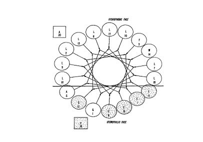
W09~'4C~ PCT/U',6~679
Figure 5 represents expression results of JTS-1
mediated gene delivery.
Figure 6 is a representation of K8 peptides and various
R group substitutions.
Figure 7 is a representation of K8 variations by
changing side chain length and charged groups.
Figure 8 is a representation of pseudopeptides
substituted at core lysine sequences of KN to improve
stability.
Figure 9 is a schematic for formation of pegylated KN
peptides.
Figure 10 is a representation of transfection
efficiency using KN peptides.
Figure 11 is a schematic formula of JTS/K8 conjugates.
Figure 12 is a representation of transfection of C2Cl2
myoblast cells with K8/JTS- 1/DNA complexes.
Figure 13 is a representation of transfection of 4Mbr-
5 bronchus cells with K6 or K7/JTS-1/DNA complexes.
Figure 14 is a representation of target ligands used
to direct delivery of JTS/K8/DNA complex to the hepatocyte.
Figure 15 is a representation of target ligands
containing carbohydrates for uptake by asialoglycoprotein
receptor.
Figure 16 is a representation of target ligands for
delivery of JTS/K8/DNA to cells with mannose or mannose-6-
phosphate receptors.
Figure 17 is a representation of RGD targeting ligands
for delivery of JTS/K8/DNA to connective tissue, wounds and
for healing.
Figure 18 is a representation of ligands useful in
delivery of JTS/K8/DNA to hepatocytes.
The drawings are not necessarily to scale. Certain
features of the invention may be exaggerated in scale or
shown in schematic form in the interest of clarity and
conciseness. In addition, the drawings of PCT publication
WO 93/18759 are hereby incorporated by reference.
CA 02222~0 l997-ll-27
wOs6'~093~ PCT~S96/05679
Detailed Description of the Invention
The following are examples of the present invention
using nucleic acid transporter systems with lysis and/or
binding molecules for delivery of nucleic acid to a cell.
These examples are offered by way of illustration and are
not intended to limit the invention in any manner.
The following are specific examples of preferred
embodiments of the present invention. These examples
demonstrate how specific lysis agents release nucleic acid
into the cellular interior. These examples also
demonstrate how specific binding molecules stabilize and
condense the nucleic acid for cell delivery. Furthermore,
these examples demonstrate how surface and nuclear ligands
can be used with a nucleic acid binding moiety to target
nucleic acid into the cellular interior and/or the cell
nucleus. Such targeted delivery is enhanced by use of the
lysis agent and binding molecules. These examples include
in vivo and in vitro techniques, various cellular or
animal models and how nucleic acid can be inserted into
cells. The utility of such nucleic acid transporter
systems is noted herein and is amplified upon in the PCT
publication WO 93/18759, by Woo et al., entitled "A DNA
Transporter System and Method of Use," hereby incorpor-
ated by reference.
Below are provided examples of specific nucleic acid
transporter systems that can be used to provide certain
functionalities to the associated nucleic acid in the
nucleic acid transporter system, and thus within a
transformed cell or animal containing such associated
nucleic acid. Those in the art will recognize that
specific moieties of the nucleic acid=transporter system
can be identified as that containing the functional region
providing the desirable properties of the nucleic acid
transporter system. Such regions can be readily minimized
using routine deletion, mutation, or modification
techniques or their equivalent.
CA 02222~0 l997-ll-27
WO 9''~958 PCT~S96/05679
JTS Pe~tides, Analoqs and Derivatives
In order to eliminate the use of adenovirus as an
endosomal lysis agent, fusogenic or membrane disruptive
peptides were designed which would increase the rate of
5 delivery of nucleic acid from the endosome to the cell and
ensure that higher concentrations of the endocytosed
nucleic acid would be released and not degraded in the
endosomes. A number of fusogenic/lytic peptides have been
previously described, including the amino terminal
sequence of the vesicular stomatitis virus glycoprotein
and the synthetic amphipathic peptide GALA. O~cius et
al., TIBS, 16 :225-229 (1991); Doms et al., Membrane
Fusion, pp. 313-335 (Marcel Dekker, Inc., N.Y. 1991);
Subbarao et al., Biochemistry, 26 :2964-2972 (1987) .
Short synthetic peptides from the hemagglutinin HA2
subunit of influenza have been studied with artificial
lipid membranes. Wharton et al., J. Gen. Virol ., 69: 1847-
1857 (1988) . These peptides give both membrane fusion and
leakage of liposomal contents similar to whole
20 hemagglutinin molecules. However, the rates are quite
slower.
In order to increase the low efficiency rate by
endosomal lysis with influenza peptides, new peptides were
created. In creating these new peptides for endosomal
25 lysis, four factors were considered: (1) the content and
spacing of the hydrophilic and hydrophobic amino acid
residues along the ~-helix to direct organized oligomer
association of the peptides after their insertion into the
membrane; (2) covalent attachment of the peptide to a
30 binding molecule and preclusion of oligomer formation and
the necessary aggregation; (3) sufficient aggregation of
several oligomeric structures necessary to achieve lysis;
and (4) presence of hydrophilic carboxyl and amino side
chain and terminal groups to create the pH sensitive
35 endosomal processing.
It is well known that the distribution of the amino
acid side chA ~ n-~ along the peptide chain determines the
CA 02222~0 l997-ll-27
WOs6/~C958 PCT~S96/05679
secondary and tertiary structure of a protein. For
membrane associating proteins, the amphipathic profile
created by the hydrophobic and hydrophilic residues is a
principal determinant of the function o~ the protein.
Analysis of the region of the influenza hemagglutinin
responsible for fusion of the viral envelope with the
plasma membrane of cells reveals that a large hydrophobic
surface is formed when the protein becomes ~-helical (see
discussion below).
In the present invention, a number of lytic peptides,
e . g., JTS peptides, have been designed and tested for
endosomal lytic activity. In order for these peptides to
be functional, they must have the following parameters.
These peptides are amphipathic membrane associating
peptides. These amphipathic peptides were designed as an
~-helix, containing a sequence of amino acids such that
the side chains are distributed so that the peptide has a
hydrophobic and hydrophilic side. The hydrophobic side
contains highly apolar amino acid side chains, while the
hydrophilic side contains an extensive number of glutamic
acids.
In general, the amphipathic membrane associating
peptides usually contain 21 amino acids or fewer. The
design criteria requires that the amino acids have a high
probability of forming amphiphilic species. This can be
exhibited in the secondary structure of the membrane
associating peptides, i. e ., helices, turns, bends, loops,
~-sheets, and their oligomeric aggregates and other super
secondary structures defined in the literature, e . g.,
helix-turn-helix. In addition, the amino acids should
have a high probability of being found in an ~-helix and
a low probability of forming a ~-sheet or turn structure.
Leucine, lysine and glutamate are appropriate amino acids
for such characteristics. For example, lysine positioned
on the lateral face of the ~-helix and glutamate residues
opposite leucine provide optimal charge distribution for
lipid interaction. Furthermore, lysines and glutamates
CA 02222~0 l997-ll-27
WO9f~10~5~ PCT~S96/05679
33
can be positioned to take advantage of potential helix
stabilization. Helix dipole stabilization is optimized by
removing the charge at the NH2 and COOH-termini so NH2
termini and COOH-terminal amides are useful. Such
probabilities can be determined from secondary structural
predictions or analogous methods to optimize secondary
structural design. Unnatural amino acid which have been
described for their propensity to induce helix structures
in peptides are also used.
The hydrophobic or lipophilic face has a great effect
on lipid-peptide interactions. Thus, the lipophilic face
is modeled after peptides known to interact with lipids.
Hydrophobic and lipid interactive residues (Ala, Leu, Met,
Val, Phe, Trp, Tyr, Cys, Pro) when substituted on the
lipophilic face either singularly or collectively promote
a similar membrane associating e~fect. Similarly, an acid
group and/or hydrophilic group (Glu, Gln, His, Lys, Gly,
Ser, Asp, Asn, Pro, Arg) can be placed on the hydrophilic
face to achieve the objective. The lipophilic and
hydrophilic faces can also contain residues which promote
lipid interaction and/or induce endosomal lysis at acidic
pH. Such an interaction is not limited to an ~-helix
promoting residue since glycine and serine positioned on
the hydrophilic ~ace have been shown to favorably
influence activity as seen with the examples below.
One in particular, the JTS-1 peptide, GLFE~T~TT~TTT~ L-
WF.T.T.T.T~.~, has a hydrophobic face which contains only
strongly apolar amino acids, while the hydrophilic face is
dominated by negatively charged glutamic acid residues at
physiological pH values. At the amino terminus end, the
JTS-1 peptide uses the Gly-Leu-Phe sequence at amino acid
positions 1-2-3, respectively, as a fusogenic or membrane
disruptive sequence. For increased pH sensitivity Glu is
added at amino acid position 4. In addition, at positions
12-15, Ser-Leu-Trp-Glu is used as a lipid binding site.
The remaining sequences are arranged to provide the
hydrophobic and hydrophilic face of JTS-1. The helical
CA 02222~0 l997-ll-27
W09G/~0~58 PCT/~,G~'~5G79
wheel of the amphipathic membrane associating peptide JTS-
1 can be found in Figure 1. This figure shows the
division of the hydrophobic and hydrophilic faces within
the JTS-1 helical structure. Amino acids 16, 9, 2, 13, 6,
17, 10, 3, 14, 7 and 18 form the hydrophobic face. Amino
acids 5, 12, 1, 8, 15, 4 and 11 form the hydrophilic face.
The following JTS peptides were constructed and
characterized for lytic activity:
Molecular Parent
Sequence Weiqht Ions
JTS-1 GLFE~ T .T .FT .T .F.~LWRT .T .T .R~ 2301.8 2302.2
JTS-2 GLFE~T ~T .FT ~T .F.. ~LWRT ~T .T ~RT .YA 2578.2 2578.4
JTS-3 GLFE~T.T.RT.T.RF.T.WRT.T.T.R~ 2343.8 2342.9
JTS-4 GLFE~T.T.F.T.T.FF.T.WEALLEA 2301.8 2301.8
15 JTS-6 GLFE~T.T.F.T.T.R.~LWFT.T.T.F.~GGGGC 2633.2 2633.8
JTS-7 SLFE~T.T.F.T.T.R.~LWRT.T.T.~ 2331.8 2332.4
JTS-8 GLFE~T .T .FT .T ~F-~LYRT .T .T .R~ 2278.8 2279.3
JTS-9 GLFEAL~FT.T.F........ ~LWEALLEA 2217.6 2218.3
JTS-10 GLFE~T.T.RT.T.F...... sPWF.T.T.T.R~ 2285.8 2285.0
20 JTS-11 GLFE~T.T.RT.T.F.~LWEFLLEA 2335.8 2336.2
JTS-12 GLFE~TT.F.T.T.R.~LWFT.T.T.F.~ 2369.8 2302.2
JTS-12a GLFEALLELWEA 1390.6 ---
JTS-13 GLFEALLESLWEA 1477.7 1477.8
JTS-14 GLFEALLEILEsLwF~TlTlTlF~ 2369.8 ---
25 JTS-15 ~GLFEALLELWEA 1390.6 1390.8
JTS-16 GLFE~T ~T .RT ~T .F.~LWEA 1833.2 1834.0
JTS-17 GLFE~ T ~T ~~T ~T .F.~LWEFFLEA 2369.8 2370.4
JTS-18 GLFEALLELFESLWELLEA 2335.8 ---
JTS-19 GLFEST.T.F.T.T.R.~LWRT.T.T.R~ 2317.8 ---
30 JTS-20 GLFE~T.T.F.T.T.R.~LWELLKEA 2315.3 ---
JTS-24 GLFE~T.T.RT.T.R.~LWFT.T.T.F.~AEEA 2702.2 2703.4
JTS-lOK8 GLFE~T.T.RT.T.R.~PWF.T.T.T.F.~GGGSG-
SGSGSGSGYKAKKKKKKKK~K 4937.1 4937.7
JTS-15KPam
GLFEALLELWEAKNH2~-Pam 1756.2 1757.0
CA 02222~0 l997-ll-27
WO9-/1358 PCT/U~3~5G79
JTS-16KBIO
GLFE~T~T.FT~T~ LWEAKNH2 ~ -
BIOHX 2298.6 2300
JTS-lKBIO
GLFE~T~T~T~T~F~LW~T~T~T~KNH2~-
BIOHX 2642.8 ---
acJTS-1 acGLFE~TT.T~'T.T.T~LW~TT.T.~2345.8 2344.2
DMGJTS-1 Me2GLFE~T T.T~'.T.T ~.~LW~T .T.T.~ 2350 ___
desGJTs-l LFEAT.T.T~'.TT~LW~TTT.F~ 2244.7 ---
10 JTS-16KKCC14
GLFE~T .T .~T .T ~LWEAAAKLSKLEK-
KLSKLEK 1833.2 ---
GALA18 WEAALAEALAEALAEHLA 1879.1 1879.0
GALA30 WEAALAEALAEALAEHLAEALAEAL-
EALAA 3030.6 3032.8
In addition to the above, n-acyl tetrapeptides with
~usogenic or membrane destabilizing activity can be
constructed. The structure of these is set ~orth in
Figure 2. The tetrapeptide sequence when substituted with
the appropriate amino acids as discussed above are capable
of interacting with lipid bilayers and thereby
destabilizing. The acyl chain can be lengthened or
shortened depending on structure/~unction requirements.
Furthermore, shorter ~-helical peptides were also
synthesized with the above design moti~s in mind to retain
the lytic properties as discussed above. Figure 3 shows
the helical wheels o~ smaller ~usogenic peptides. For
example, LLEKLLEWLE (number IV in Figure 3) is a shorter
~-helical peptide with lytic properties. Leucine is used
~or hydrophobic properties and ~-helical movement.
Glutamic acid residues are used ~or lytic activity. These
residues also have the propensity to ~orm an ~-helical
structure at pH 4Ø Furthermore, a COOH-terminal amide
is used to provide helix-dipole optimization. When in an
~-helical structure the hydrophobic ~ace appears at
positions 4, 7, 3, and 10.
CA 02222~0 l997-ll-27
W096/40958 PCT/U~,G~SG79
36
To provide the Gly-Leu-Phe fusogenlc or membrane
disruption activity to the above ~-helical peptide in
Figure 3, the peptide was lengthened to an 11-mer. Adding
the additional amino acid to form the following peptide,
Suc-GLFKLLEEWLE, allowed the activity of the three
glutamic acids to be retained. In addition, the peptide
was succinylated at the amino terminus to afford an i to
i+4 salt bridge with lysine which is designed to stabilize
the helix.
Synthesis, Purification and Characterization of JTS-1
Peptides
JTS-1 peptides are synthesized by the solid phase
method as developed by Merrifield et al., Sol id Phase
Peptide Synthesis, Academic Press (N.Y. 1980). In
addition, a modified polystyrene (Sparrow, J. Org. Chem.,
41:1350-1353 (1976)) and/or polyamide resin (Sparrow et
al., Int. J. Peptide Prot., 38:385-391 (1991)) with fast
HBTU/HOBT coupling is used. It should be noted that the
following procedure was used to synthesize INF-7 as well.
The procedures involved in solid phase synthesis include:
(1) attachment of the protected carboxyl terminal amino
acid to the solid support through the oxymethyl-
phenylacetamide linkage; (2) deprotection of the N-
terminal amino group; (3) neutralization of the amino
group; (4) coupling of the next N-protected amino acid to
the peptide resin; and (5) after completion of the
synthesis, removal of the peptide from the solid support.
Specifically, the carboxyl terminal amino acid
protected with the N-~butyloxycarbonyl group is esterified
to bromomethyl-phenylacetic acid and coupled directly to
aminomethyl-polystyrene resin or to a resin containing a
long spacer chain between the point of attachment and the
polystyrene backbone or directly to the amino-propyl
polyamide resin. In the fast HBTU/HOBT synthesis protocol
the above procedure is modified as follows to give a total
program time of 45 minutes. Trifluoroacetic acid (100~)
CA 02222~0 1997-11-27
WO9f'~035~ PCT~S96/05679
is used to deprotect the amino group in 6 minutes. The
resulting salt is neutralized by the excess
diisopropylethylamine used to activate the N-
tbutyloxycarbonyl amino acid with HBTU/HOBT in
dimethylformamide ("DMF"). This combines the coupling and
neutralization steps. The coupling reaction is allowed to
proceed for 15 minutes and the resin washed extensively
with DMF and dichlormethane ("DCM"). These steps are then
repeated until the sequence of interest has been
synthesized.
To protect side chains, the following are used: (1)
2,6-dichlorobenzyl for the hydroxyl of tyrosine; (2)
benzyl for the hydroxyl of serine and threonine; (3)
benzyl esters for the ~- and y-carboxyl of aspartic and
glutamic acids; (4) 2-chlorobenzyloxycarbonyl for the ~-
amino of lysine; (5) p-methoxybenzyl or acetamidomethyl
for the sulfhydryl of cysteine; (6) formyl for the indole
of tryptophan; (7) benzyloxymethyl for the imidazole of
histidine; (8) trimethyl benzenesulfonyl for the guanidino
of arginine; and (9) xanthanyl for the amido of glutamine
and asparagine.
The peptide is cleaved from the solid support by
treatment of 1 g of resin with 60 ml of anhydrous hydrogen
fluoride containing 10~ anisole and 1~ ethanedithiol for
30 minutes at 0~C. In the case of peptides containing
arginine, the cleavage is performed at -20~C for 3 hours.
The hydrogen fluoride is evaporated under vacuum at 0~C
and the peptide precipitated with ether. The peptide and
resin are filtered off and washed with ether. The peptide
is then extracted with trifluoroacetic ("TFA") (3 x 30 ml)
and the TFA evaporated under vacuum. The peptide is
precipitated with ether and the precipitate collected by
centrifugation. The precipitate is suspended in 10 ml 1
M TRIS and 6 M GnHCl. An additional 30 ml of 6 M GnHCl is
added to completely dissolve the peptide. The pH of the
solution is adjusted to 8Ø The peptide is desalted on
a column of BioGel P-2 equilibrated in 0.1 M ammonium
CA 02222~0 l997-ll-27
WO9~/~0~58 PCT~S96/05679
38
bicarbonate for the lytic peptides. The peptide fractions
are located by absorbance at 254 nm and 280 nm, pooled and
lyophilized. The lyophilized peptide is dissolved in 25
ml of 6 M GnHCl for the lytic peptides.
A280 is used to determine peptide concentration. This
is performed in 6 M guanidine HCl for JTS-1 or other
peptides so that aggregation is not observed. The
following molar extinction coefficient is used for JTS-1
in 6 M guanidine HCl - 5 6 0 0 .
The peptide is purified by reversed phase HPLC. The
following procedures were used. The peptide (50-100 mg in
5 mI of 6 M GnHCl) is diluted with 20 ml of 0.1 M ammonium
phosphate in 6 M GnHCl, pH 6 . 7 and the pH confirmed. This
solution is pumped onto a 2.5 x 25 cm Vydac C4 column
15 (214TP152022; 300 A pore size) equilibrated in 0.01 M
ammonium phosphate at a flow rate of 20 ml/min. Two
buffers are used to elute the peptide, Buffer A (0.01 M
ammonium phosphate in ddH2O, pH 6 . 7) and Buffer B (2-
propanol 100~). The peptide is eluted with a linear
gradient between Buffer A and 30~ Buf~er B, and Bu~fer A
and 50~ Buffer B. The gradient program used is Buf~er A
to 50~ Buffer B for 45 minutes, with retention time being
40-41 minutes. The peptide is detected by absorbance at
254 and 280 nm.
The peptide containing fractions are pooled, the pH
adjusted to 8.0 with ammonium hydroxide and desalted on a
BioGel P-2 column equilibrated in 0.1 M ammonium
bicarbonate. The peptide containing fractions are pooled
and lyophilized. The peptide is dissolved in water after
lyophilization. Diluted ammonium hydroxide is added to
adjust the pH to 7 . 5 in order to completely dissolve the
peptide.
The following criteria were applied to evaluate the
purity of the synthetic products: 1) analytical high
performance liquid chromatography using a linear gradient
between 0.01 M ammonium phosphate, pH 3.0 or pH 6 . 8 and 2-
propanol and 0.1~ TFA and 2-propanol, 2) fast atom
CA 02222~0 1997-11-27
WO 9~':1D358 PCT/US96/05679
bombardment or electrospray mass spectrometry, 3) amino
acid analysis for correct composition, 4) automatic amino
acid sequencing, and 5) capillary electrophoresis. For
amino acid analysis of the peptides for correct
composition, decomposition and purity, quantitation of the
molar ratios of the peptide components determines peptide
purity. With fast atom bombardment mass spectrometry
("FAB") purity, molecular weight can be determined. A
single peak for JTS-1 occurs at the expected mass of 2302
amu.
Secondary structure is determined by circular
dichroic and FTIR spectroscopy. These standard methods
are used to confirm the secondary structure of JTS-1.
Since guanidinium HCl is used to solubilize the JTS-1
during purification, guanidinium contamination must be
tested. Guanidinium contamination in JTS-1 is detected by
the Sakaguchi method for determining arginine
(guanidinium). This method is well known in the art.
SYnthetic Membrane Disru~tive Behavior of JTS-1
In order to test the synthetic membrane disruptive
behavior of JTS-1, a comparison of turbidity was performed
with liposomes containing either JTS-1,
polyvinylpyrrolidone ("PVP") or Tween80 (a detergent).
The liposomes were made from dimyristoylphospha-
tidylcholine ("DMPC") by methods well known in the art.
Phosphatidylcholine vesicles containing calcein were
prepared by sonication. Briefly, 10 mg/ml
phosphatidylcholine was dried down under a stream of
nitrogen. The lipid was resuspended in 100 mM calcein
(adjusted to pH 7.3 with sodium hydroxide) and sonicated
with a probe sonicator for 20 minutes in an ice bath.
Liposomes were separated from unentrapped calcein using a
Sephadex G25 column. Calcein release was measured at 520
nm (excitation at 470 nm) using a fluorescence
spectrophotometer. For leakage assays, liposomes were
diluted 1000-fold in 150 mM NaCl, 15 mM sodium citrate, pH
CA 02222~0 l997-ll-27
W095/~03~ PCT~S96/05679
7.0 or 5Ø Peptide was added at a concentration of 1
~g/ml. Fluorescence was measured before and 10 minutes
after addition of peptide. 100~ leakage was determined by
adding Triton X-100 to a final concentration of 0.5~. The
turbidity was monitored using apparent absorbance
measurements at 400 nm on a visible-wavelength
spectrophotometer. This experiment was performed at
various pHs.
Initially, all liposomes were turbid. The clearance,
10 i . e., decrease in turbidity, as a function of time after
adding the test samples to the liposomes was then
measured. Both JTS-1 and Tween80 showed fusogenic or
membrane disruptive behavior, i . e., decrease in turbidity,
whereas PVP did not. JTS-1 lysed phosphatidylcholine
liposomes to a greater extent at pH 5.0 than at pH 7Ø
It should be noted that Tween80 was used as a
positive control. Tween80 is a sur~ace active agent which
concentrates at oil-water interfaces causing an
emulsi~ying action. Such properties allow Tween80 to
disrupt membranes of synthetic liposomes. PVP was used to
see if there was any activity. PVP is an amorphous
powder, is compatible with hydrophobic and hydrophilic
residues. Although it can be used as a detergent, PVP has
colloid protective properties. These properties allow PVP
to act as a surface-active substance that prevents the
dispersion of a suspension, i . e ., liposomes, from
coalescing by forming a thin layer on the surface of each
particle. In addition, the above assay was also performed
using INF-7 instead of JTS-1 (see description of INF-7,
below). INF-7 showed pH dependent membrane activity on
phosphatidylcholine liposomes. The activity of INF-7 was
approximate 8-fold lower than that of JTS-1.
Hemolysis Activit~ of JTS Pe~tides
The JTS peptides were studied for their ability to
lyse erythrocytes. Erythrocytes were isolated by methods
known in the art. Hemolysis assays were performed
CA 02222~0 l997-ll-27
WO 9~'1DS~. PCT/U',~ 79
41
according to the literature. Erythrocyte lysis assays
have been previously used to determine the membrane
activity of bacterial toxins and membrane active peptides.
Human erythrocytes were washed three times with phosphate
buffered saline and were resuspended in 150 mM NaCl, 15 mM
sodium citrate, pH 7.0 or 5.0, at a concentration of
7xlO7/ml. Peptides were diluted serially in 150 mM NaCl,
15 mM citrate pH, 7.0 or 5.0, in a 96-well plate. Next,
75 ,ul erythrocyte suspension was added to each well. The
10 plates were incubated at pH 7.0 and pH 5.0 for 60 minutes
at 37~C with occasional shaking. Unlysed erythrocytes
were pelleted and the extent of hemolysis was determined
visually. One hemolytic unit (HU) was defined as the
amount of protein necessary to induce >50~ hemolysis. All
15 hemolysis assays were performed in duplicates.
JTS-1 was hemolyticly active at pH 5.0 and not at pH
7Ø At pH 5.0, the specific hemolytic activity was lx107
erythrocytes lysed per ~g of peptide. JTS-3 had a
specific hemolytic activity at pH 5.0 of 8x10⁷ erythrocytes
20 lysed per ~g of peptide, whereas JTS-9 was 3x10⁷
erythrocytes lysed per ,ug of peptide.
In a separate hemolysis assay, it was determined that
the hemolysis activity of JTS peptides decreased as the pH
increased. Hemolysis activity was stronger at pH 5.0 and
25 is undetectable at pH 7Ø These studies show that the
JTS-peptides exert enhanced lysis activity at pH 5Ø
Hemolysis Assav ComParinq JTS-1 and Influenza ("INF-7")
Peptide
JTS-1 and INF-7 peptides were studied for their
30 ability to lyse erythrocytes. INF-7 is the active region
of HA2, influenza hemagglutinin, responsible f~or ~usion o:E
the influenza viral envelope with the plasma membrane of
cells. INF-7 has the following amino acid sequence,
GLFEAIEGFIENGWEGMID. Amino acid numbers 5, 16, 9, 2, 13,
35 6, 17, 10, 3 and 14 of the helical wheel form the
CA 02222~0 l997-ll-27
WOs6/1C~5~ PCT~S96/05679
hydrophobic face. Amino acid numbers 7, 18, 11, 4, 15, 8,
1 and 12 of the helical wheel form the hydrophilic face.
Erythrocytes were isolated by methods well known in
the art. Serial dilutions of INF-7 and JTS-1 peptides
were incubated with washed human erythrocytes at pH 7.0
and pH 5Ø After one hour, the unlysed erythrocytes were
pelleted. The concentration at which 50~ lysis occurred
was determined by visual reading. INF peptides showed no
hemolytic activity at either pH 7.0 or pH 5Ø Just as
above, JTS-1 was hemolyticly active at pH 5.0 and not at
pH 7Ø At pH 5.0, the specific hemolytic activity was
lx107 erythrocytes lysed per ~g peptide.
JTS Peptide-Liposome Leakaqe AssaY
Liposome membrane activity was measured by testing
liposomal leakage. This assay measures the release of
calcein, a fluorescent dye, from phosphatidylcholine
("PC") vesicles. Briefly, calcein is encapsulated into
liposomes by well known procedures at a concentration
where the fluorescence of the dye is greatly reduced
(self-quenchlng). When the liposomes are destroyed by the
lysis agent, the fluorescent dye leaks out of the
liposomes and is diluted in the incubation buffer. This
causes a great increase of fluorescence; (dequenching)
which can be followed in a fluorescence spectrophotometer.
Liposomes were incubated with monomeric forms of JTS-
1, JTS-3 and JTS-9 peptides at a concentration of 0.5
~g/ml in a sodium-citrate buffer with a pH ranging from
5.0 to 7Ø Before and 5 minutes after the addition of
the lysis agent peptides, the fluorescence was determined.
The fluorescence corresponding to 100~ leakage was
determined by complete lysis of the liposomes with a
detergent (Triton X-lOO; final concentration 0.5~) and the
values obtained were plotted as percentage~leakage.
JTS-1 lysed phosphatidylcholine vesicles, as well as
erythrocytes, in a pH-dependent manner. No membrane
CA 02222~0 1997-11-27
wOs~'~055~ PCT/U~5~5G79
43
activity was observed at pH 7Ø A sharp increase of
membrane activity was observed at a pH lower than 6Ø
In comparison, use of the influenza fusion peptide,
INF-7, showed pH-dependent membrane activity on liposomes.
In contrast to JTS-l, only very little hemolytic activity
was observed. Furthermore, the JTS-9 peptide was shown to
be more potent than the activity of JTS-l and JTS-3.
JTS-l Mediated Expression in Cell Lines
The ability of JTS-l to mediate expression in cells
in vi tro was tested using DNA complexes containing a CMV-
~-galactosidase expression vector with transferrin/poly-L-
lysine and unmodified poly-L-lysine to create a positive
particle. The cell lines tested were obtained from ATCC
and cultured by well known methods in the art.
Poly-L-lysine ("PLL") Mwt. 20,500, was coupled to
transferrin in a l to 2.0 ratio by using l-ethyl-3-(3-
dimethylaminopropyl)carbodiimide ("EDC") at pH 7.3. The
reaction was incubated for 24 hours at room temperature
after which it was concentrated and resuspended in 2 M
Guanidine-HCl, 50 mM HEPES, pH 7.3, and fractionated by
gel filtration on a Superose 6 column with a Fast Protein
Liquid Chromatography system ("FPLC"). Once the conjugate
was made and purified, fractions from the FPLC were
analyzed on an SDS-PAGE gel to determine those fractions
that contained modified transferrin ("TF/PLL conjugate")
only. These fractions were then pooled and dialyzed
against 150 mM NaCl, 20 mM HEPES, pH 7.3 prior to complex
formation.
The DNA plasmid CMV/~-gal containing the E. col i ~-
galactosidase gene under the control of the CMV enhancerand promoter was used as a reporter gene. The plasmid was
isolated and purified by double CsCl banding. This
plasmid has been thoroughly described in the art. In
addition, unmodified poly-L-lysine was added to help
create a positive particle. The JTS-l peptides were added
and bound to the DNA complex through ionic interactions.
CA 02222~0 l997-ll-27
W096/~035S PCT~S96/05679
44
Conjugate/DNA complexes were prepared by diluting the
conjugate in 150 ~l of HBS (150 mM NaCl per 20 mM HEPES,
pH 7.3) and diluting 6 ~l of DNA, in 350 ~l of HBS. The
diluted DNA was added directly to the diluted conjugate
while mixing. The reaction was allowed to incubate at
room temperature for 30 minutes before analysis.
Immediately following the incubation, all complexes were
analyzed on 0.8~ agarose gels and electrophoresed in TBE.
Six ~g of DNA complex was incubated with 3x10⁵ tissue
culture cells. Before adding the complex to the tissue
culture cells, the complete media was removed and replaced
with 1 ml of Low Glucose DMEM containing, 5 mM Ca2' and 2
fetal calf serum. After a 2 hour incubation at 37~C, 1.5
ml of complete media was added to the tissue culture cells
and the lncubation continued for 24 hours at 37~C.
After 24 hours, analysis of ~-galactosidase (~-gal)
activity was performed by staining the cells, using X-gal
as a substrate. The following procedures were used to
analyze ~-galactosidase activity. The cells are washed
with lX PBS twice. The cells are then fixed for 15
minutes in a solution containing 1~ glutaraldehyde (from
50~ stock), 100 mM sodium phosphate buffer, pH 7.0, and 1
mM MgCl2.
The cells are washed with lX PBS twice and incubated
at 37~C for 30-60 minutes with the solution containing
0.2~ X-gal, 10 mM sodium phosphate buffer, pH 7.0, 150
NaCl, 1 mM MgCl2, 3.3 mM K4Fe (CN)63H2O and 3.3 mM K3Fe
(CN) 6'
The staining solution is removed and the cells washed
with lX PBS twice. The stained cells can be identified
under a phase-contrast microscope (400x). To quantify the
actual amount of ~-galactosidase produced, ONPG can be
used as a substrate with aliquots of cell extracts.
Figures 4 and 5 show the expression results. With
MCA-26 cells, up to 40~ of the cells were stained blue;
3T3 cells, 30~; Sol 8 cells, 20~; 4MBR-5, 50~; 293 cells,
CA 02222~0 l997-ll-27
WO9~'lC3~ PCT~S96/05679
90%; human fibroblast, 30%; and SKOV3 cells, 1%. Without
peptides no blue cells were observed.
In addition to the above, the expression of JTS-1
mediated expression was compared with an INF-7 peptide
using the same procedures as above and transfecting Sol 8
myoblast cells. Up to 5% of the cells were positively
stained blue where JTS-1 was part of the complex. In the
absence of peptides, no cells were stained blue. With
INF-7 peptides as part of the complex, only a few blue
cells (~0.01%) were observed.
Gene Expression Usinq Various Ratios of JTS-1 to
Transferrin/PLL
DNA complexes were made by condensing a CMV-~-
galactosidase expression vector with transferrin/poly-L-
lysine and unmodified poly-L-lysine to create a positive
particle. The same procedures as described above were
used. The lytic peptides were then added and were bound
to the DNA complex through ionic interactions as discussed
above. Various concentrations o~ DNA complex (5-20 ~gm)
and ratios of JTS-1 to transferrin-PLL (2.5-42.5 ~gm) were
incubated with 3x10⁵ Sol 8 myoblast cells. In addition, in
one series of experiments 12-18 ~gm of PLL was also added.
After 24 hours, ~-galactosidase activity was determined.
Significant ~-galactosidase activity was observed at all
ratios of JTS-1/transferrin-poly-L-lysine tested. In
almost all cases, increasing JTS-1 concentration enhanced
transfection and therefore expression of ~-galactosidase.
Human Fibroblast UPtake and Dearadation of LDL/JTS
Complexes
To form the l25I-LDL/JTS complexes, 125I-LDL was
dissolved in dimethylsulfoxide and incubated with 10-fold
excess of l-ethyl-3-(3-dimethylaminopropyl)carbodiimide
("EDC") for 30 minutes at room temperature. A 30-fold
excess was added to JTS peptides (89 KBg/~g) in phosphate
buffer and incubated for 4 hours at room temperature. The
CA 02222~0 l997-ll-27
WO9f/l0~i~ PCT~S96/05679
46
reaction was quenched with a 50-~old excess of
ethanolamine. Free JTS was separated from the l25I-LDL/JTS
complex by passing the reaction mixture over a Sephadex G-
25 column equilibrated with phosphate buffered saline, at
pH 7.4.
A 10:1 molar ratio of JTS peptides bound to 125I-LDL
was incubated with human fibroblasts for 5 hours at 37~C.
Fibroblasts were isolated and cultured by using methods
well known in the art. JTS-1, JTS-3j JTS-8 and JTS-9
peptides were used for the uptake/degradation studies. As
a control, l25I-LDL without JTS peptides were also incubated
with the same fibroblast and analyzed accordingly.
A~ter incubation ~or 5 hours, cells were harvested to
determine internalized radioactivity. Cells were washed
once with ice cold PBS, followed by a 30 second wash with
ice cold acid saline (0.15 M NaCl, adjusted to pH 3.0 with
glacial acetic acid) to remove sur~ace bound LDL and then
harvested by scraping in ice cold PBS. The cells were
pelleted, washed with PBS, and then dissolved in 0.1 M
NaOH. One-hal~ was used for protein determination (BCA-
Protein assay) and the other half was counted in a scin-
tillation counter. For competition, a 100-fold molar
excess o~ LDL was added prior to incubation with the 125I-
LDL-JTS complexes.
Binding and uptake of l25I-LDL was not affected, while
JTS peptides decreased degradation by 50~. To insure LDL
uptake was speci~ic ~or the JTS peptides, the experiment
was repeated in the presence o~ 20-~oId excess cold LDL.
Under these conditions, more than 90~ of the cellular
30 uptake of l25I-LDL-JTS was inhibited. Cells receiving 125I-
LDL showed intense perinuclear ~luorescence. Most cells
receiving both JTS peptides and 125I-LDL had diffuse
intracellular fluorescence. Using chloroquine to block
endosome acidi~ication, fluorescence was perinuclear, the
same as that observed without JTS peptide.
The same studies were performed with HAEC cells.
HAEC were isolated and cultured using methods well known
CA 02222~0 1997-11-27
WO 96/~0~5v PCT/U' ,G/'~679
in the art. Binding and uptake of 125I-LDL was not
affected, while JTS-1 and JTS-8 peptides decreased
degradation by 30~ depending on the amount of JTS peptide
used. To show that LDL uptake was specific for the JTS
peptides, the experiment was repeated in the presence of
20-fold excess cold LDL. Under these conditions, more
than 90~ of the cellular uptake of 125I-LDL-JTS was
inhibited.
KM Peptide Analoqs and Derivatives
A variety of peptides with the formula YKA(K)NWK,
wherein N = 1-40, have been synthesized. This general
structure for KN peptides, more specifically K8, is shown
in Figure 6. The lysines (i . e., "K") act as the binding
molecule. The tyrosine partially contributes to the A280
and also allows for the iodination for tracking in vitro.
Tryptophan increases the stability of interaction with DNA
through intercalation and also provides a fIuorophore
which quenches upon interaction with the DNA. When R in
Figure 6 is tryptophan, smaller particles are obtained and
improved transfection occurs. In addition, the R group
can also be substituted with other R groups to achieve the
same effects. See Figure 6 for additional R groups for
improving peptide activity. Alanine provides a linker
that allows the tyrosine and nearest neighbor lysine
residues to be wrapped around the DNA in a more helical
fashion resulting in a more stable complex.
Extensive characterization of the interactions of
these peptides with DNA were performed for each of the
peptides. In these experiments, the dependence of time,
peptide concentration, and number of lysines in the
peptide on the size of the peptide-DNA complexes formed
were examined. Condensates formed between KN peptides and
DNA polymers have a higher propensity to aggregate when
the relative charge ratios of peptide lys:DNA phosphates
are near 1:1. This is more pronounced for the lower
molecular weight peptides. Higher molecular weight
CA 02222~0 1997-11-27
WO~6'10~58 PCT~S96/05679
48
peptides tend to result in particles that are smaller and
more monodisperse. At a constant lys:phosphate ratio,
condensed particle size tends to increase with increasing
concentrations of DNA.
Variations to optimize K8 activity are also useful.
The K8 peptide contains an octamer of polylysine. To
optimize the nucleic acid condensing activity of the
cationic peptide, variations, as shown in Figure 7, of the
side chain length and charged groups are made.
Modifications introduced at the core cationic oligomer
decrease or increase the number of methylenes placed
between the side chain cationic group and the peptide
backbone. For instance, Figure 7 shows the use of a
peptide backbone spacer that varies the distance between
cationic groups, where the cationic group is the ~-amino
group of the substituted amino acid, e.g., for K8 it would
be the ~-amino of lysine. Likewise, NH2- and COOH-
terminal substitutions and deletions can also be used to
optimize DNA binding of the molecule. For example,
variation o~ a NH2 or COOH terminal acyl group may yield
enhanced activity. Functionalities of the charged or
cationic groups include guanidinium, amine or imidazole.
Furthermore, variations of the NH2- and COOH- termini
include esters, acyl groups and amides, as well as
deletions.
In addition to the above, amino charged groups can be
substituted in the peptide backbone ~or condensing the
associated nucleic acid. Pseudopeptides of the formula
~[CH2NH] when substituted within the core lysine sequences
(see Figure 8), improves stability and enhances
electrostatic interactions with the nucleic acid
phosphates. Other possible substitutions using ~[(CH2)nX]
where X is a heteroatom can help optimize the
intermolecular ionic interactions important for condensing
the nucleic acid.
Other modifications include pegylated KN peptide
analogs to increase plasma half-li~e, resistance to
CA 02222~0 l997-ll-27
WO96'1D358 PCT~S96/05679
49
degradation, solubility and decreased antigenicity and
immunogenicity. Figure 9 outlines a scheme for pegylation
of lysine. Once the Fmoc-Lys-N-(~-PEG)-OH is synthesized,
it can be used for the solid phase synthesis O~ KN-
pegylated peptides.
The following KN peptides were constructed and
characterized:
Molecular Parent
Condensinq PePtides Weiqht Ions
10 K4 YKAKKKKWK 1207.5 1207.8
K5 YKAKKKKK~K 1335.7 1336.1
K5FK YKAKKKKK~K 1296.7 1296.5
K5LK YKAKKKKK~K 1262.6 ---
K6 YKAKKKKKK~K 1463.9 ---
15 K7 YKAKKKKKKK~K 1592.1 ---
K8 YKAKKKKKKKK~K 1720.2 1719.7
FK8 YKAKKKKKKKK~K 1681.2 1680.1
R4K8 YKAKKKRKKKKWK 1748.3 1747.4
R8 YKARRRRRRRRWR 1972.3 ---
20 GSK8 GSGSGSGSGSGYKAKKKKKKKK~K 2498.1 ---
CGSK8 CGSGSGSGSGSGYKAKKKKKKKK~K 2601.2 ---
WK8 WKAKKKKKKKK~K 1743.3 1742.8
WK10 K~KKKKKKKKKK~K 1928.6 1928.4
K10 YKAKKKKKKKKKK~K 1977 ---
25 K12 YKAKKKKKKKKKKKK~K 2233 ---
KA20 YKAKAKAKAKAKAKAKAKAKAKAKA-
KAKAKAKAKAKAKAKAKAKWK
K40 YKAKKKKKKKKKKKKKKKKKKKKKK-
KKKKKKKKKKKKKKKKKK~K 5822 5820.6
30 KKCC14 KLSKLEKKWSKLEK 1744.1 1744.3
KKCC21 KLSKLEKKLSKLEKKWSKLEK 2570.6 2571.2
A4KlO KAKKAKKKAKKAKWK 1770.3 1769.8
S4K10 KSKKSKKKSKKSKWK 1834.3 ---
SSK8 YKAKKKKN~(cH2)2ss)cH2)2coK-
KKKWK 1720.2 ---
("Short NLS")
CA 02222~0 l997-ll-27
W09.'~9S~ PCT~S96/OS679
SHNLSK6 STPPKKKRKVEDPKDFPSELLSAKKK-
KKKWK 4044.2 ---
SHNLSK8 STPPKKKRKVEDPKDFPSELLSAYKA-
KKKKKKKK~K 4300.2 4299.3
("Long NLS")
LGLSK6 SSDDEATADSQHSTPPKKKRKVEDPK-
DFPSELLSKKKKKK~K 4925.7 4924.9
LGLSK8 SSDDEATADSQHSTPPKKKRKVEDPK-
DFPSELLSAYKAKKKKKKKK~K 5544.5 ---~0 K40 - Short NLS (attached at the ~-amino of Tyr and
~-amlno group of Lys-2)
K40 - Long NLS (attached at the ~-amino of Tyr and
~-amino group of Lys-2)
Synthesis, Purification and Characterization of K~T Peptides
Peptides were synthesized as discussed above for the
JTS peptides. In addition, the peptides were also cleaved
from the solid support as discussed above for JTS peptides
with the following changes. A~ter precipitation of the
peptide with ether, the precipitate is suspended in the
same solution; however, water replaces the 6 M GnHCl and
the pH is adjusted to 3.5 instead of 8.0 as used above.
In addition, the peptides are desalted on the BioGel P-2
column equilibrated with 5~ acetic acid instead of 0.1 M
ammonium described above for the JTS peptides. After the
collected peptide is lyophilized, it is dissolved in 25 ml
o~ water instead of 6 M GnHCl for the lytic peptides.
A280 is used to determine peptide concentration. This
is performed in water for KN peptides so that aggregation
is not observed. The following molar extinction
coefficient is used for K~ - 6860.
The peptide is purified by reversed phase HPLC. The
peptide (50-100 mg in 5 ml of water) is diluted with 20 ml
of 1~ TFA and the solution pumped onto a 2.5 x 25 cm Vydac
C18 column (218TP152022, 300 A pore size) equilibrated in
0.1~ TFA (Buffer A). The peptide is eluted with a linear
gradient between 0.1~ TFA and 0.1~ TFA, 10~ 2-propanol
CA 02222~0 1997-11-27
WO 9~ 9.)~ PCT/U' ,. '~'~;79
51
(Buffer B) at a flow rate of 20 ml/min. The gradient used
is 100~ A to 10~ B for 30 minutes.
Then the column is washed with 10~ B to 90~ B in 5
minutes, 90~ B for 5 minutes, then 90~ B to 100~ A in 5
minutes. The retention time is 6.0 to 6.8. The peptide
is detected by absorbance at 254 and 280 nm. The peptide
containing fractions are pooled, frozen and lyophilized.
The peptide is then dissolved in water. Purity is
confirmed by analytical reversed phase HPLC.
The purity and molecular weight is also determined by
electrospray mass spectrometry ("ESMS"). ESMS has been
performed on K8 peptides. A single peak for K8 occurs at
the expected mass of 1720 amu. Decomposition and purity
is also determined by amino acid analysis of the peptides.
Quantitation of the molar ratios of the peptide components
determines peptide purity.
Secondary structure is determined by circular
dichroic and FTIR spectroscopy. These standard methods
are used to confirm the secondary structure of K8 and other
KN peptides.
KN CYtotoxicity Studies
Prior studies using poly-L-lysines with DNA/protein
complexes have been shown to be toxic in nM concentrations
to living cells. This limits the general applicability of
such poly-L-lysines. Such peptides were 50-200 in average
chain length. In order to avoid such toxicity, KN peptides
were constructed. These shorter polylysine peptides were
synthesized as discussed above, such as K8 which contains
a central cluster of eight lysines. To show that KN
peptides are not cytotoxic to cells, HepG2 cells were
incubated with K8 and with PLL. HepG2, a hepatocyte cell
line, were cultured using standard methods known in the
art. HepG2 cells were incubated at 37~C for 24 hours with
increasing concentrations of K8or poly-L-lysine (100 mer),
after which viable cells were counted. Poly-L-lysine
concentrations of greater than 0.1 ~M led to complete cell
CA 02222~0 l997-ll-27
W09~ 3~8 PCT~S96/05679
death of HepG2 cells. In contrast, no cytotoxicity was
observed for up to 100 ~M of K8, the highest concentration
tested. This indicates that K8 is at least 1000-fold less
toxic than poly-L-lysine for HepG2 cells.
DNA/KM Transfection Efficiency in C2Cl7 Myotubes
In order to determine KN peptide transfection
efficiency, C2C12 myotubes were transfected with KN/DNA
complexes.
DNA was added to each well in a volume containing
300 ~l per well in Fisherbrand culture tubes. The KN
peptide is added to each well with DNA and then vortexed
before the two solutions mix. Samples then set for at
least 30 minutes.
If JTS-1 peptide is to be added as in the studies
below, then JTS-1 is added to each of the DNA-KN peptide
samples and vortexed before the two solutions mix. The
samples then set for at least 30 minutes.
C2Cl2 myotubes in DMEM containing 10~ FBS are incubated
for 30-50 minutes prior to transfection. Then, 300 ~l o~
the solution complex is added to each well in a 24-well
plate and incubated for 5 hours at 37~C. After 5 hours,
1 ml of DMEM containing 10~ FBS is added.
Cell extracts are prepared by adding 100 ~l lysis
solution to each well (24 well-plate). Cells are
transferred to a microfuge tube and centrifuged for 10
minutes at 4~C to pellet any debris. Supernatant is
transferred to a fresh microfuge tube and cell extracts
are frozen at -70~C for future use.
The cell extracts are diluted 50 times by ddH2O. Cell
extracts in 5-20 ~l aliquots are diluted so that the total
volume is 20 ~l. Reaction Bu~fer (200 ml) is added with
sample to luminometer tube and mixed, and then incubated
at room temperature for 2-3 hours. Then, 300 ~l of
Accelerator is injected and the sample counted.
CA 02222~0 l997-ll-27
WO9C'~C~3~ PCT/U~,~/O'C79
The cells are harvested two days later and viewed for
~-Galactosidase activity. Chemiluminescent assays are
then performed as discussed.
Figure 10 is a graph showing the effects of KN peptide
molecular weight on transfection efficiency in C2C12
myotubes. Of the peptides used (K5, K6, K7 and K8), K8
provided the highest transfection efficiency in C2C12
myotubes.
Nuclear Localization Sequence ("NLS")/KN
In addition to the above characteristics, K8 also has
nuclear targeting capabilities. The nuclear localization
ligand containing peptide GYGPPKKKRKVEAPYKA(K)NWK was used
to form a nuclear binding molecule by the same procedures
as described above. The tyrosine can be used for
incorporation of 125I to quantify binding parameters and to
determine stoichiometry of the DNA complex. Binding of
the peptide to DNA quenches tryptophan fluorescence and
allows the kinetics and thermodynamics of complex
formation to be determined. The function of the EAP
sequence is to extend the nuclear localization sequence,
GYGPPKKKRKV, at right angles to the lysine backbone. The
peptide is homogenous by reversed phase HPLC and has the
expected molecular weight, determined by electrospray mass
spectroscopy.
Cou~linq or Association of JTS-1 to K~
JTS and K8 can be associated by covalently linking
JTS-1 and K8 together to form a bifunctional
condensing/endosomal peptide as depicted in Figure 11.
JTS-1 peptides were combined with K8 peptides at a
concentration of 16 ~M, along with 1-ethyl-3-(3-
dimethylaminopropyl)carbodiimide (EDC) at a final
concentration of either 130 ~M (low EDC) or 2600 ~M (high
EDC) in a final volume of 4 ml. After incubation on ice
for four hours after which time the unreacted components
were removed by ultra-centrifugation (150,000xg) for 18
CA 02222~0 l997-ll-27
WO9~/~0338 PCT/U',.r/~JG79
hours on a CsCl gradient at a CsCl concentration o~ 1.35
g/ml. JTS-1/K8 complexes can be shown in Figure 11. The
DNA used in complex formation was the plasmid pCMV/~Gal,
which contains the ~-galactosidase gene under the control
o~ the CMV promoter. This complex was constructed as
follows.
The JTS-1/K8/DNA complexes were made by adding 10 ~g
of DNA, in 250 ~l of HBS, to the JTS-1/K8 conjugate in 250
~l, with continuous mixing, followed by incubation at room
temperature for 30 minutes. The JTS-1/K8 conjugate was
synthesized and purified as described above. Sufficient
JTS-1/K8 conjugate to neutralize 75~ of the charge on the
DNA molecule was used. After complex formation, the
complexes were either analyzed by electron microscopy or
used with the cell line for analysis.
In addition to the above, LDL-receptor gene/K8/JTS-1
complexes were also constructed as described above.
LDL/K8/JTS-1 complexes were transfected with fibroblasts
using the transfection procedures above. These complexes
led to functional expression of LDL-receptor in Watanabe
~ibroblasts (see below). DNA/K8/JTS-1 complexes mediated
high levels of gene expression in a variety of cell lines
(see below).
JTS-1 and K8 can also be associated noncovalently.
Nucleic acid, K8 and JTS-1 were associated by calculating
dif~erent ratios of DNA, K8 and JTS-1 by only considering
the negative charges of DNA (phosphate groups) and JTS-1
(5~-carboxylic groups), and the positive charges of K8
(10~-amino groups). Six ~g of DNA in 500 ml 250 mM
sucrose were mixed at a phosphate to amino group ratio of
1:2, 1:3 or 1-:4. After incubation for 30 minutes at room
temperature, 7-38 mM JTS-1 was added to create either
positively charged, neutral or negatively charged
DNA/K8/JTS-1 complexes. Positively charged complexes had
an overall +/- charge ratio of 0.66 to 0.7, neutral
complexes a ratio of 1:1 and negatively charged complexes
a ratio of 1:1.5. DNA/K8/INF-7 could also~be prepared as
CA 02222~0 l997-ll-27
WO9f/403~8 PCT/U'3f.,C'~79
above. For effective transfection, the preferred
embodiment utilizes a relative charge ratio of nucleic
acid/K8/JTS-1 at 1/x/y, where x > 1, and 0.25 c y c 2
(x = K8; y = JTS-1). Optimal transfections occur when
using 2 c x ~ 6 and 0.75 ~ y < 2.
The Effect of ~H on Particle Size of the DNA/Ke/JTS-1
Complex
The following is useful in characterizing the
DNA/K8/JTS-1 complex. The effects of pH on the particle
size of 100 ~g/ml pDNA/K8/JTS-1 complex was examined. The
complex was formed as discussed above. When the pH was
dropped to below pH 7, JTS-1 caused the complex to
precipitate. When the pH was above 9, K8 deprotonates and
loses its ability to bind DNA efficiently. Thus, the
complex falls apart. Therefore, the optimal pH for the
complex is between pH 7-9.
The Effects of NaCl Concentration on the Particle Size of
DNA/K8/JTS-l
The following is useful in characterizing the
DNA/K8/JTS-1 complex. From 0.1-0.4 M NaCl, increasing salt
concentration reduces electrostatic repulsion between
complexed particles by lowering the Debye length through
ion atmosphere screening. This increases the chances of
particle coagulation. The particle is completely
decomplexed by 0.4 M NaCl as indicated by a large decrease
in counts per second.
The Effects of Different Isotonic Solution on the Particle
Size of the DNA/Ke/JTS-1 Complex
The following is useful in characterizing the
DNA/K8/JTS-1 complex. Both 100 and 250 ~g/ml DNA were
tested in different isotonic solutions to determine if
there was any effect on particle size. Mannitol solutions
provided the smallest sized particles over time. Particle
CA 02222~0 l997-ll-27
WO9f'in~3~ PCT/U~3~aS679
size remained similar at one (1) hour after addition to
isotonic trehalose, sucrose, lactose, mannitol or NaCl.
The Effects of Tween80 on the 100 ~q/ml DNA/Ke/JTS-1
Complex
The following is useful in characterizing the
DNA/K8/JTS-1 complex. The effects of Tween80 on particle
size was examined. The complex was dissolved in water for
injection ("WFI") and 5~ mannitol. Tween80 did not
enhance particle size.
The Effects of Filtration of Peptide-DNA Complexes on
Particle Size
The following is useful in characterizing the
DNA/K8/JTS-1 complex. The effects of filtration of
peptide-DNA complexes on particle size were studied. DNA
15 preparation in the range of 100-500 ~g/ml were used.
Results show large particles still exist after filtration
of the K8/DNA/JTS-1 complex.
The Effects of Successive Addition and Filtration of
- e /DNA/JTS-1 Complexes
The following is useful in characterizing the
DNA/K8/JTS-1 complex. The effects of successive addition
and filtration of K8/DNA/JTS-1 complexes were examined.
DNA preparations in 100 ~g/ml increments were tested.
Small particles (<200 nm) were obtained but a solubility
limit in the amount of DNA and peptide material in
solution exists. All particles appear to be stable and
suitable for injection.
The Effects of Centrifuaation of Aqqreaates to Obtain
Small Complexes of DNA/K~/JTS-1
The following is useful in characterizing the
DNA/K8/JTS-1 complex. The effect of centrifugation of
aggregates was ~x~ml ned. Particle size can be reduced by
CA 02222~0 l997-ll-27
W096'~-3~8 PCT/~,G/'~5679
using centrifugation at selective speeds ranging from
ll,OOO to 14,000 rpm.
The Effects of JTS-1 Concentration on DNA/Ke/JTS-1 Particle
Size and on Transfection Efficiency
The following is useful in characterizing the
DNA/K8/JTS-1 complex. The concentration of JTS-1 was
altered to examine the effects on particle size and
transfection efficiency in myotubes. Particle size was
measured at 400 ~g/ml DNA in 5~ mannitol, or at 100 ~g/ml
DNA in water. It is apparent from these experiments that
particle size increases significantly once the JTS-1/DNA
ratio is greater than 0.3.
DNA transfection at different JTS-1 ratios was also
analyzed. Transfection efficiency decreases if the JTS-
1/DNA ratio was < 0.5. These results can be seen inFigure 12.
Transfection of 4Mbr-5 Bronchus Cells with K6 or K,
PePtides and JTS-1 Pe~tide
Figure 13 examines the transfection of 4Mbr-5 (monkey
bronchis epithelial) cells using K6 or K, peptides in
conjunction with JTS-1. DNA complexes were made by
condensing a CMV-~-galactosidase expression vector with
transferrin/K6 or K7 and unmodified K6 or K7 to create a
positive particle. The lytic peptides were then added and
were bound to the DNA complex through ionic interactions.
Transfection experiments using the 4Mbr cells followed the
procedures as discussed above. Between 3-12 ~gs of DNA
complex was incubated with 3x10⁵ 4Mbr cells. After 24
hours, the cells were stained with X-gal for ~-
galactosidase expression (see above). Up to 50~ of thecells were positively stained blue where 9 ~g of DNA
complex with K7 was used, as compared to 40~ using K6. In
the absence of peptides, no cells were stained blue.
CA 02222~0 l997-ll-27
W096/40958 PCT~S96/05679
Cell Tranafections of C2C,~ Muscle Cells
Additional studies u~ing C2C12 muscle cells consider
the effects of charge ratios and serum additions, as well
as other factors, on transfection. Using the above
procedures, C2C12 cells were transfected with DNA/K8/JTS-1
complexes, using a 1/3/1 ratio. Up to 60~ of the cells
were positively stained blue using 20 mg of DNA complex.
In the absence of peptides, no cells were stained blue.
When the ratios were changed from 1/3/1 to 1/6/1
transfection rates dropped 2-fold. Transfection rates
dropped as much as 75~ when serum was added to the assay
using 10 mg of DNA per well and the charge ratio was
changed from 1/3/1 to 1/6/1; with 20 mg of DNA under the
same parameters, transfection rates dropped by 40~; 90
using 30 mg of DNA; and 80~ using 40 mg of DNA complex.
DNA complexed with lipofectamine showed no transfection
rates at all. Lipofectamine was used as a control.
Transfection of RAW264 Cells Usinq K8/JTS-1/DNA
The effects of DNA~K8/JTS-1 complexes on DNA
transfection in macrophage (RAW264) cells were studied, as
well as transfection o~ K8/JTS-1/DNA complexes in
synovialcytes (HIG82). The same transfection procedures
as discussed above were used.
With RAW264 cells, DNA transfection efficiency can be
correlated at least qualitatively with the following
parameters: (1) the presence of the binding molecule,
i.e., the condensing component; (2) the presence of a
fusogenic component; and (3) DNA dose.
DNA/K8/JTS-1 Mediated Gene Delivery Into HepG2 Cells
DNA/K8/JTS-1 complexes were associated noncovalently
by ionic interaction as described above. This procedure
allowed the addition of more membrane active peptide per
DNA complex. The different ratios of DNA, K8 and JTS-1
were calculated by considering the negative charges of DNA
(phosphate groups) and JTS-1 (carboxylic groups), and the
CA 02222~0 1997-11-27
WO 9-'~C~i~, PCT/US96/05679
positive charges of K8 (~-amino groups). DNA was mixed
with K8 at a phosphate to amino group ratio of 1:2, 1:3 and
1:4. Next, JTS-1 was added to form positively, neutral or
negatively charged DNA/K8/JTS-1 complexes. These complexes
had the tendency to form microaggregates slowly over time.
To determine if these complexes would allow gene
expression in HepG2 cells, the Photinus pyral is luciferase
gene under the control of the early cytomegalovirus
("CMV") enhancer and promoter was used as a reporter gene.
Cells were incubated with DNA/K8/JTS-l complexes containing
increasing amounts of K8 and JTS-1. Twenty-four hours
after gene delivery, the cells were harvested and cellular
extracts were analyzed for luciferase activity.
When cells were incubated with DNA or DNA and JTS-1,
no significant increase of luciferase activity was
observed. Incubating cells with DNA/K8 complexes led to a
50- to 100-fold increase of luciferase activity. When
DNA/K8/JTS-I complexes were incubated with cells, a
dramatic increase of gene expression was observed.
Maximal gene expression was at least 100,000-fold over
background and was achieved with positively charged and
neutral DNA/K8/JTS-I complexes having a phosphate to amino
group ratio of 1:3 or 1:4. When cells were incubated with
DNA or DNA and maximal amounts of JTS-1, no increase in
gene expression over background was observed. Moreover,
incubating cells with DNA/K8 complexes led only to a 50- to
100-fold increase of luciferase expression. Thus,
efficient gene expression was obtained using K8, as well as
JTS-1.
Gene transfer was independent of a receptor ligand,
i.e., receptor ligand. For high levels of gene expression
no receptor ligand was necessary. Since under all
conditions tested, positively charged and neutral
DNA/K8/JTS-l complexes led to 2- to 4-fold higher levels of
gene expression than negatively charged complexes, it is
likely that cell binding ionic interaction at the cell
surface is important. This is similar to DNA/cationic
CA 02222~0 l997-ll-27
W096~09sv PCT/U~,G~3679
liposome complexes, which bind to anionic groups on the
cell surface and enter the cell by phagocytosis or other
unknown mechanism. Receptor independent gene expression
has been also reported for DNA/poly-L-lysine/influenza
peptide complexes. The presence of a ligand like
transferrin only made a 1.5- to 8-fold difference in gene
expression. Moreover, the effect of the ligand was cell
type dependent.
To investigate the correlation between DNA in complex
form and level of gene expression, a dose response curve
was performed. lx105 HepG2 cells were incubated with 0, 1,
3, 6, 9, 12, 15, 18 ~g of DNA in complex form and 24 hours
after gene delivery luciferase activity was determined.
Incubation of cells with increasing amounts of DNA
produced a non-linear response of gene expression. With
1 ~g of DNA lx103 light units/mg protein were achieved,
with 3 ~g of DNA lX107 light units/mg protein were
achieved, and with 6 to 15 ~g of DNA lx1O8 light units/mg
protein were achieved. In fact, with 6 to 9 ~g of DNA
maximal levels of gene expression was achieved. No
further increase was observed for higher DNA amounts.
This lack of increase was not due to cytopathic effects of
DNA/K8/JTS-1 complexes, since the morphology of the cells
was unchanged and the protein concentration of cell
homogenates did not decrease significantly. Therefore,
there is a threshold, which has to be overcome to achieve
high levels of gene expression. It is possible that a
critical number of membrane active peptides have to be
present in an endosomal compartment to mediate DNA release
into the cytoplasm.
To determine what percentage of cells expressed the
reporter gene after gene delivery, a plasmid containing a
CMV-E. coli ~-galactosidase expression cassette was used.
Cells were incubated with positive DNA/K8/JTS-1 complexes
and 24 hours after the gene delivery the cells were
stained with X-gal. Twenty-five to 30~ of cells were
CA 02222~0 l997-ll-27
Wos.~409s8 PCT/U~,6~v~C79
positive for ~-galactosidase expression. In contrast, no
blue cells were observed in control cells.
Comparison of Gene Transfer Activity of DNA/K~/JTS-1
Complexes With DNA/K~/INF-7 Complexes and Recombinant
Adenoviral Vectors
JTS-1 mediated gene delivery for DNA/K8 complexes was
compared with the gene transfer activity of INF-7.
Positively, neutral and negatively charged complexes were
formed similar to DNA/K8/JTS-1 complexes and 24 hours after
gene delivery luciferase activity was determined. For
every DNA/K8/INF-7 complex condition tested, the achieved
level of gene expression was at least 1000-fold lower in
direct comparison to DNA/K8/JTS-1 complexes. This
difference in gene transfer activity corresponded well to
the observed differences in the hemolytic activity of JTS-
1 and INF-7 using the hemolysis assay protocol described
herein. However, the membrane activity of peptides is not
the only factor which determines gene transfer activity.
Single amino acid substitutions in the JTS-1 sequence
which do not affect the membrane activity of the peptide
will lead to considerable differences in gene transfer
activity.
To compare the novel DNA/K8/JTS-1 complexes with a
known viral gene delivery system, HepG2 cells were
infected with a recombinant adenovirus containing the same
CMV-luci~erase expression cassette (Adv/CMV-luc) as the
plasmid. The adenovirus was grown in 293 cells and
purified by double banding on CsCl gradients. The
concentration of the virus was determined by ultraviolet
spectrophotometric analysis and plaque assay, and the
virus was stored in 10~ (v/v) glycerol at -70~C.
Adenovirus was thawed and used immediately for
experiments.
HepG2 cells were incubated with increasing M.O.I. of
Adv/CMV-luc. Twenty-four hours after infection the cells
were harvested and luciferase activity was determined.
CA 02222~0 1997-11-27
W O 96/40958 PCTAUS96/05679
There was a linear increase in gene expression from a
M.O.I. of 0.1 to 100. At an M.O.I. of 1000 no further
increase was observed, due to cytopathic effects of the
recombinant adenovirus. The maximal achieved level of
gene expression was around 109 light units/mg protein.
This was 10- to 50-fold higher than gene expression
achieved with DNA/K8/JTS-l complexes. This result
indicates the potency of DNA/K8/JTS-l complexes for the use
of gene transfer in cultured cells. The observed
difference between the recombinant adenovirus and
DNA/K8/JTS-l complexes can be due to a number of reasons.
For example, after entry of adenovirus particles into the
cytoplasm, they are translocated to the nuclear pore for
efficient viral DNA delivery into the nucleus. This
finding could be significant, since the incorporation of
a nuclear localization sequence into DNA vectors increased
gene expression 5- to 10-fold.
DNA/K8/JTS-l Mediated Gene Deliverv Into Mammalian Cells
Cell lines from different species and organs were
tested. Cells were incubated with positive DNA/K8/JTS-l
complexes as prepared above using the E. coli ~-
galactosidase as a reporter gene (see description above).
Twenty-four hours after gene delivery cells were stained
with X-gal and the percentage of blue cells was
determined. The transfection efficiencies in 14 cell
lines varied between 1 and 50~ with a mean of 23~. These
results indicate that DNA/K8/JTS-l complexes can be used to
transduce a broad range of cell lines in vitro. However,
the efficiency varies from cell line to cell line as
observed with other non-viral and viral delivery systems.
For receptor dependent gene delivery, the transduction
efficiency of cells correlates well with expression levels
of the specific receptor. For receptor independent gene
delivery the basic mechanism for cell type variation is
poorly understood, but has been documented, especially for
DNA/cationic liposome complexes. The type o~ cells tested
CA 02222~0 l997-ll-27
WO9~'13~ PCT/U'~ 79
included fibroblast, glioma, myoblast, colon carcinoma,
hepatoma, ovarian cancer and embryonic kidney. Cell lines
tested included 3T3, Watanabe, 9L, C6, C2C12, Sol8, MCA-26,
HCT-116, ML3, HepG2, Skov3 and 293.
DNA/K~/JTS-1 Mediated Delivery of the Rabbit LDL-Receptor
Gene Into Watanabe Fibroblasts
LDL-receptor ("LDL-R") deficiency is one of the most
devastating lipid disorders leading to coronary
atherosclerosis and myocardial infarction. Recombinant
adenoviral vectors containing the LDL-R gene have been
used to transiently correct the cholesterol levels in two
animal models for hypercholesterolemia. To access i~ non-
viral vectors can be utilized to deliver the LDL-R into
cells, Watanabe fibroblasts were incubated with DNA/K8/JTS-
1 complexes. Watanabe fibroblasts were derived from skinbiopsies of Watanabe rabbits, which bear an inframe
deletion of 12 nucleotides that eliminate four amino acids
from the cysteine-rich ligand binding domain of the LDL-R.
This deletion prevents LDL-R mediated uptake and
degradation of LDL particles, resulting in dramatic
increases of plasma cholesterol levels. The plasmid CMV-
rbLDL-R containing the rabbit LDL receptor was constructed
by digestion of the plasmid pAdL1/RSV-rbLDL-R with Xba I
and Hind III. The isolated fragment was cloned into the
plasmid pcDNA3, which contains a CMV expression cassette.
DNA/K8/JTS-1 complexes were used to deliver the rabbit
LDL-R ("rbLDL-R") gene under the control of the CMV
enhancer and promoter element into Watanabe fibroblasts.
Twenty-four hours after gene delivery the cells were
incubated with [1Z5I]-labeled human LDL for five hours and
LDL binding/uptake and degradation was determined.
Studies were per~ormed in the absence and presence of 200-
fold excess unlabeled LDL. In comparison to control
cells, there was a 4- to 5-fold increase of specific
binding and uptake of LDL in Watanabe fibroblast after
rbLDL-R gene/K8/JTS-1 mediated gene delivery. Furthermore,
CA 02222~0 l997-ll-27
WO9f/~C~ PCT~S96/05679
64
a 2-foid increase o~ LDL degradation was observed,
indicating that the DNA/K8/JTS-1 complexes can be utilized
to express functional therapeutic genes. This level of
LDL receptor replacement is low in comparison to
adenovirus mediated LDL receptor replacement in
hepatocytes of LDL deficient mice or rabbits. However, it
demonstrates that DNA/peptide complexes can correct
metabolic diseases.
Tarqetinq/DNA/K~/JTS-1 Complexes
10 Figures 14-18 set forth various surface ligands that
can be coupled to binding molecules, such as K8, or coupled
to JTS-1 to direct delivery of the nucleic acid to a
specific cell, see below. For delivery to hepatocytes,
peptides containing carbohydrates for uptake via the
asialoglycoprotein receptor were constructed (Figures 14
and 15). For delivery to cells with mannose or mannose-6-
phosphate receptors, ligands in Figure 16 were coupled to
JTS-1 or K8. The following is a list o~ other receptor
ligands coupled to K8 or JTS-1 that have also been
constructed and characterized.
Molecular Parent
Condensinq Pe~tides Weiqht Ions
FolateK8
FOLATECGSGSGSGSGSGYKAKKKKKK-
KKWK --- ---
ERPJTS1
SHLRKLRKRLLRAASLFEST.T.T~T.T.T~.~L-
W~T.T.T.~ 4048 4044.3
CS35K8
EWSPCSVTCGNGIQVRIKPGSGSGSGS-
GSGYKAKKKKKKKK~K 4554.6 4554.5
SPDPK8
SPDPGSGSGSGSGSGYKAKKKKKKKKI~K --- ---
Man-6-PO4K8
Man-6-PO4-SCGSGSGSGSGSGYKAK-
KKKKKKK~K 2990 2989.6
-
CA 02222~0 l997-ll-27
wog~'~o33a PCT~S96/05679
In addition to the above, RGD targeting ligands can
also be attached to K8 peptides as set forth in Figure 17.
Such a ligand is useful in delivery of therapeutic genes
to connective tissue, wounds, and for healing. Likewise,
the lipids in Figure 18 can be used for delivery to
hepatocytes.
Cell Transformation
One embodiment of the present invention includes
cells transformed with nucleic acid associated with the
nucleic acid transporter systems described above. Once
the cells are transformed, the cells will express the
protein, polypeptide or RNA encoded for by the nucleic
acid. Cells included, but are not limited to, liver,
muscle and skin. This is not intended to be limiting in
any manner.
The nucleic acid which contains the genetic material
of interest is positionally and sequentially oriented
within the host or vectors such that the nucleic acid can
be transcribed into RNA and, when necessary, be translated
into proteins or polypeptides in the transformed cells.
A variety of proteins and polypeptides can be expressed by
the sequence in the nucleic acid cassette in the trans-
formed cells. These products may function as intracellu-
lar or extracellular structural elements, ligands,
hormones, neurotransmitters, growth regulating factors,
apolipoproteins, enzymes, serum proteins, receptors,
carriers ~or small molecular weight compounds, drugs,
immunomodulators, oncogenes, tumor suppressors, toxins,
tumor antigens, antigens, antisense inhibitors, triple
strand forming inhibitors, ribozymes, or as a ligand
recognizing specific structural determinants on cellular
structures for the purpose of modifying their activity.
Transformation can be done either by in vivo or ex
vivo techniques. One skilled in the art will be familiar
with such techniques for transformation. Transformation
by ex vivo techniques includes co-transfecting the cells
CA 02222~0 l997-ll-27
WO96/1C35~ PCT/u~,~/~SG79
66
with DNA containing a selectable marker. This selectable
marker is used to select those cells which have become
transformed. Selectable markers are well known to those
who are skilled in the art.
For example, one approach to gene therapy for hepatic
diseases is to remove hepatocytes from an affected indi-
vldual, genetically alter them i~ vitro, and reimplant
them into a receptive locus. The ex vivo approach
includes the steps of harvesting hepatocytes, cultivating
the hepatocytes, transducing or transfecting the hepato-
cytes, and introducing the transfected hepatocytes into
the affected individual.
The hepatocytes may be obtained in a variety of ways.
They may be taken from the individual who is to be later
injected with the hepatocytes that have been transformed
or they can be collected from other sources, transformed
and then injected into the individual of interest.
Once the ex vivo hepatocyte is collected, it may be
transformed by contacting the hepatocytes with media con-
taining the nucleic acid transporter and maintaining thecultured hepatocytes in the media for sufficient time and
under conditions appropriate for uptake and transformation
of the hepatocytes. The hepatocytes may then be
introduced into an orthotopic location (the body of the
liver or the portal vasculature) or heterotopic locations
by injection of cell suspensions into tissues. One
skilled in the art will recognize that the cell suspension
may contain: salts, buffers or nutrients to maintain
viability of the cells; proteins to ensure cell stability;
and factors to promote angiogenesis and growth of the
implanted cells.
In an alternative method, harvested hepatocytes may
be grown ex vivo on a matrix consisting of plastics,
fibers or gelatinous materials which may be surgically
implanted in an orthotopic or heterotopic location after
transduction. This matrix may be impregnated with factors
to promote angiogenesis and growth of the implanted cells.
CA 02222~0 l997-ll-27
WO 9~/1C~3~ PCT/U' _ -1,)~679
Cells can then be reimplanted. The above are only
examples and are nonlimiting.
A~m;n;stration
~m;n;stration as used herein refers to the route of
introduction of the nucleic acid transporters into the
body. ~m;n;stration includes intravenous, intramuscular,
topical, or oral methods of delivery. A~m;n;stration can
be directly to a target tissue or through systemic
delivery.
In particular, the present invention can be used for
administering nucleic acid for expression of specific
nucleic acid sequence in cells. Routes of administration
include intramuscular, aerosol, olfactory, oral, topical,
systemic, ocular, intraperitoneal and/or intratracheal.
A preferred method of administering nucleic acid
transporters is by intravenous delivery. Another
preferred method of administration is by direct injection
into the cells.
Transfer of genes directly has been very effectlve.
Experiments show that administration by direct injection
of DNA into joints and thyroid tissue results in expres-
sion of the gene in the area of injection. Injection of
plasmids containing IL-1 into the spaces of the joints
results in expression of the gene for prolonged periods of
time. The injected DNA appears to persist in an uninte-
grated extrachromosomal state. This means of transfer is
one of the preferred embodiments.
In addition, another means to administer the nucleic
acid transporters of the present invention is by using a
dry powder form for inhalation. One compound which can be
used is polyvinylpyrrolidone ("PVP"), an amorphous powder.
PVP is a polyamide that forms complexes with a wide
variety of substances and is chemically and
physiologically inert. Specific examples of suitable
PVP's are Plasdone-C~15, MW 10,000 and Plasdone-C~30, MW
50,000. Furthermore, administration may also be through
CA 02222~0 l997-ll-27
WO9f/1053~ PCT~S96/05679
68
an aerosol composition or liquid ~orm into a nebulizer
mist and thereby inhaled.
The special delivery route of any selected vector
construct will depend on the particular use for the
nucleic acid associated with the nucleic acid transporter.
In general, a specific delivery program for each nucleic
acid transporter used will focus on uptake with regard to
the particular targeted tissue, followed by demonstration
of efficacy. Uptake studies will include uptake assays to
evaluate cellular uptake of the nucleic acid and
expression of the specific nucleic acid of choice. Such
assays will also determine the localization of the target
nucleic acid after ,uptake, and establishing the
requirements for maintenance of steady-state
concentrations of expressed protein. Efficacy and
cytotoxicity is then tested. Toxicity will not only
include cell viability but also cell function.
Incorporated DNA into transporters, as described
herein, which undergo endocytosis increases the range o~
cell types that will take up foreign genes from the
extracellular space.
The chosen method o~ delivery should result in
cytoplasmic accumulation and optimal dosing. The dosage
will depend upon the disease and the route of administra-
tion but should be between l-lO00 mg/kg of body weight/
day. This level is readily determinable by standard
methods. It could be more or less depending on the
optimal dosing. The duration of treatment will extend
through the course o~ the disease symptoms, possibly
continuously. The number of doses will depend upon
disease delivery vehicle and efficacy data from clinical
trials.
Establishment o~ therapeutic levels of~DNA within the
cell is dependent upon the rate of uptake and degradation.
Decreasing the degree of degradation will prolong the
intracellular hal~-life of the DNA.
CA 02222~0 1997-11-27
WO 9G'IC~3X PCT/U' ,. ~G79
69
Methods of Use
Direct DNA Delivery to Muscle
The muscular dystrophies are a group of diseases that
result in abnormal muscle development, due to many differ-
ent reasons. These diseases can be treated by using thedirect delivery of genes with the nucleic acid
transporters of the present invention resulting in the
production of normal gene product. Delivery to the muscle
using the present invention is done to present genes that
produce various antigens for vaccines against a multitude
of infections of both viral and parasitic origin. The
detrimental effects caused by aging can also be treated
using the nucleic acid delivery system described herein.
Since the injection of the growth hormone protein promotes
growth and proliferation of muscle tissue, the growth
hormone gene can be delivered to muscle, resulting in both
muscle growth and development, which is decreased during
the later portions of the aging process. Genes expressing
other growth related factors can be delivered, such as
Insulin Like Growth Factor-1 (IGF-1). Furthermore, any
number of different genes may be delivered by this method
to the muscle tissue.
IGF-1 can be used to deliver DNA to muscle, since it
undergoes uptake into cells by receptor-mediated endocyto-
sis. This polypeptide is 70 amino acids in length and isa member of the growth promoting polypeptides structurally
related to insulin. It is involved in the regulation of
tissue growth and cellular differentiation affecting the
proliferation and metabolic activities of a wide variety
of cell types, since the polypeptide has receptors on many
types of tissue. As a result, the nucleic acid
transporter delivery system of the present invention
utilizes IGF-1 as a ligand for tissue-specific nucleic
acid delivery to muscle. The advantage of the IGF-
1/nucleic acid delivery system is that the specificity andthe efficiency of the delivery is greatly increased due to
a great number of cells coming into contact with the
CA 02222~0 l997-ll-27
W096/40958 PCT~S96/05679
ligand/nucleic acid complex with uptake through receptor-
mediated endocytosis. Using the nucleic acid described
above in the delivery systems of the present invention
with the use of specific ligands for the delivery of
nucleic acid to muscle cells provides treatment of
diseases and abnormalities that affect muscle tissues.
In addition to the above, Factor IX can also be
delivered to the muscle cells. DNA encoding Factor IX can
be delivered using the nucleic acid transporters of the
present invention. As a result, the nucleic acid
transporter delivery system of the present invention
utilizes nucleic acids encoding Factor IX to treat cells
which are Factor IX deficient and are susceptible to
disease and abnormalities due to such a deficiency. DNA
encoding Factor IX can be coupled or associated with K8 and
JTS-1 as described above. The complex can then be
delivered directly to muscle cells for expression. The
preferred ratio of DNA to K8 to JTS-1 is 1:3:1. Direct
injection of the above complex is preferred. Use of the
above nucleic acid delivery system of the present
invention for the delivery of nucleic acid expressing
Factor IX to muscle cells provides treatment of diseases
and abnormalities that affect muscle tissues.
Direct DNA Delivery to Osteoqenic Cells
There are many other problems that occur during the
aging process, but one major problem is osteoporosis,
which is the decrease in overall bone mass and strength.
The direct nucleic acid delivery system of the present
invention can be used to deliver genes to cells that
promote bone growth. The osteoblasts are the main bone
forming cell in the body, but there are other cells that
are capable of aiding in bone formation. The stromal
cells of the bone marrow are the source o~ stem cells for
osteoblasts. The stromal cells differentiate into a
population of cells known as Inducible Osteoprogenitor
Cells tIOPC), which then under induction of growth
CA 02222~0 l997-ll-27
WO96'1C~;~ PCT~S96/05679
factors, differentiate into Determined Osteoprogenitor
Cells (DOPC). It is this population of cells that mature
directly into bone producing cells. The IOPCs are also
found in muscle and soft connective tissues. Another cell
involved in the bone formation process is the cartilage-
producing cell known as the chondrocyte.
The factor that has been identified to be involved in
stimulating the IOPCs to differentiate is known as Bone
Morphogenetic Protein (BMP). This 19,000 MW protein was
first identified from demineralized bone. Another factor
similar to BMP is Cartilage Induction Factor (CIF), which
functions to stimulate IOPCs to differentiate also,
starting the pathway of cartilage formation, cartilage
calcification, vascular invasion, resorption of calcified
cartilage, and finally induction of new bone formation.
Cartilage Induction Factor has been identified as being
homologous to Transforming Growth Factor ~.
Since osteoblasts are involved in bone production,
genes that enhance osteoblast activity can be delivered
directly to these cells. Genes can also be delivered to
the IOPCs and the chondrocytes, which can differentiate
into osteoblasts, leading to bone formation. BMP and CIF
are the ligands that can be used to deliver genes to these
cells. Genes delivered to these cells promote bone forma-
tion or the proliferation of osteoblasts. The polypep-
tide, IGF-1 stimulates growth in hypophysectomized rats
which could be due to specific uptake of the polypeptide
by osteoblasts or by the interaction of the polypeptide
with chondrocytes, which result in the formation of
osteoblasts. Other specific bone cell and growth factors
can be used through the interaction with various cells
involved in bone formation to promote osteogenesis.
Nonlimiting examples of genes expressing the
following growth factors which can be delivered to these
cell types are Insulin, Insulin-Like Growth Factor-1,
Insulin-Like Growth Factor-2, Epidermal Growth Factor,
Transforming Growth Factor-~, Transforming Growth Factor-
CA 02222~0 l997-ll-27
W096/~C95~ PCT~S96/05679
72
~, Platelet Derived Growth Factor, Acidic Fibroblast
Growth Factor, Basic Fibroblast Growth Factor, Bone
Derived Growth Factors, Bone Morphogenetic Protein,
Cartilage Induction Factor, Estradiol, and Growth Hormone.
All of these factors have a positive e~fect on the
proliferation of osteoblasts, the related stem cells, and
chondrocytes. As a result, BMP or CIF can be used as
conjugates to deliver genes that express these growth
factors to the target cells by the intravenous injection
of the nucleic acid/Protein complexes of the present
invention. Using the nucleic acid described above in the
delivery systems of the present invention with the use of
specific ligands for the delivery of nucleic acid to bone
cells provides treatment of diseases and abnormalities
that affect bone tissues.
Direct DNA Delivery to the Synovialcytes
The inflammatory attack on joints in animal models
and human diseases may be mediated, in part, by secretion
of cytokines such as IL-1 and IL-6 which stimulate the
local inflammatory response. The inflammatory reaction
may be modified by local secretion of soluble fragments of
the receptors for these ligands. The compIex between the
ligand and the soluble receptor prevents the ligand from
binding to the receptor which is normally resident on the
surface of cells, thus preventing the stimulation of the
inflammatory effect. Therapy consists of the construction
of a vector containing the soluble form of receptors for
appropriate cytokines (for example, IL-1), together with
promoters capable of inducing high level expression in
structures of. the joint and a formulation which enables
efficient uptake of this vector. This DNA is then used
with the DNA transporters of the present invention. This
DNA is injected into affected joints where the secretion
of an inhibitor for IL-1 such as a soluble IL-1 receptor
or natural IL-I inhibitor modifies the local inflammatory
response and resulting arthritis.
CA 02222~0 1997-11-27
WO9f'lt~ PCT/U~,6~679
This method is useful in treating episodes of arth-
ritis which characterize many "autoimmune" or ~collagen
vascular" diseases. This method can also prevent dis-
abling injury of large joints by inflammatory arthritis.
In addition to the above, the present invention can
also be used with the following method. Current therapy
for severe arthritis involves the administration of
pharmacological agents including steroids to depress the
inflammatory response. Steroids can be administered
systemically or locally by direct injection into the joint
space.
Steroids normally function by binding to receptors
within the cytoplasm of cells. Formation of the steroid-
receptor complex changes the structure of the receptor so
that it becomes capable of translocating to the nucleus
and binding to specific sequences within the genome of the
cell and altering the expression of specific genes.
Genetic modifications of the steroid receptor can be made
which enable this receptor to bind naturally occurring
steroids with higher affinity, or bind non-natural,
synthetic steroids, such as RU486. Other modifications
can be made to create steroid receptor which is
"constitutively active" meaning that it is capable of
binding to DNA and regulating gene expression in the
absence of steroid in the same way that the natural
steroid receptor regulates gene expression after treatment
with natural or synthetic steroids.
Of particular importance is the effect of
glucocorticoid steroids such as cortisone, hydrocortisone,
prednisone, or dexamethasone which are the most important
drugs available for the treatment of arthritis. One
approach to treating arthritis is to introduce a vector in
which the nucleic acid cassette expresses a genetically
modified steroid receptor into cells of the joint, e.g.,
a genetically modified steroid receptor which mimics the
effect of glucocorticoids but does not require the
presence of glucocorticoids for effect. This is termed
CA 02222~0 l997-ll-27
WO96/10~58 PCT~S96/05679
74
the glucocortico-mimetic receptor. This is achieved by
expression of a constitutively active steroid receptor
within cells of the joint which contains the DNA binding
domain of a glucocorticoid receptor. This induces the
therapeutic effects of steroids without the systemic
toxicity of these drugs. Alternatively, steroid receptors
which have a higher affinity for natural or synthetic
glucocorticoids, such as RU486, can be introduced into the
joint These receptors exert an increased anti-
inflammatory effect when stimulated by non-toxic
concentrations of steroids or lower doses of pharmaco-
logically administered steroids. Alternatively, consti-
tution of a steroid receptor which is activated by a
novel, normally-inert steroid enables the use of drugs
which would affect only cells taking up this receptor.
These strategies obtain a therapeutic effect ~rom steroids
on arthritis without the profound systemic complications
associated with these drugs. Of particular importance is
the ability to target these genes di~erentially to
specific cell types (for example synovial cells versus
lymphocytes) to a~fect the activity of these cells.
As described in U.S. Patent No. 5,364,791 to Vegeto,
et al., entitled "Progesterone Receptor Having C Terminal
Hormone ~3inding Domain Truncations," and U.S. Application,
Serial No. 07/939,246, entitled "Mutated Steroid Hormone
Receptors, Methods for Their Use and Molecular Switch for
Gene Therapy," Vegeto, et al., filed September 2, 1992,
both hereby incorporated by reference (including
drawings), genetically modified receptors, such as the
glucocortico-mimetic receptor, can be used to create novel
steroid receptors including those with glucocortico-
mimetic activity. The steroid receptor family of gene
regulatory proteins is an ideal set of such molecules.
These protelns are ligand activated transcription ~actors
whose ligands can range ~rom steroids to retinoids, fatty
acids, vitamins, thyroid hormones and other presently
unidenti~ied small molecules. These compounds bind to
CA 02222~0 1997-11-27
WO ~f ' ~0~ ~ PCT/U' 5 ~ iG79
receptors and either up-regulate or down-regulate
transcription.
The preferred receptor of the present invention is
modification of the glucocorticoid receptor, i.e., the
glucocorticoid-mimetic receptor. These receptors can be
modified to allow them to bind various ligands whose
structure differs from naturally occurring ligands, e.g.,
RU486. For example, small C-terminal alterations in amino
acid sequence, including truncation, result in altered
affinity and altered function of the ligand. By screening
receptor mutants, receptors can be customized to respond
to ligands which do not activate the host cells own
receptors.
A person having ordlnary skill in the art will
recognize, however, that various mutations, for example,
a shorter deletion of carboxy terminal amino acids, will
be necessary to create useful mutants of certain steroid
hormone receptor proteins. Steroid hormone receptors
which may be mutated are any of those receptors which
comprise the steroid hormone receptor super family, such
as receptors including the estrogen, progesterone,
glucocorticoid-~, glucocorticoid-~, mineral corticoid,
androgen, thyroid hormone, retinoic acid, and Vitamin B3
receptors. Furthermore, DNA encoding for other mutated
steroids such as those which are capable of only
transrepression or of only transactivation are also within
the scope of the above embodiment. Such steroids could be
capable of responding to RU486 in order to activate
transrepression.
In addition to the above, the present invention can
also be used with the following method. Drugs which
inhibit the enzyme prostaglandin synthase are important
agents in the treatment of arthritis. This is due, in
part, to the important role of certain prostaglandin in
stimulating the local immune response. Salicylates are
widely used drugs but can be administered in limited doses
which are often inadequate for severe ~orms of arthritis.
CA 02222~0 l997-ll-27
W03~4C9S8 PCT~S96/05679
Gene transfer using the present invention is used to
inhibit the action of prostaglandin synthase specifically
in affected joints by the expression of an antisense RNA
for prostaglandin synthase. The complex formed between
the antisense RNA and mRNA for prostaglandin synthase
interferes with the proper processing and translation of
this mRNA and lowers the levels of this enzyme in treated
cells. Alternatively RNA molecules are used for forming
a triple helix in regulatory regions of genes expressing
enzymes required for prostaglandin synthesis. Alterna-
tively, RNA molecules are identified which bind the active
site of enzymes required for prostaglandin synthesis and
inhibit this activity.
Alternatively, genes encoding enzymes which alter
prostaglandin metabolism can be transferred into the
joint. These have an important anti-inflammatory effect
by altering the chemical composition or concentration of
inflammatory prostaglandin.
Likewise, the present invention is useful for
enhancing repair and regeneration of the joints. The
regenerative capacity of the joint is limited by the fact
that chondrocytes are not capable of remodelling and
repairing cartilaginous tissues such as tendons and
cartilage. Further, collagen which is produced in
response to injury is of a different type lacking the
tensile strength of normal collagen. Further, the injury
collagen is not remodeled effectively by available
collagenase. In addition, inappropriate expression of
certain metalloproteinases is a component in the
destruction of the joint.
Gene transfer using promoters specific to chondro-
cytes (i.e., collagen promoters) is used to express
different collagens or appropriate collagenase for the
purpose of improving the restoration of function in the
joints and prevent scar formation.
Gene transfer for these purposes is affected by
direct introduction of DNA into the joint space where it
CA 02222~0 1997-11-27
WO 96'~1195~ PCT/US96/05679
comes into contact with chondrocytes and synovial cells.
Further, the genes permeate into the environment of the
joint where they are taken up by ~ibroblasts, myoblasts,
and other constituents of periarticular tissue.
Direct Delivery to the Lunqs
Nucleic acid transporters of the present invention
can also be used in reversing or arresting the progression
of disease involving the lungs, such as lung cancer. One
embodiment involves use of intravenous methods of adminis-
tration to delivery nucleic acid encoding for a necessarymolecule to treat disease in the lung. Nucleic acid
transporters which express a necessary protein or RNA can
be directly injected into the lungs or blood supply so as
to travel directly to the lungs. Furthermore, the use of
an aerosol or a liquid in a nebulizer mist can also be
used to administer the desired nucleic acid to the lungs.
Finally, a dry powder form, such as PVP discussed above,
can be used to treat disease in the lung. The dry powder
form is delivered by inhalation. These treatments can be
used to control or suppress lung cancer or other lung
diseases by expression of a particular protein encoded by
the nucleic acid chosen.
One skilled in the art would readily appreciate that
the present invention is well adapted to carry out the
objects and obtain the ends and advantages mentioned, as
well as those inherent therein. The nucleic acid
transporter systems along with the methods, procedures,
treatments, molecules, specific compounds described herein
are presently representative of preferred embodiments are
exemplary and are not intended as limitations on the scope
of the invention. Changes therein and other uses will
occur to those skilled in the art which are encompassed
within the spirit of the invention are defined by the
scope of the claims.
It will be readily apparent to one skilled in the art
that varying substitutions and modifications may be made
CA 02222~0 1997-11-27
WO96/~-9J~ PCT~S96/05679
78
to the invention disclosed herein without departing from
the scope and spirit of the invention.
All patents and publications mentioned in the speci-
fication are indicative of the levels of those skilled in
the art to which the invention pertains. All patents and
publications are herein incorporated by reference to the
same extent as if each individual publication was specif
ically and individually indicated to be incorporated by
reference.
CA 02222~0 l997-ll-27
WO 9~/~0~3~ PCT~US96/05679
S~U~N~ LISTING
(1) GENERAL INFORMATION:
(i) APPLICANT: Smith, Louis C.
Sparrow, James C.
Woo, Savio L.C.
(ii) TITLE OF lNv~NLlON: NUCLEIC ACID TRANSPORTERS
FOR DELIVERY OF NUCLEIC
ACIDS INTO A CELL
(iii) NUMBER OF ~U~N~S: 39
(iv) CORRESP~N~N-~ ADDRESS:
(A) ADDRESSEE: LYON & LYON
(B) STREET: 633 West Fi~th Street,
Suite 4700
(C) CITY: Los Angeles
(D) STATE: Cali~ornia
(E) COUNTRY: U.S.A.
(F) ZIP: 90071-2066
(v) COMPUTER READABLE FORM:
(A) MEDIUM TYPE: 3.5" Diskette, 1.44 Mb
storage
(B) COMPUTER: IBM PC compatible
(C) OPERATING SYSTEM: IBM MS-DOS (Ver. 5.0)
(D) SOFTWARE: WordPer~ect (Ver. 5.1)
(vi) CURRENT APPLICATION DATA:
(A) APPLICATION NUMBER: Not yet assigned
(B) FILING DATE: Herewith
(C) CLASSIFICATION:
(vii) PRIOR APPLICATION DATA
Prior Applications Total,
including application
described below: None
CA 02222~0 l997-ll-27
WO ~6/1035~ PCTAJS96/05679
(A) APPLICATION N~MBER:
(B) FILING DATE:
(viii) A~llOKN~Y/AGENT INFORMATION:
(A) NAME: Knight, Matthew W.
(B) REGISTRATION NUMBER: 36,846
(C) REFERENCE/DOCKET NO.: 211/270
(ix) TELECOMM~NICATION INFORMATION:
(A) TELEPHONE: (213) 489-1600
(B) TELEFAX: (213) 955-0440
(C) TELEX: 67-3510
(2) INFORMATION FOR SEQ ID NO: 1:
(i) SEQUENCE CHARACTERISTICS:
(A) LENGTH: 20 amino acids
(B) TYPE: amino acid
(C) STRANDEDNESS: single
(D) TOPOLOGY: linear
(ii) MOLECULE TYPE: amino acid
(Xi) ~U~N~ DESCRIPTION: SEQ ID NO: 1:
GLFEALLELL ESLWELLLEA 20
(2) INFORMATION FOR SEQ ID NO: 2:
( i ) ~U~N~ CHARACTERISTICS:
(A) LENGTH: 20 amino acids
(B) TYPE: amino acid
(C) sTR~Nn~nNR~s: single
(D) TOPOLOGY: linear
(ii) MOLECULE TYPE: amino acid
(Xi) ~QU~N~ DESCRIPTION: SEQ ID NO: 2:
CA 02222~0 l997-ll-27
W 0 96/40958 PCT/U',G/'~_679
81
GFFEALLELL ESLWELLLEA 20
(2) INFORMATION FOR SEQ ID NO: 3:
(i) ~U~N~: CHARACTERISTICS:
(A) LENGTH: 22 amino acids
(B) TYPE: amino acid
(C) STR~NnF~n~.~S: single
(D) TOPOLOGY: linear
(ii) MOLECULE TYPE: amino acid
(xi) SEQUENCE DESCRIPTION: SEQ ID NO: 3:
10 GLFEALLELL ESW~T .T .T .~T .F EA 22
(2) INFORMATION FOR SEQ ID NO: 4:
(i) SEQUENCE CHARACTERISTICS:
(A) LENGTH: 20 amino acids
(B) TYPE: amino acid
(C) STRANDEDNESS: single
(D) TOPOLOGY: linear
(ii) MOLECULE TYPE: amino acid
(xi) SEQUENCE DESCRIPTION: SEQ ID NO: 4:
GLFEALLELL EKLWELLLEA 20
(2) INFORMATION FOR SEQ ID NO: 5:
( i ) ~U~N~ CHARACTERISTICS:
(A) LENGTH: 13 amino acids
(B) TYPE: amino acid
(C) STR~NnT~nNT~.~c single
(D) TOPOLOGY: linear
CA 02222~0 1997-11-27
W O 9~/~C~ PCT~US96/05679
82
(ii) MOLECULE TYPE: amino acid
(xi) ~QU~N~ DESCRIPTION: SEQ ID NO: 5:
YK~ K K K K K K K KWK 13
(2) INFORMATION FOR SEQ ID NO: 6:
(i) SEQUENCE CHARACTERISTICS:
(A) LENGTH: 21 amino acids
(B) TYPE: amino acid
(C) STR~Nn~nN~s: single
(D) TOPOLOGY: linear
lû (ii) MOLECULE TYPE: amino acid
(xi) SEQUENCE DESCRIPTION: SEQ ID NO: 6:
GLFEALLELL ESLWELLLE A 21
(2) INFORMATION FOR SEQ ID NO: 7:
(i) SEQUENCE CHARACTERISTICS:
(A~ LENGTH: 20 amlno acids
(B) TYPE: amino acid
(C) STRANu~N~SS: single
(D) TOPOLOGY: linear
(ii) MOLECULE TYPE: amino acid
20 (xi) ~Qu~N~ DESCRIPTION: SEQ ID NO: 7:
GLFEALLELL ESLWELLLEA 20
(2) INFORMATION FOR SEQ ID NO: 8:
(i) ~QU~N~ CHARACTERISTICS:
CA 02222~0 l997-ll-27
WO 9f'10358 PCT~US96/05679
83
(A) LENGTH: 22 amino acids
(B) TYPE: amino acid
(C) STR~N~ S: single
(D) TOPOLOGY: linear
(ii) MOLECULE TYPE: amino acid
(Xi ) ~QU~N~ DESCRIPTION: SEQ ID NO: 8:
GLFEALLELL ESLW~T~T~T~T~ YA 22
(2) INFORMATION FOR SEQ ID NO: 9:
(i) SEQUENCE CHARACTERISTICS:
(A) LENGTH: 20 amino acids
(B) TYPE: amino acid
(C) STRANnT~nN~.~S: single
(D) TOPOLOGY: linear
(ii) MOLECULE TYPE: amino acid
(xi) SEQUENCE DESCRIPTION: SEQ ID NO: 9:
GLFEALLELL EELWELLLEA 20
(2) INFORMATION FOR SEQ ID NO: lO:
(i) SEQUENCE CHARACTERISTICS:
(A) LENGTH: 20 amino acids
(B) TYPE: amino acid
(C) sTR~NnT~nN~.~s single
(D) TOPOLOGY: linear
(ii) MOLECULE TYPE: amino acid
(xi) SEQUENCE DESCRIPTION: SEQ ID NO: lO:
25 GLFEALL~LL EELWEALLEA 20
CA 02222~0 l997-ll-27
WO 9~ 3~ PCT/U~,G~0;C79
84
(2) INFORMATION FOR SEQ ID NO: 11:
( i ) ~yU~N~ CHARACTERISTICS:
(A) LENGTH: 25 amino acids
(B) TYPE: amino acid
(C) STR~Nn~n~S: single
(D) TOPOLOGY: linear
(ii) MOLECULE TYPE: amino acid
(xi) SEQUENCE DESCRIPTION: SEQ ID NO: 11:
GLFEALLELL ESLWELLLEA GGGGC 25
lû (2) INFORMATION FOR SEQ ID NO: 12:
(i) SEQUENCE CHARACTERISTICS:
(A) LENGTH: 20 amino acids
(B) TYPE: amino acid
(C) STRANDEDNESS: single~
(D) TOPOLOGY: linear
(ii) MOLECULE TYPE: amino acid
(xi) SEQUENCE DESCRIPTION: SEQ ID NO: 12:
SLFEALLELL ESLWELLLEA 20
(2) INFORMATION FOR SEQ ID NO: 13:
(i) ~yu~N~ CHARACTERISTICS:
(A) LENGTH: 20 amino acids
(B) TYPE: amino acid
(C) STRA~N~SS: single
(D) TOPOLOGY: linear
(ii) MOLECULE TYPE: amino acid-
(xi) SEQUENCE DESCRIPTION: SEQ ID NO: 13:
CA 02222~0 l997-ll-27
W QgG/~S~ PCT/U',G~G79
GLFEALLELL ESLYELLLEA 20
-
(2) INFORMATION FOR SEQ ID NO: 14:
( i ) S~yU~'N~ CHARACTERISTICS:
(A) LENGTH: 20 amino acids
(B) TYPE: amino acid
(C) STRANDEDNESS: single
(D) TOPOLOGY: linear
(ii) MOLECULE TYPE: amino acid
(xi) SEQUENCE DESCRIPTION: SEQ ID NO: 14:
10 GLFEALAELL ESLWEALLEA 20
(2) INFORMATION FOR SEQ ID NO: 15:
(i) ~QU~N~ CHARACTERISTICS:
(A) LENGTH: 20 amino acids
(B) TYPE: amino acid
(C) sTR~Nn~nN~s single
(D) TOPOLOGY: linear
(ii) MOLECULE TYPE: amino acid
(xi) SEQUENCE DESCRIPTION: SEQ ID NO: 15:
GLFEALLELL ESPWELLLEA 20
(2) INFORMATION FOR SEQ ID NO: 16:
(i) SEQUENCE CHARACTERISTICS:
(A) LENGTH: 20 amino acids
(B) TYPE: amino acid
(C) sTR~Nn~nN~.~s: single
(D) TOPOLOGY: linear
CA 02222~0 l997-ll-27
WO S''10~Jj PCTrUS96/05679
86
(ii) MOLECULE TYPE: amino acid
(Xi) ~U~N~ DESCRIPTION: SEQ ID NO: 16:
GLFEALLELL ESLWEFLLEA 20
(2) INFORMATION FOR SEQ ID NO: 17:
(i) ~U~'N~'~ CHARACTERISTICS:
(A) LENGTH: 20 amino acids
(B) TYPE: amino acid
(C) STRANDEDNESS: single
(D) TOPOLOGY: linear
(ii) MOLECULE TYPE: amino acid
(xi) SEQUENCE DESCRIPTION: SEQ ID NO: 17:
GLFEAILELL ESLWELLLEA 20
(2) INFORMATION FOR SEQ ID NO: 18:
(i) SEQUENCE CHARACTERISTICS:
(A) LENGTH: 12 amino acids
(B) TYPE: amino acid
(C) STR~NnR~NR~S: single
(D) TOPOLOGY: linear
(ii) MOLECULE TYPE: amino acid
(xi) SEQUENCE DESCRIPTION: SEQ ID NO: 18:
GLFEALLELW EA 12
(2) INFORMATION FOR SEQ ID NO: 19:
(i) SEQUENCE CHARACTERISTICS:
(A) LENGTH: 13 amino acidg
CA 02222~0 l997-ll-27
W O 9C/~C358 PCT~US96/05679
87
(B) TYPE: amino acid
(C) STR~Nn~nN~-~S: single
(D) TOPOLOGY: linear
(ii) MOLECULE TYPE: amino acid
(xi) ~yU~N~ DESCRIPTION: SEQ ID NO: 19:
GLFEALLESL WEA 13
(2) INFORMATION FOR SEQ ID NO: 20:
(i) SEQUENCE CHARACTERISTICS:
(A) LENGTH: 20 amino acids
(B) TYPE: amino acid
(C) sTR~Nn~nNR.cs: single
(D) TOPOLOGY: linear
(ii) MOLECULE TYPE: amino acid
(xi) SEQUENCE DESCRIPTION: SEQ ID NO: 20:
15 GLFEALLEIL ESLWELLLEA 20
(2) INFORMATION FOR SEQ ID NO: 21:
(i) SEQUENCE CHARACTERISTICS:
(A) LENGTH: 12 amino acids
(B) TYPE: amino acid
(C) sTR~Nn~nN~.~s: single
(D) TOPOLOGY: linear
(ii) MOLECULE TYPE: amino acid
(Xi) ~U~N~h' DESCRIPTION: SEQ ID NO: 21:
GLFEALLELW EA 12
(2) INFORMATION FOR SEQ ID NO: 22:
CA 02222~0 l997-ll-27
WO ~/lC3~ PCT/U~ G79
88
( i ) ~QU~N~ CHARACTERISTICS:
(A) LENGTH: 16 amino acids
(B) TYPE: amino acid
(C) STRA~ N~ S: single
(D) TOPOLOGY: linear
(ii) MOLECULE TYPE: amino acid~
(xi) ~OU~N~ DESCRIPTION: SEQ ID NO: 22:
GLFEALLELL ESLWEA 16
(2) INFORMATION FOR SEQ ID NO: 23:
(i) ~QU~N~ CHARACTERISTICS:
(A) LENGTH: 20 amino acids
(B) TYPE: amino acid
(C) STRANDEDNESS: single
(D) TOPOLOGY: linear
(ii) MOLECULE TYPE: amino acid
(xi) SEQUENCE DESCRIPTION: SEQ ID NO: 23:
GLFEALLELL ESLWEFFLEA 20
(2) INFORMATION FOR SEQ ID NO: 24:
( i ) S~yu~Nc~ CHARACTERISTICS:
(A) LENGTH: 19 amino acids
(B) TYPE: amino acid
(C) STRA~n~nN~S: single
(D) TOPOLOGY: linear
(ii) MOLECULE TYPE: amino acid
(xi) ~yu~N.~ DESCRIPTION: SEQ ID NO: 24
GLFEALLELF ESLWELLEA 19
CA 02222~0 l997-ll-27
W O 9~'~C93X PCTAUS96/05679
(2) INFORMATION FOR SEQ ID NO: 25:
(i) ~u~ CHARACTERISTICS:
-
(A) LENGTH: 20 amino acids
(B) TYPE: amino acid
~ 5 (C) STR~NnT1~nN~.~s single
(D) TOPOLOGY: linear
(ii) MOLECULE TYPE: amino acid
(xi) SEQUENCE DESCRIPTION: SEQ ID NO: 25:
GLFT2-~T~T~T~T~T~ ESLWELLLEA 20
(2) INFORMATION FOR SEQ ID NO: 26:
(i) SEQUENCE CHARACTERISTICS:
(A) LENGTH: 20 amino acids
(B) TYPE: amino acid
(C) STRANDEDNESS: single
(D) TOPOLOGY: linear
(ii) MOLECULE TYPE: amino acid
(xi) SEQUENCE DESCRIPTION: SEQ ID NO: 26:
GLFEALLELL ESLWELLKEA 20
(2) INFORMATION FOR SEQ ID NO: 27:
(i) ~QU~N~ CHARACTERISTICS:
(A) LENGTH: 24 amino acids
(B) TYPE: amino acid
(C) sTRANnT1~n~-~s: single
(D) TOPOLOGY: linear
(ii) MOLECULE TYPE: amino acid
CA 02222~0 1997-11-27
W O 96/40958 PCT~US96/05679
(xi) SEQUENCE DESCRIPTION: SEQ ID NO: 27:
GLFEALLELL ESL~FT.T.T.~ AEEA 24
(2) INFORMATION FOR SEQ ID NO: 28:
(i) SEQUENCE C~ARACTERISTICS:
(A) LENGTH: 46 amino acids
(B) TYPE: amino acid
(C) STRANDEDNESS: single
(D) TOPOLOGY: linear
(ii) MOLECULE TYPE: amino acid
(xi) SEQUENCE DESCRIPTION: SEQ ID NO: 28:
GLFEALLELL ESPWELLLEA GGGSGSGSGS GSGYKAKKKK KKKKWR 46
(2) INFORMATION FOR SEQ ID NO: 29:
(i) SEQUENCE CHARACTERISTICS:
(A) LENGTH: 14 amino acids
(B) TYPE: amino acid
(C) sTRANn~nN~s: single
(D) TOPOLOGY: linear
(ii) MOLECULE TYPE: amino acid
(ix) FEATURE:
(D) OTHER INFORMATION: /note= X - ~-Pam and
attaches to the NH2 o~ K
(xi) SEQUENCE DESCRIPTION: SEQ ID NO: 29:
GLFEALLELW EAKX 14
(2) INFORMATION FOR SEQ ID NO: 30:
(i) ~yu~N~ CHARACTERISTICS:
CA 02222~0 l997-ll-27
WO 96/1O9J~ PCTAJS96/05679
91
(A) LENGTH: 18 amino acids
(B) TYPE: amino acid
(C) STR~NnR~R~S single
(D) TOPOLOGY: linear
(ii) MOLECULE TYPE: amino acid
(ix) FEATURE:
(D) OTHER INFORMATION: /note= X = ~-BIOHX and
attaches to the NH2 of K
(xi) SEQUENCE DESCRIPTION: SEQ ID NO: 30:
10 GLFEALLELL ESLWEAKX 18
(2) INFORMATION FOR SEQ ID NO: 31:
( i ) ~QU~N~ CHARACTERISTICS:
(A) LENGTH: 22 amino acids
(B) TYPE: amino acid
(C) STRANDEDNESS: single
(D) TOPOLOGY: linear
(ii) MOLECULE TYPE: amino acid
(ix) FEATURE:
(D) OTHER INFORMATION: /note= X = ~-BIOHX and
attaches to the NH2 of K
(xi) SEQUENCE DESCRIPTION: SEQ ID NO: 31:
GLFEALLELL ESLWELLLEA KX 22
(2) INFORMATION FOR SEQ ID NO: 32:
(i) S~U~N~ CHARACTERISTICS:
(A) LENGTH: 21 amino acids
(B) TYPE: amino acid
(C) STR~NDEDNESS: single
(D) TOPOLOGY: linear
CA 02222~0 l997-ll-27
WO 9~'l03~ PCT~US96/05679
92
(ii) MOLECULE TYPE: amino acid
(ix) FEATURE:
(D) OTHER INFORMATION: /note= X = ac
(Xi) ~U~N~'~ DESCRIPTION: SEQ ID NO: 32:
5 XGLFEALLEL LESLWELLLE A 21
(2) INFORMATION FOR SEQ ID NO: 33:
( i ) ~U~N~'~ CHARACTERISTICS:
(A) LENGTH: 21 amino acids
(B) TYPE: amino acid
(C) STRANDEDNESS: single
- (D) TOPOLOGY: linear
(ii) MOLECULE TYPE: amino acid
(ix) FEATURE:
(D) OTHER INFORMATION: /note= X = Me2
(xi) SEQUENCE DESCRIPTION: SEQ ID NO: 33:
XGLFEALLEL LESLWELLLE A 21
(2) INFORMATION FOR SEQ ID NO: 34:
(i) SEQUENCE CHARACTERISTICS:
(A) LENGTH: 19 amino acids
(B) TYPE: amino acid
(C) STR~N~ )N~:.cs single
(D) TOPOLOGY: linear
(ii) MOLECULE TYPE: amino acid
(xi) SEQUENCE DESCRIPTION: SEQ ID NO: 34:
25 LF~.~T.T.T~T.T.T~ ~T.~iT~T.T.T,~ 19
CA 02222~0 l997-ll-27
W O 9~ 3~8 PCT/U'3GI'~'C79
93
(2) INFORMATION FOR SEQ ID NO: 35:
(i) SEQUENCE CHARACTERISTICS:
(A) LENGTH: 32 amino acids
(B) TYPE: amino acid
(C) sTR~NnRnN~s~s: single
(D) TOPOLOGY: linear
(ii) MOLECULE TYPE: amino acid
(xi) ~U~N~ DESCRIPTION: SEQ ID NO: 35:
GLFEALLELL ESLWEAAAKL .sKr.RKRT.. sRT. EK 32
(2) INFORMATION FOR SEQ ID NO: 36:
(i) SEQUENCE CHARACTERISTICS:
(A) LENGTH: 18 amino acids
(B) TYPE: amino acid
(C) STRANDEDNESS: single
(D) TOPOLOGY: linear
(ii) MOLECULE TYPE: amino acid
(xi) SEQUENCE DESCRIPTION: SEQ ID NO: 36:
WEAALAEALA EALAEHLA 18
(2) INFORMATION FOR SEQ ID NO: 37:
(i) SEQUENCE CHARACTERISTICS:
(A) LENGTH: 30 amino acids
(B) TYPE: amino acid
(C) sTR~NnRnNR~s~s: single
(D) TOPOLOGY: linear
(ii) MOLECULE TYPE: amino acid
(xi) ~U~N~ DESCRIPTION: SEQ ID NO: 37:
CA 02222~0 l997-ll-27
WO 9~/~03a~ PCTAJs96/05679
94
WEAALAEALA EALAEHLAEA LAEALEALAA 30
(2) INFORMATION FOR SEQ ID NO: 38:
(i) SEQUENCE CHARACTERISTICS:
(A) LENGTH: 12 amino acids
(B) TYPE: amino acid~
( C ) S TR ~ N ~ ) N ~ s single
(D) TOPOLOGY: linear
(ii) MOLECULE TYPE: amino acid
(ix) FEATURE:
(D) OTHER INFORMATION: /note= X = Suc
(xi) SEQUENCE DESCRIPTION: SEQ ID NO: 38:
XGLFKLLEEW LE 12
(2) INFORMATION FOR SEQ ID NO: 39:
ti) SEQUENCE CHARACTERISTICS:
(A) LENGTH: l9 amino acids
(B) TYPE: amino acid
(C) STRANn~nN~S: single
(D) TOPOLOGY: linear
(ii) MOLECULE TYPE: amino acid
(xi) ~yu~:N~ DESCRIPTION: SEQ ID NO: 39
GLFEAIEGFI ENGWEGMID 19