Note: Descriptions are shown in the official language in which they were submitted.
CA 02222742 1997-11-28
WO 96/38083 PCT/US96/08088
METHOD OF INTERNAL MAGNETIC RESONANCE IMAGING AND
SPECTROSCOPIC ANALYSIS AND ASSOCIATED APPARATUS
BACKGROUND OF THE INVENTION
1. Field of the Invention
The present invention relates to an improved system for magnetic
resonance imaging and spectroscopic analysis of a wide variety of specimens
and is in
one embodiment employable with small blood vessels in determining the presence
of
atherosclerotic plaque and the composition thereof.
2. Description of the Prior Art
The advantageous use of magnetic resonance technology in providing safe,
rapid images of a patient has long been known. It has also been known to
employ
magnetic resonance technology in producing chemical shift spectra to provide
information
regarding the chemical content of a material.
In a general sense, magnetic resonance imaging involves providing bursts
of radio frequency energy on a specimen positioned within a main magnetic
field in order
to induce responsive emission of magnetic radiation from the hydrogen nuclei
or other
nuclei. The emitted signal may be detected in such a manner as to provide
information
as to the intensity of the response and the spatial origin of the nuclei
emitting the
responsive magnetic resonance signal. In general, imaging may be performed in
a slice
or plane or multiple planes or three-dimensional volume with information
corresponding
to the responsively emitted magnetic radiation being received by a computer
which stores
the information in the form of numbers corresponding to the intensity of the
signal. The
pixel value may be established in the computer by employing Fourier
Transformation
which converts the signal amplitude as a function of time to signal amplitude
as a
function of frequency. The signals may be stored in the computer and may be
delivered
with or without enhancement to a video screen display, such as a cathode-ray
tube, for
example, wherein the image created by the computer output will be presented
through
black and white presentations varying in intensity or color presentations
varying in hue
and intensity. See, generally, U.S. Patent 4,766,381.
U.S. Patent 5,170,789 discloses an MR coil probe that is said to be
insertable within a specimen, which has an opening, for purposes of nuclear
magnetic
resonance spectroscopy. It also discloses the use of a probe in the nature of
an
endoscope. The two component probe has a portion which is insertable into the
body
CA 02222742 1997-11-28 Pffu/[~S (~LV' OOp O88
/9EA~U7 ra 0 4A1SG '97
-R2-
cavity and an external portion. As the tuning and matching circuit is outside
the body,
this limits the permitted extent of insertion into the body. Also, the body
has an elliptical
or circular shape that may deform during insertion and, as a result, require
that the coil
be tuned after insertion. If the coil were made of a very rigid material,
insertion
problems would also occur. A further limitation of this disclosure is that the
coil axis
cannot be placed along the z axis, i.e., the direction of the main magnetic
field,
otherwise, it would have a practically zero sensitivity. Finally, the coil has
no receive
only mode and, as a result, limits its application to spectroscopy. See, also,
U.S. Patents
4,932,411 and 4,672,972 which have the same inadequacies as the system in U.S.
Patent
5,170,789.
U.S. Patent 4,932,411 discloses a solenoidal RF coil which is insertable
into the body. The coil, while not disclosed in great detail, is generally
similar to the
coil of U.S. Patent 5,170,789 except that a solenoidal coil is used instead of
a single turn
coil.
U.S. Patent 4,672,972 discloses an NMR probe disposed at the dkstal end
of a catheter or endoscope for obtaining NMR spectra from within a patient.
The multi-
turn probe has a parametric amplifier and/or a gate-array attached to it and
also has a coil
cooling system. The small parametric pre-amplifier and the gate-array could
tend to
create a significant amount of electrical noise to the received signal and,
thereby, create
a problem.
U.S. Patent 5,271,400 discloses the use of an MR active specimen placed
in an RF coil within a catheter. The frequency of the coil received signal
provides
information as to the position of the coil. It is not employed to provide MR
imaging and
spectroscopic analysis. U.S. Patent 5,307,808 has a similar disclosure which
employs
the signal coming from the surrounding tissue.
One of the beneficial end uses of the present invention is in connection
with atherosclerotic disease which is a major cause of mortality and morbidity
in the
United States. Localized forms of the disease, such as the deposit of plaque
on the walls
of blood vessels, can restrict local blood flow and require surgical
intervention in some
instances. While angiography is an effective means for detecting the luminal
narrowing
caused by plaque. it does not provide information regarding the nature of the
process
leading to blood tlow reduction. Unfortunately, therapeutic methods, such as
= r
CA 02222742 1997-11-28
WO 96/38083 PCT/US96/08088
-3-
intravascular intervention, may experience failure partially due to the lack
of valid animal
models and lack of sufficiently precise imaging methods. An imaging system
capable of
providing detailed, qualitative and quantitative data regarding the status of
vascular walls
at the time of surgical intervention, could favorably influence the outcome by
enabling
the selection of the intervention method to be customized to the particular
need. It would
also serve to provide precise guidance for various forms of localized therapy.
It has been
known to use angioplasty and intravascular ultrasound for imaging plaques.
See,
generally, Spears et al., "In Vivo Coronary Angioscopy," Journal of the
American
College of Cardiology, Vol. 1, pp. 395-399 (May, 1993), and Waller et al.,
"Intravascular Ultrasound: A Histological Study of Vessel During Life,"
Circulation,
Vol., 85, pp. 2305-2310 (1992). Intravascular ultrasound, however, provides
several
drawbacks, including the insensitivity to soft tissue and the inability to
reliably detect
thrombus and discriminate thrombus (new or organized) superimposed upon plaque
from
soft lipid-laden plaques. Also, the presence of artifacts related to
transducer angle
relative to the vessel wall, and an imaging plane limited to the aperture of
the transducer
in variable resolution at different depths of view are further problems with
this approach.
The feasibility of identification of atherosclerotic lesions by employing MR
microimaging in vitro has previously been suggested. See, for example,
Pearlman et al.,
"Nuclear Magnetic Resonance Microscopy of Atheroma in Human Coronary
Arteries,"
Angiology, Vol. 42, pp. 726-733 (1991); Asdente et al., "Evaluation of
Atherosclerotic
Lesions Using NMR Microimaging," Atherosclerosis, Vol. 80, pp. 243-253 (1990);
and
Merickel et al.,"Identification and 3-d Quantification of Atherosclerosis
Using Magnetic
Resonance Imaging," Comput. Biol. Med., Vol. 18, pp. 89-102 (1988).
It has also been suggested that MRI can be used for quantification of
atherosclerosis. See, generally, Merickel et al., "Noninvasive Quantitative
Evaluation
of Atherosclerosis Using MRI and Image Analysis," Arteriosclerosis and
Thrombosis,
Vol. 13, pp. 1180-1186 (1993).
Yuan et al, "Techniques for High-Resolution MR Imaging of
Atherosclerotic Plaques," J. Magnetic Resonance Imaging, Vol. 4, pp. 43-49
(1994)
= 30 discloses a fast spin echo MR imaging technique to image atherosclerotic
plaques on an
isolated vessel that has been removed by carotid endarterectomy. As the signal-
to-noise
ratio (SNR) decreases with the decrease in imaging time and increase in
resolution,
CA 02222742 1997-11-28
WO 96/38083 PCT/US96/08088
-4-
special RF receiver coils were designed. The article suggests that by the use
of special
MR hardware at 1.5T using various Ti and T2-weighted pulse sequences, it is
possible
to discriminate foam cells, fibrous plaque organized thrombus, new thrombus,
loose
necrosis and calcium.
It has also been suggested that the fat content of atherosclerotic plaque in
excised tissue samples can be determined using chemical shift imaging or
chemical shift
spectroscopy. See, generally, Vinitski et al., "Magnetic Resonance Chemical
Shift
Imaging and Spectroscopy of Atherosclerotic Plaque," Investigative Radiology,
Vol. 26,
pp. 703-714 (1991), Maynor et al., "Chemical Shift Imaging of Atherosclerosis
at 7.0
Tesla," Investigative Radiology, Vol. 24, pp. 52-60 (1989), and Mohiaddin et
al.,
"Chemical Shift Magnetic Resonance Imaging of Human Atheroma," Br. Heart J.,
Vol.
62, pp. 81-89 (1989).
The foregoing prior art articles in the aggregate could lead one skilled in
the art to conclude that MR, while having potential for fully characterizing
vessel wall
disease, suffers from low anatomic resolution unless used in vitro on small
specimens
with high resolution methods.
MR compatibility characteristics of various catheter and guide wire systems
for use in interventional MR procedures, has been considered. See Dumoulin et
al.,
"Real-time Position Monitoring of Invasive Devices Using Magnetic Resonance,"
Magnetic Resonance in Medicine, Vol. 29, pp. 411-415 (Mar. 1993) and Koechli
et al.,
"Catheters and Guide Wires for Use in an Echo-Planar MR Fluoroscopy System,"
R.
79th Scientific Meeting, editor, Radiology, Vol. 189 (P), p. 319 (Nov. 1993).
It is
known that in order to obtain the desired high-resolution imaging and
spectroscopy of
arteriosclerotic plaques, a coil must be placed close to the target blood
vessel.
In Kantor et al., "In vivo 31P Nuclear Magnetic Resonance Measurements
in Cacine Heart Using a Catheter-Coil," Circulation Research, Vol. 55, pp. 261-
266
(Aug. 1984), there is disclosed an effort to improve the signal-to-noise ratio
in the 31P
spectroscopy of a dog myocardium using an elliptical coil. This coil is rigid
and rather
bulky. Further, as it was designed for spectroscopy of the myocardium, it is
not ideal
for vessels.
Disclosures of efforts to develop catheter coils for imaging vessel walls are
contained in Martin et al., "MR Imaging of Blood Vessel with an Intravascular
Coil,"
CA 02222742 1997-11-28
WO 96/38083 PCT/US96/08088
-5-
J. Magn. Reson. Imaging, Vol. 2, pp. 421-429 (1992) and Hurst et al.,
"Intravascular
(Catheter) NMR Receiver Probe: Preliminary Design Analysis and Application to
Canine
Iliofemoral Imaging," Magn. Reson. Med., Vol. 24, pp. 343-357 (Apr. 1992).
These
disclosures employ two tiny diameter, back-to-back solenoid coils to produce a
good axial
profile when the coils are placed along the main magnetic field. The magnetic
fields
detected by these coils is perpendicular to the long axis of the catheter.
Martin et al., "Intravascular MR Imaging in a Porcine Animal Model,"
Magn. Reson. Med., Vol. 32, pp. 224-229 (Aug. 1994) discloses use of the
system
disclosed in the above-cited Martin et al. article for high-resolution images
of live
animals. See, also, Abstract, McDonald et al.,"Performance Comparison of
Several
Coil Geometries for Use in Catheters," R. 79th Scientific Meeting, editor,
Radiology,
Vol. 189(P) p. 319 (Nov. 1993). A strong disadvantage of these disclosures is
that
multislice acquisition cannot be carried out because the longitudinal coverage
of the
sensitive regions is limited to a few millimeters. Also, these designs
require, in order
to function effectively, that the long axis of the coils be parallel to the
main magnetic
field. Unfortunately, for most vessels of interest, such as coronary arteries
or veins, for
example, the vessels are tortuous and oblique to the magnetic field. Further,
to the
extent that the coil itself does not have desired flexibility while
maintaining the desired
efficiency of data acquisition, they are also unsuitable for the purposes of
the present
invention.
There remains, therefore, a very real and substantial need for an improved
means for MR imaging and spectroscopic analysis of specimens in a manner which
provides efficient data acquisition with maximum SNR while permitting in vivo
or in
vitro acquisition from small vessels, as well as other body openings and a
wide range of
other types of specimens.
SUMMARY OF THE INVENTION
As used herein, the term "specimen" shall refer to any object placed in the
main magnetic field for imaging or spectroscopic analysis and shall expressly
include, but
not be limited to members of the animal kingdom, including humans, test
specimens,
- 30 such as biological tissue, for example, removed from such members of the
animal
kingdom and inanimate objects which may be imaged by magnetic resonance
techniques
or contain water or sources of other sensitive nuclei.
CA 02222742 1997-11-28
WO 96/38083 PCT/US96/08088
-6-
As used herein, the term "patient" shall mean human beings and other
members of the animal kingdom.
The present invention has met the above described need.
The method of the present invention includes positioning a specimen within
a main magnetic field, introducing an invasive probe having an elongated
receiver coil
into or adjacent to the specimen, imposing a main magnetic field on the region
of interest
of the specimen, applying RF pulses to the region of interest to excite
magnetic resonance
signals within the specimen, applying gradient magnetic pulses to the region
of interest
to spatially encode the magnetic resonance signals with a receiver coil
receiving the
magnetic resonance signals, and emitting output signals to computer means
which convert
them into image or spectra information which is provided to display means for
display
of a corresponding image or spectra.
The receiver coil employed in one preferred embodiment has at least one
pair of elongated conductors which are preferably parallel to each other,
disposed within
a dielectric material, and having a pair of ends electrically connected to
each other. The
receiver coil for most embodiments is preferably flexible so as to permit
efficient
movement through or adjacent to the specimen openings and other specimens to
be
analyzed regardless of whether the path is straight or not. The coil is
designed to have
data acquisition capability substantially over all coil orientations relative
to the main MR
magnetic field, thereby permitting data acquisition even at oblique angles.
The coil is also adapted to be employed in chemical shift imaging through
acquisition of spatially localized chemical shift information.
In this manner, the method enables both imaging and chemical shift
analysis which may also be advantageously employed substantially
simultaneously with
surgical intervention.
The coil has substantial length and may be on the order of about 2 cm to
50 cm and may have a relatively small maximum outer dimension of about 0.5 mm
to
2 cm.
In one embodiment the receiver coil also functions as a transmitting coil
to provide the RF signals and, thereby, provide enhanced efficiency of
operation for
certain uses.
CA 02222742 2006-08-04
52319-13
- 7 -
The method may also employ additional elements,
such as a decoupling circuit, a Faraday shield, and a
tuning/matching circuit in order to provide enhanced
operation.
In a preferred embodiment, the coil may have more
than two pairs of conductors with each conductor in a pair
being electrically connected to the other and the additional
pairs being angularly offset with respect to the first pair.
The invention may be summarized as one aspect as a
method of internal magnetic resonance analysis of an
in vitro specimen comprising positioning said specimen
within a main magnetic field, introducing an invasive probe
having an elongated receiver coil into said specimen,
employing as said receiver coil an elongated resiliently
flexible coil having at least one pair of elongated
electrical conductors disposed within a resiliently flexible
dielectric material and having a pair of ends electrically
connected to each other, maintaining the spacing between
said pair of elongated electrical conductors substantially
uniform throughout said analysis, imposing said main
magnetic field on region of interest of said specimen,
applying RF pulses to said region of interest to excite
magnetic resonance signals within said specimen, applying
gradient magnetic pulses to said region of interest to
spatially encode the magnetic resonance signals, said
receiver coil receiving said magnetic resonance signals and
emitting responsive output signals, receiving and processing
by processing means said output signals and converting them
into MR information, and employing display means for
receiving MR information from said processing means and
displaying the same as an image or as chemical shift
spectra.
CA 02222742 2006-08-04
52319-13
- 7a -
According to another aspect the invention provides
magnetic resonance specimen analysis apparatus comprising
magnetic field generating means for establishing a main
magnetic field on said specimen, magnetic field gradient
generating means for establishing gradients in said main
magnetic field, RF signal generating means for emitting
pulsed RF signals to at least portions of said specimen
disposed within said main magnetic field, an elongated
flexible receiver coil having at least a pair of elongated
electrical conductors disposed within a resiliently
dielectric material and having a pair of ends electrically
connected to each other and having means for receiving
signals emitted from said specimen responsive to said
RF pulses and emitting responsive output signals, processing
means for receiving and processing said output signals from
said receiver coil and creating MR information related
thereto, visual display means for displaying said
MR information received from said processing means as an
image or as chemical shift spectra, and probe means for
positioning said receiver coil with respect to said specimen
for insertion of said receiver coil into said specimen.
According to another aspect the invention provides
a magnetic resonance coil assembly comprising a resiliently
flexible elongated receiver coil for internal magnetic
resonance analysis of a region of interest of a specimen by
insertion of the receiver coil into the specimen to receive
magnetic energy emitted by nuclei disposed within a specimen
which is positioned within a main magnetic field responsive
to bursts of radio frequency energy and gradient magnetic
pulses which are both applied to the specimen and emitting
responsive signals, and said receiver coil having at least
one pair of elongated electrically connected conductors
CA 02222742 2006-08-04
52319-13
- 7b -
disposed within a flexible dielectric material and having
means for receiving signals emitted from a specimen and
emitting responsive output signals.
According to another aspect the invention provides
magnetic resonance specimen analysis apparatus comprising
magnetic field generating means for establishing a main
magnetic field on said specimen, magnetic field gradient
generating means for establishing gradients in said main
magnetic field, RF signal generating means for emitting
pulsed RF signals to at least portions of said specimen
disposed within said main magnetic field, elongated receiver
coil means having at least a pair of elongated electrical
conductors disposed within a dielectric material and having
a pair of ends electrically connected to each other and
having means for receiving signals emitted from said
specimen responsive to said RF pulses and emitting
responsive output signals, processing means for receiving
and processing said output signals from said receiver coil
means and creating MR information related thereto, visual
display means for displaying said MR information received
from said processing means as an image or as chemical shift
spectra, probe means for positioning said coil with respect
to said specimen for insertion into said specimen, said pair
of conductors disposed generally parallel to each other,
said processing means having means for converting said
output signals into spatially localized chemical shift
spectra, said visual display means having means for
displaying said spectra, and said receiver coil having a
plurality of capacitors relatively spaced along said coil
and secured to a flexible substrate, whereby the presence of
said capacitors will not preclude flexing of said receiver
coil.
CA 02222742 2006-08-04
52319-13
- 7c -
According to another aspect the invention provides
magnetic resonance specimen analysis apparatus comprising
magnetic field generating means for establishing a main
magnetic field on said specimen, magnetic field gradient
generating means for establishing gradients in said main
magnetic field, RF signal generating means for emitting
pulsed RF signals to at least portions of said specimen
disposed within said main magnetic field, elongated receiver
coil means having at least a pair of elongated electrical
conductors disposed within a dielectric material and having
a pair of ends electrically connected to each other and
having means for receiving signals emitted from said
specimen responsive to said RF pulses and emitting
responsive output signals, processing means for receiving
and processing said output signals from said receiver coil
means and creating MR information related thereto, visual
display means for displaying said MR information received
from said processing means as an image or as chemical shift
spectra, probe means for positioning said coil with respect
to said specimen for insertion into said specimen, and said
electrical conductors in said receiver coil means being
helically intertwined with each other to enhance said
MR response signals.
According to another aspect the invention provides
magnetic resonance specimen analysis apparatus comprising
magnetic field generating means for establishing a main
magnetic field on said specimen, magnetic field gradient
generating means for establishing gradients in said main
magnetic field, RF signal generating means for emitting
pulsed RF signals to at least portions of said specimen
disposed within said main magnetic field, elongated receiver
CA 02222742 2006-08-04
52319-13
- 7d -
coil means having at least a pair of elongated electrical
conductors disposed within a dielectric material and having
a pair of ends electrically connected to each other and
having means for receiving signals emitted from said
specimen responsive to said RF pulses and emitting
responsive output signals, processing means for receiving
and processing said output signals from said receiver coil
means and creating MR information related thereto, visual
display means for displaying said MR information received
from said processing means as an image or as chemical shift
spectra, probe means for positioning said coil with respect
to said specimen for insertion into said specimen, said pair
of conductors disposed generally parallel to each other,
said processing means having means for converting said
output signals into spatially localized chemical shift
spectra, said visual display means having means for
displaying said spectra, decoupling circuit means disposed
intermediate said receiver coil means and said processing
means to resist undesired current induction during said
RF pulses, a Faraday shield disposed around said receiver
coil to resist undesired dielectric losses, and said Faraday
shield including a plurality of electrically conductive
ring-like elements secured to the dielectric material of
said receiver coil in relatively axially spaced position
from each other.
According to another aspect the invention provides
magnetic resonance specimen analysis apparatus comprising
magnetic field generating means for establishing a main
magnetic field on said specimen, magnetic field gradient
generating means for establishing gradients in said main
magnetic field, RF signal generating means for emitting
pulsed RF signals to at least portions of said specimen
CA 02222742 2006-08-04
52319-13
- 7e -
disposed within said main magnetic field, elongated receiver
coil means having at least a pair of elongated electrical
conductors disposed within a dielectric material and having
a pair of ends electrically connected to each other and
having means for receiving signals emitted from said
specimen responsive to said RF pulses and emitting
responsive output signals, processing means for receiving
and processing said output signals from said receiver coil
means and creating MR information related thereto, visual
display means for displaying said MR information received
from said processing means as an image or as chemical shift
spectra, probe means for positioning said coil with respect
to said specimen for insertion into said specimen, said
processing means having means for locating the coil position
with respect to said main magnetic field, calculating the
sensitivity map of said coil, and means for employing said
sensitivity map to enhance the display of acquired images.
According to another aspect the invention provides
magnetic resonance specimen analysis apparatus comprising
magnetic field generating means for establishing a main
magnetic field on said specimen, magnetic field gradient
generating means for establishing gradients in said main
magnetic field, RF signal generating means for emitting
pulsed RF signals to at least portions of said specimen
disposed within said main magnetic field, elongated receiver
coil means having at least a pair of elongated electrical
conductors disposed within a dielectric material and having
a pair of ends electrically connected to each other and
having means for receiving signals emitted from said
specimen responsive to said RF pulses and emitting
responsive output signals, processing means for receiving
and processing said output signals from said receiver coil
CA 02222742 2006-08-04
52319-13
- 7f -
means and creating MR information related thereto, visual
display means for displaying said MR information received
from said processing means as an image or as chemical shift
spectra, probe means for positioning said coil with respect
to said specimen for insertion into said specimen, said pair
of conductors disposed generally parallel to each other,
said processing means having means for converting said
output signals into spatially localized chemical shift
spectra, said visual display means having means for
displaying said spectra, and said coil being tunable by
adjusting the length of the coil.
According to another aspect the invention provides
magnetic resonance specimen analysis apparatus comprising
magnetic field generating means for establishing a main
magnetic field on said specimen, magnetic field gradient
generating means for establishing gradients in said main
magnetic field, RF signal generating means for emitting
pulsed RF signals to at least portions of said specimen
disposed within said main magnetic field, elongated receiver
coil means having at least a pair of elongated electrical
conductors disposed within a dielectric material and having
a pair of ends electrically connected to each other and
having means for receiving signals emitted from said
specimen responsive to said RF pulses and emitting
responsive output signals, processing means for receiving
and processing said output signals from said receiver coil
means and creating MR information related thereto, visual
display means for displaying said MR information received
from said processing means as an image or as chemical shift
spectra, probe means for positioning said coil with respect
to said specimen for insertion into said specimen, said pair
of conductors disposed generally parallel to each other,
CA 02222742 2006-08-04
52319-13
- 7g -
said processing means having means for converting said
output signals into spatially localized chemical shift
spectra, said visual display means having means for
displaying said spectra, and said coil having as said
plurality of capacitors a plurality of relatively spaced
electrically conductive elements each of which cooperate
with another of said elements to function as a capacitor.
According to another aspect the invention provides
magnetic resonance specimen analysis apparatus comprising
magnetic field generating means for establishing a main
magnetic field on said specimen, magnetic field gradient
generating means for establishing gradients in said main
magnetic field, RF signal generating means for emitting
pulsed RF signals to at least portions of said specimen
disposed within said main magnetic field, elongated receiver
coil means having at least a pair of elongated electrical
conductors disposed within a dielectric material and having
a pair of ends electrically connected to each other and
having means for receiving signals emitted from said
specimen responsive to said RF pulses and emitting
responsive output signals, processing means for receiving
and processing said output signals from said receiver coil
means and creating MR information related thereto, visual
display means for displaying said MR information received
from said processing means as an image or as chemical shift
spectra, probe means for positioning said coil with respect
to said specimen for insertion into said specimen, said pair
of conductors disposed generally parallel to each other,
said processing means having means for converting said
output signals into spatially localized chemical shift
spectra, said visual display means having means for
CA 02222742 2006-08-04
52319-13
- 7h -
displaying said spectra, and said capacitors having a
plurality of electrically conductive elements secured to a
dielectric substrate.
According to another aspect the invention provides
magnetic resonance specimen analysis apparatus comprising
magnetic field generating means for establishing a main
magnetic field on said specimen, magnetic field gradient
generating means for establishing gradients in said main
magnetic field, RF signal generating means for emitting
pulsed RF signals to at least portions of said specimen
disposed within said main magnetic field, elongated receiver
coil means having at least a pair of elongated electrical
conductors disposed within a dielectric material and having
a pair of ends electrically connected to each other and
having means for receiving signals emitted from said
specimen responsive to said RF pulses and emitting
responsive output signals, processing means for receiving
and processing said output signals from said receiver coil
means and creating MR information related thereto, visual
display means for displaying said MR information received
from said processing means as an image or as chemical shift
spectra, probe means for positioning said coil with respect
to said specimen for insertion into said specimen, said pair
of conductors disposed generally parallel to each other,
said processing means having means for converting said
output signals into spatially localized chemical shift
spectra, said visual display means having means for
displaying said spectra, and said plurality of spaced
capacitors having a plurality of axially spaced ring shaped
capacitors.
CA 02222742 2006-08-04
52319-13
- 7i -
According to another aspect the invention provides
a magnetic resonance coil assembly comprising an elongated
receiver coil having at least one pair of elongated
electrically connected conductors disposed within a
dielectric material and having means for receiving signals
emitted from a specimen and emitting responsive output
signals, said coil being resiliently flexible, said coil
having a plurality of capacitors spaced along said coil, and
secured to a flexible substrate.
Corresponding apparatus is provided.
It is an object of the present invention to
provide a method and apparatus for providing high-resolution
and spectroscopic imaging of the interior of specimens,
including in vivo and in vitro real-time imaging of patients
and patient derived specimens.
It is a further object of the present invention to
provide such a system which will permit rapid imaging of
walls of small, tortuous blood vessels with high-resolution,
as well as other specimens, and will permit the use of
multislice data acquisition techniques.
It is a further object of the present invention to
employ elongated, flexible coils in such a system to provide
both qualitative and quantitative data and to facilitate use
of the same substantially simultaneously with medical
intervention to correct undesired conditions.
It is a further object of the present invention to
provide such a system which facilitates acquiring
morphological information about soft tissue and plaque.
CA 02222742 2006-08-04
52319-13
- 7j -
It is a further object of the present invention to
provide such a system which facilitates acquiring chemical
information about soft tissue and plaque.
It is a further object of the present invention to
provide such a system wherein the flexible coil may function
only as a receiver coil or may function as a coil for both
excitation and detection of MR signals.
It is a further object of the present invention to
provide such a system wherein the coil may be placed within
a catheter or secured to an endoscope, a biopsy needle, or
other probe-type medical devices.
It is a further object of the present invention to
provide such a system wherein no tuning of the system is
required after insertion into a specimen.
CA 02222742 1997-11-28
WO 96/38083 PCTIUS96/08088
-8-
It is a further object of the present invention to provide a system having
such a coil which is sensitive to magnetic resonance signals even in oblique
positions and
provides generally uniform sensitivity along the coil.
It is a further object of the invention to provide such a system which may
be employed with conventional hardware.
These and other objects of the present invention will be more fully understood
from the following description of the invention with reference to the
illustration appended hereto.
BRIEF DESCRIPTION OF THE DRAWINGS
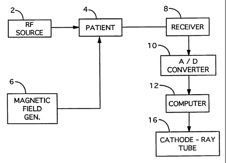
Figure 1 is a schematic illustration of a magnetic resonance imaging
system.
Figure 2 is a cross-sectional illustration of a form of coil usable in the
present invention.
Figure 3 is a left end view of the coil of Figure 2.
Figure 4 is a fragmentary partially schematic illustration of the coil of
Figure 2 and associated electronic components positioned in a blood vessel.
Figure 5A is a circuit diagram of a catheter coil.
Figures 5B and 5C illustrate other suitable tuning and matching circuits.
Figure 6A is a sensitivity map of the catheter coil with the coil oriented
parallel to main magnetic field B,
Figure 6B is a sensitivity map of the catheter coil of Figure 2 with the coil
oriented perpendicular to the main magnetic field B,
Figure 6C is a sensitivity map of the coil of Figure 2 with the coil
positioned generally tangential to main magnetic field Bo.
Figure 7 is a perspective partially schematic view of the coil of Figure 2
provided with a Faraday shield.
Figure 8 is a modified form of coil of the present invention having two
pairs of electrodes rather than the single pair electrode of the coil of
Figure 2.
Figure 9 is a left end view of the coil of Figure 8 without the end
connectors shown.
Figure l0A is an MR signal sensitivity map of the coil of Figure 8 with
the coil oriented parallel to the magnetic field B,
CA 02222742 1997-11-28
WO 96/38083 PCT/US96/08088
-9-
Figure lOB is a sensitivity map of the coil of Figure 8 with the coil
oriented generally perpendicular to the magnetic field Bo.
Figure 11 is a schematic illustration of the sensitivity volume of a coil,
such as that of Figure 2 of the present invention.
Figure 12 is a representation of the spectra of three adjacent voxels along
the length of the catheter coil of Figure 11.
Figure 13 is a schematic illustration of a coil of the present invention
secured to an endoscope.
Figure 14 is a cross-sectional illustration through 14-14 of the endoscope
of Figure 13.
Figure 15 illustrates a form of tuning and matching circuit for the
endoscope of Figure 13.
Figure 16 is a schematic cross-sectional illustration showing the coil of the
present invention employed with a biopsy needle.
Figure 17A illustrates a form of capacitive tuning arrangement of the
present invention.
Figure 17B illustrates an embodiment of the invention wherein distributed
capacitance is employed in connection with a two conductor coil.
Figure 18 is a schematic illustration of a distributed capacitance coil
wherein two pairs of conductors are employed.
Figure 19 is an alternate form of distributed capacitance wherein a plurality
of axially spaced rings are employed as the capacitive elements.
Figure 20 is a schematic illustration of a two conductor distributed
capacitance coil having a common ground.
Figure 21 A illustrates schematically an alternate form of Faraday shield
employing short lengths of electrical conductor.
Figure 21B-21D show cross-sectional views of several specific approaches
to the embodiment of Figure 21A.
Figure 22 represents a twisted pair coil of the present invention.
= 30 Figure 23 is a schematic flow diagram showing a method of correcting
signal variation by the sensitivity map related to the coils of the present
invention.
Q
CA 02222742 1997-11-28 PGT/US QLV/ Ovp O8Cj
!p(EA/U7s 0 4 auG'97
-R 10-
DESCRIPTION OF THE PREFERRED EMBODIMENTS
Figure 1 shows a schematic representation of the general concept of
magnetic resonance imaging as employed with a patient. An RF source 2 provides
pulsed
radio frequency energy to the specimen to excite MR signals therefrom which,
in the
form shown, is a patient 4 disposed in the main magnetic field which is
created by a
magnetic field generator 6. The specimen is generally aligned with the main
magnetic
field and the RF pulses are imposed perpendicular thereto. Where oblique
imaging is
employed, the angle of impingement of the vector representing the spatial
gradient of the
magnetic field will be angularly offset from either the x, y, or z directions.
This
arrangement results in excitation of the nuclei within the area or volume to
be imaged and
causes responsive emission of magnetic energy which is picked up by receiver
8.
The receiver 8 may be a coil which has a voltage induced in it as a result
of such responsive emissions of magnetic energy. As a practical matter,
separate coils
or identical coils may be employed as the RF source 2 and receiver 8. T4e
signal
emerging from receiver 8 is amplified, phase-sensitive detected, and passes'
through
analog-to-digital (A/D) convertor 10 and enters computer 12. Within computer
the
Fourier Transformations of signals convert the plot of amplitude versus time
to a map of
the distribution of frequencies by plotting amplitude versus frequency. The
Fourier
Transformations are performed in order to establish the intensity value
locations of
specific pixels and to obtain chemical shift spectra at those locations. These
values may
be stored, enhanced or otherwise processed, and emerge to be displayed on a
suitable
screen, such as a cathode-ray tube 16, for example.
Referring to Figures 2 and 3, there is shown a preferred form of coil 22
of the present invention. The coil 22 has a pair of electrodes 24, 26 which in
the form
shown, are generally parallel and spaced from each other a distance d which is
about 0.1
mm to 30 mm and, preferably, about 0.1 mm to 10 mm. The coil 22 has a
dielectric
material 30 which serves to reduce dielectric losses of the coil 22 and of the
specimen.
Ends of conductors 24, 26 of the coil 22 are electrically connected by wire
32. The coil,
as measured along the portion of the conductors 24, 26 contained within
dielectric 30,
has an overall length X which may be about 2 cm to 50 cm and, preferably about
5 cm
to 20 cm. The coil has a width D which is the major dimension of about 0.5 mm
to 2
cm and, preferably about 0.5 mm to 15 mm. The conductors 24, 26 may have an
AME(VD.ED SHEET
CA 02222742 1997-11-28 RUIUS 9 6 / 0 8 0 8 8
ITEAMS 0 4 Ai1G 97
-R11-
individual diameter of about 0. 1 mm to 3 mm and, preferably about 0.1 mm to 1
mm.
It is preferred that the separation d of the conductors 24, 26 be a dimension
substantially
less than the length X. The conductors 24, 26 are preferably made of a good
electrical
conductor, such as copper, silver, or aluminum, for example. Because of the
skin effect,
however, wherein only about an 8 m outer layer of the conductor carries
electrons at RF
frequencies, a material plated with a good conductor will also function
effectively. The
dielectric material 30 should be resilient so as to permit flexing of the coil
22 and return
to its original configuration. Any suitable dielectric material having the
properties
required to function in this environment may be employed. While the thickness
of the
dielectric will depend to an extent on the specific material selected, in
general, it is
preferred that the conductors 24, 26 be covered by at least 0. 1 mm of
dielectric material.
A suitable dielectric may, for example, be a bio-compatible plastic material,
or blend
having the desired properties. The dielectric material employed may, for
example, be
tetrafluoroethylene, which is sold under the trade designation, "Teflon. " It
is k~own for
its fine electrical insulating properties, does not interact with any
components in water,
and can be safely used in blood vessels.
It will be noted that as is shown in Figures 2 and 3, the dielectric 30 is
preferably, sufficiently rigid to resist undesired deformation involving
significant
alterations in the spacing d of the electrical conductors 24, 26 which, in the
form shown,
are impregnated therein. It resists deformation other than through resilient
flexing of the
entire coil 22.
With the coil shown in Figures 2 and 3, the inductance can be calculated
by Formula 1.
L= Z tan 2Tcl
w
wherein "L" equal inductance, "Zõ" is the characteristic impedance of the
wire, and is
a function of the separation and diameter of the conductors, the "w" is the
larmor
frequency in radians per second. For 1.5 Tesla, larmor frequency is
approximately 4x10g
AMENOED SHEET
(
CA 02222742 1997-11-28 P~T/~jJ~$,7 (~ 0 E 0 8 f
--~v ~~,
AU '97
-R12-
radian/sec.,"1" is the length of the cable, and X is the wavelength between
the wires,
which is approximately 1.5 meters with the coil inserted into a blood vessel.
In one method of employing the present invention in combination with a
catheter to image or spectrographically analyze a blood vessel, a guide wire
may first be
inserted into the vessel. The motion of the guide wire can be observed by the
method
described in U.S. Patents 5,271,400 or 5,307,808, or by any other suitable
method. A
catheter is then inserted into the vessel employing the guide wire. The guide
wire is then
removed and the catheter coil of the present invention is inserted. The
catheter is then
removed. This positions the coil within the vessel as shown in Figure 4. If
desired,
other means of introducing the coil into the vessel may be employed.
Referring to Figure 4, there is shown in cross-section of a blood vessel 40
having an interior bore 42 filled with blood (not shown). The blood vessel 40
has one
or more atherosclerotic plaque deposits, such as plaque deposit 44 which is
secured to
the interior surface 45 of the vessel 40. The coil 22, in the form shown,~~ is
fully
embedded within dielectric 30 with the connecting wire 32 also contained
within the
dielectric 30. The tuning/matching circuit 50 is also embedded in the
dielectric material
30 and is electrically connected to the coil. A coaxial cable 48 is connected
to the
tuning/matching circuit 50 which will be described hereinafter in greater
detail and serves
to match impedance of coil 22 with the impedance of the coaxial cable 48. A
decoupling
diode 52 is provided with coaxial cable 48.
Referring to Figure 5A, a specific example of the invention will be
considered.
Example 1
A coil 70 has two conductors similar to those shown in Figures 2 through
4 made of 30 AWG 7.5 cm conductor wire having a dielectric insulator made of
Teflon
with the conductors being silver-plated copper conductors shorted at one end.
This was
employed as the catheter coil. For tuning and matching at the time of
manufacture,
tuning/matching circuit 72 is provided with capacitors Cl and C2, which are
electrically
connected to the coil 70. The capacitors may be provided on a microchip having
a
dimension of about 1.5 mm x 1.5 mm x 1.4 mm, for example.
The other end of the tuning/matching circuit 72 is electrically connected
to coaxial cable 76 which has a 2 mm outer diameter and a 50 ohm resistance
and is
AMEfVD-ED SHEET
CA 02222742 1997-11-28 PCT/llS 9 b/ 0 8 0 8 8
IPEA/US U 4 AU6 '97
-R13-
used to carry the magnetic resonance signal from the coil to the processor
(not shown in
this view). If desired, a Teflon tape may be employed to cover both the coil
70 and the
capacitor circuit 72.
In order to decouple the transmit and the catheter coil, in the form shown,
a PIN diode 80 is placed in the coaxial cable 76. The diode turns on during RF
transmission using a DC current applied by the scanner hardware. If desired,
other
means of decoupling may be employed. The coaxial cable length "1" is precisely
adjusted
so that when the diode 80 is on, the coaxial cable 76 behaves like an inductor
and
resonates with capacitor C2 to disable a current through the receiver coil 70.
In the
decoupling circuit, in order to resist current induction in the receiver coil
during RF
transmission, the MR scanner may provide a positive DC pulse to the coil 70
for this
purpose. This normally turns on PIN diode 80. When this PIN diode 80 is on, no
current from the coil 70 is allowed to pass. This can be incorporated into the
cathode
coil probe assembly by placing the diode 80 as a shunt to a coaxial cable at a
predetermined distance "1" from the tuning circuit 72. When the diode 80 is
on, the
coaxial cable behaves like an inductor and resonates with the parallel
capacitor C2 that
disables the induced current flow from the catheter coil 70. As it is
desirable to put only
a small number of electronic components in the vessel, a~,/2 cable length may
be added
to this length and the diode 80 located outside the blood vessel.
The tuning/matching circuit 72 maximizes RF power transfer from the
receiver coil 70 to the preamplifier 82 as shown in Figure 5A. Preamplifier 82
receives
and amplifies the output of PIN diode 80. The tuning/matching circuit 72 is
preferably
placed next to the coil 70 to minimizes losses. In the catheter coil
embodiment, it may
be in the catheter closed end 42 (FIG. 4). The capacitors Cl, C2 may be
relatively small
fixed capacitors whose values are chosen to resonate coil 70 at the MR
frequency of the
nuclei of interest and match the coil to the optimum input impedance of
preamplifier 82.
For example, if " f " is the MR resonance frequency, the coil may be tuned to
resonate at
f by adjusting the values of the capacitors Cl and C2 in Figure 5A such that
the condition
f=(2TcV[L C])-' is met, where L is the coil inductance with the coil in the
sample, and
C is the sum of the tuning capacitance, Ct = Cl C2/(C1 + C2), and may stray
capacitance along the length of the coil that may result from interactions
between the coil
conductor and the specimen in which it is inserted. Stray capacitance can be
reduced by
AMEN4ED SHEET
CA 02222742 1997-11-28 } 'OUS-9J 08!Y)~ aa
~7 ~ '
~~EAP, ~ 0 QUG 97
-R14-
reducing the length of the coil 70. The value of capacitor C2 may be adjusted
so that the
impedance of the coil at resonance in the specimen, as viewed by the
preamplifier 82,
is optimally matched to maximize the signal-to-noise ratio of the preamplifier
82, for
example, it may be adjusted to be 50 SZ at resonance. The arrangement of
tuning/matching network 72 in Figure 5A is not limiting and it will be
understood that
other tuning and matching arrangements will be evident to those skilled in the
art,
including those illustrated in Figures 5B and 5C. In Figures 5B and 5C,
capacitor values
C3, C4, and values C5, C6, C7, respectively, are adjusted to meet the same
criteria so
as to resonate the coil at the desired MR frequency, and to match the coil
impedance at
resonance to optimize the MR signal-to-noise ratio.
In another test, a specimen in the form of an aorta from the rabbit's left
femoral artery was employed. The catheter coil 70 was inserted into aorta. An
image
with a 3 mm thickness, a field of view (FOV) of 50x25mm and a 512x256 matrix,
4NEX, TR/TE 2000/50 ms was obtained using a fast spin echo pulse sequeilce. An
image showing the two conductors and the vessel wall around the coil, as well
as the
structures around the vessel was visualized successfully.
In another experiment, an isolated dog heart was employed. The catheter
coil was placed in the circumflex artery of the heart from the aorta. The
isolated heart
was placed in a saline solution. The orientation of the heart was adjusted so
that the long
axis of the coil was aligned with the main magnetic field (z direction). The
axial images
of the heart were obtained using fast spin echo imaging techniques. An image
resolution
of l00gm x 100 m with a 3mm slice thickness was obtained with a 4 NEX data
acquisition. The vessel wall and myocardium were visualized in the image. When
the
coil was placed with the long axis orthogonal to the magnetic field, there
remains
sufficient x and y components of the coil's magnetic field to permit detection
of a
magnetic resonance signal.
Referring to Figures 6A, 6B and 6C, there is shown a sensitivity map or
plot of the catheter coil. Assuming that the sensitive region in an axial
field produced
by the coil is much smaller than the coil length X (FIG. 2) and that the
diameter of the
conductors_24, 26 is much less than the separation "d" between conductors 24,
26, the
RF field sensitivity of the coil 22 can be calculated from the Biot-Savart law
with z as
AMEN[}ED SFlEET
CA 02222742 1997-11-28 --F-C~~= --9-6 / 0 8 0 8 8
IPEA/US o 4 AUG '97
-R15-
the direction of a magnetic field Bo and the net field perpendicular to Bo is
the transverse
field. MR detection and excitation ornly involves the transverse field. In
general, a
catheter MR coil of the present invention may be oriented in any direction
relative to Bo.
The wires 24, 26 of the coil 22 are held at fixed separation "d" such that
length X is
much greater than d. The transverse field amplitude produced by the coil
oriented in the
3 orthogonal Cartesian directions is shown in Figures 6A-6C. It is seen from
these
figures that the coil is insensitive to magnetic field changes along its long
axis and
produces a transverse field in any orientation with respect to the magnetic
field, although
its sensitivity profile varies with the orientation relative to the Bo
magnetic field. If the
conductors 24, 26 are oriented in the same direction as the Bo field as in
Figure 6A, the
sensitivity is angularly uniform. Sensitivity generally drops approximately in
proportion
to 1/r along the radial axis where "r" is the distance from the center of the
coil.
It is noted in Figure 6B wherein the field Bo is perpendicular to the coil
that the transverse magnetic field map is altered, but data is still received.
Finally, with
respect to Figure 6C, wherein the B. field is tangential to the coil, there is
further
modification of the map, but meaningful data is nevertheless obtained. As a
result of this
characteristic of coil 22 may function effectively when the coil is not
ideally located with
respect to the main magnetic field Bo. Such would be the case in passing the
coil through
a tortuous path, such as in a small blood vessel.
A suitable MR scanner usable in the practice of the present invention is the
G.E. 1.5T signa magnetic resonance scanner.
As a result of high dielectric losses resulting from interaction of the coil
with the vascular environment, the quality factor (Q) of the coil may tend to
drop as the
length of the coil increases and, in addition, the tuning of the coil may be
altered. In
order to reduce the risk of such undesired dielectric losses and detuning
effects, a
Faraday shield may be employed. It serves to decrease the electric field and,
therefore,
the dielectric losses of the coil when in situ. As shown in Figure 7, a coil
of the two-
conductor type shown in Figure 2, but with a cylindrical configuration, has a
pair of
generally parallel, straight conductors 90, 92 having a shunt 94 electrically
connecting
ends thereof, and having the conductors 90, 92 passing through a flexible
dielectric
material 96. The Faraday shield, in the form shown, consists of a plurality of
rings, such
aMENDED SHEET,
CA 02222742 1997-11-28
WO 96/38083 PCT/US96/08088
-16-
as rings 100, 102, 104, 106, for example, which are positioned about the
circumference
of the dielectric material 96 and are axially spaced from each other. The
rings 100, 102,
104, 106 may be continuous or discontinuous as by being a slit or annularly
discontinuous.
Referring to Figures 8 and 9, a preferred form of coil of the present
invention will be considered. This embodiment provides improved SNR and
increased
uniformity of sensitivity. This coil is generally similar to that of Figures 2
through 4,
except that instead of having a single pair of conductors 24, 26, which are
electrically
connected to each other by a shunt 32, two pairs of electrical conductors
which are
positioned in planes orientated at 90 relative to each other are employed. In
this
embodiment, a first pair of conductors 110, 112 extends through a dielectric
material 114
which, preferably, is generally flexible and encloses the conductors 110, 112.
The ends
of conductors 110, 112 are shunted by electrical connector 118. Similarly, a
second pair
of conductors 124, 126 are contained within the dielectric material 114 and
are positioned
90 out of alignment with a plane passing through conductors 110, 112 as shown
by
angle A in Figure 9. The ends of conductors 124, 126 are shunted by electrical
connector 125. As shown in Figure 10A, the transverse magnetic field map, when
the
main magnetic field Bt, is oriented generally parallel with the orientation of
coils of
conductors 110, 112, 124, 126 is somewhat similar to that of Figure 6A, but
offers the
additional advantage of higher sensitivity. This embodiment is advantageous as
compared
with the two conductor coil with respect to improved uniformity in the
vicinity of the
conductors and the uniformity is less sensitive to coil orientation with
respect to the main
magnetic field B,
Figure lOB shows the conductors 110, 112, 124, 126 oriented
perpendicular to the main magnetic field B,,. Figure lOB shows that the coil
remains
sensitive even when the main magnetic field B, is not aligned with the
direction of the
conductors 110, 112, 124, 126.
It will be appreciated that, if desired, additional numbers of pairs of
conductors, generally, equally spaced from each other, much the same way the
two
conductors of Figure 6, and the four conductors of Figures 8 and 9 are spaced,
may be
employed. For example, if desired, a total of three pairs of conductors, each
embedded
within the dielectric and equally spaced from each other in a circumferential
sense, might
CA 02222742 1997-11-28 rL#1i/u%,) q/08M
- iKNUGI 94 AUG '97
-R17-
be employed. An advantage of the use of more conductors in the coil is the
further
improvement in sensitivity around the coil and enhanced independence of the
sensitivity
on coil orientation. A disadvantage is the increased minimum width or diameter
of the
coil that is achievable with a large number of conductive elements.
Referring to Figure 11, there is shown a coil 130 generally of the type
shown in Figures 2 and 3 with conductors 132, 134 being generally parallel and
passing
through a dielectric 136. The coil 130 may be employed to provide a set of
spatially
resolved chemical shift spectra. As the sensitivity of the coil is restricted
to regions close
to coil conductors (see FIGS. 6 and 10), one-dimensional chemical shift
imaging or 1-D
spectroscopy may be employed without localization pulses. The imaging voxel,
such as
140, depicted here as a cylindrical as shown in Figure 11, is actually the
shape of the
sensitivity profiles shown in Figures 6A-6C for a two-conductor coil or in
Figures 10A-
lOB for the four-conductor coil. A TR of 2000, 140mm FOV and 64 phase encoding
steps along the coil with 2 NEX was used in another experiment in which the
body coil
was employed to transmit RF pulses and the catheter coil employed to receive
the same.
The spectra of three adjacent voxels is shown in Figure 12 with peaks 142,
144, 146
representing water signals from the three regions and peaks 145, 147 and peaks
145, 147
from lipid signals in or adjacent to the vessel walls. The bandwidth was 1000
Hz, with
1,024 point resolution, and the z dimension of the voxel 140 is 2.2 mm. The
radial
dimension of the voxel is determined by the sensitivity of the coil. Water and
lipid peaks
will tend to vary between normal and atherosclerotic vessels. As the spectra
of the three
adjacent voxels represented in Figure 12 are the result of the phase of coil
sensitivity
above the coil and under the coil, opposing each other, some signal
cancellation results.
While in the first embodiment of the invention, the coil of the present
invention was employed solely as a receiver of magnetic resonance signals
emanating
from the specimen, in another embodiment of the invention, employing the same
coil
with certain modifications, if desired, the coil may serve both as an RF
transmitter and
as an RF receiver. In such an approach, the system would function essentially
as before,
in both imaging and 1-D spectroscopy, except the source of the MR excitation
RF
magnetic field would be the coil rather than transmitter coil in the standard
MR scanner.
If this approach is taken, the transmitter power may be introduced at the
diode 80 of
AMEiVDED SHEET
CA 02222742 1997-11-28 ~CT/US 7n'o/f(~8OQQ
~+ IPEA/US01a,4~ AUG'V9IV
-R18-
Figure 5A may be eliminated with the preamplifier 82 connected to the same
point by a
cable tuned to be of a length X /4 at the MR frequency.
Referring to Figures 13 through 15, the coil of the present invention may
be mounted on a conventional flexible endoscope 180 which has conventional
external
tubular connections 182, 184. The coil 190 may be fixedly secured about the
circumference of the lower portion 192 of the endoscopic tube with an adjacent
tuning/matching circuit 194 provided. As shown in Figure 14, the coil 190 is
of annular
configuration and surrounds the lower portion 192 of the endoscope 180 which
lower
portion 192 is composed of an MR compatible material. The elongated coil
conductors
191, 193 are embedded in annular dielectric material 189 which is intimately
secured to
lower end 192.
As shown in Figure 15, the tuning/matching circuit 194 has diode 198 and
capacitors 200, 202.
It will be appreciated with the present invention that the catheter toil may
be employed, for example, in a blood vessel to provide an image and 1-D
spectroscopic
analysis of plaque built up on the interior of the vessel wall with multislice
imaging being
provided in an efficient manner due to the long coil being employed. It may be
employed to examine many other characteristics, such as fatty streaks,
calcification,
sclerosis, and thrombosis, for example. It will be appreciated that
substantially
simultaneously with the use of the coil and the catheter, medical intervention
as, for
example, by laser destruction of the undesired plaque, may be employed.
Similarly, any
normal diagnostic or therapeutic measures undertaken with the aid of endoscope
180, may
be accomplished substantially simultaneously with the use of the coil for
imaging and/or
spectroscopic analysis.
Referring again to Figure 16, there is shown the use of the invention in
connection with biopsy needle 220 which is composed of a material which is
magnetic
resonance compatible, such as a ceramic material as distinguished from a steel
material,
for example. In this embodiment, the specimen 224 contains a lesion 226 from
which
a sample 228 has been obtained by the biopsy needle. The coil 232, which is
fixedly
secured to the exterior of the needle sheath 240 may be a two or four
conductor coil
having the general configuration shown in Figure 14, for example. The tuning
and
matching circuit 244 is electrically connected to both the coil 232 and a
preamplifier 246
AMENDED SHEET
CA 02222742 1997-11-28 P~T/U{~ 9 O/ 0 Q O Q(
i,PEA/~JS 0 4 A(U~G '97
-R19-
which serves to amplify the signal before it enters the computer (not shown)
for further
processing. In this embodiment, the coil 232 need not be flexible and the
apparatus need
not enter a natural passageway within the patient. The coil may be secured to
the needle
by a suitable glue or resin or in the case of a ceramic needle, by depositing
the conductor
onto the ceramic by methods well known to those skilled in the art of
electronic
integrated circuit fabrication. The conductors are then sheathed with
insulating material.
Referring to Figures 17A and 17B, another approach to reduce the
dielectric losses is to distribute the tuning capacitance as distributed
series capacitance
along the length of the catheter coil. For example, instead of short-
circuiting the two
-conductors, as shown in Figure 2, by conductor 32, a tiny chip capacitor
having a
capacitance- selected such that in combination with tuning capacitors Cl and
C2 of Figure
5, the coil is correctly tuned to the MR frequency. Other approaches will be
considered
with respect to the embodiments illustrated in Figures 17A through 21D.
Considering
Figure 17A wherein the output end 250 of coil 252 has an impedance matching
'capacitor
254, a plurality of capacitors, such as capacitors 256, 257, 258, 259, for
example, spaced
throughout the longitudinal extent of the coil 252, with individual values
chosen so that
the coil is tuned to resonate at the MR frequency of the nuclei of interest.
For example,
if " f " is the MR resonance frequency, the coil may be tuned to resonate at f
by allowing
the values of the capacitors 256, 257, etc., each to be substantially equal to
a value C;,
and adjusting C; such that the condition f=(2Ts-/[LC;/n])-1 is met, where "L"
is the coil
inductance with the coil in the specimen, and n is the number of capacitors
distributed
along the length of the coil. The capacitors are connected by flexible
conducting
elements 266, 267, 268, 269, etc., with spacing "d" substantially constant
along the
length of the coil, as in Figure 3. This embodiment has the advantage over
that depicted
in Figure 5, that in order to meet the tuning condition for a particular value
of "f," the
values of C; will generally be much larger than those of C1-C7 in Figure 5 and
also, be
sufficiently large so as to minimize dielectric losses that result from
interactions between
coil conductor elements 266, 267, 268, 269, etc., and the specimen. The
distributed
capacitance of this embodiment of the invention may be achieved by a number of
means
-- including (a) the use of discrete circuit capacitive elements of
sufficiently small
dimensions to meet the desired aforementioned dimensional specifications of
the coil, and
(b) by direct deposition of conductor onto a flexible dielectric substrate
that forms the
AMEiVDED SHEET -
CA 02222742 1997-11-28 PCTJUM'S 8 O 8 8
;. tPEAIUS 0 4 AUG'97
-R20-
body of the coil, or (c) by etching away of conductor from a flexible
dielectric substrate
using techniques well known in the manufacture of printed circuit boards and
integrate
electronic circuit devices. An embodiment that is electrically equivalent to
Figure 17A
that provides a distributed capacitance coil by method (b) is depicted in
Figure 17B.
Considering Figure 17B, wherein the output end 280 of coil 282 has an
impedance
matching capacitor 284, a plurality of capacitors formed by conductive
elements 290,
292, 294 and where they are proximal to conductive elements 310, 312, 314, for
example, are spaced throughout the longitudinal extent of the coil 282 and a
plurality of
capacitors formed by conductive elements 296, 298, 300 in close proximity to
conductive
elements 316, 318, 320, for example. The remaining unnumbered conductive
elements
will similarly provide a plurality of capacitors. Here, the capacitors formed
by conductor
290, 292, 294 in proximity to conductors 310, 312, 314, etc., may be
fabricated by
deposition or etching of conducting material on two sides of a flexible
dielectric substrate
material such that sections 290, 292, 294 are on one side of the dielectric
mat~rial and
sections 310, 312, 314, etc., are on the other side. It will be appreciated
that 'adjacent
pairs of the conductive plates will provide the two conductors which form a
capacitor and
in the aggregate provide a plurality of capacitors along the coil. With
reference to the
conductors numbered for purposes of example, conductors 294 and 314 cooperate
to form
a capacitor as do conductors 294 and 312. In this manner, the capacitance is
distributed
along the coil and yet the coil preserves its desired flexibility.
Figure 18 shows a similar construction wherein the output 356 and
impedance matching capacitor 352 are employed in a construction wherein four
conductors are used as in Figures 8 and 9. The coil 354 has a plurality of
capacitors,
such as is formed by flexible conductive elements 356, 358 and underlying
flexible
conductive elements 360, 362 associated with a first conductor and a plurality
of
capacitors formed by elements 370, 372, for example, associated with flexible
elements
374, 376. On a third conductor, a series of capacitors formed by elements 380,
382 are
associated with flexible conductive elements 384, 386. Finally, the fourth
conductor has
a plurality of capacitors, such as are formed by elements 390, 392 which are
associated
with flexible elements 394, 396. As an alternative to_the capacitors shown in
Figure 18,
a plurality of ring-shaped capacitors, such as 400, axially spaced along the
coil may be
employed in a coil as shown in Figure 19.
AME.MED SHEET
CA 02222742 1997 11 28 TIUS 9 6 / 0 8
IPEAlUS 0 4 .AU0 V
-R21-
A further embodiment of the distributed capacitance concept is shown in
Figure 20 wherein the output end 410 of coil 412 has an impedance matching
capacitor
414 and a plurality of first capacitors formed by conductive elements 420,
422, 426, in
association with conductive elements 440, 442, 446, 427, for example, and a
plurality
of second capacitors formed by conductive elements 430, 432, 434, for example,
associated with the common capacitance rail 440, 442, 446, 435.
If desired, once the dielectric losses have been decreased, for example by
distributing the capacitance, the catheter RF receiver coil length can be
increased to
increase coverage on the coil. In addition, a coil fabricated from lengths of
conductor
incorporating capacitance distributed at a particular value of capacitance per
unit length,
for example, by etching or deposition as discussed hereinbefore, can be turned
to
different specific MR frequencies by adjusting coil length. If the coil length
is increased
to X/4 where X is the wavelength in the coil with the coil disposed within the
pecimen
at the MR frequencies, the coil will self-resonate and additional resonance
tuning
capacitors may be eliminated.
Referring to Figures 7 and 21, an alternate version of Faraday shield is
shown in Figure 21A, wherein a coil 460, in lieu of having ring-like
conductors, has a
plurality of short length conductors 462, 464, 466, 468, for example, embedded
in a
flexible substrate 470 oriented substantially perpendicular to at least one
pair of elongated
electrical conductors that form the basic receiver coil. Examples of specific
approaches
to the embodiment of Figure 21A are provided in Figures 21B-21D. As shown in
Figure
21B, dielectric 470' has as conductors 472' and 474' with a plurality of
elongated
conductors 462' positioned generally parallel to a plane passing through the
conductors
472' and 474' and embedded within dielectric 470'. A plurality of such
conductive
elements are spaced axially along the coil. In Figure 21C, conductors 472",
474" are
positioned within dielectric 470" and the elongated conductive elements, such
as 462"
are oriented generally perpendicular to a plane passed through conductors
472", 474".
In Figure 21D, the elongated conductor 462"' are positioned generally
perpendicular to
a plane passed through conductors 472"', 474"' and is positioned between such
conductors 472"', 474"' with all of the conductors being disposed within
dielectric
470"'.
AMEN'DED SHEET
CA 02222742 1997-11-28 P0VUS 9 6/ 0 8 0 8 8
-R22-
Referring to Figure 22, there is shown a two conductor coil 500 having
two conductors 502, 504 associated therewith and an electrical conductor 506
connecting
the same. Associated capacitors 510, 512, which comprise the tuning/matching
circuit
and the diode 516, are electrically connected to the coil 500. An advantage of
this
embodiment of the invention, wherein the conductors 502, 504 would be encased
in a
suitable dielectric (not shown), is the sensitivity to the far field will
drop. The number
of turns per unit length of the coil 500, may be adjusted as to provide the
same near field
sensitivity and, thereby, improve the signal-to-noise ratio. The preferred
number of turns
per centimeter of length is about 1 to 2 turns.
Referring to Figure 23, a feature of the software of the present invention
will be considered. As noted hereinbefore, the RF sensitivity of the coil
roughly drops
by 1/r, wherein "r" equals the radial distance to the center of the coil.
Although the
signal-to-noise ratio is high, the dynamic range for the visualization of the
signal is so
high it is desirable to provide an image processing technique in order to
facilitate display
of all existing information. In one such approach the image is divided pixel
by' pixel by
the sensitivity map. For reasons where the sensitivity is low, i.e, the signal
is
comparable or lower than the noise level, a value of 0 is assigned to the
corresponding
pixel. After this process, unlike the usual MR images, noise will be space
dependent.
As shown in Figure 23, first the position of the RF coil in the image needs
to be determined (step 550) for sensitivity map registration purposes. This
may be done
either manually or automatically by a computer, for example. To minimize or
completely
eliminate user interaction, this can be accomplished by finding two dark spots
near the
pixel with the highest signal intensity. Locating the coil position, with
respect to the
main magnetic field, should be determined in order to calculate the
sensitivity map of the
coil (step 552). This can be automated by tracking the coil position in a
multislice
acquisition or assuming that the image plane is always perpendicular to the
coil. One
then divides the acquired images with the sensitivity map (step 554). Step 556
involves
masking or filling the regions of the image with zero where the SNR is lower
than a
threshold value which is typically within the range of 1 to 5. This processing
corrects
the RF field inhomogeneity of the acquired image data.
It will be appreciated, therefore, that the present invention provides an
improved method and associated apparatus for enhanced MR imaging and 1-D
chemical
AMEMDED SHEU
CA 02222742 1997 11 28 7'6 / 01,
V.#
-R23-
shift analysis of the interior of a specimen. This is true even in situations
where a
tortuous path in a blood vessel requires that the receiver coil be flexible
and multislice
data acquisition is desired. The system also functions effectively by
providing desired
sensitivity to the MR signal even in oblique positions. Further, the coil
provides uniform
sensitivity along the coil and, as a result of the use of longer coils,
facilitates a longer
portion of the specimen being imaged with one coil position. Further, no
tuning is
required after insertion of the coil into a specimen. In a preferred
embodiment, one or
more pairs of conductors are embedded within a dielectric material which is
flexible so
as to facilitate desired bending of the same in providing images and
spectroscopic
analysis. The coil, in addition to serving solely as a receiver coil in one
embodiment,
may in another embodiment function as a transmitter coil and a receiver coil.
The
invention may be employed generally simultaneously with medical intervention,
such as,
for example, laser removal of blood vessel plaque.
The invention also contemplates enhanced efficiency through theRuse of a
at least one of the following features: a tuning/matching circuit; a
decoupling circuit; a
Faraday shield; as well as distributed capacitance along the length of the
coil; self-
resonance; and a twisted pair coil.
While for clarity of disclosure reference has been made herein to display
means for displaying an image, it will be appreciated that the image
information may be
stored, printed on hard copy, be computer modified, or be combined with other
data.
All such processing shall be deemed to fall within the terms "display" or
"displaying" as
employed herein.
Whereas particular embodiments of the present invention have been
described herein for purposes of illustration, it will be appreciated by those
skilled in the
art that numerous variations of the details may be made without departing from
the
invention as described in the appended claims.
AMENDED SHEET