Note: Descriptions are shown in the official language in which they were submitted.
CA 0222280~ 1997-12-01
W O 96/39999 PCT~US96/08794
INTRALUMINAL GRAFTING SYSTEM
This application is a continuation-in-part of application
Serial No. 109,162 filed August 19, 1993, which is a divisional
of application Ser. No. 553,530 filed July 13, 1990, now U.S.
Pat. No. 5,275,622, which is a continuation-in-part of
application Ser. No. 166,093 filed on March 9, 1988, now U.S.
Pat. No. 5,104,399, which is a continuation-in-part of
application Ser. No. 940,907 filed on Dec. 10, 1986, now U.S.
Pat. No. 4,787,899, which is a continuation of application Ser.
No. 559,935 filed on Dec. 9, 1983, now abandoned. The contents
of each of these applications are hereby incorporated by
reference.
BACKGROUND OF THE INVENTION
This application relates to endovascular grafting
apparatus, system and method and devices for use therewith.
The state of the art is described in the background of the
invention in U.S. Pat. No. 4,787,899.
SUMMARY OF THE INVENTION
In general, it is an object of the present invention to
provide an endovascular grafting apparatus, system and method
and devices for use therewith which overcome the disadvantages
of the prior art apparatus, systems and devices.
Another object of the invention is to provide an apparatus
and system of the above character which utilizes a pusher rod
assembly which is constrained so that relatively great forces
can be applied by the pusher rod assembly.
Another object of the invention is to provide an apparatus
and system of the above character in which the capsule is
flexible so that it can negotiate bends in the vessels of a
patient.
~ 30 Another object of the invention is to provide a grafting
apparatus and system which utilizes a flexible capsule which
can contain a graft with hook-like elements without any danger
of the hook-like elements penetrating the capsule.
CA 0222280~ 1997-12-01
W O 96/39999 PCT~US96/08794
--2--
Another object of the invention is to provide an apparatus
and system of the above character in which the graft
automatically springs into an open or expanded position when it
is released from the capsule.
Another object of the invention is to provide an
apparatus, system and method of the above character in which a
pushing force is applied to the distal extremity of the balloon
for advancing a graft out of the capsule.
Another object of the invention is to provide an apparatus
and system of the above character in which a fixed wire or an
over-the-wire guide wire system can be used.
Another object of the invention is to provide an apparatus
and system of the above character in which the graft can be
compressed to a very small size in a ~lexible capsule.
Additional objects and features of the invention will
appear in the following description in conjunction with the
accompanying drawings.
Another feature of the present invention is a novel
attachment system that comprises a sinusoidal wire frame and V-
shaped lumen piercing members. The sinusoidal frame has twoends and alternating base apices and protruding apices. The
protruding apices protrude outward and are mounted onto the
graft to extend outward past the end of the graft. The base
apices are oriented inside the lumen of the graft and points
inward from the end of the graft. The portion of the wire
~rame connecting the protruding apices to the base apices are
struts.
In one embodiment, the two ends o~ the wire frame are
welded together to obtain circular continuity of the wire
frame. In another embodiment, the wire frame has one
additional protruding apex and the ends of the wire frame
terminate ln helices generally aligned with the base helices.
The frame is mounted by overlapping the two ends of the wire
including a pair o~ protruding apices adjacent the end. The
wire frame is sewn to the body of the gra~t at various points
over the entire wire frame. The lengths of the struts may be
adjusted to stagger the apices so that the profile o~ the wire
frame and the graft can be minimized to ~it into a smaller
CA 0222280~ 1997-12-01
WO 96~9999 PCT~US96/08794
--3--
delivery capsule.
In addition to the wire frame, the attachment system
further includes a plurality of lumen piercing members affixed
to the struts. The lumen piercing members are con~igured to
protrude radially outward from the attachment system to engage
the lumen wall of a blood vessel and secure the graft in place
to prevent migration of the graft along the blood vessel. The
lumen piercing member of one embodiment includes a wire arm
that has an outwardly protruding hook constructed of stainless
steel wire. The hooks are aligned with and welded to the
struts of the wire frame.
Another embodiment of the lumen piercing members
eliminates the need for welds to secure the lumen piercing
members to the graft. Each lumen piercing member is bent into
a V-shape and each have an apex and two arms that extend in a
direction parallel to the struts of the wire frame. The arms
terminate in radially outward protruding hooks that are
configured to engage the wall of the vessel. The lumen
pielcing member is secured to the graft in close proximity to
the wire frame and is responsive to the outward bias of the
wire frame. Another embodiment of the attachment system of
the present invention con~igured ~or use in the iliac arteries
in a bifurcated graft includes two sinusoidal wire frames that
have alternating base apices and protruding apices. Each of
the iliac wire ~rames have two end arms that extend
longitudinally outward to engage the iliac artery wall. The
wire arms are configured as lumen piercing members which extend
as struts from the end base apices. The two wire frames are
joined together by overlying the end base apex of one of the
wire :Erames with the end base apex ~rom the other wire ~rame
such that each of the wire arms extend parallel to an adjacent
strut. The end arms are twisted around the adjacent struts and
bent behind the protruding strut that is integrally connected
to the adjacent strut. The ends of the lumen piercing member
is hook-like to securely engage the vessel wall. The hooks are
secured to the vessel wall when an additional radially outward
force presses the vessel into the lumen wall, such as from a
deployment balloon.
CA 0222280~ 1997-12-01
W O 96/39999 PCTAUS96/08794
--4--
Another feature of the present invention includes a device
to substantially eliminate leaks around the perimeter of the
graft at the ends where the attachment system engages the lumen
wall. The outside of the graft is textured with a plurality of
filaments or fibers that are spun, woven, knotted, pressed or
otherwise loosely associated to form a puffed textured filler
or tuft that is sewn to or affixed to the outside of the graft
proximal to the end of the graft. The ends of the fibers may
be frayed to increase the surface area of the tuft.
Alternatively, strands of loosely spun synthetic yarn are
cross-stitched around the perimeter of the graft proximate the
attachment system.
Another feature of the present invention includes a graft
that is crimped radially along at least a portion of the length
of the graft. The crimps form a generally corrugated tubular
surface defining a plurality of radially outwardly protruding
ribs that are separated longitudinally by alternating inwardly
directed folds or pleats. The crimping occurs along the length
of the graft between the two attachment systems. The crimping
may be configured over the entire length or over only a portion
of the graft.
Other features and advantages of the present invention
will become apparent from the following detailed description,
taken in conjunction with the accompanying drawings, which
illustrate, by way of example, the principles of the invention.
BRIEF DESCRIPTION OF THE DRAWINGS
FIGURE 1 is an isometric view of an endovascular grafting
apparatus and system incorporating the present invention.
FIG. 2 is a side elevational view partially in cross
section of a capable catheter incorporating the present
invention.
FIG. 3 is a side elevational view partially in cross
section showing a balloon catheter assembly incorporating the
present invention.
FIG. 4 is a partial side elevational view in cross section
of a portion of an alternative balloon catheter assembly
CA 0222280~ 1997-12-01
WO 96/39999 PCT/U~,G~_t;~
--5--
incorporating the present invention showing the use of a
movable pusher button capsule o~ sliding over a limited range.
FIG. 5 is a side elevational view partially in cross
section of another alternative embodiment of a balloon catheter
assembly incorporating the present invention showing the use of
a movable guide wire.
FIG. 6 is a cross sectional view taken along the line 6-6
of FIG. 5.
FIG. 7 is a side elevational view partially in cross
section of a pusher rod assembly incorporating the present
invention.
FIG. 8 is a side elevational view partially in cross
section of another embodiment of a pusher rod assembly
incorporating the present invention.
FIG. 9 is a cross sectional view partially in cross
section showing in combination a balloon catheter and a pusher
rod assembly and a movable guide wire.
FIG. 10 is a side elevational view of a graft
incorporating the present invention.
FIG. 11 is an enlarged isometric view showing one of the
spring attachment means utilized on the graft.
FIG. 12 is a partial enlarged view o~ an alternative hook-
like element utilized in the spring attachment means of FIG.
11 .
FIG. 13 is an enlarged view showing another embodiment of
a hook-like element used in the spring attachment means o~ FIG.
11 .
FIG. 14 is a side elevational view partially in cross
section showing the manner in which the graft is held in the
capsule after e~ection of the proximal extremity o~ the gra~t
from the capsule.
FIG. 15 is a view similar to FIG. 14 but showing the
proximal and distal extremities of the graft outside of the
capsule with the balloon retracted so that it is within the
graft and inflated to force the distal attachment means into
the vessel wall.
FIG. 16 is an enlarged side elevational view of one strut
and lumen engaging member of FIG. 30.
CA 0222280~ 1997-12-01
W O 96/39999 PCT~US96/08794
--6--
FIG. 17 is a graph showing the compression and tension
forces on the strut of FIG. 16.
FIG. 18 is an isometric view of an endovascular graft
incorporating the attachment system of FIG. 30, further showing
the tufts and stitching on the outside and inside of the graft.
FIG. 19 is a plan view of the inside of an endovascular
graft cut longitudinally, showing the wire frame, separate
lumen engaging members and stitching of the attachment system.
FIG. 20 is a plan view of the outside of an endovascular
graft cut longitudinally, showing in partial hidden view the
wire frame and the separate lumen engaging members of the
attachment system and further showing the tufts attached to the
outside of the graft.
FIG. 21 is a plan view of the inside of an endovascular
graft cut longitudinally, showing the wire frame, lumen
engaging members and stitching of the attachment system.
FIG. 22 is a top plan view of an endovascular graft having
an attachment system as shown in FIG. 20, showing the pleats
and tufts of the graft secured within the vessel lumen.
FIG. 23 is an enlarged top plan view of the area shown
along curve 23 of FIG. 22.
FIG. 24 is a side elevational view of an iliac attachment
system, wherein the base apices are sewn to the end edge of a
leg of a bifurcated graft.
FIG. 25 is a side elevational view of an iliac attachment
system, wherein the base apices are sewn within a leg of a
bifurcated graft and below the end edge of the leg.
FIG. 26 is a plan view of the graft and attachment system
of FIG. 24 cut longitudinally, showing the wire frame of the
iliac attachment system having lumen engaging members.
FIG. 27 is a partial cross-sectional view of a leg of a
bifurcated graft and an iliac attachment system secured within
a vessel having an enlarged vessel wall and constricted lumen.
FIG. 28 is a partial cross-sectional view of a leg of a
bifurcated graft and an iliac attachment system secured within
a vessel having a bulge in the vessel wall.
FIG. 29 is a side elevational view of an endovascular
graft incorporating an attachment system of the present
CA 0222280~ 1997-12-01
W O 96/39999 PCT~US~ 3/94
--7--
invention.
FIG. 30 is an enlarged isometric view showing one of the
spring attachment systems shown in the graft of FIG. 29.
FIG. 31 is an enlarged perspective view of a duck-billed
configured hook of a lumen engaging member.
DETAILED DESCRIPTION OF THE PREFERRED EMBODIMENTS
In general, the endovascular grafting system is comprised
of a capsule catheter having a flexible elongate tubular member
with proximal and distal extremities and a capsule mounted on
the distal extremity of the tubular member. The capsule is
generally cylindrical in shape and is formed of a helical wraps
of a metal ribbon. Means is provided for bonding said wraps
into a unitary capsule while permitting bending of said unitary
capsule. A graft is disposed within the capsule. The graft is
comprised of a tubular member having proximal and distal ends.
Hook-like attachment means is secured to the proximal and
distal ends of the tubular member and face in a direction
outwardly towards the inner wall of the capsule. Push rod
means is disposed within the capsule catheter and engages the
graft whereby upon relative movement between the push rod means
and the capsule catheter, the graft can be forced out of the
capsule.
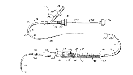
More in particular, the endovascular grafting apparatus
and system 11 and the devices for use therein are shown in
FIGS. 1-10. This apparatus and system 11 includes a capsule
catheter 12 (see FIG. 2) which consists of a flexible elongate
tubular member 16 formed of a suitable plastic material such as
Nylon of a suitable length as, for example, 40 to 100
centimeters and preferably approximately 43 centimeters for the
abdominal aortic artery and approximately 70 centimeters for
the thoracic aortic artery. The tubular member 16 can have a
suitable size such as an outside diameter of .187 inches and an
inside diameter of .125 inches. The tubular member 16 can be
produced in a certain color such as blue. In order to make it
radiopaque under x-rays, the flexible tubular member 16 is
loaded with a suitable radiopaque material such as bismuth
CA 0222280~ l997-l2-Ol
W 096/39999 PCT~US96/08794
--8--
subcarbonate or barium sulfate. By way o~ example, the
flexible elongate member 16 can be compounded with
approximately 20~ o:E the radiopaque material by weight.
An inner liner 17 is mounted within the tubular member 16.
The liner 17 is sized so that it will ~it within the tubular
member 16. The liner is pre~erably ~ormed of a lubricous
material such as Tefzel (ethylene tetra~luoroethylene) or
Teflon FEP (fluorinated ethylene polypropylene). It can have
an inside diameter of . 085 inches and an outside diameter of
.125 inches and a length as, ~or example, 41 centimeters which
is slightly less than that o~ the tubular member 16. If
desired, the inside diameter o~ the liner 17 can be in the
range of .075 to .120 inches. The liner 17 is provided with a
lumen 18 which extends the length thereo~. The liner 17
reduces the inside diameter of~ the lumen 18 for a purpose
hereina~ter described. The liner 17 is made of a radiation
stable material so that the catheter can be radiation
sterilized. Te~zel, or Te~lon FEP, which is a polymer is such
a radiation sterilizable material. The inner liner 17 also
serves to provide additional columnar strength to the catheter
12.
A wye adapter 21 is secured to the proximal extremity o~
the :Elexible tubular member 16. The side arm 22 o~ the adapter
21 has a stop cock 23 mounted therein which is movable between
open and closed positions. The stop cock 23 is provided with
a Luer fitting 24 which is adapted to be secured to a syringe
which can be utilized ~or injecting a dye, or medications such
as a vasodilator.
As shown in FIG. 2 with continued re~erence to FIG. 1, the
central arm 26 of the adapter 21 is connected to a Touhy Borst
adapter 27 and includes a i~emale part 28 that carries an O-ring
29 which is adapted to be engaged by a protrusion 31 forming a
part o:E the male part 32.
The capsule catheter 12 has a capsule 36 incorporating the
present invention mounted on the distal extremity of~ the
flexible elongate tubular member 16. The capsule 3 6 when used
in humans has a diameter ranging i~rom 4 to 8 millimeters. The
flexible elongate tubular member 16 which also serves as a
CA 0222280~ 1997-12-01
WO 96/39999 PCT~US96/08794
_g_
shaft for advancing the capsule 36 as hereinafter described and
should have a diameter which is less than that of the capsule
and therefore has an outside diameter ranging from 3 to 7
millimeters.
The capsule 36 is a composite structure and is formed of
an inner layer 37 and an outer layer 38. The inner layer 37 is
formed of a stainless steel ribbon 39 with the ribbon having a
width of .150 inches and a thickness ranging from .002 to .004
inches and preferably approximately .003 inches. The ribbon is
spiral wound on a mandrel ~not shown) so that each wrap of the
ribbon overlaps the preceding wrap by approximately 30 to 50~
of the width of the ribbon. Viewing the capsule 36 from the
left hand end, the ribbon is wrapped in a clockwise or
counterclockwise direction so that the edges 41 face distally
or in the direction which is toward the right as shown in FIG.
2 for a purpose hereinafter described. By winding the ribbon
37 at high tension, it is possible to deform it over the
adjacent wrap which contributes to the flexibility of the
capsule and also at the same time makes it possible to provide
a capsule having a low profile. The stainless steel for the
ribbon 39 can be of any suitable type, however, it has been
found that it is desirable to select a stainless steel which
can be heat treated. This enables one to wind the capsule with
a ribbon in a ductile state and heat treat the capsule after
winding to obtain a spring-like temper. One such stainless
steel is 17-7 PH supplied by Brown Metals Company of Santa Fe
Springs, California.
In order to prevent elongation of the capsule 36 and also
to prevent one wrap separating from another of the inner layer
37, a plurality of elongate flexible strands 43 are provided
which extend from one end to the other of the capsule. It has
been found that the use of four strands has been sufficient
with the strands being spaced apart circumferentially by 90~.
The strands 43 can be formed of a suitable material such as a
Kevlar aramid fiber, 195 denier. These four strands 43 are
bonded to the proximal and distal extremities of the capsule by
a suitable adhesive such as a cyanoacrylate ester at points 44.
The outer layer 38 which overlies the strands 43 and the
-
CA 0222280~ 1997-12-01
W O 96/39999 PCT/U~G~ 4
--10--
wrapped ribbon inner layer 37 is in the form of a jacket formed
of a suitable material such as heat shrinkable polyethylene.
This jacket can have a wall thickness ranging from .001 to .006
inches and preferably a thickness of approximately .004 inches.
The polyethylene jacket which forms the outer layer 38 serves
to contain the Kevlar strands 43 in close proximity to the
inner layers 37 and also serves to prevent elongation of the
capsule 36 while permitting the capsule to bend during use as
hereinafter described. The outer layer or jacket 38 serves
also to provide a smooth surface for the exterior of the
capsule 36 by enclosing the edges 41 of the wraps of ribbon 39.
In addition, ~he proximal and distal extremities of the capsule
36 are bonded together by a solder in the regions 46 as
indicated in FIG. 2. The solder can be of a suitable type,
such as a tin silver solder comprised of 95~ tin and 5~ silver.
When constructed in this manner, the capsule 36 can have an
inside diameter of .175 inches to .300 inches with a nominal
wall thickness of .0012 inches.
The capsule 36 is secured to the distal extremity of the
flexible elongate tubular member 16 by a capsule adapter 51 of
a suitable material such as a polycarbonate. The capsule
adapter 51 is secured in the proximal extremity of the capsule
36 by suitable means, as a press fit or alternatively, in
addition, by the use of a suitable adhesive such as a
cyanoacrylate ester. The other extremity of the capsule
adapter 51 is also mounted ill a suitable manner such as by a
cyanoacrylate ester adhesive to the distal extremity of the
flexible elongate tubular member 16. The capsule adapter 51 is
provided with a hole 52 of a suitable diameter such as 1/16th
of an inch.
The capsule 36 made in accordance with the present
invention has a number of desirable features. It is
particularly desirable because it is flexible and can be bent
through an angle of 70 to 120~ in a length of 8-20 centimeters.
In order to prevent hangups on the inside edges 41 of the
ribbon, the inside edges are rounded and polished, preventing
damage to capsule contents during ejection as hereinafter
described. The Kevlar strands 43, which are also contained by
CA 0222280~ 1997-12-01
W O 96/39999 . PCT/U~ /94
--11--
the outer jacket or layer 38, serve to maintain the wrap,
prevent stretching or elongation and prevent discontinuities
from being formed in the capsule during use of the same. In
addition, the Kevlar strands prevent the capsule from being
flexed beyond a predetermined angle, as, for example, 120~.
Thus, it can be seen that a capsule 36 has been provided
which is very flexible, yet is still very hard and has great
strength which inhibits crushing or collapsing while being bent
or flexed. In other words, it is kink resistant. It is also
puncture proof due to the use of the metal ribbon 37. The
capsule 36 is semi-radiopaque and is radiation sterilizable.
As shown in FIG. 3, the endovascular grafting apparatus
also includes a balloon catheter assembly 61 which consists of
a shaft in the form of a flexible elongate element 62 formed of
a suitable material such as irradiated polyethylene tubing
extruded to a larger diameter of .160 inches outside diameter
and .090 inches inside diameter and then reduced in size by
heating and elongating the same to provide an inside diameter
of .020 inches and an outside diameter of .050 inches.
However, the inside diameter can range from .015 to .025 inches
and the outside diameter can range from .035 to .065 inches for
a single lumen balloon catheter assembly. The single balloon
inflation lumen 63 extends the length of the catheter. The
catheter can have a suitable length as, for example, 50 to 130
centimeters. The lumen 63 can also serve as an injectate lumen
and a pusher wire lumen as hereinafter described.
A separate balloon 64 formed of suitable material such as
polyethylene is secured to the distal extremity of the flexible
elongate member 62 in a manner hereinafter described. A pusher
button 66 is provided which is formed of a suitable material
such as 300 series stainless steel. The pusher button 66 can
have a diameter ranging from .120 inches to .200 inches and
preferably an outside diameter of approximately .140 inches.
Stainless steel is utilized to achieve radiopacity.
The pusher button 66 is mounted on a fixed position on the
catheter shaft 62 and is spaced a predetermined distance from
the proximal extremity of the balloon 64 as, for example, a
distance of 2 to 3 centimeters. The pusher button 66 is
CA 0222280~ l997-l2-Ol
W 096~9999 PCT~US96/08794
-12-
retained in this position longitudinally o~ the sha~t 62 by
annular bulbs 67 and 68 which are ~ormed by localized heating
in those areas of the shaf~t 62 which causes it to expand
radially in an attempt to achieve its original cize to trap the
pusher button 66 in that position to the shaft 62. Thus, it
can be seen that the pusher button 66 can be mechanically
trapped in place without the use o~ an adhesive and without
changing the size o~ the lumen 63 which extends therethrough.
An alternative embodiment in which the pusher button 66 iS
movable between the proximal extremity oi~ the balloon 64 and a
single bulb 67 is shown in FIG. 4.
A small stainless stee] tube 69 iS disposed within the
balloon 64 and has its proximal extremity seated within the
distal extremity oi~ the sha~t or flexible elongate member 62.
15 The tube 69 has a suitable inside diameter such as . 022 inches,
an outside diameter of . 032 inches and a suitable length as,
~or example, 7. 5 centimeters. As can be seen ~rom FIG. 3, the
tube 69 extends through the balloon 64 and terminates in the
distal extremity oE the balloon. The pro~r; m~l extremity o~ the
20 tube 69 iS i~lared slightly so that it is firmly retained within
the shaEt 62 when the proximal extremity o:E the balloon is
~used to the shaEt 62 by the use of heat. The tube 69 serves
to provide stif~ness to the balloon 64 o~ the balloon catheter
assembly 61 and is provided with a lumen 71 extending
25 therethrough through which a ~luid such as a gas or liquid can
be introduced i~rom the lumen 63 into the lumen 71 to inflate
the balloon and to thereai~ter de~late the balloon 64 by
withdrawing the gas or liquid. The balloon 64 can vary in
diameter i~rom 12 to 35 millimeters in diameter and can have a
30 wall thickness ranging ~rom .001 and .005 inches. The
polyethylene utilized ~or the balloon is irradiated to achieve
an appropriate balloon size. One balloon made in accordance
with the present invention had an outside diameter o~ 16
millimeters and had a wall thickness of approximately .003
35 inches. In addition, the balloon when de~lated is twisted into
a helix and heated so as to provide it with a memory which
~acilitates its introduction into a vessel of a patient as
hereina~ter described.
CA 0222280~ l997-l2-Ol
W096~9999 PCTAUS96/08794
-13-
A very flexible guide wire 74 iS secured to the distal
extremity of the balloon 64. The guide wire can have a
suitable diameter such as .052 inches in outside diameter and
can have a suitable length, as for example, 7 centimeters. The
guide wire 74 can be a spring formed from wire having a
suitable diameter such as .009 inches so that it will be
radiopaque and thus readily observable under x-rays when being
used. The guide wire is provided with a rounded tip 76 which
can be formed from a suitable material such as a tin silver
solder of 95~ tin and 5~ silver. The solder tip 76 has bonded
therein the distal extremity of a safety ribbon 77 which
extends towards the proximal extremity of the spring guide wire
74 and is secured to the proximal extremity thereof by suitable
means 'such as the same tin silver solder hereinbefore
described. The guide wire 74 can range in diameter from . 036
inches to . 060 inches. The ribbon 77 can be formed of a
suitable material such as stainless steel and have a thickness
of .003 inches and a width of .010 inches.
As can be seen from FIG. 3, the proximal extremity of the
spring guide wire 74 has been stretched longitudinally beyond
the yield point so that there is a space or interstice between
each turn of the wire forming the proximal extremity of the
spring. A plug 78 of a non-irradiated polyethylene is placed
within the proximal extremity of the spring guide wire 74 but
remote from the distal extremity of the tube 69. The plug 78
and the distal extremity o~ the balloon 64 are then heated to
cause the non-irradiated polyethylene to melt and ~low into the
interstices of the stretched spring 74 to bond the spring 74 to
the distal extremity of the balloon 64 and to seal the distal
extremity of the balloon so that gas cannot escape therefrom.
The guide wire 74 iS easily observed using x-rays due to
its width and stainless steel composition. Since the pusher
button 66 is also formed of stainless steel, it also is an easy
marker to :Eollow. The pusher button 66 and guide wire 74 help
indicate the position of the balloon 64 because the balloon 64
is positioned between the pusher button 66 and the guide wire
74. The balloon 64 itselE can be observed under x-rays because
the blood in the patient's vessel is more opaque than the gas
CA 0222280~ l997-l2-Ol
W O 96/39999 PCT/U',-/~8794
-14-
used for inflating the balloon. However, increased visibility
of the balloon 64 can be obtained by inflating the balloon 64
with a diluted radiopaque contrast solution. In addition, if
desired as shown in FIG. 3, two radiopaque bands 79 and 80 of
a suitable material such as platinum or a platinum tungsten
alloy can be placed on the proximal and distal extremities or
necked-down portions of the balloon 64 to aid in ascertaining
the position of the balloon ~4.
It should be appreciated that although a separate balloon
64 has been provided, if desired, an integral balloon can be
provided which is formed of the same tubing from which the
flexible elongate tubular member 62 is made. This can be
readily accomplished, as is well known to those skilled in the
art, by using an additional radiation dose for the balloon
region o~ the tubing.
In FIGS. 5 and 6 there is shown an alternative balloon
catheter assembly 81 which utilizes a multi-lumen flexible
shaft 82 having a balloon 84 secured to the distal extremity of
the same. The flexible shaft 82 is provided with a guide wire
lumen 86 of a suitable size, as for example, .040 inches which
extends the entire length of the shaft and through the balloon
84. It is also provided with a balloon inflation lumen 87 of
a smaller size such as .010 to .015 inches which opens through
a notched recess 90 into the interior of the balloon 84. The
lumen 87 can be connected to a suitable syringe or other device
for inflating and deflating the balloon 84. A pusher button 88
is mounted on the shaft 82 which is held in place by a bulb 89
formed on the shaft 82. A conventional guide wire 91 can then
be inserted into the lumen 86 of the catheter assembly 81 and
utilized in a conventional manner to advance the balloon
catheter into tortuous vessels. Thus it can be seen that
applicants~ balloon catheter assembly 61 can be utilized in an
over-the-wire system which is commonly used in angioplasty.
The proximal and distal extremities of the balloon 84 can be
fused by heat to the shaft 82 so that the balloon 84 can be
inflated and deflated. With the guide wire 91 removed the
lumen 86 can be used as an injectate lumen.
The endovascular grafting apparatus also includes a pusher
CA 0222280~ l997-l2-Ol
WO 96/39999 . . PCT~US96/08794
-15-
rod assembly 96 which is shown in FIG. 7. It consists of a
rigid thin wall tube 97 formed of a suitable material such as
stainless steel. It has a suitable length as, for example, 21
centimeters and has an outside diameter of .065 inches and an
inside diameter of .053 inches. An elongate solid flexible
wire 98 of a suitable diameter as, ~or example, .018 inches is
provided which extends centrally into the bore 99 of the tube
for the entire length of the rigid tube 97. The wire 98 is
secured by suitable means such as an adhesive into a male Luer
cap 101 mounted on the proximal end of the tube 97.
Reference is now made to FIGS. 1 and 3-4 with continued
reference to FIG. 7. The outside of the tube 97 is small
enough so that it can slide inside the lumen sleeve 18 of the
liner 17 of the catheter 12. The bore 99 of the rigid tube 97
is large enough so that it can receive the balloon catheter
sha~t 62 with the wire 98 extending into the lumen 63 of the
shaft 62. The wire 98 is long enough so that it can extend
through the balloon sha~t 62 and through the balloon 64 and the
tube 69 to engage the plug 78 provided at the distal extremity
of the balloon 64. Typically, the pusher rod assembly 96 has
a total length of approximately 75 centimeters.
An alternative pusher rod assembly 106 is shown in FIG. 8
with additional reference to FIG. 1 and consists of a rigid
tube 107 similar to the tube 97 with a .018 wire 108 extending
into the same and being connected to a male Luer cap 109. A
Touhy Borst O-ring adapter 111 is secured to the proximal
extremity of the tube 107 and is provided with an O-ring 112.
A ~emale Luer ~itting 113 is mounted on the Touhy Borst adapter
111. In use of the pusher rod assembly 106, the sha~t 62 of
the balloon catheter assembly 61 is threaded into the tube 106
over the wire 108 and through the O-ring 112. The proximal
extremity of the shaft 62 is flared slightly over the O-ring
after which the Touhy Borst adapter 111 can be tightened to
seal the O-ring 112 around the balloon catheter sha~t 62.
After certain operations are accomplished as hereina~ter
described, the male Luer cap 109 and the wire 108 attached
thereto can be removed and a syringe (not shown) can be placed
on a ~emale Luer adapter 113 to in~late the balloon.
CA 0222280~ l997-l2-Ol
W 096~9999 PCT/U',5.'~/,5
-16-
An alternative embodiment of a pusher rod assembly 116
cooperating with the balloon catheter assembly 81 shown in FIG.
5 is shown in FIG. 9. The pusher rod assembly 116 is comprised
of a flexible relatively rigid tubular sleeve 117 of stainless
steel which has a bore of a diameter to accommodate the shaft
82 of the catheter assembly 81 through which the guide wire 91
extends. A wye adapter 118 is secured to the pro~;m~l
extremity of the sleeve 117. A stop 119 is mounted in the side
arm of the adapter 118 and a Touhy Borst adapter 120 is mounted
10 in the central arm of the adapter 118. The guide wire 91
extends through the guide wire lumen 8 6 and through the wye
adapter 118 and the Touhy Borst adapter 120 so that it can be
readily engaged by the hand for advancing and retracting the
guide wire 91. The balloon 84 can be inflated and deflated
15 through the stop cock 119. By pushing on the adapter 118 a
force is applied to the pusher button 88 by the coaxial sleeve
117 for a purpose hereinafter described.
The endovascular grafting apparatus 11 also includes an
expandable intralt~m; n~31 vascular graft 121 shown in FIGS. 10
20 and 11 for implanting in a body vessel. The graft 121 consists
of a deformable tubular member 122 which is provided with first
and second ends 123 and 124 and a cylindrical or continuous
wall 126 extending between the first and second ends 123 and
124. The continuous wall 126 can be woven of any surgical
25 implantable material such as a Dacron-type 56 ~iber. One
material found to be satisfactory is DeBakey soft woven Dacron
vascular prosthesis (uncrimped) sold by USCI. In order to
prevent unraveling of the woven material at the ends, the ends
can be melted with heat to provide a small melted bead of
30 Dacron on each end. The tubular member 122 can have a suitable
length as, for example, 8 to 15 centimeters with 10 centimeters
being typical. The tubular member 122 can have a maximum
expandable diameter ranging from 14 to 30 millimeters and a
minimum diameter in a collapsed condition of .175 to . 300
35 inches. Expandable spring means 131 iS provided on each of the
first and second ends 123 and 124 of the tubular member 122 and
is secured to the tubular member. The spring means serves to
yieldably urge the tubular member 122 from a first compressed
CA 0222280~ l997-l2-Ol
W 096~9999 PCTrUS96/08794
-17-
or collapsed position to a second expanded position. The
spring means 131 is formed of a plurality of vees 132 with the
apices 133 of the vees 132 being formed with helical coil
springs 136 to yieldably urge the legs 137 and 138 of each of
the vees 132 outwardly at a direction at right angles to the
plane in which each o:E the vees lie. The spring means 131 iS
shown more in detail in FIG. 11 and as shown therein, the
spring means is comprised of a single piece of wire which is
formed to provide the vees 132 and also to define the helical
coil springs 136 between the legs 137 and 138. In the
construction shown in FIG. 10, it can be seen that the spring
means 131 have apices lying in three longitudinally spaced-
apart parallel planes 141, 142 and 143 which are spaced with
respect to the longitudinal axis o:E the tubular member 122.
The two ends of the single piece o~ wire can be welded together
in one of the legs 137 and 138 to provide a continuous spring
means.
The spring means 131 iS secured to the first and second
ends 123 and 124 of the tubular member by suitable means such
as Dacron polyester suture material 146 which is utilized for
sewing the spring means onto the tubular member. This can be
accomplished by a sewing operation with the suture material 146
extending into and out of the wall 12 6 of the tubular member
and in which knots 147 are formed on each of the legs or struts
137 and 138 in such a manner so that the apices lying in the
plane 141 extend outwardly and are spaced from the end on which
they are mounted and in which the apices lying in the plane 142
extend just beyond the outer edge of the tubular member and in
which the apices in the third plane are positioned inwardly
from the outer edge.
Hook-like elements 151 are provided on the apices lying in
planes 141 and 142 and are secured to the vees 132 in the
vicinity of the apices by suitable means such as welding. The
hook-like elements 151 can have a suitable diameter such as
.OlO to O.14 inches and a length ~rom .5 to 3 millimeters. The
hook-like elements are sharpened to provide conical tips. The
hook-like elements 151 should have a length which is su~icient
for the hook to penetrate into the vessel wall, but not through
CA 0222280~ l997-l2-Ol
W 096/39999 PCTAUS9~ 3/Y4
-18-
the vessel wall.
The spring means 131 with the hook-like elements 151
secured thereto are formed of a corrosion resistant material
which has good spring and ~atigue characteristics. One such
material found to be particularly satisfactory is Elgiloy which
is a chromium-cobalt-nickel alloy manu~actured and sold by
Elgiloy of Elgin, Illinois. The wire can have a diameter
ranging from .010 to . 015 inches in diameter with the smaller
diameter wire being utilized for the smaller diameter tubular
members as, ~or example, 12 to 15 millimeters in diameter and
the larger tubular members as, for example, those having a 30
millimeter diameter using the larger wire sizes.
It has been ~ound that the spring force created by the
helical coils 136 at the api.ces 133 iS largely determined by
the diameter of the wire. The greater the diameter of the
wire, the greater the spring ~orce applied to the struts or
legs 137 and 138 of the vees. Also, the longer the distances
are between the apices lying in planes 141 and 142, the smaller
the spring Eorce that is applied to the legs or struts 137 and
138. It therefore has been desirable to provide a spacing
between the outer extremities of the legs or struts of
approximately one centimeter, although small or larger
distances may be utilized.
The hook-like elements 151 at the proximal and distal
extremities of the graft 121 are angled at suitable angles with
respect to longitudinal axis o~ the tubular member 122. The
hook-like elements ~ace towards each other to ~acilitate
holding the graft 121 in place in the vessel o~ the patient.
Thus, the hook-like elements 151 on the proximal extremity 123
are inclined i~rom the longitudinal axis by 55~ to 80~ and
pre~erably about 65~ toward the distal end o:E the graft 121 in
the direction o~ blood :Elow. The hook-like elements 151 on the
distal end 124 of the graft or implant 121 are inclined from
the longit-l~;n~l axis by 30~ to 90~ and preferably 85~ in a
direction towards the proximal end 123 and opposite the
direction o:E blood flow. The hook-like elements 151 serve as
attachment means at each end of the graft 121 and when
implanted oppose migration o~ the graft.
CA 0222280~ 1997-12-01
W O 96/39999 PCTAUS96/08794
--19--
The helical coil springs 136 placed at the nodes or apices
133 of the vees 132 of the spring means 131 serve to facilitate
compression of the graft when it is desired to place the same
within the capsule 36 as hereinafter described. The
compression of the graft is accomplished by deformation of the
coil springs 136 within their elastic limits. Placing the
nodes or apices 133 in different planes greatly aids in
reducing the size to which the graft can be reduced during
compression of the same by staggering or offsetting the hooks
or hook-like elements 151. This also helps to prevent the
hook-like elements from becoming entangled with each other.
The natural spring forces of the helical coil springs 136
provided in the apices of the vees serves to expand the graft
to its expanded position as soon as the graft is free of the
capsule 36 (FIG. 1). By way of example, as shown in the
drawings, three apices or nodes can be provided in the plane
141 and three apices or nodes in the plane 142 which are offset
longitll~; n~l ly with respect to the nodes in plane 141 and six
nodes in plane 143. The placement of six nodes or apices 133
in the plane 143 does not interfere with the compression of the
graft 151 because there are no hook-like elements 151 at these
nodes or apices 133 in the plane. For larger diameter grafts,
the spring means 131 can be provided with additional apices or
nodes 133 to enhance attachment as hereinafter described.
Radiopaque marker means is carried by the graft 121. The
radiopaque marker means takçs the form of four radiopaque
markers 156. The radiopaque markers are made of a suitable
material such as a platinum tungsten alloy wire of a suitable
diameter such as .003 inches which is wound into a spring coil
having a diameter of .040 inches and having a length of .125
inches. These markers 156 are secured to the tubular member
122 by the same suture material 146. Two of the radiopaque
markers 156 are located on the tubular member 122 in spaced
apart aligned positions longitudinally of and parallel to the
longitudinal axis of the tubular member 122 but are adjacent to
the apices 133 lying in the planes 143 at the opposite ends 123
and 124 of the graft 121. Thus the markers 156 are spaced a
maximum distance apart on the graft but still within the
CA 0222280~ l997-l2-Ol
W 096/39999 PCT/US~ /94
-20-
attachment means carried by the graft 121. Another set of two
markers is provided on the tubular member 122 spaced 180~ from
the first set of two markers along the same longitudinal axis
(see FIG. 15). By placing the markers in these positions, it
is possible to ascertain the position o:E the graft 121 and at
the same time to ascertain whether or not there has been any
twist in the graft between the first and second ends of the
graft. In other words when there is no twist in the graft 121
the four markers 156 form four corners of a rectangle.
However, if a twist in the graft 121 is present, then the pair
of markers 156 at orLe end of the graft 121 have a different
spacing transverse of the longitudinal axis of the gra~t then
the other pair of markers 156 at the other end.
In order to ensure that the graft 121 will not become
dislodged after it has been implanted, it may be desirable to
provide alternative hook-like elements to ensure that the graft
will remain in place after it has been implanted. An
alternative hook-like element 161 is shown in FIG. 12 in which
each of the hook-like elements 161 has been provided with a
barb 162 which extends outwardly from the main body 163 of the
hook-like element. Thus by way oi~ example, the main body 163
can be formed of a wire having a suitable diameter such as .012
inches with the diameter of the hook-like body in the vicinity
of the barb 162 having a suitable diameter such as .010 inches.
The hook-like element can have a suitable length such as 1. 5
millimeters.
Another alternative hook-like element 166 is shown in FIG.
13 which has a body 167 of a suitable diameter such as .010
inches with a conical tip 168 Outwardly extending spring-like
ribbons 169 having a suitable dimension such as . 002 inches in
thickness and a width of .008 inches are secured by suitable
means such as welding of the body 167. As shown, the spring-
like elements 169 can flare outwardly so that in the event any
attempt is made to withdraw or retract the hook-like element,
the spring-like ribbons 169 will become firmly imbedded in the
tissue to inhibit such removal. It also should be appreciated
that other means can be provided on the hook-like elements to
inhibit withdrawal of the same from tissue once they have
CA 0222280~ 1997-12-01
WO 96/39999 PCT~US9GJ'~ 4
-21-
become embedded in the same. Thus, by way of example as shown
in FIG. 13, helical or annular serrations 170 can be provided
on the hook body to inhibit such withdrawal. In each of the
embodiments with the hook-like elements it can be seen that the
profile of the hook-like element is kept to a minimum during
the time that it is penetrating the tissue.
The endovascular grafting apparatus 11 is shown assembled
for use as shown in FIG. 1 typically in the manner it would be
packaged for shipment to a hospital or doctor for use. As
shown in FIG. 1, the graft 121 has been compressed or squeezed
onto the balloon shaft 62 and is positioned within the capsule
36 with the pusher button 66 being positioned immediately to
the rear or pro~;m~l to the proximal extremity 123 of the graft
121 (FIG. 14). In this connection, it should be appreciated
that in order to ml n; mize the diameter of the graft to make use
of a capsule of minimum diameter, the balloon catheter should
be of minimum profile. The balloon shaft 62 is threaded on the
wire 98 and ex~ends into the rigid tube 97 of the pusher rod 96
(FIG. 7). The balloon 64 is disposed forwardly or distally of
the capsule 36. The wire 98 is in engagement with the plug 78
in the distal extremity of the balloon 64.
When it is desired to perform a procedure utilizing an
endovascular grafting apparatus 11 or system of the present
invention to perform the method of the present invention, an
apparatus is selected which has the appropriate size of graft
121 within the capsule 36. The length and size of the graft
121 is determined by the size of the vessel of the patient in
which the aneurysm has occurred. Typically the size of the
graft 121 is selected so that it has sufficient length to span
approximately one centimeter proximal and one centimeter distal
of the aneurysm so that the hook-like elements 151 of the graft
can seat within normal tissue of the vessel on both sides of
the aneurysm. Thus, the graft should be two centimeters longer
than the aneurysm being repaired. The diameter is selected by
measuring the vessel in a preimplant procedure by conventional
radiographic techniques and then using a graft 121 of the next
larger one millimeter size. During the preimplant fluoroscopy
procedure, using a conventional pigtail catheter, the locations
CA 0222280~ l997-l2-Ol
W 096~9999 PCT/U~,C/'0~i~4
-22-
of the renal arteries are ascertained so that they will not be
covered by the graft 121 when it is implanted.
Let it be assumed that the patient on whom the operation
is to take place has been prepared in a conventional manner by
use of a dilator with a guide wire and a sheath (not shown) to
open the femoral artery or vessel of the patient. The
apparatus 11 is inserted into the sheath which has previously
been placed in the femoral artery of the patient. This
insertion can be accomplished without a guide wire, with a
guide wire or by the use of a soft sheath previously positioned
over a guide wire. With the construction shown in FIG. 3, the
balloon 64 with its guide wire 74 followed by the capsule 36 is
introduced into the femoral artery and advanced in the femoral
artery by the physician grasping the proximal extremity of the
capsule catheter 12 and the cap of the pusher rod assembly 106
(FIG. 8). The balloon 64 is twisted into a helix to place it
in its helical memory condition to reduce its pro~ile to a
m; n; mllm . The balloon 64 and the capsule 3 6 are advanced by the
physician into the desired position by use of the guide wire
74. The physician slightly rotates the apparatus 11 in the
direction of the balloon twist to maintain the helical twist in
the balloon 64 and pushes on the apparatus 11.
Typically a desired position will be within the abdominal
aorta with the proximal extremity 123 of the graft 121 and at
least one centimeter distal to the lower renal artery. At
about the same time, the physician should rotate the capsule
catheter 12 to rotate the capsule 36 and the graft therein in
order to orient the radiopaque graft markers 156 such that the
distance between the pair of~ markers 156 at each end of the
graft 121 is mi3xim; zed. As soon the capsule 36 is in the
desired position, the Touhy Borst O-ring assembly 27 is opened
to permit free movement of the pusher rod assembly 96. With
the balloon 64 riding well beyond or just distal of the end of
the capsule 36, one hand of the physician is used for holding
the pusher rod assembly between the pusher rod assembly 96 by
engaging the cap 101 and holding the pusher rod stationary and
pulling outwardly on the capsule catheter 12 with the other
hand to cause relative movement between the pusher rod assembly
CA 0222280~ l997-l2-Ol
W096~9999 PCTAUS96/08794
-23-
96 in the inner liner 17 and the capsule 36. This causes the
wire 98 of the pusher rod assembly 96 to engage the plug 78 of
the balloon catheter assembly 61. The pusher button 66 carried
by the balloon catheter shaft 62 which is in engagement with
the proximal extremity of the graft 121 in the region of the
nodes 133 in the plane 143 forces the graft 121 out of the
capsule 36 as the capsule is withdrawn. As soon as the
prox;m~1 extremity of the graft has cleared the distal
extremity of the capsule the proximal extremity 123 of the
10 graft 121 pops outwardly under the force o~ the spring means
131 carried by the proximal extremity 123 of the graft 121 and
will spring into engagement with the vessel wall 166.
As soon as this has occurred, the pusher rod assembly 96
(FIG. 7) iS pulled out of the capsule catheter 12. While the
15 physician uses one hand to hold the capsule catheter 12
stationary, the catheter shaft . 62 which is protruding
proximally o:~ the capsule catheter 12 iS grasped by the other
hand and pulled rearwardly to position the proximal extremity
of the balloon 64 into the proximal extremity 123 oi~ the gra~t
20 121 as shown in FIG. 15. A conventional hand operated syringe
and Touhy Borst adapter (not shown) are then taken and attached
to the proximal extremity of the balloon catheter sha:Et 62.
The balloon 64 iS then expanded by introducing a suitable gas
such as carbon dioxide or a dilute radiopaque liquid from the
25 syringe to urge the hook-like elements 151 outwardly to ~irmly
seat within the vessel wall 166.
As soon as this has been accomplished, the capsule
catheter 12 iS pulled out further with the balloon 64 still
in~lated until approximately one-hal:E or more of the gra~t 121
30 has cleared the capsule 36. Leaving the balloon in~lated
provides additional security to ensure that the proximally
seated graft 121 will not move during retraction of the capsule
36. The balloon 64 is then de:Elated. The balloon 64 iS then
retracted ~urther into the gra~t and rein~lated to ensure that
35 a good attachment is made between the hook-like elements 151
carried by the spring means 131 at the proximal extremity 123
o~ the gra~t 121. The capsule 36 can then be removed in
successive steps and the balloon de~lated, retracted and
CA 0222280~ l997-l2-Ol
WO 96/39999 PCT/U~C~
-24-
reinflated. The capsule catheter 12 can then be withdrawn
completely to the distal portion of the abdominal aorta to
permit the distal extremity 124 of the graft 121 to move out
completely of the capsule 36 and to permit its distal extremity
124 to spring open and have the hook-like elements 151 move
into engagement with the vessel wall 166. Thereafter, the
balloon 64 is again deflated. The balloon catheter shaft is
then grasped by the physician's hand and pulled rearwardly to
center the balloon 64 within the distal extremity 124 of the
graft 121. The balloon 64 is reinflated to set the hook-like
elements 151 at the distal extremity of the graft into the
vessel wall 166. As soon as this has been completed, the
balloon 64 is again deflated. The balloon catheter assembly 61
is then removed from the femoral artery.
The entire procedure hereinbefore can be observed under
fluoroscopy. The relative positioning of the graft 121 and the
balloon 64 can be readily ascertained by the radiopaque
attachment means 131, radiopaque markexs 156 provided on the
graft, and the radiopaque portions of the balloon 64. If any
twisting of the graft 121 has occurred between placement of the
proximal hook-like elements and the distal hook-like elements,
this can be readily ascertained by observing the four markers
156. Adjustments can be made before ejection of the distal
extremity 124 by rotation of the capsule catheter 12 to
eliminate any twisting which has occurred. In addition, the
distance between the pairs of radiopaque markers 156
longitl~; n~l of the axis is measured on the flat plate
abdominal x-ray made during the procedure and compared with the
known distance between the pairs of markers 156 longitudinal of
the axis o~ the graft 121 ascertained during manufacture of the
graft 121. This is done to ascertain whether longitudinal
according of the graft 121 has occurred.
Post implant fluoroscopy procedures can be utilized to
confirm the proper implantation of the device by the use of a
conventional pigtail catheter. Thereafter the sheath can be
removed from the femoral artery and the femoral artery closed
with con~entional suturing techniques. Tissues should begin to
grow into the graft within two to four weeks with tissue
CA 0222280~ l997-l2-Ol
W O 96/39999 PCT/U'3~ /94
-25-
completely covering the interior side of the graft within six
months so that no portion of the graft thereafter would be in
communication with the blood circulating in the vessel. This
establishes a complete repair of the aneurysm which had
occurred.
It is apparent from the foregoing that there has been
provided a new and improved endovascular grafting apparatus,
system and method for utilizing the same. The construction of
the capsule catheter is such that it has sufficient rigidity to
ensure easy and ready placement of the capsule carried thereby.
The pusher rod assembly which is used therein is constrained
in such a manner so that relatively great forces can be applied
to the pusher rod assembly even though the pusher wire has only
a diameter of .018 inches. The tube 69 also serves to provide
a confined space for the wire 98 to sit in while a high
compressive force is being applied to the wire. The tube 69
prevents the wire from buckling or kinking within the balloon.
It also prevents the balloon from collapsing during insertion
of the apparatus 11. The capsule 36 which is provided as a
part of the catheter assembly is formed of metal which makes it
possible to utilize grafts having very sharp hook-like elements
without any danger of then penetrating the capsule during the
time that the capsule is being introduced into the vessel of
the patient. In addition, the capsule since it is flexible and
can bend through angles up to approximately 120~ in order to
readily negotiate the bends which occur in the vessel of the
patient. The balloon catheter is made in such a way that the
balloon can be readily introduced into the vessel because of
the rigid tubular member provided within the balloon while at
the same time permitting inflation and deflation of the balloon
through the same tubular member. The pusher button 66 is
mounted on the balloon catheter in such a manner so that it
cannot shift at all in one direction or proximally
longitudinally of the balloon catheter. The pusher button 66
also can only move a limited distance towards the balloon 64
until it reaches the balloon 64. In one embodiment shown in
FIG. 3 the pusher button 66 cannot move proximally or distally
whereas in another embodiment shown in FIG. 4 it cannot move
CA 0222280~ 1997-12-01
W 096/39999 PCT~US9''L9/~4
-26-
proximally but can move distally. This is an advantage when
retracting the proximal extremity of the balloon 64 into the
graft 121 for placement of the proximal hook-like elements 151
because the pusher button 66 can slide forwardly or distally of
the shaft 62 as the shaft 62 iS retracted to bring the proximal
extremity with the balloon 64 into the graft 121. Thus the
pusher button 66 will not be pulled back into the capsule 36
and catch on the collapsed distal extremity 124 of the graft
121 within the capsule 26. The balloon is also mounted on the
distal extremity of the bal]oon catheter in such a manner so
that the balloon cannot leak. The balloon catheter can be
provided with either a fixed guide wire, or if desired, a
movable guide wire so that an over-the-wire system can be
utilized.
The capsule 36 is constructed in such a manner so that it
is semi-radiopa~ue allowing it to be visualized while still
permitting observation of the graft within the capsule and the
attachment means provided on the graft. The capsule 3 6 is also
constructed in such a manner so that the hooks which are
provided on the graft will readily slide in one direction over
the wraps or turns of the capsule without hanging up or
catching onto the individual wraps of the ribbon forming the
capsule.
The graft which is provided with the helical coil springs
at each of the nodes is particularly advantageous in that it
permits compression of the graft into a very small size without
causing permanent deformation of the attachment means. Because
of the spring forces provided by the attachment means, it is
possible that the grafts can be implanted without the use of an
inflatable balloon for forcing the hook-like elements into the
tissue of the vessel. However, at the present time, it is
still believed to be desirable to utilize the balloon to ensure
that the hook-like elements are firmly implanted into the wall
of the vessel so as to inhibit migration of the graft within
the vessel.
As shown in FIG. 30 with reference to FIG. 29, the wire
attachment system 200 includes a wire frame 202 and is
generally sinusoidal in shape and surrounds the inside 204 o~
CA 0222280~ l997-l2-Ol
W096/39999 PCTAUS9''~ /~4
-27-
both ends of the graft 206. The wire frame is a single
continuous wire with a first end 208 and a second end 210. The
wire frame is formed into a sinu~oidal shape by bending the
wire around a mandrel (not shown) as known by one ordinary
skilled in the art. The wire defines alternating base apices
214 that are oriented inside the lumen of the graft. The base
apices point generally inward. Alternative protruding apices
216 are formed to point, outward, in the opposite direction of
the base apices. The protruding apices generally past the
outer extremity of the graft when the wire frame is mounted
into the graft.
The terms of reference such as radial, longitudinal, and
lateral are defined in spacial relationship to the graft 206.
For example, longitudinally outward represents a direction
parallel to the axis of the graft outward from the middle of
the graft to the end. Terms defining spacial relationships of
the attachment system 200 are oriented relative to the graft
when the attachment system is mounted into the graft. Thus a
longitudinally outward protruding apex 216 is an apex that
protrudes longitudinally outward from the graft.
Connecting each alternating apex 214 and 216 are struts
212. The wire i~rame is made of a stainless spring steel or
metal alloy with a high amount of resilience or spring. An
example of a preferred wire found to be useful is "ELGILOY"
brand cobalt-chromium-nickel alloy manufactured and sold by
Elgiloy of Elgin, Illinois. The struts are each connected to
a protruding apex 216 and a base apex 214. Each apex is
connected by a pair of struts that define an angle between such
pair of struts.
As illustrated in FIG. 30, the wire frame 202 is formed to
have circular continuity that does not destroy the generally
sinusoidal shape of the attachment system 200. The term
continuity as used herein, defines a wire frame that is affixed
end to end so that the frame is one continuous unit. The term
circular refers to the fact that the every other apex 214 and
216 is aligned in a generally circular shape.
The sinusoidal shape is retained when there are an equal
number o~ base apices and protruding apices. The apex closest
CA 0222280~ l997-l2-Ol
W 096~9999 PCT~US9G~ 4
-28-
to the ~irst end 208 iS a protruding apex and is referred to
herein as the first end apex 218. The apex closest to the
second end 210 is a base apex and is re:Eerred to herein as the
second end apex 220. Extending from the first apex to the
first end is a first partial strut 222. The strut extending
~rom the second apex to the second end is the second partial
strut 224. The first and second partial struts are aligned but
point in opposite directions. The length o~ the ~irst and
second partial struts are predetermined to permit overlap o~
the ends and are equal in length so that the portion of the
respective struts that overlap are equidistance from the first
and second end apices. The first and second partial struts are
welded at a point 226 equidistant from the first and second end
apices. Once welded together, the first and second partial
struts act as a single strut.
The struts 212 and apices, 214 and 216, are biased to
create a radially outwardly directed ~orce when the wire frame
202 is a~:Eixed to the inner perimeter o~ the graf~t 204. This
is accomplished by compressing the struts together so that the
angle between the struts generally are smaller than attached
when the wire ~rame is permitted to relax to an equilibrium
state. By compressing the struts the longitudinal pro~ile of
the attachment system decreases and the attachments system can
be a~fixed to the inside o~ the graft. When a~ixed to the
graft, the attachment system can relax and expand radially
outward to bias the sides o~ the graft against the wall of the
vessel.
The attachment system 200 i~urther includes a plurality of
lumen piercing members 228 af~fixed to the struts 212. The
lumen piercing members are designed to protrude radially
outward from the attachments system to engage the lumen wall o~
the blood vessel (not shown in FIGS. 27 and 28) and secure the
graf~t 206 in place. The lumen piercing member includes a wire
arm 232 constructed of stainless steel wire having the same
thickness as the wire in the attachment system. The wire arm
has a base end 234 that is welded to the strut of the
attachment system. The wire arm is ~ormed with a radially
outward protruding hook 236 at the opposite end of the base.
CA 0222280~ 1997-12-01
WO 96/39999 PCTrUS96/08794
-29-
The hook ends of the wire arm are positioned longitudinally
outward from the base end and extend~ outward past the adjacent
protruding apices 216. When affixed to an adjacent strut, the
wire arm is preferably aligned parallel to the strut. FIG. 30
illustrates that the lumen piercing members are affixed to
every other strut and have a point of weld generally proximal
to the outward end of the strut.
FIG. 16 likewise shows a lumen piercing member 228
attached to a strut 212 adjoining a protruding apex 216 and a
base apex 214. However, the base end 234 of the arm 232 is
affixed to the strut at a point 238 equidistance from the
adjacent protruding apex and base apex. The weld 226 is
centered between the top end 240 and bottom end 242 of the
strut. The arm of the lumen piercing member is tangential to
the strut at the point of the weld. FIG. 16. illustrates how
metal fatigue of the welded lumen piercing member can be
minimized by locating the weld equidistant from the top and
bottom of the strut.
While the example of FIG. 16 concerns a lumen piercing
member 228 welded to a strut 212, the principles described
herein apply to any weld in the attachment system including a
weld connecting two partial end struts. FIG. 17 illustrates
measurement of the compression and tension of the strut at
various points along the side of the strut 248 that is welded
to the lumen piercing member 228 during a cardiac cycle.
Tension is observed when a wire that has resilience and spring
is bent from an equilibrium state in an arch.
Compression is observed in the triangular regions
represented by numeral 244 at the top and bottom of the strut.
Compression is caused when the molecular lattice of the wire is
compressed together such that the wire molecules are closer
together than if they were in no state of equilibrium. The
force arrow 233 indicates the internal repulsion force that
biases the wire towards its equilibrium state.
Tension is observed in the triangular regions indicated by
reference numeral 246 on the top half and the bottom half of
the strut. The tension results when the molecular lattice of
the wire is pulled apart from its state of equilibrium. A
CA 0222280~ 1997-12-01
W O 96~9999 PCT/Uv~ /Y4
-30-
force internal to the wire in the direction of force arrows 235
and 237 can be observed that biases the wire back to its
original position.
FIG. 17 is a graphic representation of the tension or
compression o~ the strut 212 in FIG. 16 during a cardiac cycle.
The first curve 250 represents the tension and compression at
a point at the top of the strut 240. The second curve 252
represents the tension and compression at the top of the weld
258. The third curve 254 represents the tension and
compression at a point at the bottom of the weld 260. The
fourth curve 256 represents t_he tension and compression at the
bottom of the strut 242.
With continued re~erence to FIG. 16 and FIG. 17, the wire
frame 202 is in a partially compressed position during the
entire cardiac cycle. At least some compression of the wire
frame when mounted into a blood vessel lumen is preferred in
order to maintain a radial outward force su~icient to hold the
graft against the inner wall of the lumen. Since the wire
frame is preferably partially compressed at all times
throughout the cardiac cycle, a measurable amount of
compression and tension will exist at various points along the
strut 212. The tension is greatest at the top 240 of a
partially compressed strut than any other point along the side
248 of the strut that is welded to the lumen piercing member.
At the top of the weld 258, some tension exist, but is
considerably less than at the top of the strut. At the bottom
of the weld 260, conversely, compression rather than tension
exists and has a magnitude approximately equal to the magnitude
of tension at the top of the weld Furthermore at the bottom
of the strut 242, the amount of compression of the metal is
maximum at the bottom o~ the strut in an amount proportional to
the bottom of the weld.
As the cardiac cycle begins, the blood vessel lumen
contracts causing each strut 212 to bend slightly increasing
the tension slightly along the top hal~ of the strut and
increasing the compression along the bottom hal~ o~ the strut.
Once the compression of the blood vessel reaches a maximum 262,
the blood vessel relaxes causing the tension at the top of the
,
CA 0222280~ l997-l2-Ol
W 096/39999 PCT~US96/08794 -31-
strut 240 and the top of the weld 258 to decrease to a minimum.
Likewise, the bottom of the strut 242 and the bottom of the
weld 260 respond to the relaxation of the blood vessel with a
decrease in the amount of compression to a minimum 264. The
cardiac cycle becomes complete as the blood vessel again begins
to constrict again causing an increase in the tension at the
top of the strut and the top o~ the weld respectively, as well
as a decrease in the compression at the bottom of the vessel.
Throughout the cardiac cycle, the midpoint 238 defined as the
point along the strut that is exactly equidistant between the
protruding apex 216 at the top of the strut and the base apex
214 at the bottom of the strut. The compression of the upper
portion of the graft and tension of the lower portion of the
graft are equal in magnitude at any two given points that are
equal distance Erom the midpoint throughout the entire cardiac
cycle. Consequently, the magnitude of compression or tension
rem~; n.q constant absent any compression or tension throughout
the cardiac cycle.
Observation of the compression and tension at various
points along a strut 212 during the cardiac cycle reveals two
important ~acts. First, the magnitude of compression or
tension decreases along the strut toward the midpoint 238.
Second, the di~erential between the magnitude o~ the maximum
and minimum tension or compression during the cardiac cycle
decreases along the length o~ the strut i~rom the respective
ends 240 and 242 to the midpoint. From a practical standpoint,
the tension and compression contributes to metal fatigue of a
wire spring and particularity to a weld 226. Consequently,
metal ~atigue of the weld is minimized when the weld is located
as close to the midpoint of the strut as possible.
Another way of reducing the a~ect of metal ~atigue is to
create a wire frame 202 that has no welded parts. FIGS. 18
through 23 illustrate an attachment system for a gra~t 206 with
a lumen diameter of twenty-six millimeters which is a typical
size of an aorta. The dimension given below relate to an
attachment system 200 for a graft with a lumen diameter of
twenty-six (26) millimeters. It shall be apparent to one
skilled in the art that the dimensions may be adjusted to fit
CA 0222280~ l997-l2-Ol
W 096~9999 PCTAJS96/08794
-32-
lumens of different sizes without departing from the spirit of
the invention.
To create an attachment system 200 without welds, the
welded lumen piercing members 228 illustrated in FIGS. 30 and
16, must be replaced with a lumen piercing member that can be
included in the attachment system in such a manner that the
lumen piercing members will be responsive to the compression of
the spring without welding the lumen piercing members to the
wire frame 202. Furthermore, the attachment system must be
10 mounted to the graft 206 So that the wire ~rame effectively
exerts a constant force around the entire periphery of the
graft. One embodiment of such an attachment system can be
observed in FIGS 18 through 23.
As shown in FIG. 18 with re~erence to FIG. 19, the
15 attachment system 200 is configured to affix an end of a
tubular graft 206 that may have two or three ends. The graft
is generally tubular in shape and is designed to fit into a
blood vessel such as the aortic, thoracic, or iliac arteries
for repair of an aneurism. The general shape of the wire frame
20 iS sinusoidal. The sinusoidal frame has longitudinally
inwardly directed base apices that are affixed to the graft
longitl~;n~lly inward from the outer extremity. Alternatively
spaced between the sinusoidal frame are outwardly directed
protruding apices A1 through A9 that extend outward from the
25 end of the gra:Et. As shown in the embodiment illustrated in
FIG. 19, the wire frame has a first end 208 and a second end
210. The ~irst and second ends of the wire frame are wound
into helical coils or helices with one and a half rotations.
The helixes on the first and second ends have an inside
30 diameter of 0.031 inches (0. 79 mm) and are respectively
referred to as first end helix 266 and second end helix 268.
The sinusoidal wire frame 202 iS :Eormed with nine outward
protruding apices numbered A1 through A9 respectively beginning
at the protruding apex A1 closest to the first end helix 266.
35 Each o~ the apices are wound into a helical spring coil 270.
Apex A1 and A9 are respectively the two apices that are closest
to the first and second end helices. The alternating base
apices are numbered for reference B1 through B9 beginning with
CA 0222280~ 1997-12-01
W O 96/39999 PCT/U',5'~_/ S
-33-
the base apices closest to apex A1.
Each of the protruding apices A1 through A9 are integrally
connected to adjacent base apices B1 through B8 by struts 212.
As observed in FIG. 19, not all of the struts are of equal
length. Rather, the length of the struts are configured to
stagger the apices along different planes that are spaced
longitudinally apart and are perpendicular to the axis of the
tubular graft 206 according to the pattern described below. It
is an important objective of the present invention to create a
narrow profile for the attachment system 200 when the
attachment system is constricted radially. Since the helical
apices tend to have a greater radial width than the struts,
staggering the apices serves the purpose of creating a narrow
profile for insertion into a cap~ule. The helixes 270 located
at outward protruding apices A1 through A8 are aligned slightly
outward from the end of the graft 206. This accomplishes the
purpose of m;n;m;zing the radial profile of the graft in
collapsed position. The graft provides considerable bulk to
the attachment system 200 and positioning the apices A1 through
A8 beyond the end of the graft distributes longitudinally the
bulk of the graft and helices. To further m;n;m;ze the bulk of
radial profile of the attachment system, the struts 212 that
are adjacent to apex A9 are shortened slightly to offset apex
A9 longitudinally inward from the end of the graft.
Consequently, apex A9 when assembled and sewn into the graft is
offset longitudinally inward from apex A1 and does not overly
the same.
The helixes 270 located at the base apices B1 through B8
as well as the two end helices 266 and 268 respectively are
staggered considerably. Apices B1, B3, B5, and B7 are
configured with slightly larger diameter helices 276 to
accommodate the lumen piercing mem~bers 274 which are bent into
the shape of a vee. V-shaped lumen piercing members 274 will
fit between the struts 278 adjacent to apices B1, B3, B5 and B7
in a close proximal relationship. The lengthened struts that
connect the apices are sufficiently long to orient the apices
B1, B3, B5 and B7 0.70 inches (17.8 mm) longitudinally inward
from the protruding apices. Furthermore, the diameter of the
~ =====
CA 0222280~ 1997-12-01
W 096/39999 PCT~US96/08794 -34-
enlarged helices 276 at apices Bl, B3, B5, B7 are 0.63 inches
(1.2 mm), which is considerably larger than the diameter of
r~m~;n;ng smaller helices 282 formed in the wire frame 202.
The smaller helices 282 have a diameter of 0. 42 inches (1.1
mm). The enlarged helice,s 276, in combination with the
lengthened struts 278, create a space between the struts 278
that extend longitudinally outward from the enlarged helices
276 formed in apices B2, B4, B6 and B8 that conform in shape to
the V-shaped lumen piercing members 274 such that the lumen
piercing members can fit into the attachment system in close
proximity to the lengthened struts and the enlarged helices,
without contacting or rubbing against the same.
Apices B2 and B6 may be further staggered with respect to
apices B4 and B8. Apices B2 and B6 are oriented 0.46 inches
longitudinally inward from the protruding apices. Apices B4
and B8 are oriented 0.36 inches longitudinally inward from the
protruding apices. The first end helix 266 iS also aligned
0.36 inches from the protruding apices.
As shown in FIG. 21, it may not be necessary or desirable
under some circumstances to stagger apices B2 and B6 relative
to B4 and B8. For example, the profile of the protruding
apices Al through A8 and the hooks 236 of the attachment system
200 might be sufficiently large that even if the staggering of
helices B2 and B6 relative to B4 and B8 occurred it would not
serve to reduce the diameter of the overall capsule. When
staggering apices B2 and B6 relative to B4 and B8 would not
serve to facilitate the use of a narrower capsule or delivery
system, then aligning such apices along a fourth plane 288 that
may be oriented between 0. 36 inches to 0. 46 inches
longitudinally inward from the first plane 272.
The wire frame 202 of the attachment system 200
illustrated in FIGS. 18 through 21 is designed to fit inside a
graft 206 that has a diameter of twenty-six (26) millimeters.
When affixing the frame to the tubular graft, the wire frame is
preferably partially compressed to maintain a constant outward
bias against the wall of the graft. The two ends of the wire
frame, 208 and 210, overlap so that the first end helix 266 is
aligned longitudinally outward from apex B8. Apex A1 is
CA 0222280~ 1997-12-01
W O 96/39999 PCTAJS96/08794
-35-
longitudinally aligned with apex A9. The second end helix 268
is aligned with apex Bl longitudinally inward from apex Bl.
The attachment system 200 including the wire frame 202 and
the V-shaped lumen piercing members 274 are sutured to the
graft 206 at various points throughout the graft. The sewing
pattern can best be viewed with reference to FIGS. 19 or 21
showing the stitching from the perspective of the inside of the
graft.
In the embodiment illustrated in of FIGS. 18-20, the V-
shaped lumen piercing members 274 are not welded to the wireframe 202, but rather are sewn into the graft 206 in close
proximity to the sinusoidal wire frame and are responsive to
the compression and expansion of the wire frame. To provide
stability and flexibility, the lumen piercing members are
formed from a single strand of wire with two ends 290 and 292.
The wire is bent into a V-shape having an apex 294 and two
outwardly protruding arms 296 and 298 that form an acute angle
when in relaxed position. The two ends of the wire are bent
radially outward to form hooks that, when mounted to the graft,
are designed to pierce into the wall of the blood vessel. As
shown in FIGS. 19-21, the hooks are shown to point tangential
to the graft perimeter. These illustrations are merely to show
what the hooks look like. In actuality, the hooks would be
directed at an angle perpendicular to the paper. At such an
angle, the hooks would be difficult to illustrate.
Each hook forms an angle with its respective arm ranging
from ninety degrees to forty five degrees, but preferably
seventy (70) degrees as shown in FIG. 31 with angle Sigma (a)
303. The wire of each V-shaped lumen piercing member is wound
at the apex to form a helical coil 300. Such a helical coil
contributes to the outward bias and spring of the entire
attachment system. Absent such a design feature, the V-shaped
lumen piercing members would not be as responsive to the
contractions of the graft. Moreover, the fatigue life of the
hooks are extended because the kelical design distributes the
tension of the wire over the helix when the arms of the lumen
piercing member are subject to continual contractions caused by
the pulsing of the blood vessel during the cardiac cycle. The
CA 0222280~ l997-l2-Ol
W 096~9999 PCTrUS96/08794
-36-
diameter of the apices in the embodiment illustrated in FIG. 19
should have an outside diameter ranging between 0. 025 inches
and 0. 060 inches and preferably 0. 047 inches.
There are four pairs of V-shaped lumen piercing members
5 274 in the embodiment illustrated in FIGS. 19 and 20. The
number of V-shaped lumen piercing members mounted depends upon
the number of pairs of protruding apices 214 and base 216
apices, discounting the number of apices that overlap at the
ends of the wire frame (i.e., apex A1 overlaps with apex A9).
The V-shaped lumen piercing members are placed around the graft
equally spaced apart. They are fitted into the space between
the elongated struts 278 and are mounted adjacent to apices Bl,
B3, B5, and B7. The arms of the V-shaped lumen piercing
members extend parallel to adjacent elongated struts. The V-
shaped lumen piercing members of the embodiment illustrated in
FIG. 19 has a length of 15-17 mm and a helical diameter of
0.047 inches.
The hooks 292 have a length of two to three millimeters
and are sharpened at the tips 302. The hooks may be sharpened
with a conical tip 305 as shown in FIGS. 19 through 21 or with
a duck billed tip 307 as shown in FIG. 32. A conical tip is
formed when the wire tip is held at an angle against the
sharpening tool (not shown) and rotated. The duck bill tip is
formed by holding one side of the tip of the hook 292 against
the sharpening surface (not shown) at an angle. Not rotating
the wire results in an oblong flat surfacé 302 and a sharpened
curved cutting edge 304 that cuts into the blood vessel wall
when the hook is pressed against the vessel wall.
One possible method of attaching the V-shaped lumen
piercing members 274 to the frame can be observed with
reference to FIGS. 19 and 20. As can readily be observed, the
helices of the V-shaped lumen piercing members are located on
the outside 306 of the graft 206 while the arms 296 and 298
extend parallel to the struts along the inside 308 of the graft
206. By mounting the V-shaped lumen piercing members directly
through the fabric of the graft, the V-shaped lumen piercing
members will be mounted more firmly. Furthermore, the fabric
of the graft separates the he]ix of the V-shaped lumen piercing
CA 0222280~ l997-l2-Ol
W 096/39999 - PCT~US96/08794
-37-
member 300 from the respective adjacent enlarged helices 276
and thereby prevents the helices of the V-shaped lumen piercing
member from rubbing against the adjacent base helices.
The V-shaped lumen piercing members 274 are mounted into
the graft by pressing together the two arms 296 and 298 of the
V-shaped lumen piercing members until the hooks are separated
by a distance approximately e~ual to the outer diameter of the
helices. The hooks are then punctured through the fibers of
the graft from the outside 306 of the graft wall 206 to the
10 inside 308 of the graft 206. The entry holes made by the V-
shaped lumen piercing members are spaced longitudinally outward
by more than the outer diameter of the helices 300 of the V-
shaped lumen piercing members. The spacing apart of helices
300 of the V-shaped lumen piercing members prevents them from
15 radially overlapping the enlarged base helices 276. This
longitudinal spacing also furthers the goal of distributing the
bulk of the attachment system 300 thereby narrowing the radial
profile of the graft 206 when in a compressed state. The
apices of the lumen piercing member, prior to insertion of the
20 hooks through the graft, point outward towards the end of the
graft. The two hooks should preferably be laterally aligned so
that the entry holes 310 through the graft wall created by the
hooks are laterally aligned. The V-shaped lumen piercing
members are pressed through the puncture holes and slid inward
25 along the arms until the helix 300 contacts the outer wall of
the graft. The V-shaped lumen piercing members are inverted to
an upright position thereby orienting the hooks radially
outward to engage the wall of the blood vessel.
The arms 298 of the V-shaped lumen piercing members 274
30 are compressed before being sewn to the graft 206 to maintain
the outward bias of the graft. The distance between the arms
at the edge of the graft is preferably six to seven
millimeters. The arms are sutured to the graft parallel to and
in close proximal relationship to the struts 278 adjacent to
35 the V-shaped lumen piercing members. The arms of the V-shaped
lumen piercing members are generally not sutured directly to
the adjacent struts. The arms of the V-shaped lumen piercing
members and the adjacent struts are sutured separately in order
CA 0222280~ l997-l2-Ol
W 096~9999 . PCT~US96/08794
-38-
to prevent them from rubbing together.
The attachment system 200 is inserted into a capsule (not
shown). When the graft 206 iS deployed to the appropriate
location by a catheter delivery system (not shown), the graft
iS removed from the capsule. Immediately upon removal, the
attachment system exerts a radially outward force on the wall
of the gra~t biasing the graft against the wall of the vessel
198 (FIGS. 22 and 23). The deployment balloon (now shown) is
then used to further force the wall of the graft against the
10 wall of the vessel and to cause the hooks 236 of the attachment
system to pierce the vessel wall.
FIG. 22 iS the cross section of a blood vessel that has a
graft 206 implanted into the wall of the vessel 198. The hooks
236 are implanted into the wall but preferably do not puncture
15 through the wall of the blood vessel. The protruding apices
216 can be observed around the top of the graft 196. The
pressed portion of the graft 312 that is directly pressed
against the vessel wall by the wire frame 202 forms a seal that
assists in the prevention of fluid leaking around the end of
20 the graft. Since the wire ~rame is continuous, the portion of
the graft that is pressed directly against the vessel wall
should in most cases be continuous. The relieved portions of
the graft 314 are the parts of the graft that are not directly
pressed against the wall of the vessel. The relieved portion
25 of the graft are most vulnerable to leaks.
Leaking is also more likely to occur if the vessel is
deformed. For example, the graft may have a slightly larger
diameter than the inner dimension of the vessel or the vessel
wall may not be smooth. In such circumstances, pleats in the
30 graft are sometimes formed hetween the struts 212. Another
factor that increases the likelihood of pleating is the pulsing
of the blood vessel during the cardiac cycle as described
above. When the blood vessel is contracted, pleating may be
mildly accentuated.
The further prevention of leaks can be accomplished by
texturing the outside of the graft 306 with a plurality of
filaments or fibers that are spun, woven, knotted, pressed or
otherwise loosely associated to form a puffed textured filler
CA 0222280~ l997-l2-Ol
W 096/39999 PCTrUS96/08794
-39-
that can be sewn to or affixed to the outside of the graft
proximal to the end of the graft. The filler of the embodiment
illustrated in FIG. 18 with reference to FIG. 20 includes
stitches of a biocompatible synthetic yarn called tufts 318.
The tufts are formed by stitching two or more strands 320 of
synthetic yarn into the graft with the ends of the strands
pointing outward from the side of the graft. The strands can
be formed or knotted by employing a double knot or a square
knot. The ends of the strands can be frayed to increase the
surface area exposed and to distribute the filaments as much as
possible.
A possible pattern of orienting the fibrous tufts 318
includes covering the graft 206 proximal to the perimeter of
the attachment system with batting or by sewing loosely spun
synthetic yarn such as polyester around the perimeter of the
graft. Such a configuration would certainly fill all of the
gaps that may arise. However, when considering the competing
need of maintaining a narrow profile for the attachment system
and graft in order to fit the attachment system into a capsule,
the more spatially conservative approach of using tufts may be
preferred.
FIG. 20 illustrates an example of a pattern of placing
fibrous tufts in a more spatially conservative pattern. The
tufts 318 while conserving radial bulk of the graft, cooperate
with the attachment system to minimize leakage of blood. As
previously stated, the parts of the gra~t that are most
vulnerable to leaks are the relieved portion of the graft 314.
Therefore, the tufts will be centered in between such places.
For example, a first row 322 of tufts are sewn into the spaces
between the protruding apices three to five millimeters
longitudinally inward from the end of the graft. The tufts are
trimmed to three to five millimeters in length. The ends of
the tufts are teased or frayed to spread out the filaments
throughout the relieved portion of the graft.
A second and third row of tufts 326 are respectively sewn
to the graft 206 near the base apices. The second row of tufts
324 are two to five millimeters longitudinally outward from the
base apices B2, B4, B6, and B8. The second row has eight tufts
CA 0222280~ l997-l2-Ol
W 096~9999 PCTAUS96/08794
-40-
and each tuft is radially aligned with each protruding apex 1-
8. The second row of tu~ts are located three to seven
millimeters longitll~lin~ly inward from the first row and are
radially aligned with each of the eight base apices B1-B8.
Each of the tufts in the second and third rows are five to
seven millimeter long.
Polyester tufts are knotted directly onto each helix of
the V-shaped lumen piercing members which are located on the
outside 306 of the graft 206. The polyester tufts are designed
to seal the holes in the graft created by puncturing the hooks
236 of the V-shaped lumen piercing member through the wall of
the graft as well as provide a surface covering the apex that
can bind to tissue growth of the graft.
Referring now to FIG. 22 with continued reference to FIG.
15 23, the tufts can be observed around the outer surface 306 of
the graft 206 between the protruding apices 216. These tufts
serve two purposes. First, when a leak occurs, the fiber of
the tufts assist in the clotting of the leak. By way of
illustration, a pleat 316 is shown between two apices. A tui~t
20 318 is sutured to the graft within the pleat. A space 328 is
created between the pleated graft that is vulnerable to leaks.
The filaments or strands 320 of the tufts provide a surface to
which blood 330 may clot to fill the space and prevent i~urther
leaks. A second benefit becomes apparent once the graft has
25 been in place for a considerable period of time and the tissue
332 begins to build up along the wall of blood vessel 198. The
tissue growth 332 that builds up to the side of the graft from
the blood vessel wall further anchors the ends of the graft 206
to the wall.
Another embodiment of the present invention includes an
attachment system that is well adapted to af~ix a graft into
the iliac arteries as illustrated in FIGS. 24 through 28. The
wire frame has two wire frame parts 334 and 336 that are bent
into a generally sinusoidal shape and are respectively referred
35 to as the first and second wire frames. The first wire frame
334 terminates in a first end 338 and a second end 340. Each
frame has four base apices. The base apices of the first frame
are numbered Dl, D2, D3, and D4 beginning with the apex closest
CA 0222280~ 1997-12-01
WO 96/39999 PCT~US961'~5i~1
--41--
to the first end. The second wire frame also has four base
apices numbered D5, D6, D7 and D8 in order beginning with the
apex closest to first end 342 of the second frame and ending
with the apex closest to the second end 344. Each wire frame
has three protruding apices. The first frame has protruding
apices labeled from said first end to said second end
respectively, Cl, C2, C3. The second wire frame has protruding
apices labeled from said first end respectively, C4, C5, and
C6. All of the apices have helical coils 346 with a 0. 031 inch
diameter. The wire has a 0.010 inches (O. 25 mm) diameter for
ten to fourteen millimeter grafts and is made of an alloy as
described above.
Lumen piercing members 348 extend outward from apices Dl
and D4 towards said first end 338 and second end 340 of said
15 first wire frame 334 respectively. Similarly, lumen piercing
members extend outward from apices D5 and D8 towards said first
end 342 and second end of said second wire frame 336
respectively. Each lumen piercing member has a longitudinally
outward protruding arm 350 that is approximately one millimeter
20 in length. At the outermost extremity of the lumen piercing
member, the arms are bent in a radially outward direction to
~orm hooks which are designed to pierce the lumen to which the
graft is being affixed. While the hooks in FIG. 26 are shown
to protrude at a tangent to the circumference of the graft,
25 this representation merely shows the angle of the hooks with
the protruding arm 250. The hooks actually protrude radially
outward as shown in FIGS. 24-25.
The base apices are preferably sewn to the outer extremity
of the gra~t and are spaced equally around the circumference at
30 six points with two pairs of overlapping base apices. Base
apex Dl overlaps with base apex D8 when affixed to the grai~t.
Base apex D5 likewise overlaps apex D4.
An important feature of the present invention is how the
two wire frames 334 and 336 can be affixed together to
35 cooperate as a single wire frame unit wlthout actually welding
the two frames together. The first and second wire frames are
ai~fixed together by wrapping the arms 350 of the lumen piercing
member around the adjacent struts 352 through 358 when oriented
CA 0222280~ l997-l2-Ol
W 096~9999 PCT/U'~5/08794
-42-
with the overlapping base apices above. For example, the lumen
piercing member on the first end 338 of the first wire frame is
wrapped around the strut 352 extending between base apex D8 and
protruding apex C6. The lumen piercing member on the second
end of the second wire frame is wrapped around the strut 354
extending between base apex Dl and protruding apex C1. The
lumen piercing member on the second end 340 of the first wire
frame is wrapped around the strut 356 extending between base
apex D5 and protruding apex C4. The lumen piercing member on
the first end of the second wire frame is wrapped around the
strut 358 extending between base apex D4 and protruding apex
C3.
By wrapping around, it is meant that the arms 350 of the
lumen piercing member 348 make at least one ~ull twist around
their adjacent strut 352-358~ It is desirable that the struts
rest against the inside edge of the adjacent protruding apices
C1-C6 When the arms, after being twisted around the adjacent
struts are radially inward from the protruding apex, the spring
tension caused by twisting the wire arms together with adjacent
struts 352-358 more forcefully biases the arms outward. In
some instances, the arms can be further supported by adjacent
protruding apices by threading the arms through the respective
eyes of the adjacent protruding apices.
As shown in FIGS. 24 and 26, the pair of wire frames 334
and 336 are sewn preferably to the outward extremity of the
graft 358. When necessary to conserve length of the graft
assembly 206, the wire frame pair may be inset inside the
tubular graft two to five millimeters from the outward
extremity of the graft. The base apices Dl through D8 are
spaced equally around the gra:Et. The base helices are slightly
compressed when sewn into the graft. When the attachment
system is relaxed, the protruding apices extend radially
outward past the circumference of the graft in a generally
frusto-conical configuration as shown in FIG. 24. This
configuration is primarily due to the base apices Dl through D8
being restricted ~rom ~ully expanding to equilibrium. The
protruding apices C1- through C6 are not sewn into the graft
and expand radially outward farther than the base apices. The
CA 0222280~ l997-l2-Ol
W096/39999 PCT~US96/08794
-43-
circumference of the protruding apices of the attachment system
can be adjusted by compressing the protruding apices radially
inward. Such adjustment can be accomplished without causing
the base apices to respond in an inward direction. This
feature improves the fit of the graft in blood vessels that are
diseased or similarly do not have smooth lumens with consistent
diameters.
FIGS. 27 and 28 illustrate the attachment system of FIG.
25 positioned within two diseased blood vessels 360 and 362.
The blood vessel in FIG. 28 has a mild bulge 364 providing an
increased inside diameter vessel lumen. The normal vessel
lumen has an inner diameter comparable in size to the outer
diameter of the graft 20 at the end of the graft. The base
apices cooperate with the struts to exert an outward bias of
the attachment system against the walls of the vessel, securing
the end of the graft within the lumen. The diameter of the
vessel adjacent to the protruding apices is considerably larger
than the diameter of the graft. Since the protruding apices
are capable of extending radially outward past the diameter of
the graft, the protruding apices are capable of biasing the
hooks 348 against the larger diameter lumen.
FIG. 27 shows a vessel where the diameter of the vessel
tapers radially inward from the end of the graft to form a
constricted area 366. The attachment system 333 remains able
to conform to the abnormal shape of the vessel. The protruding
apices 346 are capable of being constricted radially inward
without eliminating the outward bias exerted by the base
helices against the outer periphery of the graft. The ability
o~ the attachment system to adjust to the various shapes of
diseased vessels stems from an important design feature that
allows each helix to act as point of rotation for the adjacent
strut pairs. Consequently, each helix is capable of
~acilitating the formation of an angle between the adjacent
struts with little or no interference caused by the angle of
the other struts. This feature in combination with the
sinusoidal shape allows the wire frame to adapt to a number of
abnormally shaped or diseased vessels.
Referring agaln to FIG. 26, the embodiment illustrated
CA 0222280~ 1997-12-01
WO 96/39999 PCTAUS96/08794
-44-
therein includes a cross stitched strand of loosely spun
synthetic yarn 368.By loosely spun, it is meant that the
individual strands of yarn have a puffy texture with a large
amount of surface area exposed. The cross stitched pattern is
5 oi~fset approximately 2 to 5 millimeters longit-7tl;n~1ly inward
:Erom the end of the graft 206. If the base apices are sewn to
the graft inward ~rom the end of the graft as illustrated in
FIG. 25, the location of the synthetic yarn will be
longitudinally displaced one to i~ive millimeters inward from
10 the row of base apices sewn to the graft. The stitching in
this embodiment occurs in a cross hatch or herringbone pattern.
The entire circumference o:E the graf~t is covered by the
synthetic yarn. Because the stitching is located
longitudinally inward from the helices, a narrow pro:Eile of~ the
15 compressed graft can be maintained. If, however, the narrow
profile o~ the attachment system can be maintained, the
synthetic yarn can be sewn around the perimeter of the graf~t
radially outward from the wire frame.
As viewed in FIG. 29, the graft 206 of the present
20 invention has a plurality of radial crimps 371 spaced
longitudinally along at least a portion o~ the length o~ the
graft. The crimps form a generally corrugated tubular sur~ace
defining a plurality of radially outwardly protruding ribs 372
that are separated longitl]~iin~lly by alternating inwardly
25 directed :Eolds or pleats 374. The distance between each crimp
is generally two to three millimeters apart. The radial depth
of the ribs is one to two millimeters.
The crimps 371 can be formed by methods known to one
skilled in the art such as heat crimping. A crimp iron (not
30 shown) with a heating desired element that is ~ormed into the
general shape of the crimping pattern may be placed into the
lumen o~ the graft 206. The graft is tied radially inward at
each pleat 374. Finally, the crimp iron is heated causing
permanent crimps in the gra~t. It must be noted that the
35 length o:~ the gra~t should be adjusted to offset the
longitudinal shrinkage caused by heat crimping the graft. For
example, a seventy millimeter crimped graft with crimps along
twenty millimeters of the graft must use an uncrimped graft
CA 0222280~ 1997-12-01
W O 96/39999 PCT~US96/08794
-45-
that will have a pre-crimped length of about eighty
millimeters.
The crimped configuration in the graft 206 has benefits.
The crimps 371 prevent kinking in the graft when the graft is
deployed in the lumen of angulated vessels. Uncrimped vessels
have a greater tendency to form kinks at the angulations,
creating an uneven surface within the vessel. Such kinks will
cause more turbulence in the vessel.
Another benefit resulting from adding crimps 371 to the
graft 206 is due to the patient's respiratory cycle. Certain
blood vessels undergo a length change due to the respiratory
cycle. The length change of blood vessels may cause
considerable stress on the attachment system implanted into the
wall of the blood vessel. Crimping of the graft allows a
certain longitudinal flexibility of the graft and blood vessel
to reduce the stress exerted upon the attachment system.
One of the considerations when determining whether or not
to crimp the graft is the goal of spatially distributing the
bulk of the graft and the attachment system longitudinally.
Because the crimping substan- tially increases the radial
profile of the graft when in a collapsed position, the amount
of crimping must be carefully considered. For example, the
graft is not crimped along the portion of the graft that
surrounds the attachment system. Also, crimps are placed along
the graft at locations which are likely to be placed adjacent
an angulation of the vessel.
It will be apparent from the foregoing that, while
particular forms of the invention have been illustrated and
described, various modifications can be made without departing
from the spirit and scope of the invention. Accordingly, it is
not intended that the invention be limited, except as by the
appended claims.