Note: Descriptions are shown in the official language in which they were submitted.
CA 02222878 1997-12-01
W O 96/41153 PCT~US96/09304
I
BIOLOGICAL CELL SAMPLE HOLDER
FOR USE IN INFRARED AND/OR
RAMAN SPECTROSCOPY ANALYSIS
S Field of the Invention
The present invention relates to sample holders that are used for holding biological
cells that are to be analyzed by infrared and/or Raman spectroscopy.
Back~round to the Invention
E~c~min:ltion of cells and tissues, referred to here as diagnostic pathology, remains
10 a critical step for reaching a medical diagnosis and selecting the most appropriate therapy
for patients. The practice of pathology is 'i mited in reaching definitive diagnoses in many
instances because of the difficulty in identifying morphological changes in individual cells
that correlate with clinical h~llm~rk.~ of disease. This is an especially significant problem
when cells and not intact blocks of tissue are available for e~min:~tion.
The accuracy and clinical value of microscopic ex~min~tions of cells, which may
form a basis for making definitive pathological and clinical ~ gnosço" is becoming
increasingly important and may provide a method of especially detecting stages of
precancer and cancer without the need for tissue. This method also is attractive because
cells are easier, safer, and cheaper to obtain than tissue, which is available usually via
20 sUrgiCal procedures~
The easy acces.~ibility to cells as compared to tissue makes it possible to use such
cells for screening healthy popnl~tif)n.c for evidence of early stages of diseases, such as
cancer. Cervical cells, for example, are examined to detect precancer and/or early stages of
cancer of the ceNix; cells in urine are examined for evidence of early stages of urogenital
cancer; cells in sputum are ex~min~d for early diagnosis of lung cancer. These kinds of
"cytological" tests are becoming increasingly important in the practice of medicine and for
public health. This is true despite the evidence that the clinical value of cytological
e~t~min~tions is limited and often suspect because of the high incidence of false-negative
results. Cytologic testing also is beset with a high incidence of false-positive results. Both
of these results impact negatively on patient confidence and add nnnece~c~rily to the costs
3~ of health care.
CA 02222878 1997-12-01
W O96/41153 PCTrUS96/09304
The incidence of cancer is rising as the incidence of other diseases decrease and
people live longer. As such, cancer will continue to be a major health problem for years to
come. The best approach to m~n~ging the burden of the cancer problem is to find the
disease in its precancerous stages and then to prevent the emergence of frank cancer from
5 precancerous cells. The way to this end is better methods for detecting cells in a
precancerous stage of disease and for showing the extent to which precancerous disease
approaches frank cancer.
An alternative to the traditional method of subjective, microscopic ç~c~min~tion of
stained cells for detecting precancerous disease and early stages of cancers is to assess the
chemical and physical properties of the molecules within cells The logic of this approach
is that normality or abnormality in the chemical and physical properties of the molecules in
cells is the basis for health and disease. Changes in the chemical and physical properties of
molecules in cells precede and underlie the changes in morphology that pathologists search
for microscopically as evidence of disease.
It is known that the vibrational spectra of whole cells, e.g., infrared spectroscopy
15 and Raman spectroscopy, are sensitive methods for measuring whether the molecules in
cells are normal or abnormal. It also is known that abnormalities in the vibrational spectra
of cells correlate with pathological diagnoses made by microscopic ex~min~tion of the
tissues and cells. Copending application serial no. , titled A System and Method for
Diagnosis of Disease by Infrared ~nalysis of Human Tissues and Cells, and filed June 7,
20 1995, demonstrates that infrared spectroscopy of cells detects disease that cannot be
detected by microscopic ex~min~tion of cells, detects the evolution of normal cells through
the continuum of the precancerous changes that eventuate in cancer, detects the evolution
of cells through stages of dysplasia that proceeds by dilferent detailed pathways in the
accumulation of genotypic and phenotypic abnormalities, and detects the presence of viral
25 infection of cells.
The use of spectroscopy for studying cells, i.e., describing in detail how light of
different frequencies interacts with the molecules in cells, is in its infancy as a medical
technolo~y. There is, however, a need for a rapid. inexpensive method for the preparation
of cells for eX~min~tion by vibrational spectroscopy. Conventional methods of preparing
cells for pathological ex~min~tion, e.g., fixing, embedding, and staining of cells, prior to
30 microscopic eX~min~tion are not particularly useful for preparing cells for spectroscopic
CA 02222878 1997-12-01
W O96/41153 PCT/U~_~'0~304
eX~min~ion. Moreover, the methods used by spectroscopists to study in~nim~te matter
were not useful for preparing cells for vibrational spectroscopy ex;~min~tiQn for medical
7 diagnosis. This is because such methods are time consuming, labor intensive, and
expensive. Also, what is being used by spectroscopists for study of cells requires a high
degree of diligence and expertise on the part of the operator, which further inhibits the
general application of the methods of vibrational spectroscopy for the diagnosis of disease
Principally, there are three known ways to prepare cells for ex~min~tion by
vibrational spectroscopy. The first is no preparation at all. This method requires that cells
in their natural state be added to a suitable sample holder and analyzed in the presence of
small amounts of water. Second, cells may be placed on a sample holder and any water
removed by drying. Third, cells may be isolated, dried, and incorporated into KBr discs.
Moreover, the method of the direct addition of cells to inl'rared windows (of BaF, ) by
cytocentrifugation also has been used.
All of these methods have si~nifi~nt problems such as expense, time, and limitedavailability of qualified people to do it. However, cells, as they are collected from tissues,
from patients, from the body fluids of patients, from cells in culture, or otherwise, cannot
be used directly in sample preparation method just described.
In order to put the problems in perspective, the specific~tion will consider, for
example, the problem of ex~mining cervical cells by infrared spectroscopy. Cells are
collected from the cervix by scraping with a brush or spatula. For conventional cytology,
the cells are smeared directly from the brush or spatula onto glass slides. This method can
not be used as preparation method for vibrational spectroscopy because the beam of light
in the spectrometer cannot cover the area of the typical smear. Moreover, there is difficulty
in controlling the thickness of cells and mucous deposited on the slide. There also may be
some difficulty in fixing cells with m:~tPn~l~ that can be washed off completely so as not to
interfere with spectral analysis of the cells. Lastly, materials that are used for slide material,
which are transparent to mid-infrared frequencies of light, are relatively expensive.
Rather than smearing, cells can be removed l'rom the collecting brushes and
spatulas by vigorously shaking them in a fluid medium. Next, the cells in the fluid medium
are concentrated and then examined. This concentration is independent of the exact set of
conditions under which spectra will be obtained. If the concentrated cells are examined
directly without drying, the amount of water relative to cells must be quite small or the
CA 02222878 1997-12-01
W O 96/41153 PCTrUS96/09304
water will detrimentally effect the result because of water's avid absorption of infrared
light.
As described, cells may be prepared by drying and then ex~mining the dried cells.
Ex~min~tion of dried cells also cannot be used for vibratory spectroscopy without finally
5 concentrating the cells. Concentration is neceSc~ry because only small volumes of cellular
suspensions (in the microliter range) can be added at one time to suitable sample holders
for vibrational spectroscopy. Adding an appropriate number of cells to suitable infrared
sample holders, for example, depends on adding, serially, several r~icroliter acquits of cells
in suspension, allowing each aliquot of the sample to dry on the sample holder before
adding the next aliquot. This is a very time consuming process that is not appropriate for
clinical use, i.e., it takes as long as 20 to 30 minutes, for small volumes of sample (about
20 ~1) to dry.
Even with the dried cells, there will be artifacts unless the cells are fixed. Fixation
of cells in this context adds considerable complexity, labor, cost, and the requirements of
skill and diligence. Fixatives that are contemplated also must be removed from the cells by
15 extensive washing prior to collecting spectra from the cells. This step or set of steps cannot
be accomplished within the confines of currently available sample holders usable for
vibratory spectroscopy.
A sample preparation method that accomplishes concentration and drying involves
the incorporation of cells into KBr disks. This method depends on the prior concentration
20 of cells followed by drying. Using this method, one gains no advantage over the direct
addition of concentrated cells to sample holders.
Concentrating cells may be accomplished by centrifugation, which isolates the cells
from the suspending fluid. The concentration of cells is not difficult to achieve by
centrifugation, but it requires specialized equipment, time, and labor. Also, the need to
25 concentrate cells by centrifugation makes it difficult to automate the process of adding cells
to the appropriate sample holders. For example, in the case of cervical cells suspended in
some type of aqueous medium or cells suspended in body fluids, the suspension of cells is
centrifuged and the supernatant removed by aspiration or decantation. In the case of
cervical cells, the cells are suspended in a relatively small volume of fluid, which is easy to
remove by centrifugation in small centrifuges. When the cells are from body fluids,
30 however, the volume of fluid can be considerable, e.g., a liter or more, which complicates
CA 02222878 1997-12-01
W O 96/41153 PCT~US96~5304
the process of concentration by centrifugation.
Once cells are concentrated as a pellet in the bottom of a centrifuge tube, a small
aliquot of concentrated cells is pipetted directly onto a variety of suitable sample holders
The cells can be ex~minPd in the wet state or the cells can be dried on the sample holder
prior to ex~min~tion~ F.x~min~tion of cells in the wet state requires a second window for
the sample holder in order to confine the wet sample to a closed system, which increases
costs and labor in cl~ning the sample holder.
A complication of ex~mining cells in the dry state, already.stated, is that drying a
sample of 20 to 40 111 on a sample holder at room temperature requires 20 to 30 minutes.
During this time unfixed cell, autodigest their components, which introduces artifacts into
the spectra collected from such cells.
The problem caused by allowing unfixed cells to stand at room temperature, even
for relatively brief times, is illustrated by the spectra shown in Figure 1. The spectrum in
the solid line is an infrared spectrum of cervical cells collected 10 minlltes prior to the
collection of the spectrum in the dashed line on the same sample of unfixed cells on the
same sample holder. Note how the spectrum in the region of 1023 cm-l was altered by the
metabolic activity of the unfixed cells.
Figure 2 compares two spectra of unfixed cervical cells obtained within a few hours
of each other. Here again, there are significant changes in the spectra of the unfixed cells
over time.
Figure 3 compares spectra of cervical cells that were unfixed (dashed line) and fixed
(solid line). The time differential between the unfixed and fixed samples was 30 minutes,
which was the duration of time required to add cells to the window of a convention infrared
sample holder and to dry them at 2(P C. Figure 3 shows how fixation of cells prevents short
term, metabolically-induced changes in the spectral features of unfixed cervical cells.
These ex~mples make plain that the spectral ex~min~tion of cells is best carried out on fixed
cells because fixing prevents the metabolic function of cells. Otherwise, there is no certain
way to control for the effects of cell viability on spectral features after cells are added to
the sample holder. These examples also demonstrate that in the absence of prior fixation,
heated-drying is not advantageous because heating speeds up the rate of autodigestion of
unfixed cells.
The foregoing provides evidence that conventionally, the ex~min~ion of human
CA 02222878 l997-l2-Ol
W O 96/411~3 PCTrUS96!09304
cells, or cells derived from any other source, via infrared spectroscopy is based on
preparing s~mples individually, one at a time, in a time con~nming way. These prior
methods not only require that multiple steps to transfer cells from suspensions to a suitable
sample holder but also end up introducing artifacts into the analytical spectra unless t'ne
5 cells are fixed. These methods are expensive, time-consuming, labor intensive, not
amenable to automation, require centrifuges, and depend on the skill and diligence of the
operator. Such methods also do not lend to the rapid preparation of large numbers of
samples at low cost with minim~l skill and diligence by the operatQr.
Conventional methods for detection of cervical cancer accounts for between
60,000,000 and 80,000,000 ex~min~tions of cervical cclls each year in the U.S. The
limitations referred to above prevent the full exercise of the capacity of the technology of
vibrational spectroscopy which could replace conventional cx~min~tions. In particular, the
limitations of the sample holder, loading of the sample holder with cells, and fixation of
these cells remain paramount issues to be solved.
A separate but related difficulty in examining cervical cells, either by standard
15 cytology or vibrational spectroscopy, is that the cervix is not a completely homogeneous
organ. For example, it is recognized and recommended that samples of cells be obtained
and examined separately from the endo- and the exocervix. This recommendation is almost
never followed because of the economics of carrying out the test. These two samples of
cells per patient require two separate ex~min~tions and double the real cost of performing
20 the test. Therefore, the method of detecting early stages of cancer of the cervix or
precancerous disease of the cervix is compromised by taking only one sample that may
contain a mixture of endo- and exocervical cells.
The method of vibrational spectroscopy is not only inherently superior to cytology
as a method for detecting disease in cells, it also is inherently cheaper to examine cervical
25 cells, or other types of cells, than standard cytological methods. But the difficulty of
preparing samples via conventional methods also will compromise the total amount of
information that can be collected from cells using infrared spectroscopy. The economics of
preparing samples for spectroscopic ex~min~tion, in the absence of better methods for
preparing these samples, will dictate the collection of only one sample of cervical cells per
patient per e~F~min~tion or that specimens from the endo- and exocervical regions will be
30 combined prior to preparing samples and examining their combined vibrational spectra. As
CA 02222878 1997-12-01
W O 96/41153 PCT/U~ 04
such, the method of preparing samples for spectroscopic ex~min~tion will have a
signifi~nt impact on the collection of spectral data and on the amount of clinically useful
information that can be derived from proper sampling of the cells of p~tient~c-
There is a need, therefore, for better methods for processing cells from the point of5 their collection from patients to their actual analysis by vibrational spectroscopy.
Summary of Invention
The present invention is a biological cell sample holder for use in infrared and/or
Raman spectroscopy. The present invention allows the addition of asuspension of cells and
other components in fluid medium to the window of an infrared sample holder that is
porous and selectively retains cells. According to the sample holder of the present
invention, cells are trapped on the surface of the window while all other components are
filtered through the window. This obviates the need to concentrate cells by some method
independent of placing them on the window. At the same time, trapping the cells on a
porous window makes it possible to wash the cells extensively, treat them ch~mic~lly in
many different ways, and then to wash away any conf~rnin~ntc that might alter the
15 vibrational spectra. This includes the ability to remove any cont~min~ntc that might be
added to the collecting medium to facilitate preparation ol the cells.
The present invention requires no change in the manner in which doctors collect
cells from patients. For example, with respect to the cervix, doctors can collect cells by the
method of the current Pap test, by fine needle aspiration of solid tissues, or with respect to
20 other areas, in conventional ways from the sputum, urine, cerebrospinal fluid, ascitic fluid,
pleural fluid, or any other body fluid. Moreover, the present invention also is directly
applicable to the collection of cells in any form that may exist or can be made to exist in a
fluid medium.
The present invention provides a novel method for adding collected cells to suitable
25 sarnple holders that hold the cells in an analytical beam of light for the purpose of obtaining
a vibrational spectrum of the cells. The vibrational spectrum can be in any range of the
infrared region and can be obtained by infrared, Raman or resonance Raman spectroscopy.
The present invention also can be applied to collecting spectra by tr~ncmicsion or
reflectance spectroscopy.
The present invention includes sample holder with a window region. Once the cells
30 to be analyzed are placed on the window region, an analytical light beam is shown through
CA 02222878 1997-12-01
W O 96/41153 PCT~US96/09304
cells and the window region. The beam of analytical light must pass without interference
through the window material. The window region is transparent to light of the frequencies
of interest for the analysis to be performed and does not react with components in the
sample holder.
In the present invention, the window region, in addition to the optical requirements
just described, serves the purpose of providing a means for concentrating the material of
interest as it is placed on the window and then a means for treating the sample on the
window in a wide range of different ways, all of which enhance the amount of spectral
information that can be collected from the cells.
Brief Description of the Drawin~s
Figure 1 shows a first comparison of spectral waveforms for unfixed cells.
Figure 2 shows a second comparison of spectral waveforms for unfixed cells.
Figure 3 shows a comparison of spectral waveforms for fixed and unfixed cells.
Figure 4A shows a top view of the sample holder of the present invention.
lS Figure 4B shows a cross-sectional view of the sample holder of the present
invention at 4B-4B of Figure 4A.
Figure 5A shows the vacuum filtration system of the present invention.
Figure 5B shows the vacuum filtration system of Figure SA with the sample holderof the present invention mounted on it.
Figure 6 shows a fluid suspension cont~ining collected cells in a syringe with the
fluid suspension being added to the sample holder of the present invention.
Figure 7 shows the sample holder of the present invention with a detachable funnel
connected to it.
Figure 8 shows a second embodiment of the system of the present invention for
collecting and concentrating cells.
Figure 9 shows a top view of the frit in the second embodiment of the system shown
in Figure 8.
Figure 10 shows a top view of the window and non-porous transport support.
Figure 1 lA shows a top view of a second embodiment of a sample holder of the
present invention.
Figure 1 lB shows a bottom view of the second embodiment of the sample holder
CA 02222878 1997-12-01
W O 96/41153 PCTrUS9~ 304
of the present invention.
Figure 12 shows an assembly for removing cells from collecting devices.
Figure 13 shows a third embodiment of the system of the present invention for
collecting and concentrating cells.
Figure 14 shows a system and method for automatic or semi-automatic collection
and concentrating cells.
Description of the Invention.
The present invention is a biological cell sample holder for. use in infrared and/or
Raman spectroscopic analysis. Tne present invention principally will be described in the
context of processing cervical cells for analysis by vibrational spectroscopy. However, it
is understood that the present invention applies to any type ol: cell or source of cells. For
example, cells from any body fluid, including blood can be processed in the same manner
as cervical cells. Further, cells in experimental systems, e.g., cells in culture, whether
human cells, animal cells, plant cells, normal cells, and ~lic~cecl cells, can be processed by
the present invention.
A top view the sample holder of the present invention is shown in Figure 4A. Figure
4B is a cross-sectional view of the sample holder of the present invention at 4B-4B of
Figure 4A. Referring to these Figures, body 102 of sample holder 100 serves as a structure
upon which a sarnple may be placed. Body 102 has stepped opening 107 located in the
center. Stepped opening 107 has upper section 108 and lower Section 110. Annular ledge
112 is formed between the two sections. Window 104 is disposed in the opening and is
supported by annular ledge 112.
Samples to bc analyzed are placed on Window 104. Analytical light illnmin~tes the
cells on the window to obtain spectral information. Window 104 of sample holder 100 is
transparent to predetermined frequencies of light. Moreover, the window is porous so that
water and materials dissolved in water will pass through it. The pores are not large enough,
however, to permit passage of cells. In fact, the pores of the window will allow fluid to pass
through the window at pressures that will not rupture window 104 or tear it from body 102
of the sample holder 100.
Body 102 also includes groove 106 disposed concentric with stepped opening 107.
Groove 109 is for the attachment of a funnel (not shown) that is used for collecting cells
from large volumes of fluid m~teri~l, as will be described. Groove 116, however, is not
CA 02222878 1997-12-01
W O 96/41153 PcT~us96J~3o4
required to practice the present invention.
Referring to Figure 5A, the vacuum filtration system of the present invention isshown generally at lS0. Vacuum filtration system lS0 is used for loading cells in
suspension onto window 104. Vacuum filtration system lS0 includes vacuum flask 152 and
frit 164 that is disposed in opening 154 at the top of vacuum flask 152.
Vacuum flask 152 has vacuum outlet 156 which is connected to a vacuum pump (no
shown) that will draw a predetermined level of vacuum in vacuum flask 152. Vacuum flask
152 also has drain outlet 158 to which drain line 160 connects. Valye 162 is disposed in
drain line 160 to control fluid drainage from vacuum llask 152.
Frit 164 has an outside contour and shape that pcrmits it to sealingly fit in top
opening 154 of vacuum flask 152. Frit 164 has top surlace 166 and opening 170 in the
bottom. Hollow nipple 168 extends upward from top surface 166. Hollow nipple is in fluid
communications with opening 170. Frit 164, prel'erably is made from sintered glass.
Referring now to Figure SB, vacuum filtration system lS0 is shown with sample
holder 100 disposed on it. As is shown, hollow nipple 168 is ~im~n.~ionally shaped to fit
lS into lower section 110 of stepped opening 107 in body 102 ol' sample holder 100, and up
against the bottom of window 104.
Even though the Figures SA and SB show frit 164 with hollow nipple 168 extendingfrom up surl'ace 166, the present invention contemplatcs other configurations of frit 164,
which includes without limited a flat frit with an opening to accommodate the size of
window 104 and window 104 is disposed flush with the bottom of body 102.
Again referring to Figures SA and SB, constructing frit 164 to the exact contour of
body 102 and window section 110 of the sample holder 100 insures efficient application of
suction pressure through window 104. As such, negative pressure that is applied through
vacuum flask 152 does not rupture the window or tear it from body 102. Therefore, all that
happens when the vacuum is applied through causing suction through vacuum outlet 156
is window 104 of sample holder 100 is drawn tightly to the surface of the sintered glass frit
164.
Cells in suspension may be added to the center of thc window by any convenient
method such as by a pipette (not shown). The existence of upper section 108 of stepped
opening 107 prevents the wetting of body 102 of sample holder 100 as suspensions of cells
are added to window 104.
CA 02222878 l997-l2-Ol
W O 96/41153 PCT~U~9CJ~,~04
As the cells in a fluid medium are added to window 104, the fluid medium and
dissolved components are drawn through the pores of window 104 by vacuum pressure.
Since the pores of window 104 have the al~plupliate size, the cells become trapped at the
surface of window 104. The fluid suspension is added to window 104 at a rate that will
5 permit the fluid medium to filter throu~h window 104 and collect in vacuum flask without
wetting the sample holder. This novel configuration allows for the sirnultaneousconcentration and addition of cells to window 104. Accordingly, there is no need to
concentrate the cells in the suspension prior to adding them to wirldow 104 of a sample
holder 100.
Another aspect of the vacuum filtration system 150 is that the simultaneous addition
of cells to window 104 and drying of such cells is accomplished by application of negative
pressure to flask 152. As such, window 104 of sample holder 100 facilitates the processing
of cells for spectral examin~tion by rapidly and inexpensively collecting and concentrating
the cells. Once cells have been added to window 104 of the sample holder 100, the cells are
analyzed using infrared and/or Raman spectroscopy.
Referring to Figures 4A, 4B, SA and SB, aspects of sample holder 100 will be
described in greater detail. Body 102 of sample holder 100 preferably is constructed of a
molded plastic. However, it is understood that other methods may be used. For example,
body 102 of sample holder 100 could be constructed of paper or cardboard, or other suitable
m~tP~i~l Window 104 may be constructed of any suitable material that has the nçcess~ry
20 optical properties for vibrational spectroscopy. This material also must be porous. The
upper limit to the pore size must be less than the diameter of cells. Examples of suitable
materials for window 104 are microporous non-woven or fibrous webs of glass,
polyethylene, polypropylene, and ~lumin~3
Window 104 may be thin for some application and thicker for others. The thickness
25 depends on the optical limitations imposed by the type of analysis that is to be conducted
and the nature of the material being analyzed. For example, windows constructed of
polyethylene have strong vibrational bands in the mid-infrared region. Il' such windows are
relatively thick in the 2-3 mm ranges, they will not transmit enough light in the mid-
infrared to be useful. However, if these m~t~ 1c are thin sheets in 10 to 20 ,um range, they
are excellent windows for infrared spectroscopy of cells and tissues. By contrast, thick
30 layers of glass, as windows, do not create any problem for near infrared spectroscopy, or
CA 02222878 1997-12-01
W O96/41153 PCTAUS96/09304
Raman or resonance Raman spectroscopy.
Before the cells are analyzed. they may be washed to remove any materials which
will impact negatively on the spectral response from the cells or materials under analysis.
Washing of the cells may be accomplished by using a pipette of wash solution of any
volume. The washing step is repeated until the undesired materials are washed from the
cells.
The fixation of cells provides a method to obtain a better response from the
vibrational spectroscopic analysis of cells. Fixation is not used primarily in structures in
which spectral ex~min ~tions are conducted immediately after the collection of cells,
otherwise fixation is a preferred method. However, in an automated system of analysis, in
which cells from several samples are prepared and allowed to stand prior to ex~min~tion
by a spectrometer, e.g., in large central laboratories, fixation is used to assist in securing the
succçc.cful use of spectroscopic methods to large numbers of samples.
Another method of processing the cells to be analyzed is to freeze them until they
are prepared for ex,.min~tit)n by vibrational spectroscopy. Once unfrozen the cells are then
prepared and m~int~in~d atlow temperatures until the time of analysis. This, however, adds
enormous complexity and expense to the infrared or Raman spectroscopic ex~min~tion of
cells and tissues.
In cases when a fixative m~teri~l is added to the cells, it is important to remove the
fixative prior to spectral ex~min~.tion so that artifacts based on the fixative are not present.
The fixative may be removed by washing cells as extensively as desired once they are
trapped on window 104 of sample holder 100. Specifically, the fixative is removed by
washing window 104 with appropriate solutions.
In collecting of cervical cells, the cells are scraped from the cervix with brushes and
spatulas. In preparing standard cytological smears, the cells attached to the brushes and/or
spatulas are smeared onto glass slides. For the purpose of spectral analysis, however, the
collecting devices are placed in capped bottles cont,-ining a buffered salt solution plus a
f1xative. Vigorous shaking of the sealed bottles displaces the cells from the brushes and
cp~t~ c and suspends the cells in the fluid in the collecting bottles. The suspension of cells
can be aspirated from the bottles and added to window 104 of sample holder 100. The cells
are fixed when they are removed from the bottles in which they are collected.
Cells collected by fine needle aspiration from thc bottles are aspirated into a fluid-
CA 02222878 1997-12-01
W O 96/41153 PCT/US96/09304
filled syringe. The cells collected in this manner can be added directly to window 104 of
sample holder 100. This may be done within seconds of cell collection. This action is
shown in Figure 6 at 200.
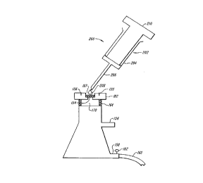
In Figure 6, fluid 208 from syringe 202 is expelled from reservoir 204 through
needle 206 upon positive pressure on plunger 210 of syringe 202. Cells can be fixed once
they are placed on window 104 of sample holder 100 to avoid an intermediate step of
adding the cells to fixative and then adding the suspension of cells in fixative to window
104. Preferably, lixation is accomplished by the addition of fixative to window 104 of
sample holder 100. The vacuum is turned off to control the duration of contact between the
fixative and the cells. After this period, the vacuum is turned on to remove the fixative. The
excess fixative is then washed from the cells under vacuum by an ~llplol),iate wash
solution. Also, once fixation is complete a series of washing steps take place to remove
fixative from the cells.
The present invention f~cilit~tes the addition of cells to window 104 of sample
holder 100 in a manner to speed up and simplify sample preparation for spectral analysis.
The present invention also enhances the user's ability to remove biological substances from
sample holder 100 that are of no interest spectrally or that might interfere with the spectral
analysis of the cells. This could be, for example, the desire to rliminich the amount of
mucous in the sample on window 104. The user could do this by repeatedly washing the
cells trapped on window 104 with large volumes of water or with a solution of normal
saline. An alternative to simply washing away "con1~min~ting material" of no spectral
interest or material that might confound the spectra of the cells is to wash the window with
chemical mixtures that react with Cont~min~nt.~ For example, again in the case of the desire
to remove mucous, the cells trapped on the window can be washed with mucolytic agents
to remove this mucous. In this regard, the contact time between wash liquid and cells
25 trapped on window 104 can be controlled by varying the strength of the applied vacuum;
this is especially ~aluable when the body is nonwettable as in the case of polyethylene,
polypropylene, or other suitable hydrophobic materials.
According to the present invention, the preparation of the cells trapped on window
104 may be modified for desired purposes. Solutions cont~ining vibrationally useful probes
of surface molecules can be reacted with cells trapped on the window and then washed
30 away prior to spectral analysis of the cells. This will provide means for enhancing or
CA 02222878 1997-12-01
W O 96/41153 PCT/U~C!'~3304
14
suppressing desired spectral aspects of the cells.
Also according to the present invention, it is possible to remove small cells from
large cells on window 104. Samples of cervical cells often contain blood cells, which are
quite small as compared with the size of cervical epithelial cells. Proper selection of the
pore size in window 104 will allow for the separation of epithelial cells from blood cells.
This will enhance the spectral analysis of the small numbers of epithelial cells in the
presence of large numbers of conl~min~ting blood cells.
The above examples apply to proce~ing cells in relatively small volumes of body
fluids, e.g. 1 to 2 ml or less. The present invention applies equally to processing cells in
extremely dilute suspension in large volumes of body tluids. This is true even when there
are liter amounts of fluids such as urine, ascites, pleural ~luid, and cerebrospinal fluid, and
the like in which a small number of cells reside. These larger fluid amounts can be easily
added directly to window 104 of sample holder 100 without prior tre~tment of the cells or
efforts to concentrate them in any way. The entire volume of fluid up to several liters, and
all the cells suspended in a large volume, can be added to window 104 of sample holder
100, as will be described.
Referring to Figure 7, generally at 260, when large volumes of dilute suspensions
of cells are to be processed, the sample holder and vacuum filtration system include
detachable funnel 262 that is disposed in groove 106 in the top surface of body 102 of
sample holder 100. Funnel 262 has bottom edge 208 that is dimensioned to fit into groove
106 in body 102. Spaced up from bottom edge 208 is circular flange 266 which is used for
handling funnel 262. Funnel 262 prevents spillage and loss of cells.
Figure 8 at 300 shows a second embodiment of the system for adding and
concentrating cells on window 304 for vibrational spectroscopic analysis. In Figure 8,
window 304 is not attached perm~n~ntly to body 308 of filter holder 302 but is free and is
2~ inserted into filter holder 302. Window 304 is disposed within filter holder 302 and filter
holder 302 can be opened after use to remove window 304. Filter holder 302 may be made
of any suitable material.
Preferably filter holder 302 includes frit 308 and cap 314. The frit and cap are made
preferably from molded plastic. Cap 314 has a hollow member that extends upward from
the top. Cap 314 and frit 308 sealably and detectability mate. Filter holder 302 holds
window 304 in place and prevents it from tearing when positive pressure is applied.
CA 02222878 1997-12-01
W O 96/41153 PCT/U~ 3~04
Referring to Figure 8, window 304 is bordered by non-porous frame 312 that is
disposed on frit 308. Frame 312 restricts the cells collected on window 304 to small area
and facilitates manipulation of window 304 after it is loaded with cells.
Figure 9 shows a top view of frit 308. Frit 308 is disposed below window 304 andhas a plurality of openings 310 in fluid connections with the flask 152.
The member that extends upward from the top of cap 314 connects to syringe 320
via Luer lock 322 or any other suitable attachment mech~ni~m Plunger 324 is disposed in
the top end of syringe 320 for forcing fluid with cells down to window 304. Morespecifically, the cells suspended in fluid medium within the barrel of the syringe 320 are
collected on the window by applying positive pressure to plunger 324 of syringe 320 and
filtering the suspension through window 304 that is held in filter holder 302. Once this is
accomplished, window 304 with attached cells is removed from filter holder 302 by
grasping window 304 via non-porous border 312. Filter holder 302 is discarded if it is made
of plastic or other inexpensive m5~ ri~1c or it can be washed for reuse if it is made of
sf~inl~ss steel or other expensive material.
Window 304 with trapped cells is mounted on body 352 of disposable sample
holder 350 that is shown in Figures 1 lA and 1 lB. Figure 1 lA shows the top view and
Figure 1 lB shows the bottom view of the sample holder. Other methods for mounting
window 304 on body 352 of sample holder 354 can be used. For example, window 304 may
be mounted magnetically to a steel sample holder.
Window 304 with trapped cells is removed from filter holder 302 and placed in
infrared transparent support 380. This transparent support shown in Figure 10, preferably
is made from crystalline CaF2 or other crystalline, infrared-transparent materials. Window
304 and support 380 may be mounted in standard infrared sample holder 354.
Referring to Figure 12, an assembly is shown for removing cervical cells from
brushes or spatulas. With plunger not removed from barrel 402 of modified syringe 400,
the brush or spatula with cells attached is placed in the fluid medium in the barrel. Cap 406
is placed on the top of barrel 402 and the assembly is shaken to dislodge the cells from the
collecting devices into the fluid medium. Cap 406 is removed and the collecting devices
are removed from barrel 402. Next, the syringe is fitted with plunger 404. Plunger 406 is
used to apply positive pressure on the fluid medium cont~ininE the cells.
A third embodiment of the system for collecting and concentrating cells is shown
CA 02222878 1997-12-01
W O 96/41153 PCTrUS9G~ 304
16
in Figure 13. This embodiment has an attached metal channel on cap 314 for puncturing the
bottom of syringe 400 (Figure 12). Positive pressure on plunger 404 of the syringe 400 will
cause cells in the fluid mt~ m to be applied to window 304. AltPrn~tively, cervical cells
in suspension, or any other type of cell in any type of collecting tube, are aspirated into a
standard syringe. The standard syringe then is attached to filter holder 302 as in Figure 8,
and the cells in suspension are added to the window held within the filter holder.
Any of the manipulations of cells described with respect to embodiment of the
invention depicted by Figures 4A, 4B, 5A and SB can be applied to the embo-1iment.~ in
Figures 8 -13. For ~x~mpl~, cells can be fixed after they are trapped on the window within
the filter holder by ch"nging syringes and treating the window with appropriate fixative.
The filter holder and syringe systems shown in Figures 8-13 can be used with fixed cells.
When fixed cells are used, residual fixative is washed from the cells by ch~nging the
syringe to app,o~,iate wash solution. Additionally, fixative or other reagents of potential
use in the analysis of the cells can be present in the syringes to which cells are added.
Referring to Figure 14 at S00, the system and method (automatic and semi-
automatic) of the present invention will be described. After vigorous shaking to transfer
cells from collecting devices to fluid suspension 552 in the collecting device 502, the
system shown generally at S00 is used for manual or automatic collection and concentrating
of cells for analysis. First end 509 of tube 506iS disposed in fluid suspension 552 in
collecting device 502. Pump 516 causes the fluid medium to flow in direction '~A" is tube
506. The cells are delivered to window 532 of sample holder 530 from end 507 of tube 506.
The vacuum suction in flask 520 draws the fluid associated with the cells through the
window and frit into the flask. This action may take place autom~tically. Preferably, the
rate of aspiration for automatic operation substantially matches the rate of filtration, or the
former is slower than the latter, to prevent spillage or waste of cells. All or the part cells
may be added to window 532 in this way. The amount of cells to be added can be controlled
by a device that controls the volume of the aspirate. Even this feature of the device can be
controlled automatically, which is especially useful for controlling the amount of cells
added to the window.
According to Figure 14, there is continuous monitoring through a suitable optical
window 512 of the aspirated stream in the flow path between aspirating pipette 506 and
window 532 of sample holder 530. Window 512 may be observed visually or via the
-
CA 02222878 1997-12-01
W O 96/41153 PCT~US96~9304
sensors at 540 and 542. The simplest method, in this regard, and the preferred embodiment
of the present invention is to monitor turbidity by light sC~tt~oring Other ways to monitor
the amount of cellular material in the stream are by absorption of protein or D~A, for
example, but light scattering will be intense at short wavelengths. Independent of the
optical method used to monitor the amount of cellular material in the stream of flow to
window 532 of sample holder 530, a built-in program correlates flow rate for aspiration and
the measurement of turbidity (or another optical property) to calculate the volume of
suspension that must be added to window 532 to yield a sample with an optimal number of
cells on window 532. When this volume of cells is added, aspiration is cut-off.
To facilitate a uniform distribution of cells in suspension, after shaking to displace
them from the collecting devices, the collecting fluid is maintained at high density by
addition of sucrose or other suitable material. Whatever material is used to increase the
density of the collecting fluid and thereby to decrease the rate of settling of cells, it must be
washed from the window after the requisite number of cells (based on measurements of
turbidity) has been added to window 532. In the case that an inadequate number of cells has
15 been added, after aspirating all the suspension, the system can provide a print out, a fl~hin~
light, or other suitable alarm (not shown) to show this condition.
In a large clinical pathology laboratory, complete automation of sample preparation
can be obtained. In such operations, the bottles containing the cells are shakenautomatically. Two sample holders, one for the endo- the other for the exocervical samples,
20 are imprinted autom~t~ ly with the same identifier code. Then the suspensions of endo-
and exocervical cells are aspirated and added to the appropriate windows. Control of the
number of cells added to each substrate is effected as described above and illustrated in
Figure 14. Once the cells are added, routines are car;ied out for washing cells and/or for
treating the cells with fixatives, and for specific chemical treatments for specific
2~ modification of the cells, as described.
In Figure 4. window 104 of sample holder 100 preferably has a diameter of
approximately 3 mm. The beam of light in a standard infrared spectrometer is reduced to
as small as 1.3 mm. With microscopic attachments, smaller light beams can be
accomplished, down to the limit of diffraction effects. In the mid-infrared, the diffraction
limi~ is greater than the dimensions of a single cell. In the near infrared, however, light
30 beams the size of a cell can be produced, which applies as well to spectroscopy by Raman
CA 02222878 1997-12-01
W O 96/41153 PCTAJS~6/'03304
18
scattering.
Generally, spectra collected on cells represent an average spectrum of all cells in a
sample. The capacity of vibrational spectroscopy to detect disease in cells and stage the
severity of disease can be maximized by examining cells one at a time. To do so, requires
5 that cells be added to a window such that the cells are spread out across the surface of the
window. According to the present invention, the application of cells to the window
provides a controlled method of adding cells to the window so that vibrational spectra can
be collected simultaneously on a minim~l number of cells at any one time or even one cell
at a time. To do this, the aspirated cells in suspension are added to the window as shown in
Figure 14.
The drop-wise addition is controlled, as regards the size of the drops (which depend
in part on the concentration of cells in the suspension) and their location on the window, by
computer software that drives the tip of the pipette in small increments (as small as 1 ~m,
for example) across the horizontal and vertical dimensions of the window. Since the
amount of fluid added at any one time is small and not allowed to spread across the window,
15 the cells become "stuck" where they are deposited. The coordinates of the window at which
cells are deposited are then used as a basis for washing thc cells, again in drop-wise fashion
or for treating cells as discussed above. Finally, the coordinates of location of cells on the
window are fed on-line to the spectrometer, which has microscopic optics. The
spectrometer collects and co-adds interferograms from each coordinate moving across and
20 sampling multiple regions of the surface of the window Software may direct the collection
in this way on the basis of the stored coordinates for the positions of cells on the window.
Each co-added spectrum is analyzed separately and simultaneously with the scanning of the
window until the entire window has been scanned - cell by cell in some cases - or until a
definitive diagnosis of disease can be made.
The terms and expressions which are used herein are used as terms of expression
and not of limitation. There is no intention in the use of such terrns and expressions of
excluding the equivalents of the features shown and described, or portions thereof, it being
recognized that various modif1cations are possible in the scope of the present invention.