Note: Descriptions are shown in the official language in which they were submitted.
CA 02223061 1997-12-02
WO 96/39535 PCT/IB96/00543
METi-fOD OF AND APPARATUS FOR DIAGNOSTIC DNA TESTING
The present invention relates to the diagnostic testing of DNA using
polymerase
chain reaction (PCR) amplification followed by electraphoretic separation of
the resulting
fragments to detect possible gene variants of mutational defects and the like;
being more
particuiariy directed to new and improved multipiex PCR techniques in
combination with
preferably two-dimensionai electrophoretic separation in denaturing gradient
gels.
SUBSTITUTE SHEET (RULE 26)
CA 02223061 1997-12-02
WO 96/39535 PCT/IB96/00543
2
This invention pertains particularfy to tests for the presence of DNA
mutations in =
patients with inherited diseases, inciuding birth defects (e.g. cystic
fibrosis) and genetic
predispositions to adult chronic diseases (e.g., cancer). More specifically,
the invention
relates to the quick preparation, by a novel two-step poiymerase chain
reaction (PCR)
ampiification, of gene fragments and their subsequent efficient and accurate
examination
for mutations.
BACKGROUND OF INVENTION
Genes with mutational defects (gene variants or aileles) can be identified by
DNA
diagnostic testing. Gene variants can be transmitted from parents to children.
Some gene
variants have a very strong effect and are, by themselves, capable of causing
disease.
Examples are many mutationai variants of the cystic fibrosis transmembrane
conductance
reguiator (CFTR) gene that cause cystic fibrosis. Other gene variants act in
combination
with gene variants from other loci. Exampies inciude many of the common
(poiygenic)
diseases, like heart disease and cancer. It is possible to test for gene
defects in an early
stage; that is, in cells from the embryo (pre-natai testing), but aiso at a
much later stage
in young, aduit or oid individuais.
By DNA diagnostic testing, information is obtained about a disease, sometimes
2 p before it has become manifest. This greatly facilitates management of the
disease, e.g.,
prevention, treatment. For example, it is possible to test for the presence of
particular
gene variants in cancers or infectious disease agents in order to predict,
e.g., the course
SUBSTITUTE SHEET (RULE 26)
CA 02223061 1997-12-02
WO 96/39535 PCT/IB96/00543
3
of the disease, response to therapy. It is aiso possible to test individuals
for carrying a
particular gene variant, which they would not like to see transmitted to their
offspring
(carrier testing). Finally, it is possible to test individuals at any time
(from pre-natal to late
age) for inherited gene-encoded predispositions to disease. An example from
one end of
the spectrum is cystic flbrosis, for which pre-natal testing and carrier
screening has
already become relatively common. The other end of the spectrum involves late-
onset
diseases, like cancers and neurodegenerative diseases.
DNA diagnostic testing invoives an analysis of the sequence integrity of
individuai
genes. At present, this is costly since accurate testing requires sequencing,
or decoding,
of the gene, which is labour intensive. Thusfar no cost-effective universaily
appiicabie
standardized system for DNA diagnostics has become availabie (for a review,
see Cotton,
1993, Current methods of mutation detection, Mutat. Res. 285:125-144). In
order to be
cost-effective and widely accepted a DNA diagnostic system must be accurate
(more than
95%), have a high throughput, and not be labour intensive.
GENERAL BACKGROUND OF ANALYSIS TECHNIQUE
It is initially in order briefly to review the general techniques involved in
PCR
amplification and in electrophoretic separation of fragments, and where the
art has
applied and is currently applying the same.
A sample of cells, such as derived from blood, is first chemically and
physicaily
treated to extract DNA strands carrying genes that occupy only about two
percent of the
SUBSTITUTE SHEET (RULE 26)
CA 02223061 1997-12-02
WO 96/39535 PCT/IB96/00543
4
total DNA material in a ceil genome, with the remainder of DNA material
ciuttering up the background. While each cell has two copies of each gene and
such can be identified by
building bioclcs or basepairs identified by sequences of letters A, C, G and T
as a letter
code, they constitute such a smail part of the long DNA strands that they must
be
amplified by making many copies of the same to permit their inspection. This
is effected
by heat-separating or denaturing of the DNA strand pairs, mixing with
appropriate primers
to bind or anneal to the beginning and the end of the gene fragments (e.g.,
gene exons
or coding regions) to be investigated, later more fuily discussed, and adding
sufficient
building blocks to generate copies of the gene exons. The successive repeating
of this
cycie of steps effects a cumulative copying of the exons to produce a purified
and
ampiified quantity - a process generaily referred to as the before mentioned
polymerase
chain reaction or PCR ampiification - and more fully reviewed, for example, in
Moiecuiar
Pathology, Hein and Siiverman, Carolina Academic Press, 1994, Chapter 2,
Moiecuiar
Techniques and Their Automation in the Clinical Laboratory, pages 5-31 (Winn-
Deen).
At this stage, it is then in order to inspect or analyze the gene exons to
determine
if there are mutations from normalcy. This is generally done by
electrophoretically
separating the DNA purified fragments, preferably on the basis both of size
and base-pair
sequence, as more fully described by co-appiicant Vijg and A.G. Uitterlinden
in Two-
dimensional DNA typing: A Paraile! Approach to Genome Analysis, Ellis Horwood,
1994,
2 0 particuiariy at pages 33-40.
Electrophoresis has been used not oniy for DNA fragment separation but aiso
for =
separating other substances than genes, such as, for example, for protein
analysis, as
SUBSTITUTE SHEET (RULE 26)
CA 02223061 2003-09-15
J
described in Electrophoresis 1993, 14, 1091-1198. Machines are provided for
one-
dimensional DNA separation by electrophoresis with fluorescent dye labeling,
such as the
ABI Prism 377TM DNA sequencer of Perkin Elmer; and for two-dimensional DNA
typing, as
described by co-applicant Vijg and E. Mullaart et al. in Nature, 365, 30
September, 1993,
Parallel genome analysis by two-dimensional DNA typing, pages 469-471,
describing
apparatus of lngeny B.V. of The Netlherlands. The first dimension (say
horizontal)
application of the electric field to an appropriate gel matrix (later
discussed) into which the
purified DNA fragments have been introduced, causes separation of the
fragments by
size, larger particles moving slower than smaller particles. By applying the
electric field
1 o in an orthogonal direction (vertically) with a chemical gradient as of
successiveiy more
concentrated urea/formamide disposed in the gel, or a temperature gradient
established
therealong, the DNA fragments will migrate (this time vertically) untii they
melt and are
locked in position in the gel matrix at particular verttcal sequence-
determined locations.
To prevent meiting of the entire DNA fragments, the latter can be attached
(before the
electrophoresis process) to a number of only G's and C's, which are more
resistant to
melting than A's and T's. This so-called GC-clamping, effectively locks each
fragment in
position which is determined entirely by the sequence of the exon-part of the
fragments.
Is this sequence changed at only one position, for example, by the
substitutlon of an AT
couple for a GC couple, it will melt later or earlier and hence become locked
in a different
vertical position than the normal reference fragment
Such two-dimensional gene scanning (TDGS) has promise for becoming a cost-
effective and widely accepted DNA diagnostic system. In this system, as above
described,
CA 02223061 1997-12-02
WO 96/39535 PCT/IB96/00543
6
a large number of DNA fragments obtained through poiymerase chain reaction
(PCR)
amplification from a given DNA sample are eiectrophoreticaily separated on the
basis of
both size and basepair sequence. This system is highiy accurate (i.e., 99%),
since the
second dimension separation is based an denaturing gradient gel
electrophoresis
(DGGE). Indeed, DGGc is the oniy system with such high accuracy (Sheffield et
al.,
1993, The sensitivity of singie-strand conformation poiymorphism analysis for
the
detection of singie base substitutions, Genomics 16, 325-332; Grompe, 1993,
The rapid
detection of unknown mutations in nucfeic acids, Nature Genet. 5, 111-117;
Guldberg et
a1.,1993, Moiecuiar analysis of phenyiketonuria in Denmark: 99% of the
mutations
detected by denaturing gradient gel electrophoresis, Genomics 17, 141-146).
Automatic
instrumentation for TDGS is parrtiy availabie and partly under development.
TDGS allows
the detection of all possibie mutations in DNA fragments obtained from one or
more
genes simuitaneously at a high throughput and with a minimum of manual
interference.
One major hindrance to the widespread appiication of TDGS in DNA diagnostics
is the difficuity of amplifying many fragments simuitaneousiy in the same
reaction tube
by PCR (muitipiex PCR). In fact, it is often not even possibie to find PCR
primer sites that
amplify the reievant gene fragments and simuitaneousiy fuifil requirements for
both PCR
and denaturing gradient gel electrophoresis, i.e., optimal PCR reactions and
optimal
meiting behaviour of the amplified fragments. The current procedure begins
with
ampiifying regions of the target DNA, usually the protein-coding regions
(exons) of a
gene, by PCR. These amplification reactions are conducted separateiy, e.g., if
27 exons in a gene are being analyzed, then 27 separate PCR reactions must be
conducted. In
SUBSTITUTE SHEET (RULE 26)
CA 02223061 1997-12-02
WO 96/39535 PCT/IB96/00543
7
= practice, it is usually possible to conduct a few PCR reactions together in
one tube (e.g.,
Edwards and Gibbs, 1994, Muitiplex PCR: advantages, development, and
appiication,
PCR Methods and Appiications 3, S65-S75).
Clearly, with a large number of individuais to be tested and when more than
one
gene is tested simuitaneausly in the same TDGS test, the totai number af
pipetting steps
and individuai reactions to be carried out can become very high. This
increases the labour
intensity of the test, but makes it aisa more complicated with a higher chance
of human
error. Indeed, in view of this complexity, even complete lab automation where
ail pipetting
steps are done automaticaily will not solve this problem.
The problem of not being able to PCR-ampiify multiple fragments simuitaneously
under identical reaction conditions in the same tube is an important technical
hurdle for
TDGS to meet criteria for ciinicai testing, i.e., laboratory user-
friendliness. To reduce the
number of PCR reactions via muitipiexing, i.e., conducting several PCR
amplifications in
one reaction by empioying muitipie sets of primers, is a non-trivial
development.
Current approaches for multiplexing are sometimes as simple as combining a few
sets of primers for which reaction conditions have been determined separately.
However,
in most cases multiplex PCRs must be developed with careful consideration for
the
regions to be amplified, the relative sizes of the fragments, the dynamics of
the primers,
and the optimization of PCR experimental conditions to accommodate muitipie
fragments.
A key problem is the positioning of the primers. For gene diagnosis one
generally
aims at ampiifying the exon sequences, splice sites and regulatory regions.
Primers for
exon-ampiifying PCR reactions are ideally placed in intronic sequences
adjacent to the
SUBSTITUTE SHEET (RULE 26)
CA 02223061 1997-12-02
WO 96/39535 PCT/IB96/00543
8
exons. This provides some margin for adjustment of fragment length or
ampiification quality as well as information about mutations affecting splice
sites. These are the first
limits to primer choice.
Then, primers shouid be positioned so that non-specific amplification at other
sites
than the target sequences does not occur. Indeed, the human genome is 3 x 10S
basepairs long, which provides ampie opportunity for fortuitous sequence
homoiogy
between the target and other non-target sequences. This problem is not typical
for
multiplexing, but can aiso occur when one wishes to amplify oniy one fragment
at a time.
This is the second limitation to primer choice.
For muitipiexing, primers should be selected so that their predicted
hybridization
kinetics are similar to those of other primers in the muitipiex reaction. This
is a third
limitation to primer choice. These limitations to primer choice, with totai
genomic DNA as
template, are the reasons why multipiex groups are usuaily small (typicaily
less than 5
fragments).
For optimal separation in TDGS there is a fourth formidable limit to primer
choice.
TDGS requires DNA fragments of 100-600 bp on average. One of the two primers
should
be coupled to a GC-rich fragment to provide for a GC-ciamp as highest melting
domain
in the fragment to be generated by PCR. This is essential to guarantee the
highest
sensitivity to detect mutations in the second (denaturing gradient) dimension
gel (Myers
et ai., 1985, Neariy ail single base substitutions in DNA fragments joined to
a GC-ciamp
can be detected by denaturing gradient gel eiectrophoresis, Nucl. Acids Res.
13, 3131-
3145; Myers, et a1.,1987, Detection and localization of singie base changes by
denaturing
SUBSTITUTE SHEET (RULE 26)
CA 02223061 1997-12-02
WO 96/39535 PCT/IB96/00543
9
gradient gel eiectrophoresis, Meth. Enzymol., 155, 501-527). Then, primers
shouid be
positioned in such a way that the target fragment comprises only one single
domain, that
melts earlier (at lower urealformamide concentration or temperature) than the
GC-ciamp
attached to it It tumed out that optimal PCR conditions for both PCR (let
alone
muitipiexing) and optimai meiting profiles are difficuit, if not impossible,
to realize (e.g.,
compare the R8 DGGE design by Blanquet et al., 1993, Identification of
germline
mutations in the R131 gene by denaturant gradient gel eiectrophoresis and
poiymerase
chain reaction direct sequencing, Hum. Moiec. Genet. 2, 975-979, with our
present
design).
The present invention involves a combination of so-called fong-PCR with short-
PCR. Recently, PCR ampiification methods were developed allowing the
amplification of
large fragments (up to 40 kb) from the genomic DNA. We have taken advantage of
this
development by using long-PCR to first amplify aiI the coding regions of the
target
gene(s) in the smallest number of fragments possible. Using these long
ampiicons as
tempiate we then PCR-amplify the smaii fragments, required for the TDGS, in a
muitipiex
fonnat In this way the target sequence is first amplified away from the
contaminating
genomic DNA, which ailows to obtain the smail PCR fragments under identical
conditions
from this pre-purified template.
Using the above procedure, primer sets selected oniy on the basis of optimal
meiting behaviour of the PCR ampiicons, aiso exhibited optimal behaviour in
the PCR and
even allowed extensive muitipiexing. In part this phenomenon can be ascribed
to the pre-
purification by the long-PCR. Indeed, the long-PCR greatly increases the
amounts of
SUBSTITUTE SHEET (RULE 26)
CA 02223061 1997-12-02
WO 96/39535 PCT/IB96/00543
target sequence relative to other genomic DNA sequences, thereby greatly
decreasing
compiexity of the reaction and increasing its specificity.
Although the above described phenomenon can be explained by the reduction of
complexity through the preparation of a purified tempiate, an exact
explanation can not
5 be given. Indeed, the sheer magnitude of the effect is surprising and
necessitates a
reevaiuation of the factors involved in PCR optimization. One thing is ciear,
however. The
present invention alone enables one to design and perform an efficient TDGS
test,
because primers can now be selected on the basis of melting profile alone and
multiplexing is greatly facilitated.
10 The invention is aiso generally appiicabie. Indeed, selection of primers in
every
PCR-based diagnostic reaction is an important issue and many potentiai priming
sites tum
out to give poor results. Muitipiexing then generates additional problems,
which is the
reason that it is not widely used. The long-PCR/short-PCR two-step
ampiification system
offers an immediate and simple solution to this problem.
OBJECTS OF INVENTION
An object of the invention, accordingiy, is to provide a new and improved
method
and apparatus for diagnostic DNA testing that obviate the above described
difficulties.
A further object is to provide for novel detection of mutations in genes by a
two-
step muftipiex polymerase chain reaction amplification using long and short
muitipiex PCR
followed by two-dimensional eiectrophoretic separation of the fragments an the
basis of
SUBSTITUTE SHEET (RULE 26)
CA 02223061 1997-12-02
WO 96/39535 PCT/IB96/00543
11
both size and basepair sequence.
Other objects wii( be explained hereinafter and pointed out in connection with
the
appended cfaims.
BRIEF DESCRIPTiON OF THE FIGURES
The invention will now be described in connection with the accompanying
drawings
in which:
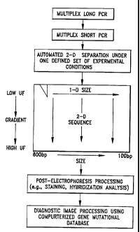
FiC. 1 illustrates the sequence of process steps for performing the invention.
FiC. 2 shows the melting curves for R8 exon 12- with and without the CC-ciamp
(i.e. retinal blasroma. gene).
FiC. 3 shows maps of the tumor suppressor gene R8 indicating the positions of
the PCR primers for the long- and short-PCR reactions.
FIG. 4 shows a computer print with the predicted positions of the short PCR
fragments in a 2-D gei eiectrophoresis pattern with the indicated
specifications.
FIG. 5 shows the actuai gei separation pattem indicating correspondence with
the
theoreticaiiy predicted pattem.
FiC. 6 shows that with the long-PCR product for exons 18-23 as tempiate all 6
of
the short PCR fragments are obtained (lanes 7-12), whereas with total genomic
DNA as
2 0 tempiate most products are missing (lanes 1-6). It aiso shows that oniy 5
ng total
genomic DNA is sufficient as starting matetiai for the long-PCR (lane 13), and
that all
short PCR products are obtained with the long-PCR products as tempiate (lanes
20-24).
FIG. 7 shows details of wildtype (homozygous normai) fragments and several
SUBSTITUTE SHEET (RULE 26)
CA 02223061 1997-12-02
WO 96/39535 PCT/IB96/00543
12
heterozygous mutants.
SUMMARY OF THE INVENTION
In summary, in an important aspect, the invention embraces a method of
analyzing
predetermined gene exons derived from DNA, that comprises, adding primer pairs
to
successive groups of the gene exons followed by effecting polymerase chain
reaction
amplifications thereof in a common tube, as a first step and reiativeiy long
multiplex
poiymerase chain reaction; adding further primer pairs to each of the gene
exons and
lo effecting poiymerase chain reaction ampiifications thereof in the common
tube as a
second step and short multiplex polymerase chain reaction; and
electrophoreticaily
separating the gene fragments.
Preferred and best mode techniques will now be described.
DESCRIPTION OF PREFERRED EMBODIMENTS
The present invention, as before stated, invoives the design of an accurate
and
efficient mutation detection test on the basis of a minimal number of two-step
multipiex
PCR reactions in combination with automatic two-dimensional separation of the
fragments
to detect ail possible mutations in the gene(s) simuitaneousiy.
Table 1 lists the different steps in the design of a TDGS test, with the RB
(retinoblastoma) tumor suppressor gene as a modei. First, the gene sequences
are
SUBSTITUTE SHEET (RULE 26)
CA 02223061 2003-09-15
13
retrieved from a database (e.g. Genbank) and the target regions, i.e., exons,
splice sites,
regulatory regions, are defined. Then, primers are positioned to obtain all
target regions
as the smallest possible number of fragments that can still be amplified
through long-
PCR, i.e. up to at least 20 kb (TaKaRa LA PCRT" Kit. Product Insert). Some
general
guidelines in choosing primer sequences for long-PCR have been described
(Foord and
Rose, 1994, Long-distance PCR. PCR Methods and Applications 3, S149-S161), but
empirical determination of optimal primers remains necessary.
Table 1. Design of a TDGS test.
1 o 1- Retrieve sequence from database.
2. Position primers for long-PCR to cover all desired regions (e.g.. coding
sequences,
splice sites, regulatory regions, mutation hotspots) by the smallest possible
number
of amplicons.
3. Position primers for short-PCR according to the following criteria:
a. the desired target sequences should be covered by amplicons of between 100
and 600 bp
b. amplicons should have optimal melting behaviour, i.e., consist of one
lowest-
melting domain in addifion to the GC-clamp attached to one of the primers.
c. optimal amplicon distribution over the 2-0 gel
d. similar reaction ldnetics
4. Set up PCR conditfons separately for each primer set with the long-PCR
products
as template.
5. Develop muitiplex co-ampiification conditions by grouping primer sets and
adjusting
reaction components.
CA 02223061 2003-09-15
14
As listed in Table 1, item 3, then, using the long-PCR fragments as template,
primers for short-PCR are selected to yield fragments of between 100 and 600
bp. The
main selection criterion here is necessarily the melting behaviour of the
fragments. In the
ideal situation, each amplicon should comprise only one meiting domain, which
should
be lower (less stable) than the GC-clamp attached to it. Attachment of -a 30-
40 bp GC-
clamp is accomplished by making it part of one of the primers (Sheffield et
al., 1989, The
sensitivity of single-strand conformation polymorphism analysis for the
detection of single
base substitutions, Genomics 16, 325-332). Optimal melting behaviour is
determined of
each candidate target sequence by using a computer program (e.g. MELT87TM;
Lerman
and Silverstein, 1987, Computationai simuiation of DNA melting and its
application to
denaturing gradient gel electrophoresis, Meth. Er+zymot. 155, 482-501). An
example of
an amplicon with optimized melting behaviour through GC-clamping is shown in
Fig. 2,
in connection with exon 12 for R8.
In general, a collection of primers is selected that allow an optimal
distribution, in
both size and DGGE dimension, over the 2-D gel. Due to the high resoiution of
2-0 gels
(5-1 0-bp size differences are easily resolved) this is generally not too
difficult. Indeed, with
50 fragments or less, spot distribution is hardly an issue and primers can
simply be
selected according to their meiting behaviour.
Fig. 3 shows the collection of amplicons selected for the RB gene, together
with
the long-PCR fragments that served as templates. Together the short-PCR
fragments
represent more than 90% of the RB coding region.
Figs. 4 and 5 show the theoretical and the empirical spot distribution for the
24
CA 02223061 1997-12-02
WO 96/39535 PCT/IB96/00543
exons of the RB gene covered by the ampiicons shown in Fig. 3. Although there
are
differences, most notably the spot representing exon 11, our conciusion is
that overall the
melting program accurately predicts spot positions.
It is important to realize that without the first long-PCR step, the optimai
melting
5 criterium is usually in conflict with other primer design criteria appiied
to PCR with total
genomic DNA as tempiate. Indeed, for the RB gene it was found to be impossibie
to
select conditions suitable for bath optimai separation in DGGE and optimai
priming in
PCR. The pre-purification step represented by the long-PCR is apparently a
conditio sine
qua non for the design of an optimai set of PCR primers in TDGS.
10 When the test format is established, the two-step PCR amplifications are
carried
out in a muitipiex format. It is the possibility to design and perform
multiplex PCR
reactions that represents the care of the present invention. The necessity of
the first,
long-PCR step for a successfui muitipiex PCR is demonstrated by the results
shown in
Fig. 6. In Fig. 6, the 6 lanes on the left contain PCR products obtained after
performing
15 a muitipiex PCR of exans 18-23 (6 fragments) of the RB gene, using
different amounts
of totai genomic DNA as template. Cleariy, virtually no products of the
desired lengths are
obtained. The latter is in contrast to lanes 7-12, in which the products were
applied of the
same multiplex PCR reaction, but this time with the iong-PCR product as
template. The
long-PCR was performed at different cycies and it is ciear that only 5-10
cycies are
needed to generate enough template for a successful muitipiex PCR.
Lanes 13 to 19 contain the muitiplex short-PCR products obtained with the long-
PCR products as template, at different amounts of startrng material, i.e.,
different amounts
SUBSTITUTE SHEET (RULE 26)
CA 02223061 1997-12-02
WO 96/39535 PCT/IB96/00543
16
of totat genomic DNA used in the long-PCR reaction. Interestingly, 5 ng total
genomic
DNA is sufficient to obtain all the products. Since ciinical materiai is
sometimes not
available in plentiful amounts (e.g., breast cancer needle biopsies) this is
an important
resuit, indicating that a successfui test can be performed with very small
amounts of DNA.
Finaily, lanes 20-24 of Fig. 6 contain the products of the 5 multiplex PCRs
corresponding to the 6 long-PCR sets (lang-PCR groups 1 and 6 were combined;
see
also Fig. 3 and later discussed Table 2). Further adjustments of PCR
conditions and/or
primers shouid make it possible to obtain an even smailer number of multiplex
sets for
this gene. Indeed, there is no reason why the entire RB gene coding region
couidn't be
amplified in only one singie PCR reaction. After the second PCR, the fragments
are
allowed to undergo one complete round of denaturation/renaturation to
facilitate the
formation of heterodupiexes. There are presented in Table 2, a listing of the
primer pairs
for TDGS for the case of R8; the exon numbers being listed in the left most
table, with
long PCR primer codes for six exon groups (0 through 24-27) and short PCR
primers for
the individuai 27 exons.
Subsequent to the PCR, the mixture of fragments is subjected to 2-D
etectrophoresis in a denaturing gradient gel (Fig. 1). The availability of an
automated
instrument greatly simplifies this process. The instrument used here ailows 10
gels at a
time to run without manuai interference, i.e., cutting out lanes and loading
these onto a
second get. All experiments involving optimization of the experimentai
conditions were
carried out using manual instruments. After 2-0 electrophoresis the gels are
reieased
from between the giass piates and stained with ethidium bromide or any other
stain.
SUBSTITUTE SHEET (RULE 26)
CA 02223061 1997-12-02
WO 96/39535 PCT/IB96/00543
17
Table 2. Primer pairs for TDGS of RB
RB: Long-PCR
crons primers S'-3' size
0-2 TGTCAGGCCTGCCTGACAGACTTCTATTCAGCA 4.5 kb
ATGTTAGCAGAGGTAAATTTCCTCTGGGTAATGG
3-6 GCAGTCATTTCCCAACACCTCCCCTCTGT 9 kb
AAGCCAAGCAGAGAATGAGGGAGGAGTACATTAC
7-11 TCAGCAGTTfCTCCCTCCAAGTCAGAGAGGC 10 kb
GAGACC,AGAAGGAGCAAGATCAGGTAGTAG
12-17 ACCATTCCCCCTACTCTCCATGGTCCATG 12.4 kb
CTCACAGGAAAAATACACAGTATCCTGTTTGTGTGGC
18-23 CCAGCCTTGCATTCTGGGGATGAAGC 14 kb
AGTCGTAAATAGATTTTCTTCACCCCGCCCC
24-27 GCCTTTGCCCTCCCTAAATATGGGCAATGG 7.3 kb
CTGGGTTATCAGGACTCCCACTCTAGGGCC
RB: Short-PCR
cson priners 5=3' srse Tm(3SiIF) multfpler set
2 [GCI] TTGATTTATAAGTATATGCCA 229bp 30 E
CAAAACGTTTTAAGAAAATCC
3 [GC1] CCAGTGTGTGAATTAT'*TAA 239bp 27 A
CCI'I'ITATGGCAGAGGCTTATA
4 [GC1] GAATTGAAATATCTATGATT 270bp 24 A
ATCAGAGTGTAACCCTAATA
5 [GCl] TACTATGACTTCTAAATTACG 157bp 27 A
GTGAAAAATAACATTCTGTG
6 TGGAAAACTTTCT'lTCAGTG 237bp 17 A
[GCIJ GAAT'ITAGTCCAAAGGAATGC
7 [GCl] CCTGCGATITTCTCTCATAC 257bp 26 B
GCAACTGCTGAATGAGAAAG
8 GTTCTTATCTAATTTACCACT 229bp 27 B
[GC1] TITTAAAGAAATCATGAAGTT
9 [GCl] AGTCAAGAGATTAGATITIY'i 227bp 20 B
ATCCTCCCTCCACAGTC
10 [GC1] GACATGTAAAGGATAATTGT 222bp 21 B
GCAAATCAATCAAATATACC
SUBSTITUTE SHEET (RULE 26)
CA 02223061 1997-12-02
WO 96/39535 PCT/IB96/00543
17/1
11 AGTATGTGAATGAC'I'PCACT 174bp 21 B
[GC 1 ] TATAATATAATTAAAAGTAGG
12 CTCCCTTCATTGCTI'AACAC 211bp 24 C
[GCI; TITCTTTGCCAAGATATI'AC
13 [GCl] GA'ITACACAGTATCCTCGAC 224bp 34 C
GCAGTACCACGAATTACAATG
14 [GC1] GTGATTI'TCTAAAATAGCAGG 179bp 35 C
ACCGCGCCCGGCTGAAAT
17 [GC1] 'I'I'CIITGTCTGATAATAAC 380bp 26 C
CTCTCACTAACAATAATTTC'TT
18 [GCl] GAC."I'iTtAAATTGCCACTGT 393bp 33 D
ATTCCCTACAGTTTCTITAT
19 [GC1J CAACTTGAAATGAAGAC 248bp 34 D
CLiTCCCGCTGCTCITGAAAATAATCATC
20 [GC1] AAAATGACTAATTrTI'CTTATTCCC 227bp 44 D
AGGAGAGAAGGTGAAGTGC
21 [GC2] CATTCTGACTACZTI'TACATC 201bp 28 D
CGGGCTTACTATC-GAAAATTAC
22 [GC3] CIZTITACTGTTCTTCC 194bp 33 D
CCAATCAAAGGATAC'ITITG
23 [GCI] TCTAATGTAATGGGTCCACC 281bp 38 D
CCCTAC'I'I'CCCTAAAGAGAAAAC
24 CGGAATGATGTATTTATGC'PCA 195bp 22 E
[GCI] TTCTITfATACTTACAATGC
25 [GCl] ATGATITAAAGTAAAGAATTCT 245bp 38 E
CATCTCAGCTACTGGAAAAC
26 [GC 1] TCCAT'ITATAAATACACATG 161bp 32 E
ATTTCGTi rACACAAGGTG
27 [GC1] TACCCAGTACCATCAATGC 191bp 43 E
TCCAGAGGTGTACACAGTG
GC-clampa:
GCl: CGCCCGCCGCGCCCCGCGCCCGTCCCGCCC (30mer)
GC2: CGCCCCGCGCCGCCGCCCCGCCCCCGCCCGTCCCGCCC(38mer)
GC3: CGCCCCGCCGCGCCCCGCGCGCCCGGTCCCCGCGC(3Smer)
The resulting patterns were dociunented and evaluated (by eye and image
analysis) for the occurrence of mutations.Under the conditions applied
ie, GC-clamping and heteroduplexing, heterozygous mutations result in 4
spots: the 2 hcmoduplex variants =
SUBSTITUTE SHEET (RULE 26)
CA 02223061 1997-12-02
WO 96/39535 PCT/IB96/00543
18
and the 2 heteroduplex variants (illustrated in Fig. 7). The latter are not
always separated.
Since mutations may also occur in a homozygous state it can be necessary to
mix each
sample before PCR with a controi sample to make sure that heterodupiex
molecules will
be present.
The foilowing provides details of the manner in which the embodiments of the
present invention may be made and used in order to achieve the accurate and
efficient
preparation and examination of gene fragments in DNA diagnostic testing. This
description, which is focused on an iitustrative or model gene, i.e., the
tumor suppressor
gene RB previously discussed, is not to be construed as specifically limiting
the invention.
The same procedure may be used on other genes and/or to combine even more PCR
fragments in the same tube, within the purview of one skilled in the art, and
are to be
considered to fali within the scope of this invention.
A. Design of the RB Two-Step PCR TDGS Test
Sequence Retrieval. The sequence of the RB gene is retrieved from a database,
i.e., Genbank. The target regions, i.e., exons, splice sites, regulatory
regions are defined.
PrimerSelectfon formultiplexLong-PCR. Primer pairs for long-PCR are positioned
in such a way as to cover all target regions by the smaiiest possible number
of fragments
that can still be ampiified through long-PCR. Long-PCR primers are aiso
selected for
highest specificity, optimai anneaiing temperature and minimal self-
complementation for
SUBSTITUTE SHEET (RULE 26)
CA 02223061 1997-12-02
WO 96/39535 PCT/IB96/00543
19
muitipiex long-PCR, e.g., by using primer design software. T he design of the
fong-PCA
muitipiex is reiatively easy in view of the ampie positioning space of the
primers.
Primer Selection for Multipiex Short-PCR. Primer pairs for short-PCR are
seiectea
an the basis of the following criteria:
a. the desired target sequences st'tauid be covered by ampiicans of between
100 and
600 bp
b= ampiicans should have aptimai meiting behaviour, i.e., consist of ane
lowest
meiting domain in addition to the GC-ciamp attached to one of the primers.
c. optimal ampiican distribution over the 2-D gei
d. similar reaction kinetics
Criterion b, above, is frequently in conflict with standard primer design
crtteria, if
applied on totai genomic DNA. Indeed, the present invention proved to be both
necessary
and sufficient for the design and performance of TDGS as a rapid, accurate and
practical
tool for mutation detection in the R8 gene.
Multiplex groups are seiected empiricaily on the basis of the behaviour of the
primers in various multipiex reactions. For RS multipiex groups were made
according to
SUBSTITUTE SHEET (RULE 26)
CA 02223061 2003-09-15
the long-PCR. That is: all short PCRs with one long-PCR fragment as template
were
amplified together as one multiplex group. Two long-PCR groups were actuaJly
combined
as one muitipiex group. All short-PCR fragment may also be amplified together.
As before
explained, Table 2 lists the primer pairs used for the long and short PCRs,
fragment
5 sizes, annealing temperatures, melting temperatures and the five different
muitipiex
groups.
B. PCR Reactions and Heteroduplexing
10 Primers (deprotected and desalted) can be obtained from various sources.
Our
primers were obtained from Gibco BRL For long-term storage, primers should be
kept
for example in a stock solution of 100 uM in ultrapure water, at -20 C. For
short term
use, we kept them at -20 C as a soiution of 12.5 .M in ultrapure water.
We carried out our PCR reactions in thermowell tubes (Costar, Cambridge, MA)
15 in a GeneE TM thermocycler (Techne, Cambridge, UK) fitted with a Heated
Lid, removing the
need for an oil overlay on the samples. Muitiplex long PCR reactions (6
fragments) were
carried out in a 100 l volume with 5-500 ng genomic DNA as template and 0.2
M of
each primer, using the LA PCR kit (TaKaRa). PCR reactions are performed
according to
the manufacturer's instruccfions. The conditions were as follows. First, one
cycle of 94 C,
20 1 min, followed by 30 cycles of 98 C. 20 sec / 68 C, 12 min with 10 s
increment per
cycle, and finally one cycle of 72 C, 12 min. The PCR products are stored at -
20 C for
further use.
CA 02223061 1997-12-02
WO 96/39535 PCT/1B96/00543
21
Short PCR reactions are carried out, using the same GeneE thennocycier, in a
50
i vaiume with 2 l long-PCR product, 0.2-0.5 M of each primer, 0.25 mM dNTPs,
2.5-
4.5 mM MgC12, 3 units of Taq polymerase (Gibco BRL or Promega). The PCR
conditions
are as follows. One cycie of 94 C, 2 min, then 30 cycies of 94 C, 40 sec /
41 C, 40 sec
/ 69 C, 2 min (with a 2-sec increment increase per cycie) and finally one
cycie of 72 C,
min.
After the short PCR, fragments are heterodupiexed by one complete round of
denaturation/renaturation. That is, 98 C, 10 min / 55 C, 30 min / 41 C, 30
min.
After PCR and heterodupiexing the contents of the tubes are mixed and 1/10
1o voiume of loading buffer is added. Based on ethidium bromide staining,
there is usually
enough sampie for severai nlns. When the totai volume is too large for the
slot capacity,
the sample (prior to adding loading buffery has to be ethanol-precipitated and
re-dissolved
in a smailer voiume.
C. Two-Dimensional Eiectrophoresis.
Instruments for both manuai and automatic 2-D slectrophoresis were from the
before
mentioned lngeny B.V. (Leiden, The Netherfiands). For manual eiectrophoresis,
the
mixtures of DNA fragments were first subjected to size separation using a 0.75
mm thick
9% PAA gel at 45 C for 5-6 h. The separation pattem was visualized by
ethidium
bromide staining for 10 min and UV transiliumination of the gel, which lies on
a glass
plate to protect the DNA fragments from damage by the UV light The 100 to 600
bp
SUBSTITUTE SHEET (RULE 26)
CA 02223061 1997-12-02
WO 96/39535 PCT/IB96/00543
22
region in the middle part of the lane (so not inc:uding the edges) was quickly
cut out and
applied to a 1-mm thick 9% PAA gel containing a 0-60% (RB) or 30-90% (p53)
urea/formamide (UF) gradient. Gradients were poured using a simpie gradient
former
(Gibco BRL). Eiectrophoresis was for 7.5 - 11 h at 60 C and 200 V. After
eiectrophoresis
the geis were stained with 0.5 g/mi ethidium bromide for 15-20 min and
destained in
water for another 15 min. The pattems were documented under UV illumination
using a
polaroid camera.
For automatic 2-0 electrophoresis, gels were poured, ten at a time, in the gel-
casting device that comes with the automated 2-0 electrophoresis instrument
according
to the manufacturer's instructions (Ingeny B.V., Leiden, The Netherlands).
After
poiymerization the gels (between glass plates) are removed from the gel-
casting box and
cleaned with a wet tissue. They are then placed in the instrument according to
the
manufacturer's instructions, that is, in two gel-holding cassettes with
silicone-side
sealings. The instrument containing buffer heated to 450C is put in the 1-0
mode with the
power switched off. After adding loading buffer, samples (up to 40 l) are
loaded in the
V-shaped wells of the gels in the automated 2-0 electrophoresis instrument.
Gels of 9%
acrylamide, 0.25 % TAE were used with a gradient of 0-60% urea/formamide. The
first
dimension is run at 180 V for 4 h at 450C. The second dimension was run at 200
V for
7.5 - 11 h at 60 C. After electrophoresis the geis were stained with ethidium
bromide and
the pattems documented under UV illumination as described for the manual
instruments.
SUBSTITUTE SHEET (BULE 26)
CA 02223061 1997-12-02
WO 96/39535 PCT/IB96/00543
23
In summary, while the present invention is described in connecfion with two-
step
(iong and short) multiplex poiymerase chain reaction ampiifications followed
by two-
dimensional electrophoretic separation, such is also useful with one-
dimensional
electrophoresis or with other methods for mutation detection that require PCfl-
ampiified
target sequences.
Further modifications will also occur to those skilled in this art and such
are
considered to fall within the spirit and scope of the invention as defined in
the appended
ciaims.
SUBSTITUTE SHEET (RULE 26)