Note: Descriptions are shown in the official language in which they were submitted.
CA 02223160 1997-12-02
WO 96/40010 PCT/US96/09626
-1-
PROSTHETIC HEART VALVE WITH INCREASED LUMEN
FIELD OF THE INVENTION
The present invention relates to prosthetic
heart valves. More particularly, the invention relates
to an increased valve lumen of a prosthetic heart valve
to improve hemodynamic performance.
BACKGROUND OF THE INVENTION
Prosthetic heart valves are used as a
replacement for natural heart valves of patients. A
standard implantable mechanical heart valve typically
includes an annular valve housing or body (often called
an "orifice") to provide a lumen or passageway
therethrough for blood flow. One or more occluders
mounted to the valve are movable between an open
position, allowing blood flow, and a closed position
which blocks blood flow. In many mechanical valves, the
occluders are essentially plate-like members called
"leaflets." Typical configurations include one, two or
three leaflets in the valve body.
An attachment mechanism typically surrounds
the valve body and is used to secure, typically with
sutures, the valve to the patient's heart tissue. While
some early prosthetic valves used hooks or barbs for
attachment, a fabric suture or sewing cuff which is
secured to the annular valve housing is typically used.
Attachment of the suture cuff to the valve may be
through any of a number of different retention
techniques, some of which provide rotatable coupling.
For example, U.S. Patent No. 5,360,014 shows a separate
stiffening ring which carries a suture cuff and which is
clipped to the valve body by a lock wire between the
valve body and the stiffening ring.
CA 02223160 1997-12-02
WO 96/40010 PCT/US96/09626
-2-
There has been an ongoing effort to improve
the efficiency of prosthetic heart valves. One critical
factor in heart valve efficiency is the total area of
the lumen when the leaflets are in an open position.
For patients with small aortic roots (typically defined
as a tissue annulus diameter of between about 17 mm and
about 21 mm) , there have been indications that available
prosthetic valves are stenotic when compared to the
healthy native valve. The orifice or lumen area of
typical prosthetic valves is so small that the left
ventricle may be unduly burdened in maintaining an
adequate cardiac output. The effective orifice area is
further reduced by the hydrodynamic impedance of the
valve. It has been found that currently available small
prosthetic aortic valves are associated with decreased
tolerance to exercise, reduced rate of regression of
left ventricular hypertrophy and a higher incidence rate
of congestive heart failure. (See "Prosthetic Valves
for the Small Aortic Root," Journal of Cardiac Surgery,
1994; 9[suppl] : 154-157, by H.B. Barner, A. J. Labovitz
and A.C. Fiore.)
One technique which provides a less stenotic
replacement valve involves enlargement of the aortic
root and tissue annulus by the surgeon. However, such
procedures introduce additional risk to the patient
because they require greater manipulation and excision
of tissue. Further, these procedures require an
increased duration of heart-lung bypass, thereby
imposing additional risks to the patient from that
procedure. Another surgical approach for implanting a
less stenotic valve has been to implant tissue valves
such as allografts and stentless heterografts in these
patients. However, for many patients, the well-
CA 02223160 1997-12-02
WO 96/40010 PCT/US96/09626
-3-
established durability of mechanical heart valves is
preferred.
To meet the need for less stenotic small
prosthetic heart valves, changes in mechanical valve
sewing cuff configurations have been introduced. This
has allowed implantation of valves having a lumen
diameter typically one size (2 mm) larger than has been
previously possible. For example, the tissue annulus of
the standard mechanical heart valve from St. Jude
Medical, Inc., of St. Paul, Minnesota, lies on sewing
cuff fabric which extends from a pyrolytic carbon
orifice ring. In the Hemodynamic Plus (HP) Series
mechanical heart valve also available from St. Jude
Medical, Inc., the sewing cuff lies entirely between
cuff retaining rims of the orifice ring so that the cuff
is implanted supra-annularly and the upstream retaining
rim periphery or circumference constitutes the valve
surface (the "valve tissue annulus") engaging or
apposing the heart's tissue annulus which remains after
excision of the native valve. The intra-annular and
subannular projection of this valve reduces the
potential for tissue overgrowth of the valving mechanism
and maintains the patency of the valve and tissue
lumens.
Another prior art prosthetic heart valve is
depicted in U.S. Patent No. 5,360,041, issued November
1, 1994. In this configuration, the valve is completely
supra-annular. The suture cuff forms a brim which
surrounds the extreme edge of the upstream annulus of
the orifice ring. Although this may allow for increased
valve and lumen size, the high supra-annular profile of
the valve has, in at least some patients, blocked the
right coronary ostium. Further, the position of the
suture cuff may render the valving mechanism relatively
CA 02223160 1997-12-02
WO 96/40010 PCT/US96/09626
-4-
vulnerable to tissue overgrowth. In addition, there is
no intra-annular barrier to retard growth of tissue into
the valve lumen.
While recent developments in prosthetic heart
valves, such as those described above, have provided
improvements, they remain stenotic compared to the
healthy native valve. Improvements to further decrease
the transvalvular pressure gradients of forward blood
flow would be beneficial to patients. Although small,
non-stenotic replacement valves are typically needed for
the aortic position, there is also a need for such
valves for the mitral position, typically in pediatric
cases.
Another problem which may be associated with
replacement heart valves with small lumens relates to
formation of thrombus and thromboembolism. Thrombus and
thromboembolism are known complications of mechanical
heart valves and can result in serious disability or
death. To help prevent these complications, a common
treatment involves life-long anticoagulant therapy.
However, anticoagulant therapy itself leads to an
increased risk of anticoagulant-related hemorrhage.
Factors which influence the risk of thrombus
and thromboembolism formation for mechanical heart valve
patients include the nonphysiological surfaces and blood
flow introduced by mechanical valves. Further, typical
mechanical heart valves subject the blood to high shear
stress, largely because the relatively small lumens of
such valves tend to produce high velocity forward flow
as the heart strives to maintain adequate cardiac
output. Since the blood flow velocity immediately
adjacent to the walls of the valve lumen and the
occluders must be zero, large velocity gradients are
generated during forward flow as a consequence of the
CA 02223160 1997-12-02
WO 96/40010 PCT/US96/09626
-5-
high mean velocity. The shear stresses are proportional
to the velocity gradients. High shear stresses are
known to activate blood platelets and damage red blood
cells. Such damaged red blood cells release a
biochemical agent, adenosine 5'-diphosphate (ADP), which
further activates platelets. The activated platelets
have the potential to be deposited on the valve or
downstream from the valve and to aggregate into thrombi.
Furthermore, the activated platelets and the released
biochemical agents initiate a coagulation cascade.
Therefore, valves with mean forward flow velocities and
peak shear stresses which are lower than prior art
valves would be beneficial to patients.
SUMMARY OF THE INVENTION
A heart valve prosthesis for implantation in
the heart of a patient includes a valve housing or body
providing a lumen therethrough. At least one occluder
in the lumen coupled to the valve body is movable
between an open position allowing blood flow through the
lumen and a closed position in which blood flow through
the lumen is blocked. The valve housing includes a
first annulus and a second annulus spaced apart from the
first annulus. The first and second annuli are on
opposite ends of the valve housing. A suture cuff is
provided for attaching the valve housing to heart tissue
of a patient.
A cuff retention mechanism is positioned
between the first and second annuli for attaching the
suture cuff to the valve housing. The suture cuff and
at least the part of the cuff retention mechanism nearer
the tissue annulus is spaced apart from the first
annulus and the second annulus, providing tissue
impingement barriers therebetween. The absence of
suture cuff and cuff retention mechanism from the
CA 02223160 2006-09-25
-6-
impingement barrier at the tissue annulus facilitates
efficient lumenal utilization of the available tissue
annulus area and thereby provides a significant
beneficial feature.
In one embodiment, the cuff retention
mechanism includes first and second rims which protrude
from the valve housing. In another embodiment, the
retention mechanism includes a single rim protruding
from the valve housing. The cuff retention mechanism
supplies support to the valve housing thereby
strengthening the valve housing.
According to an aspect of the=invention there is
provided a heart valve prosthesis for replacing a native
valve in a tissue annulus of a heart of a patient,
comprising:
a monolithic single piece valve orifice housing providing
a lumen therethrough and having an outer circumference, a
distal annulus, pivot guards, and upstream and downstream
rims integral with the rest of the housing and which define
a middle surface therebetween and formed on the monolithic
single piece housing, the rims having rim diameters and
extending around the outer circumference of the housing
adapted to provide additional stiffness to the single piece
valve orifice housing, the pivot guards positioned to
provide stiffness to the valve orifice;
at least one occluder coupled to the orifice housing
movable about a pivot axis between an open position and a
closed position in which flow through the lumen is
substantially blocked, wherein the pivot axis is configured
to be positioned on an upstream side of the tissue annulus
of the heart and on the pivot guards of the orifice
housing;
a flexible suture cuff configured to couple to the
orifice housing between the rims around the middle surface
CA 02223160 2006-09-25
-6a-
and to a proximal side of the tissue annulus of the heart
by a cuff retention mechanism; and
a lip formed with the single piece valve housing and
defined in the outer circumference of the housing between
one of said rims and the distal annulus and having a
diameter less than the rim diameter, the lip configured to
extend through the tissue annulus and generally conforming
to the tissue annulus whereby the rims and suture cuff do
not substantially limit the area of the lumen of the
housing.
According to another aspect of the invention there is
provided a heart valve prosthesis for replacing a native
valve in a tissue annulus of a heart of a patient,
comprising:
a monolithic single piece valve orifice housing providing
a lumen therethrough and having an outer circumference, a
distal annulus, and upstream and downstream rims integral
with the rest of the housing and which define a middle
surface therebetween and formed on the monolithic signal
piece housing, and the rims having rim diameters and
extending around the outer circumference of the housing
adapted to provide additional stiffness to the single piece
valve orifice housing;
at least one occluder coupled to the orifice housing
movable about a pivot axis between an open position and a
closed position in which flow through the lumen is
substantially blocked, wherein the pivot axis is configured
to be positioned on an upstream side of the tissue annulus
of the heart;
a flexible suture cuff configured to couple to the
orifice housing between the rims around the middle surface
and to a proximal side of the tissue annulus of the heart
by a cuff retention mechanism; and
CA 02223160 2006-09-25
-6b-
a lip formed with the single piece valve housing and
defined in the outer circumference of the housing between
one of said rims and the distal annulus and having a
diameter less than the rim diameter, the lip configured to
extend through the tissue annulus and generally conforming
to the tissue annulus whereby the rims and suture cuff do
not substantially limit the area of the lumen of the
housing.
BRIEF DESCRIPTION OF THE DRAWINGS
Figure lA is a top plan view of a heart valve
without a suture cuff in accordance with the invention.
Figure 1B is a cross-sectional view of the
heart valve shown in Figure lA.
Figure 1C is a cross-sectional view of the
heart valve depicted in Figure lA.
Figure 2A is a cross-sectional view showing a
heart valve in accordanre with the invention implanted
in a heart.
Figure 2B is a cross-sectional view of the
heart valve of Figure 2A rotated 900 attached to a
heart.
Figure 3 is a cross-sectional cutaway view of
a portion of a heart valve in accordance with another
embodiment of the invention.
Figure 4 is a cross-sectional view of a heart
valve in accordance with another embodiment attached to
a heart.
Figure 5 is a cross-sectional view showing a
suture cuff attached to the heart valve depicted in
Figure lA.
CA 02223160 1997-12-02
WO 96/40010 PCT/US96/09626
-7-
Figure 6 is a cross-sectional view showing a
suture cuff attached to the heart valve depicted in
Figure 4.
Figure 7 is a cross-sectional view showing a
suture cuff attached to a heart valve in accordance with
another embodiment.
Figure 8 is a cross-sectional view showing a
suture cuff attached to a heart valve in accordance with
another embodiment.
Figure 9 is a cross-sectional view showing a
suture cuff attached to a heart valve in accordance with
another embodiment.
Figure 10 is a cross-sectional view showing a
suture cuff attached to a heart valve in accordance with
another embodiment.
Figure 11 is a cross-sectional view of a heart
valve prosthesis in accordance with another embodiment.
Figure 12 is a cross-sectional view of a heart
valve prosthesis in accordance with another embodiment.
Figures 13A, 13B, 14A and 14B are cross-
sectional views of heart valve prostheses used to
illustrate one aspect of the invention.
Figure 15A, 15B and 1SC are perspective and
side plan views of heart valve prostheses in accordance
with another embodiment.
Figure 16 is a cross-sectional view of a heart
valve prosthesis having rims in accordance with another
embodiment.
DETAILED DESCRIPTION OF THE PREFERRED EMBODIMENTS
For implantation of a prosthetic valve in the
aortic position, a surgeon typically opens the aorta and
excises the native valve. The surgeon then inserts the
prosthetic valve through the opening in the aortic wall
and secures the prosthesis at the junction of the aorta
CA 02223160 1997-12-02
WO 96/40010 PCT/US96/09626
-8-
and the left ventricle. The inflow annulus of the valve
faces the left ventricle and, relative to the surgeon's
perspective, may be termed the distal annulus, while the
outflow annulus of the valve faces the aorta and may be
termed the proximal annulus.
For implantation of a prosthetic valve in the
mitral position, a surgeon typically opens the left
atrium and excises the native valve. The surgeon then
inserts the prosthetic valve through the opening in the
atrial wall and secures the prosthesis at the junction
of the left atrium and the left ventricle. The inflow
annulus of the valve faces the left atrium and, relative
to the surgeon's perspective, may be termed the proximal
annulus, while the outflow annulus of the valve faces
the left ventricle and may be termed the distal annulus.
Thus, the distal portion of the valve may be defined as
the portion of the valve typically seated intra-
annularly, for either the aortic or mitral position.
The invention provides an improved heart valve
prosthesis with an increased valve lumen achieved
through a thin intra-annular barrier and placement of
cuff and retention members supra-annularly to the tissue
annulus. A cuff retention mechanism is provided between
a first inflow annulus and a second outflow annulus of
the orifice housing of the valve. In one embodiment,
the cuff retention mechanism includes first and second
rims which protrude from the valve orifice housing, with
each rim spaced apart from its respective nearer
annulus, thereby allowing the valve to be used for
either aortic or mitral replacement while maintaining
all the invention's beneficial features. In a second
embodiment, the retention mechanism is a single rim
protruding from the valve orifice housing and spaced
apart from either annulus. In a third embodiment with
CA 02223160 1997-12-02
WO 96/40010 PCT/US96/09626
-9-
two rims, only one rim is spaced apart from its nearer
annulus while the other rim extends along its nearer
annulus. This embodiment maintains all the beneficial
features of the invention only when used either as an
aortic replacement, for the case when the upstream rim
is spaced from its annulus, or as a mitral replacement,
for the case when the downstream rim is spaced from its
annulus. In a fourth embodiment, the cuff retention
mechanism includes a metal or polymer cuff retaining
ring, the inner surface of which includes at least one
radial projection, such as a key or rim, which mates
with at least one circumferential groove or slot on the
exterior of an orifice housing without rims, and spaced
apart from the annuli, so as to prevent significant
motion of the cuff retention mechanism parallel to the
central or flow axis of the valve after assembly. In a
fifth embodiment, the groove or slot lies in a thicker
section of the orifice which is spaced apart from an
annulus. In a sixth embodiment, a thin section or lip
extends intra-annularly from a suture cuff retention
ring which captures the valve housing. In at least one
embodiment, the cuff retention mechanism provides
support and stiffness to the valve housing, thereby
helping assure that the occluders are not inadvertently
released by surgical manipulations. In another
embodiment, a rim may be interrupted or discontinuous or
a groove may be formed between the rims.
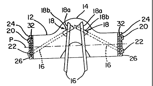
Figures 1A, 1B and 1C are top plan and cross-
sectional views, respectively, of heart valve 10 in
accordance with the invention with the suture cuff not
illustrated. Valve 10 includes a generally ring-shaped
orifice support housing (also referred to as an orifice,
orifice ring or orifice body) 12 forming a lumen 17 and
having pivot guards 14. Pivot guards 14 include
CA 02223160 1997-12-02
WO 96/40010 PCT/US96/09626
-10-
occluder mated spherical pivots 18 having opening stop
18a and closing stop 18b for occluders or leaflets 16.
In Figure 1A, leaflets 16 are shown in an open position
while in Figure 1B leaflets 16 are shown in the open
position and in the closed position in phantom.
As shown in Figure 1B, orifice body 12
includes generally circumferential body protrusions (or
rims) 20 and 22. Protrusions 20 and 22 are spaced apart
from either annulus of the orifice and toward a central
plane P of orifice 12 to provide thin projections or lip
portions 24 and 26. Lips 24 and 26 provide an
engagement surface for the tissue annulus of a heart.
For aortic and mitral replacement valves, respectively,
the peripheries of lips 24 and 26 are tissue impingement
barriers. Lips 26 and 24 serve as barriers to tissue
encroachment into the valve lumen from the tissue which
grows into the suture cuff. A sewing ring or suture
cuff 50 (shown in Figures 2A and 2B) is attached between
rims 20 and 22.
Generally, in preferred embodiments described
herein, the orifice may consist of a pyrolytic carbon
coating 30 which is deposited onto a graphite substrate
32 by a chemical vapor deposition (CVD) process.
Turning now to Figures 2A and 2B, aortic
implantation of heart valve 10 in heart 40 is shown in
cross section. Figure 2B is similar to Figure 2A except
valve 10 is rotated 90 . Heart 40 includes aorta 42,
left ventricle 44 and coronary ostium 46. Valve 10 is
shown positioned in heart tissue annulus 48. Valve 10
includes an inflow annulus 27 and an outflow annulus 29.
Lip 24 is adapted to receive tissue annulus 48 between
rim 20 and the inflow annulus 27 of orifice 12 proximate
the left ventricle 44. Figures 2A and 2B also show
suture cuff 50 secured between rims 20 and 22. Suture
CA 02223160 1997-12-02
WO 96/40010 PCTIUS96/09626
-11-
cuff 50 is used to suture valve 10 to heart tissue,
thereby securing valve 10 in position as shown in
Figures 2A and 2B and preventing perivalvular leakage.
As shown in Figures 2A and 2B, lips 24 and 26
act as tissue impingement barriers to prevent ingrowth
of heart tissue into orifice 12. Lip 24 provides an
orifice annulus for engagement or apposition with the
tissue annulus 48 of heart 40. The upstream 51 and
downstream 53 planes of sewing cuff 50 are generally
located within the confines of rims 20 and 22. Cuff 50
and rims 20 and 22 are entirely supra-annular in
implanted valve 10. Lip 24 provides an extension of the
orifice 12 into the plane of the tissue annulus 48. The
outside diameter of orifice 12 at lip 24 generally
conforms to the inside diameter of tissue annulus 48.
Additionally, a portion of lip 24 is intra-annular with
pivot guards 14 extending subannularly. The intra-
annular projection of lip 24 reduces the probability of
overgrowth of tissue from tissue annulus 48 into the
valve lumen. This is advantageous since such tissue
overgrowth tends to reduce the lumen area, disturbs the
flow and may encroach on the valve mechanism, reducing
the effectiveness of the heart valve. The subannular
extension of pivot guards 14 reduces the height of
orifice 12 protruding into the aortic root thereby
reducing the likelihood of blockage of coronary ostium
46. Lip 26 may be used to reduce tissue growth
progressing from the cuff 50 onto the outflow annulus 29
and into the valve lumen 17.
For the mitral position, lip 26 is positioned
intra-annularly, and lip 24 and pivot guard 14 are
positioned supra-annularly. Lip 24 may be used to
reduce the probability of tissue growth progressing from
CA 02223160 1997-12-02
WO 96/40010 PCT/US96/09626
-12-
cuff 50 onto the inflow annulus 27 and into the valve
lumen 17.
Figure 3 shows a cross-sectional view of a
portion of a valve 100 in accordance with a second
embodiment. Valve 100 includes orifice housing 102,
including single protrusion rim 104. Single rim 104 is
positioned proximate plane P through the approximate
center of orifice 102. Tissue impingement barrier lips
106 and 108 are formed on either side of rim 104 between
rim 104 and the ends of orifice 102. A suture cuff 110
(shown in Figure 4) is attached to rim 104.
Figure 4 is a cross-sectional view of valve
100 implanted in heart 40. Numbering of similar
elements in valve 100 is consistent with those elements
in valve 10. In Figure 4, valve 100 includes suture
cuff 110 which is used by a surgeon to suture valve 100
to tissue of heart 40. As shown in Figure 4, the
seating and engagement of valve 100 in tissue annulus 48
is similar to that of valve 10 shown in Figures 2A and
2B. Cuff 110 and the majority of orifice 102 is supra-
annular. For aortic implantation, tissue impingement
barrier lip 106 is intra-annular while pivot guards 14
extend subannular.
Figure 5 is a cross-sectional view of valve
10, as shown in Figures 1A, 1B, 1C, 2A and 2B, which
shows attachment of suture cuff 50 to orifice 12. A
metal, polymer or other biocompatible material
attachment ring 120 fits between rims 20 and 22 and
pinches or clamps cuff 50. Cuff 50 comprises, for
example, a polyester or PTFE knit or a PTFE felt, or
other soft, conformable material known in the art.
Figure 5 shows the initiation of tissue ingrowth 122
into cuff 50 from the heart tissue adjacent tissue
annulus 48. Assembly of the suture cuff to the orifice
CA 02223160 1997-12-02
WO 96/40010 PCT/US96/09626
-13-
may be through any appropriate technique known in the
art. In one embodiment, ring 120 is initially in a
flattened condition such that the tips of the "U" shape
are spread apart. Ring 120 is placed between rims 20
and 22 using a relatively uniform expansion technique in
which ring 120 is slid over a conical mandril (not
shown) and over one of the two rims 20,22 until it is
positioned as shown in Figure 5. Ring 120 is a
stiffener for the orifice and can be used to attach the
cuff in a rotatable manner Cuff 50 is placed around the
outer circumference of ring 120 and the sides of ring
120 are bent as shown in Figure 5. Friction between cuff
50 and ring 120 maintains cuff 50 in position.
Additionally, sutures, staples, pins, adhesives or other
such device or material may be used to adhere cuff 50 to
ring 120 or directly to orifice 12.
Figure 6 is a cross-sectional view of a
portion of valve 100 shown in Figures 3 and 4, providing
a detailed view showing attachment of suture cuff 110 to
orifice 102 at rim 104. A metal, polymer or other
biocompatible material attachment ring 130 is attached
to cuff 110 and crimped around and onto rim 104. Prior
to attachment, ring 130 lies relatively flat. Scoring
132 is provided on ring 130 to promote bending of ring
130 at the desired locations. Ring 130 is crimped by
applying pressure to opposing sides of ring 130 such
that ring 130 bends at scoring points 132.
Figure 7 is a cross-sectional view showing
orifice 12 having rim 220 forming tissue impingement
barriers 224 and 226. Rim 220 includes groove 230
formed therein. A mating key or rim 240 of cuff
retaining ring 250 engages mating groove 230 of orifice
12. Rim 220 of orifice 12 is of sufficient thickness to
form groove 230 therein without deleteriously decreasing
CA 02223160 1997-12-02
WO 96/40010 PCT/US96/09626
-14-
the strength of orifice 12. Cuff 260 is captured in
ring 250.
Figure 8 is a cross-sectional view of orifice
12 having a cuff retention mechanism in accordance with
another embodiment in which a projection from the cuff
retention mechanism ring itself forms a tissue
impingement barrier and inflow or outflow annuli. Cuff
350 is retained between rims 340 and 345 of ring 360.
Tissue impingement barriers 324 and 326 are formed
between extensions of ring 360. Ring 360 comprises a
biocompatible metal such as titanium or cobalt-chrome
alloy and extends past the valve housing so as to serve
as the tissue impingement barrier. Cuff 350 may be
retained by suture 355 wrapped around the annulus formed
between rims 340 and 345. Radially inward extensions
313 capture orifice 12.
Figure 9 is a cross-sectional view of orifice
102 attached to cuff 150 in accordance with another
embodiment. A spring clip ring 152 extends around the
outer circumference of orifice 102 and grasps rim 104.
Preferably, cuff 150 is formed around spring clip ring
152. The cuff clip assembly is snapped onto valve rim
104. Alternatively, ring 152 includes tips 154 which
clamp the fabric of suture cuff 150.
Figure 10 is a cross-sectional view of orifice
102 attached to suture cuff 160 in accordance with
another embodiment. Attachment mechanism 162 includes
disks 164 which extend around the outer circumference of
orifice 102. Disks 164 are connected together by band
166 which provides a friction fit with rim 104 of
orifice 102. Sewing cuff 160 is secured to band 166
between disks 164 by suture windings 168. In
alternative embodiments, disks 164 and band 166 can be
formed integrally as a single piece or separately and
CA 02223160 1997-12-02
WO 96/40010 PCT/US96/09626
-15-
attached together. This may be through the use of a
biocompatible adhesive, or similar material, or a
friction fit between protrusions from band 166 and
openings in disks 164.
In prior art, the stiffness of the orifice has
typically been increased by increasing the area of the
orifice wall section, which for a given tissue annulus
diameter reduces the area of the lumen. One aspect of
this invention includes providing the orifice stiffness
for a given tissue annulus diameter without reducing
lumen area. In one or more embodiments of the current
invention the stiffness of the orifice is enhanced by
rims projecting from the orifice. It has been
discovered and demonstrated that the size, shape and
placement of the rims enhance the stiffness.
Figure 11 shows a cross-sectional view of a
heart valve prosthesis orifice 480 in the aortic
position in accordance with another embodiment which
includes housing 482 and pivot guard 484 which carries
a pivot 486. Housing 482 is formed on substrate 485.
Rims 488 and 490 extend around the outer circumference
of housing 482 and form outflow proximal implant lip 492
and inflow distal lip 494. A middle surface 496 is
formed between rims 488 and 490. A suture cuff 498 fits
between rims 488 and 490 around middle surface 496 and
is used to attach heart valve orifice 480 to heart
tissue annulus 500. The size of orifice 480 is selected
such that tissue annulus 500 substantially conforms to
the diameter of distal lip 494. However, the majority
of orifice 480 and suture cuff 498 are positioned supra-
annular relative to tissue annulus 500.
Rims 488 and 490 have a radial height h which
is greater than that of typical prior art designs. In
a preferred embodiment, h is greater than about 0.25 mm
CA 02223160 1997-12-02
WO 96/40010 PCT/US96/09626
-16-
and is preferably about 1 mm. It has been discovered
that by increasing the dimension h, additional stiffness
is provided to housing 482. Additionally, the increase
in the h dimension of rims 488 and 490 protects the cuff
retention mechanism 499 of suture cuff 498. In one
embodiment, retention mechanism 499 comprises sutures.
However, any mechanism may be used such as a polymer or
metal band or a ring. In one or more embodiments,
retention mechanism 499 allows rotation of valve housing
482 relative to cuff 498 during the implantation
procedure. The additional protection provided by rims
488 and 490 to the retention mechanism 499 helps reduce
application of excessive pressures to mechanism 499 such
as pressure from tissue annulus 500. Such excessive
pressures tend to change the amount of torque required
to rotate housing 482 relative to cuff 498.
Furthermore, the increased height h of rims 488, 490
further reduce the likelihood of tissue ingrowth from
tissue annulus 500 into the lumen 497 of orifice 480.
Further still, the increased height h of rims 488,490
increases the ability to retain the suture cuff 498
between rims 488, 490.
Figure 12 is a cross-sectional view of another
embodiment of heart valve prosthesis orifice 510 adapted
for aortic implantation having housing 512. Housing 512
includes pivot guard 514 and pivot 516 formed therein.
Distal rim 518 and proximal rim 520 extend around the
outer circumference of housing 512 and form middle
section 522 therebetween. Rims 518 and 520 are
positioned toward the proximal side of prosthesis 510
and rim 518 forms distal lip 524 around the outer
circumference of housing 512. It has been discovered
that the offset configuration of rims 518 and 520
relative to housing 512 provides additional stiffness
CA 02223160 2006-09-25
-17-
for a given lumen. This allows the interior lumen of
housing 512 to be increased for a given stiffness.
Therefore, the lumen area is increased while providing
orifice stiffness. Furthermore, the configuration shown
in Figure 12 allows for greater length 1 of distal lip
524 which provides for deeper sub-annular placement aind
a larger intra-annular impingement barrier. It also
decreases the valve supra-annular profile to reduce the
potential for blockage of the coronary ostia. The
design shown in Figure 12 also includes an increased rim
height h as described above for the embodiment of Figure
11.
Figures 13A and 13B show orifices 610 and 510,
respectively. Orifice 610 is an embodiment adapted for
implant in the mitral position, with pivot guards 620
supra-annular (in the left atrium) and orifice 510 is an
embodiment adapted for implant in the aortic position,
with pivot guards 520 subannular (in the left
ventricular outflow tract) . Orifice 610 is shown acted
upon by hypothetical force F generated by the mitral
valve tissue annulus. Orifice 510 is shown acted upon
by hypothetical force G generated by tissues within the
left ventricular outflow tract below the aortic annulus. DD
refers to deflection. Figures 14A and 14B are cross-sectional
views of heart valve prostheses 480 and 510, respectively.
In Figs 14A and 14B, CC refers to centroid. Figures,
13A, 13B, 14A and 14B are provided to illustrate the.
relationship between the placement of the rims and the
stiffness of the prosthesis orifice. A comparison of
the stiffness of valves 510 and 480 follows.
The stiffness, or ability of the housing to
resist loading, is dependent on the orifice geometry and
material elastic modulus. The present invention
provides a technique for increasing stiffness for a
given material. The method increases resistance to both
CA 02223160 1997-12-02
WO 96/40010 PCT/US96/09626
-18-
translational and torsional loads on the orifice and to
combinations thereof. The geometric parameter that is
used to analyze and determine stiffness is the area
moment of inertia which, for a given material, is
directly proportional to stiffness. There are three
area moments of inertia associated with an area, Ix, Iy
and Jo (polar moment of inertia). The I moments are
each associated with an axis in the plane of the area,
such as x and y in Figures 14A and 14B, and the polar
moment of inertia Jo is associated with rotation, and
therefore, an axis perpendicular to the plane.
The polar moment of inertia of the area is the
simple algebraic sum:
Jo = Ix + Iy Eq. 1
Thus, if either Ix or Iy is increased, the ability of
the structure to resist rotation is increased.
Other important rules of area moments of inertia are:
The Additive rule:
For the orifice body, Ix = Ixl + Ixz + Ix3 where Ixi is
the moment of inertia of area i (where i = 1,2,3) with
respect to the x axis of the entire system.
The Parallel axis theorem:
Ixi = Ixilocal + AiDi2 where Ixilocal is the moment of inertia
of area i with respect to its centroid and AiDiZ is the
transformation for the offset in the area's axis with
respect to the system's axis. Quantity D. is the
distance from the local area's x axis and the system's
x axis and A. is the area of the local element.
Furthermore, for a rectangle Ilocal = (Width x
Height3)/12. Height and Width are relative to the axis
of the moment, i.e., the width for Iy is the height for
Ix. Equations of the same form are also true for Iy.
CA 02223160 1997-12-02
WO 96/40010 PCT/US96/09626
-19-
The difference between the orifice 480 and 510
is the distance "v" shown in Figures 14A and 14B,
respectively. For the purpose of this explanation, all
lettered dimensions are the same in orifice 480, 510,
and orifices 480, 510 are made from the same material.
This implies that the local I moments are equal for both
designs since the heights and widths of the areas do not
change. The only portion of the I moments that change
is the parallel axis portion AD2, specifically the D.
It can be seen that the Dy of the system remains
unchanged as dimension v is changed. Therefore, the
Iy's are equal for both designs. The parallel axis
portions (D;Z) of Ix changes as area A, is shifted
downward. One aspect of the invention for one or more
embodiments moves structure away from the neutral axis.
The change is described mathematically as follows:
Parallel axis theorem portion:
For the embodiment of Figure 14A, valve 480, a prime
sign ' will be used. Due to symmetry about the x axis,
D', = 0, D'Z = (z+H2)/2, and D'3 =-(z+HZ)/2
For valve 480 shown in Figure 14A:
Ix~ E Ixilocal +AiD12 + A2Dz2 + A3D32 -
I/xiIocal +A2((z+HY2)/2)2+A3 (-(2+H2)/2)2 Eq. 2
For Ix of valve 510 shown in Figure 14B:
The neutral axis of the valve 510 is shifted downward
and is assumed to be at the midpoint between the rims.
The difference in stiffness is defined below.
For any given material the difference in stiffness is
proportional to the differences in area moments of
inertia.
Area stiffness is proportional to Ix - IX'. Therefore,
if Ix > I,t' , then the design is stiffer.
CA 02223160 1997-12-02
WO 96/40010 PCT/US96/09626
-20-
Given:
Ii xilocal - Ixilocal Eq. 3
Therefore:
IX- Ilx=AlDi +AZD2 +A3D3 -
[A1D~1 + A2DI2 + A3D/3 ] Eq . 4
Since area A3 is shifted downward the centroid will also
be shifted downward thus causing D, ;d 0. Therefore;
A1Di > AD12 = 0 Eq. 5
D/2 - D/2 -( z+H3 ) 2 2 Eq. 6
2 2 z+v +H3 Z
Dz p D3 2 ) Eq. 7
Therefore:
A2D2 > A2D/2 and A3D3 > A3D/3 Eq. 8
From this it can be seen that:
Ix - I'x = [A1Di - A1D/i] +
[A2D2 - A2D/2 ] + [A3D3 - A3D/3 ] > 0 Eq. 9
The difference is positive therefore the area moment of
inertia and stiffness are greater for the aortic or
mitral specific design. The analytic derivation of the
centroids and offsets have not been shown. However, one
skilled in the art could derive these equations.
CA 02223160 1997-12-02
WO 96/40010 PCT/US96/09626
-21-
Figure 15A is a perspective view and Figures
15B and 15C are side plan views of a heart valve orifice
540 in an aortic position in accordance with another
embodiment. Orifice 540 includes housing 542, pivot
guards 544, distal rim 546 and proximal rim 548.
Proximal rim 548 is positioned similar to proximal rim
520 shown in the embodiment of orifice 510. However,
distal rim 546 has two segments, 546A and 546B. Figures
15B and 15C show a retention ring 550 which may be used
to attach, for example, a suture cuff to orifice 540.
It has been discovered that it is desirable in some
instances for ring 550 to be a continuous member.
However, if ring 550 is continuous it must be stretched
over a rim of a typical prior art prosthesis. Such a
stretchable ring may perform poorly in retaining the
cuff to the orifice. In contrast, ring 550 is a
continuous stiff ring and is placed over rim 546 by
placing ring 550 at an angle to the axis of prosthesis
540 as shown in Figure 15B. As shown in Figure 15B,
first one side of ring 550 is slipped over segment 546B
of rim 546 and then the other side of ring 550 is
slipped over segment 546A as shown in Figure 15B.
In the embodiment of prosthesis 540, proximal
rim 548 is offset similar to Figure 12 to provide the
increased stiffness as discussed above. Furthermore,
segments 546A and 546B are positioned between pivot
guards 544 to increase the stiffness in the relatively
compliant portion of housing 542. Specifically, pivot
guards 544 provide stiffness to housing 542 and segments
546A and 546B are positioned between pivot guards 544 to
provide additional stiffness in this region of housing
542. Furthermore, the proximal rim 548 of orifice 540
can resist the load of the aortic blood pressure applied
to the closed valve. Proximal rim 548 provides additional
CA 02223160 1997-12-02
WO 96/40010 PCT/US96/09626
-22-
stiffness to the proximal side of orifice 540 to
accommodate these loads during implantation. Distal lip
554 has an enlarged length 1 (see Figure 15C) to provide
a better interface with the heart tissue annulus,
similar to that discussed with respect to Figure 12.
Figure 16 is a cross-sectional view of a heart
valve prosthesis 560 in an aortic position in accordance
with another embodiment which includes housing 562 and
pivot guard 564. Proximal rim 566 and distal rim 568
extend around the circumference of housing 562 and form
a V-shaped groove 570 therebetween. Rims 566 and 568
are offset in a proximal direction with respect to the
surgeon in a typical surgical approach to provide distal
lip 572. Rims 566 and 568 extend over a relatively
large area of the outer circumference of housing 562 and
provide a slope to groove 570 which carries retention
mechanism 574. This is in contrast with a typical rim
in which there is a step thickness differential such as
in Figure 1B. Retention mechanism 574 may be any
appropriate element to couple a suture cuff to groove
570 such as a V-shaped compliant or expandable ring,
such as a spring ring.
One aspect of the invention provides an
increase in the effective orifice area of the orifice
relative to the available tissue annulus 48 area of
heart 40. As discussed above, a small prosthetic valve
lumen in the aortic position results in high systolic
transvalvular pressure gradients which excessively
burden the left ventricle. Furthermore, a small lumen
has been related to thrombus and thromboembolism
formation. Factors relating to increased risk of
thrombus and thromboembolism include the non-
physiological surfaces and blood flows introduced by
mechanical valves. Additionally, a small lumen results
CA 02223160 1997-12-02
WO 96/40010 PCT/US96/09626
-23-
in increased shear stress due to higher mean velocity in
the blood flow. An increase in lumen area as set forth
herein provides reduced transvalvular pressure gradients
and reduced mean velocity and thereby reduced shear
stress, and therefore a reduction in the potential
formation of thrombus and thromboembolism. This is
achieved by providing a valve orifice 12 with an inner
lumen diameter (d2 in Figure 1A) of a generally
cylindrical interior bounded by two generally planar
segments proximate pivot guards 14 which are generally
perpendicular to the axis of rotation of leaflets 16.
In one embodiment, the distance d, between the lumenal
planes of pivot guards 14 is not less than about 85% of
diameter d2 shown in Figure 1A. Diameter d2 is not less
than about 85% of tissue annulus diameter d3. Diameter
d3 is the diameter to the outer edge of orifice 12 but
does not include the outer diameter of rims 20 or 22.
These dimensional relationship provide increased lumen
area. However, as the relative thickness of the heart
valve orifice 12 is reduced, the stiffness of valve
orifice 12 decreases. One aspect of the invention
includes stiffening the orifice with rims as shown in
Figures 1, 3, 5, 6, 7, 8, 9, 10, 11, 12, 13, 15 and 16.
It is within the contemplation of this invention to use
a plurality of such rims or protrusions. The additional
stiffness provided by at least one rim supplements any
reduction in orifice housing stiffness which otherwise
could occur due to the thin section.
Rings 120,130,152,162,250,360,550,574 shown in
Figures 5, 6, 7, 8, 9, 10, 15 and 16 provide additional
stiffness which also allows increased lumen area. The
rings 120,130, 152,162,250,360,550,574 may be channeled
beam shapes, such as I, V, U or H configurations, which
are known in the art to provide additional stiffness.
CA 02223160 1997-12-02
WO 96/40010 PCT/US96/09626
-24-
Rings 120,130,152,162,360,550,574 extend the width of
the suture cuff to provide easier stitching during
implantation and help to prevent perivalvular leakage.
Another advantage of the retention rings described
herein is that they are easily assembled with a heart
valve. The attachment rings are well suited for an
orifice having a reduced thickness and made of
relatively low elastic modulus materials such as CVD
pyrolytic carbon. Rings 120,130,152,162,550,574 are
adapted for mechanization or automation of the assembly
process. Furthermore, rings 120,130,152,162,550,574
allow the suture cuff to rotate relative to the orif ice .
Cuff rotation torque may be controlled by controlling
friction between the cuff attachment ring and the
orifice body. Friction can be controlled by adjusting
the crimping force of rings 120,130,152,162.
The valves set forth herein may be fabricated
with any appropriate biocompatible material. In
preferred embodiments, the orifice may be of a pyrolytic
carbon-coated graphite or other material which is
thromboresistant, durable and of sufficient strength,
stiffness and fracture resistance. The orifice may
consist of a durable, blood compatible coating or film
on a substrate. In one embodiment, the coating or film
is diamond-like carbon, and the substrate is a metal.
Suitable metal substrates include, but are not limited
to, titanium and its alloys.
The present invention provides a mechanical
heart valve for a small aortic root which significantly
reduces stenosis while inaintaining an intra-annular
barrier which blocks tissue overgrowth of the valving
mechanism and lumen. The invention is applicable and
beneficial for any size aortic root and to the mitral
position. When implanted in the aortic position, the
CA 02223160 1997-12-02
WO 96/40010 PCT/US96/09626
-25-
invention beneficially decreases the work load of the
left ventricle. Anticipated patient benefits are
increased tolerance to exercise, increased rate of
regression of left ventricular hypertrophy, and lower
incident rate of congestive heart failure. The
embodiments set forth herein provide better hemodynamics
by means of a relatively low blood flow mean velocity,
thus reducing shear stress and thereby reducing the
potential for thrombosis. The relatively low mean
velocity is attained by increasing the area of the valve
lumen. Low mean velocity also provides a decreased
occluder drag, since drag is proportional to the square
of velocity, thereby further contributing to an
increased effective orifice area. Circumferential
protrusions or rims are used for attaching the heart
valve housing to a suture cuff. Cuff retention
mechanisms set forth herein, including rims or
protrusions, and attachment rings, are provided which
increase the stiffness of the valve body and which
provide rotatable coupling. The protrusions provide
stiffness to the valve housing thereby allowing the
intra-annular and sub-annular thicknesses of the valve
housing to be reduced in order to increase the lumen
diameter. The supra-annular portion of the valve is of
sufficient thickness to provide strength and stiffness.
The various embodiments set forth herein provide
increased stiffness by selective placement of the rims;
provide increased rim height for improved cuff
retention; provide increased rim height to protect the
cuff attachment and/or rotation mechanisms placed
between the rims; provide a larger tissue impingement
barrier; reduced supra-annular height to reduce the
likelihood of interference with the coronary ostia.
CA 02223160 1997-12-02
WO 96/40010 PCT/US96/09626
-26-
Although the present invention has been
described with reference to preferred embodiments,
workers skilled in the art will recognize that changes
may be made in form and detail without departing from
the spirit and scope of the invention. For example,
although this description has been largely directed to
an aortic mechanical valve, the techniques are also
applicable to mitral mechanical heart valves.