Note: Descriptions are shown in the official language in which they were submitted.
CA 02223595 1997-12-03
WO 96/40098 PC1'/US96/10211
PREV'EN77ON AND TREATMENT OF CARDIOVASCULAR PATHOL,OC'IES WiTH TAMOXIFEN
ANALOGUES
Field of the ] nvention
This invention relates generally to the prevention and treatment of
cardiovascular pathologies. More specifically, a method for treating or
preventing atherosclerosis is provided.
Background of the Invention
Many pathological conditions have been found to be associated with
smooth muscle cell proliferation. Such conditions include restenosis,
atherosclerosis, coronary heart disease, thrombosis, myocardial infarction,
stroke, smooth muscle neoplasms such as leiomyoma and leiomyosarcoma of the
bowel and uterus, uterine fibroid or fibroma, and obliterative disease of
vascular
grafts and transplanted organs. The mechanisms of abnormal smooth muscle
cell proliferation are not yet well understood.
For example, percutaneous translunainal coronary angioplasty (PTCA) is
widely used as the primary treatment modaJity in many patients with coronary
artery disease. PTCA can relieve myocardial ischemia in patients with coronary
artery disease by reducing lumen obstruction and improving coronary flow. The
use of this surgical procedure has grown rapidly, with 39,000 procedures
performed in 1983, nearly 150,000 in 1987, 200,000 in 1988, 250,000 in 1989,
and over 500,000 PTCAs per year are estimated by 1994. Stenosis following
PTCA remains a significant problem, with from 25% to 35% of the patients
developing restenosis within 1 to 3 months. Restenosis results in significant
morbidity and mortality and frequently necessitates further interventions such
as
repeat angioplasty or coronary bypass surgery. No surgical intervention or
post-surgical treatment (to date) has provert effective in preventing
restenosis.
The processes responsible for stenosis after PTCA are not completely
understood but may result from a complex interplay among several different
biologic agents and pathways. Viewed in tiistological sections, restenotic
lesions
may have an overgrowth of smooth muscle cells in the intimal lavers of the
vessel. Several possible meclianisms for srnooth muscle cell proliferation
after
CA 02223595 1997-12-03
WO 96/40098 PCT/US96/10211
2
PTCA have been suggested. For example, Barath et al. (U. S. Patent No.
5,242,397) disclose delivering cytotoxic doses of protein kinase C inhibitors,
including tamoxifen, locally by catheter to the site of the atherosclerotic
lesion.
Compounds that reportedly suppress smooth muscle proliferation in
vitro may have undesirable pharmacological side effects when used in vivo.
Heparin is an example of one such compound, which reportedly inhibits smooth
muscle cell proliferation in vitro but when used in vivo has the potential
adverse
side effect of inhibiting coagulation. Low molecular weight fragments of
heparin, while having reduced anti-coagulant activity, have the undesirable
pharmacological property of a short pharmacological half-life. Attempts have
been made to solve such problems by using a double balloon catheter, i.e., for
regional delivery of the therapeutic agent at the angioplasty site
(e.g., U.S. Pat. No. 4,824,436), and by using biodegradable materials
impregnated with a drug, i.e., to compensate for problems of short half-life
(e.g., U.S. Pat. No. 4,929,602).
In general, atherosclerosis is a cardiovascular disease in which the vessel
wall is remodeled, compromising the lumen of the vessel. The atherosclerotic
remodeling process involves accumulation of cells, both smooth muscle cells
and monocyte/macrophage inflammatory cells, in the intima of the vessel wall.
These cells take up lipid to form a mature atherosclerotic lesion. Although
the
formation of these lesions is a chronic process, occurring over decades of an
adult human life, the majority of the morbidity associated with
atherosclerosis
occurs when a lesion ruptures, releasing thrombogenic debris that rapidly
occludes the artery. When such an acute event occurs in the coronary artery,
myocardial infarction can ensue, and in the worst case, can result in death.
The formation of the atherosclerotic lesion can be considered to occur in
five overlapping stages such as migration, lipid accumulation, recruitment of
inflammatory cells, proliferation of vascular smooth muscle cells, and
extracellular matrix deposition. Each of these processes can be shown to occur
in man and in animal models of atherosclerosis, but the relative contribution
of
each to the pathology and clinical significance of the lesion is unclear.
CA 02223595 1997-12-03
WO 96/40098 PCTIUS96110211
3
Thus, a need exists for therapeutic methods and agents to treat
cardiovascular pathologies, such as atherosclerosis and other'conditions
related
to coronary artery disease.
Summary of the Invention
A therapeutic method for preventing or treating a cardiovascular or
vascular indication characterized by a decreased lumen diameter is provided.
The method comprises administering to a mammal at risk of, or afflicted with,
said cardiovascular indication, a cytostatic dose of a therapeutic agent that
elevates the level of TGF-beta, such as a compound of formula (I)
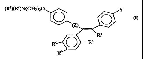
(Ri)(R2)N(CH2)20 Y
O O
R3
R O R4
wherein Z is C=0 or a covalent bond; Y is H or O(CI-Qalkyl, R' and R2 are
individually (C,-C4)alkyl or together with N are a saturated heterocyclic
group,
R3 is ethyl or chloroethyl, R4 is H, RS is I, O(C,-C4)alkyl or H and R6 is I,
O(Cj-
C4)alkyl or H with the proviso that when 1Z4, R5, and R6 are H, R3 is not
ethyl; or
a pharmaceutically acceptable salt, including mixtures thereof. The cytostatic
dose is effective to directly or indirectly i,ncrease the level of TGF-beta in
a
mammal afflicted Nvith said indication. Preferably, the effective amount
inhibits
smooth muscle cell proliferation, inhibits lipid accumulation, increases
plaque
stability, or any combination thereof. Thus, in this embodiment of the
invention,
the compound of formula (I) does not include tamoxifen, raloxifene or
droloxifene. However, in other embodiments of the invention, the compound of
CA 02223595 1997-12-03
WO 96/40098 PCT/US96/10211
4
formula(I) can include the following: R4 together with R3 is -CH2-CH2- or -S-,
RS'is OH, or R4, R5, and R6 are H and R3 is ethyl.
A therapeutic method is provided for treating or preventing
cardiovascular pathologies, such as conditions selected from the group
consisting of atherosclerosis, thrombosis, myocardial infarction, and stroke.
The method comprises the systemic or local administration of an amount of
compound of formula (I). The amount is effective to increase the level of TGF-
beta in said mammal afflicated with one of these conditions.
The administered compound of formula (1) can act on vascular smooth
muscle cells (VSMC) to inhibit the pathological activity of these smooth
muscle
cells, can inhibit the activation of endothelial cells, can inhibit lipid
accumulation by vessels, decrease lesion formation or development, and can
increase plaque stability. Preferably, the compound significantly reduces the
rate
of completion of the cell cycle and cell division, and preferably is
administered
at cytostatic, as opposed to cytotoxic, doses. A preferred embodiment of the
invention comprises treatment of atherosclerosis, wherein the compound of
formula (I), such as idoxifene or idoxifene salt, inhibits lipid accumulation
by
vascular smooth muscle cells and/or stabilizes an arterial lesion associated
with
atherosclerosis, i.e., increases plaque stability, to prevent rupture or
growth of
the lesion. As exemplified hereinbelow, orally administered tamoxifen
significantly inhibits the formation of lipid lesions, induced by a high fat
diet, in
C57B 16 mice and in the transgenic apo(a) mouse. The 90% reduction in lesion
area and number in both of these mouse models indicates that tamoxifen affects
the accumulation of lipid in the cells and stroma of the vessel wall. The
inhibition of lipid accumulation and lesion development in these treated mice
indicates that tamoxifen and analogs thereof, as well as compounds of formula
(I), may inhibit the development of atherosclerotic lesions in humans by
inhibiting lipid accumulation, in addition to decreasing smooth muscle cell
proliferation.
CA 02223595 1997-12-03
WO 96/40098 PCT/US96/10211
Other preferred embodiments of the invention comprise the local
administration of the compound of formula (I) to an arterial l'esion
associated
with atherosclerosis, and a kit to accomplish said administration.
A further embodiment of the invention is a method for preventing
5 cardiovascular pathologies in a mammal at risk of such a condition. Such
conditions include atherosclerosis, thrombosis, myocardial infarction, and
stroke. The method comprises the administration of an amount of the compound
of formula (I) to a mammal, such as a hunrian, effective to increase the level
of
TGF-beta in said mammal. The amount of the compound is administered over
time as a preventative measure. Preferably, the compound is administered
orally, in a series of spaced doses.
A further embodiment of the invention is a method for inhibiting smooth
muscle cell (SMC) proliferation associateci with procedural vascular trauma as
by the systemic or localized catheter or non-catheter administration to a
mammal, such as a human patient, subjected to said procedure, an effective
cytostatic SMC proliferation inhibitory amount of a compound of formula (I),
or
a pharmaceutically acceptable salt thereof. The systemic administration can be
accomplished by oral or parenteral administration of one of more suitable unit
dosage forms, which, as discussed below, may be formulated for sustained
release. The administration of the agents of the invention may be essentially
continuous over a preselected period of tinne or may be in a series of spaced
doses, either before, during, or after the procedural vascular trauma, before
and
during, before and after, during and after, or before, during and after the
procedural trauma.
As used herein, the term "procedural vascular trauma" includes the
effects of surgical/mechanical interventionis into mammalian vasculature, but
does not include vascular trauma due to the organic vascular pathologies
listed
hereinabove.
Thus, procedural vascular traumas within the scope of the present
treatment m--thod include (1) organ transp:lantation. such as heart, kidney,
liver
and the like. e.g., involving vessel anastoniosis; (2) vascular surgery, such
as
CA 02223595 2006-09-13
WO 96/40098 PCT/US96/10211
6
coronary bypass surgery, biopsy, heart valve replacement, atheroectomy,
throtnbectomy, and the like; (3) transcatheter vascular therapies (TVT)
including
angioplasty, e.g., laser angioplasty and PTCA procedures discussed
hereinbelow,
employing balloon catheters, and indwelling catheters; (4) vascular grafting
using natural or synthetic materials, such as in saphenous vein coronary
bypass
grafts, dacron and venous grafts used for peripheral arterial reconstruction,
etc.;
(5) placement of a mechanical shunt, such as a PTFE hemodialysis shunt used
for arteriovenous communications; and (6) placement of an intravascular stent,
which may be metallic, plastic or a biodegradable polymer. See U.S. patent
application No. 6,515,009, filed February 15, 1995.
For a general discussion of implantable
devices and biomaterials from which they can be formed, see H. Kambic et al.,
"Biomaterials in Artificial Organs", Chem. Eng. News. 30 (April 14, 1996).
In the case of organ transplantation, the entire organ, or a portion thereof,
may be infused with a solution of the compound of formula (I), prior to
implantation. Likewise, in vascular surgery, the termini of the vessels
subject to
anastomosis can be infused with the compound of formula (1), or the
antiproliferative agents can be delivered from pretreated sutures or staples.
The delivery of an agent that elevates the level of TGF-beta, e.g., TGF-
beta activators or production stimulators, to the lumen of a vessel via
catheter,
before, during or after angioplasty, is discussed in detail below. A stent or
shunt
useful in the present method can comprise a biodegradable coating or porous
non-biodegradable coating, having dispersed therein the sustained-release
dosage
form. In the alternative embodiment, a biodegradable stent or shunt may also
have the therapeutic agent impregnated therein, i.e., in the stent or shunt
matrix.
Utilization of a biodegradable stent or shunt with the therapeutic agent
irttpregnated therein is funher coated with a biodegradable coating or with a
porous non-biodegradable coating having the sustained release-dosage forcn
dispersed therein is also contemplated. This embodiment of the invention would
provide a differential rclease mte of the therapeutic agent, i.e., there would
be a
CA 02223595 1997-12-03
WO 96/40098 PCT/US96/! 02] ]
7
faster release of the therapeutic agent from t6 coating followed by delayed
rel'ease of the therapeutic agent that was irnpregnated in the stent or shunt
matrix
upon degradation of the stent or shunt matrix. The intravascular stent or
shunt
thus provides a mechanical means of maintaining or providing an increase in
luminal area of a vessel, and the antiproliferative agent inhibits the VSMC
proliferative response induced by the stent or shunt, which can cause
occlusion
of blood flow and coronary failure.
For local administration during grafting, the ex vivo infusion of the
antiproliferative agent into the excised vessels (arteries or veins) to be
used for
vascular grafts can be accomplished. In this aspect of the invention, the
vessel
that is to serve as the graft is excised or isolated and subsequently
distended by
an infusion of a solution of the therapeutic agent, preferably by pressure
infusion. Of course, grafts of synthetic fiber can be precoated with TMX
and/or
compounds of formula (I) prior to in vivo placement.
A further aspect of the invention is a method comprising inhibiting
vascular smooth muscle cell proliferation associated with procedural vascular
trauma due to organ transplantation, vascular surgery, angioplasty, shunt
placement, stent placement or vascular grafting comprising administration to a
mammal, such as a human, subjected to said procedural trauma an effective
antiproliferative amount of a compound of formula (I) or a pharmaceutically
acceptable salt thereof. Administration may be systemic, as by oral or
parenteral
administration, or local, as to the site of the. vascular trauma, or both.
Yet a further aspect of the invention provides a method comprising
inhibiting non-aortal vascular smooth muscle cell proliferation associated
with
procedural vascular trauma comprising adininistering an effective cytostatic
antiproliferative amount of tamoxifen, a structural analog thereof, a compound
of
formula (1) which includes when R4 together with R3 is -CH,-CH,- or -S-, or RS
is OH, including the pharmaceutically acceptable salts thereof, to a mammal,
such as a human, subjected to said procedural vascular trauma. Said
administration can be svstemic or by local,, catheter or non-catheter delivery
to
CA 02223595 1997-12-03
WO 96/40098 PCT/US96/10211
8
the site of the trauma. A preferred embodiment of the invention comprises
inhibiting non-aortal vascular smooth muscle cells in a non-cbronary artery.
Also provided is a kit comprising packing material enclosing, separately
packaged, a catheter, a stent, a shunt or a synthetic graft and a unit dosage
form
of an amount of a compound of formula (I) and/or tamoxifen effective to
accomplish these therapeutic results when delivered locally, as well as
instruction means for its use, in accord with the present methods.
Another embodiment of the present invention is a method for identifying
an agent which increases the level of TGF-beta, e.g., the agent is a TGF-beta
activator or production stimulator. Human vascular smooth muscle cells
(hVSMC) are cultured with an amount of the agent effective to reduce or
inhibit
the rate of hVSMC proliferation. The hVSMC are then contacted with an
amount of a moiety which specifically binds to TGF-beta in an amount effective
to block the binding of TGF-beta to the TGF-beta receptors of said hVSMC and
then the rate of proliferation is determined. The method can also include the
culture of rat aortic vascular smooth muscle cells (rVSMC) with an amount of
the same agent effective to reduce or inhibit the rate of proliferation of
rVSMC.
The rVSMC are then contacted with an amount of a moiety which specifically
binds to TGF-beta in an amount effective to block the binding of TGF-beta to
the
TGF-beta receptors of said hVSMC and then the rate of proliferation is
determined. The rate of proliferation in treated rVSMC and treated hVSMC
relative to untreated rVSMC and hVSMC, respectively, after contact with the
moiety indicates that the reduction of proliferation is due to an increase in
the
level of TGF-beta in rVSMC and hVSMC by said agent, and suggests that
rVSMC and hVSMC would be amenable to treatment by the administration of
said agent in vivo.
Agents useful in the practice of the invention include agents that elevate or
increase the level of TGF-beta, e.g., TGF-beta activators and TGF-beta
production stimulators, compounds of formula (I) which include when R' 30
together with R' is -CH.-CH2- or -S-, or RS is OI-i, tamoxifen. and structural
analogs of tamoxifen. These agents and compounds, including their salts and
CA 02223595 2006-09-13
9
mixtures thereof, may be employed in the practice of the present invention to
prevent or treat other conditions characterized by inappropriate or
pathological
activity of vascular smooth muscle cells or endothelial cells, excluding the
inappropriate proliferation or pathological activity of neoplastic vascular
smooth
muscle cells or neoplastic endothelial cell.s. Thus, it is envisioned that the
methods
of the present invention preferably do not include the treatment of neoplastic
vascular tissue.
The agents of the invention, which increase the level of TGF-beta,
inhibit abnormal activity of vascular smooth muscle cells and endothelial
cells.
Preferred agents of the invention include compounds of formula (I). Preferred
compounds of formula (I) include those wherein Z is a covalent bond, Y is H,
R3
is CICH2CH2, RS or R6 is iodo, R4 is H. R' and RZ are each CH3 or together
with N
are pyrrolidino, hexamethyleneimino or piperidino. These agents or compounds
can include structural analogs of tamoxifen (including derivatives of TMY and
derivatives of said analogs) having equivalent bioactivity. Such analogs
include
idoxifene (IDX) (E-1-[4-[2-N-pyrrolidino)ethoxy]phenyl]-1-(4-iodophenyl)-2-
phenyl-l-butene), raloxifene, 3-iodotamoxifen, 4-iodotamoxifen, droloxifene,
tomremifene, and the pharmaceutically acceptable salts thereof.
Also provided are a method and a kit to determine the presence and
amount of TGF-beta in a sample containing TGF-beta. The method for the
determination of TGF-beta in vitro can be used to identify a patient at risk
for
atherosclerosis and/or monitor a recipient that has received one or more
administrations of a therapeutic agent which increases the level of TGF-beta,
or to monitor active TGF-beta levels in an individual. Blood serum or plasma
from an individual, patient or recipient is contacted with a capture moiety to
form a capture complex of said capture moiety and TGF-beta. Preferably, the
capture moiety is an immobilized capture moiety. The capture moiety may also
be a solution phase capture moiety. The capture complex is then contacted with
a detection moiety capable of binding TGF-beta comprising a detectable label.
or a binding site for a detectable label, to form a detectable complex. More
specifically, the capture moiety may be a first antibody and the detection
moiety is a second antibody. In another embodiment, the capture or the
CA 02223595 2006-09-13
detection moiety may comprise a fusion protein comprising a TGF-beta type II
extracellular domain. More specifically, the TGF-beta type II extracellular
domain may have a methionine residue at position 5. The presence and
amount, or absence, of the detectable complex is then determined, thereby
determining the presence and amount, or absence, of TGF-beta in the blood of '
the patient or recipient.
A test kit for determining TGF-beta in vitro includes packaging material
enclosing (a) a capture moiety capable of binding TGF-beta, and (b) a
detection
moiety capable of binding to TGF-beta, where the detection moiety has a
detectable label or a binding site for a detectable label. The capture moiety
and the
detection moiety are separately packaged in the test kit. The capture moiety
may be
a~irst antibody. The detection moiety may be a second antibody. Preferably,
the
capture moiety is solid substrate-immobilized. The capture moiety may also be
present in solution. Preferably, the capture moiety is the TGF-beta type II
receptor
extracellulax domain. More preferably, the TGF-beta type II receptor
extracellular
domain is derived from a bacterial expression system. The kit can also
comprise
instruction means for correlation of the detection or determination of TGF-
beta
with the identification of the patients or monitoring discussed above.
Further provided is a method for upregulating cellular mRNA coding for
TGF-beta. Cells (e.g., smooth muscle cells) amenable to such manipulation of
mRNA accumulation are identified in the manner described herein and are
exposed to an effective amount of a TGF-beta mRNA regulator (i.e., a subset of
TGF-beta production stimulators), either free or in a sustained-release dosage
form. In this manner, TGF-beta production is stimulated.
In addition, methods for using TGF-beta to maintain and increase vessel
lumen diameter in a diseased or injured mammalian vessel are described.
Also provided is a therapeutic method of increasing the level of TGF-beta
in a mammal in need thereof. The method comprises the administration of an
effective amount of a compound of formula (I), which includes when R4 together
with R3 is -CH2-CHZ or -S-, R$ is OH, or and R4, R5, and W are H and R3 is
ethyl. A preferred embodiment of the invention is a mammal that is diabetic.
Diabetics suffer from a plethora of indications, one of which is a decrease in
the
level of TGF-beta, as described hereinbelow.
CA 02223595 2006-09-13
10a)
Diabetics are prone to vascular disease. Vascular disease includes, but is
not limited to, myocardial infarction, atherosclerosis, aneriolsclerosis, and
small
vessel disease. Moreover, the leading causes of death in diabetics are
myocardial
CA 02223595 1997-12-03
WO 96/40098 PCTlUS9612 D221
11
infarction and atherosclerosis. Thus, the present invention further provides a
method to treat diabetics at risk of, or afflicted with, vascular'disease. The
method comprises the administration of ari effective amount of an agent that
elevates the level of TGF-beta, such as a compound of formula (I) which
includes when R4 together with R3 is -CH2-CH2- or -S-, or R5 is OH, tamoxifen
or a structural analog thereof. The amount is effective to directly or
indirectly
increase the level of TGF-beta in said diabetic. The amount administered is
preferably effective to inhibit the proliferation of vascular tissue. A
preferred
embodiment of the invention includes the administration of idoxifene, 3-
iodotamoxifen, 4-iodotamoxifen, raloxifene, droloxifene, toremifene, or a
pharmaceutically acceptable salt thereof.
Description of the Drawings
Figures 1 and 2 depict pathways for the modulation of vascular smooth
muscle cell proliferation in vivo.
Figure 3A depicts the reduction iin TGF-beta binding to the TGF-beta
receptor (R2X) in the presence of increasiztg amounts of lipoprotein.
Figure 3B depicts the amount of TGF-beta necessary to half maximally
inhibit mink lung cell proliferation in the presence of increasing amounts of
lipoprotein.
Figure 4 depicts the association of TGF-beta with different lipoprotein
classes. Profile of lipoprotein particle elution measured as total cholesterol
(......)
and TGF-beta elution (open circles) following separation of the lipoprotein
fraction
(d < 1.215 g/cm3) by gel filtration chromatography. The position of the major
lipoprotein classes are marked by reference to the elution times of the major
apolipoproteins. (a) Healthy individual A. (b) Healthy individual C (c)
Diabetic
individual K (d) Diabetic individual L. Le'tters designating the individuals
shown
refer to individuals in Table 8.
Detailed Description. of the Invention
As used herein the following terms have the meanings as set forth belovv:
CA 02223595 1997-12-03
WO 96/40098 PCT/US96/10211
12
"Proliferation," means an increase in cell number, i.e., by mitosis of the
cells.
"Abnormal or Pathological or Inappropriate Activity or Proliferation"
means division, growth or migration of cells occurring more rapidly or to a
significantly greater extent than typically occurs in a normally functioning
cell of
the same type, or in lesions not found in healthy tissues.
"Expressed" means mRNA transcription and translation with resultant
synthesis, glycosylation, and/or secretion of a polypeptide by a cell, e.g.,
chondroitin sulfate proteoglycan (CSPG) synthesized by a vascular smooth
muscle cell or pericyte.
"Vascular disease" includes, but is not limited to, myocardial infarction,
atherosclerosis, arteriolsclerosis, and small vessel disease. Small vessel
disease
includes, but is not limited to, silent myocardial infarction, vascular
insufficiency
in the limbs, peripheral neuropathy and retinopathy.
"Vascular tissue," as used herein, includes non-neoplastic smooth muscle
cells and non-neoplastic endothelial cells.
The term "tamoxifen", as used herein, includes trans-2-[4-(1,2-diphenyl-
1-butenyl)pher,oxy]-N,N-dimethylethylamine, and the pharmaceutically
acceptable salts thereof, which are capable of enhancing the production or
activation of TGF-beta. The activated form of TGF-beta, in turn, inhibits
endothelial cell and vascular smooth muscle cell activity. Isomers and
derivatives of the aforementioned chemical compound are also included within
the scope of the term "tamoxifen" for the purposes of this disclosure.
The term "structural analogs thereof" with respect to tamoxifen includes,
but is not limited to, all of the compounds of formula (I) which are capable
of
enhancing production or activation of TGF-beta. See, for example, U.S. Patent
Nos. 4,536.516, 5,457,113, 5.047,431, 5,441,986, 5,426,123, 5,384,332,
5.453,442, 5.492,922, 5,462,937, 5,492,926, 5,254,594, and U.K. Patent
1.064.629. 30 Because tamoxifen (TMX) causes liver carcinogenicity in rats and
has
been correlated with an increased risk of endometrial cancer in women and may
CA 02223595 1997-12-03
WO 96140095 PCTlUS96/10211
13
increase the risk of certain gut cancers, other tamoxifen analogs may be
considered safer to administer if they are less carcinogenic. The
carcinogenicity
of TMX has been attributed to the formation of covalent DNA adducts. Of the
TMX analogs and derivatives, only TMX and toremifene have been studied for
long-term carcinogenicity in rats and these studies provide strong evidence
that
covalent DNA adducts are involved in rocient hepatocarcinogenicity of TMX.
Toremifene, which exhibits only a very low level of hepatic DNA adducts, was
found to be non-carcinogenic. See Potter et al., Carcino eg nesis. 11439
(1994).
A preferred embodiment of the invention includes the use of a compound of
formula (I) which includes when R4 together with R3 is -CH2-CH2- or -S-, or RS
is OH, that forms DNA adducts at a reduced level relative to TMX. A more
preferred embodiment of the invention includes a compound of formula (I)
which includes when R4 together with R3 is -CH7-CH2- or -S-, or RS is OH, that
does not form DNA adducts. The extent of DNA adduct formation by an agent
or a compound is determined by methods well known to the art.
It is postulated that 4-hydroxylation of TMX yields electrophilic
alkylating agents which alkylate DNA through the ethyl group of TMX. This
mechanistic hypothesis explains the low level of DNA adduct formation by the
non-TMX analogs of formula (1), includitig the TMX analog toremifene and the
absence of DNA adducts detected for the analogs 4-iodotamoxifen and
idoxifene. Thus, all of these analogs are likely to be free from the risk of
carcinogenesis in long term use. See Potter et al., supra. Idoxifene includes
(E)-
1-[4-[2-(N-pyrrolidino)ethoxy]phenyl]-1-(4-iodophenyl)-2-phenyl-l-butene and
its pharmaceutically acceptable salts and derivatives. See R. McCague et al.,
Ofganic Preparations and Procedures Int., a, 343 (1994) and S.K. Chandler et
al., Cancer Res., 51, 5 851 (1991). Beside-s its lower potential for inducing
carcinogenesis via formation of DNA adducts which can damage DNA, other
advantages of IDX compared with TMX are that IDX has reduced residual
estrogenic activity in rats and an improved metabolic profile. Thus, another
preferred embodiment of the inventior. includes the use of a compound of
formula (1) which includes when R' togcther ~%ith R3 is -CH,-CH:- or -S-, or
R'
CA 02223595 1997-12-03
WO 96/40098 PCT/US96/10211
14
is OH, that has reduced, or no, estrogenic activity. The estrogenic activity
of an
agent or a compound of formula (I) can be determined by methods well known
to the art. A more preferred embodiment of the invention includes the use of a
compound of formula (I) which includes when R4 together with R3 is -CH2-CH2-
or -S-, or RS is OH, that forms DNA adducts at low frequency, or preferably
not
at all, and has low, or preferably no, estrogenic activity. IDX is a preferred
embodiment of the present invention.
Also included within the scope of the term tamoxifen are the TMX
structural analogs toremifene and raloxifene, metabolites or pharmaceutically
acceptable salts thereof. Other "antisteroids" or "steroidal antagonists" can
also
be useful as agents that increase the level of TGF-beta or lead compounds,
including other known stilbene-type antisteroids including cis- and trans-
clomiphene, droloxifene, (1-[4-(2-dimethylaminoethoxy)phenyl]-1-(3-
hydroxyphenyl)-2-phenyl-2-butene (see U.S. Patent No. 5,384,332), 1-nitro-l-
phenyl-2-(4-hydroxyphenyl or anisyl)-2-[4-(2-pyrrol-N-ylethoxy)-
phenyl]ethylene(CN-55,945),trans-1,2-dimethyl-l,2-(4-
hydroxyphenyl)ethylene(trans-dimethylstilboestrol), trans-diethylstilboestrol,
and 1-nitro-l-phenyl-2-(4-hydroxyphenyl)-2-[4-(3-
dimethylaminopropyloxy)phenyl-ethylene (G1680).
Known 1,2-diphenylethane-type antisteroids include cis- 1,2-anisyl- 1 -[4-
(2-diethylaminoethoxy)phenyl] ethane (MRL-37), 1-(4-chlorophenyl)1-[4-(2-
diethylaminoethoxy)phenyl]-2-phenylethanol (WSM-4613); 1-phenyl-1 [4-(2-
diethylaminoethoxy)phenyl]-2-anisylethanol (MER-25); 1-phenyl-l-[4-(2-
diethylaminoethoxy)phenyl)-2-anisyl-ethane, mesobutoestrol (trans-1,2-
dimethyl-1,2-(4-hydroxyphenyl)-ethane), meso-hexestrol, (+)hexestrol and (-)-
hexestrol.
Known naphthalene-type antisteroids include nafoxidine, 1-[4-(2,3-
dihydroxypropoxy)phenyl ]-2-phenyl-6-hydroxy-1,2,3,4-tetrahydro-naphthalene,
1-(4-hydroxyphenyl)-2-phenyl-6-hydroxy-1,2,3,4-tetrahydronaphthalene, 1-[4-
(2-pyrrol-N-ylethoxy)-phenyl]-2-phenyl-6-methoxy-3,4-dihydronaphthalene
(U 11, 100A). and 1-
_
CA 02223595 1997-12-03
WO 96/40098 PCT/US96120221
[4-(2,3-dihydroxypropoxy)phenyl]-2-phenyl-6-methoxy-3,4-dihydronaphthalene
(U-23, 469).
Known antisteroids which do not fall anywhere within these structural
classifications include coumetstrol, biochanin-A, genistein, methallenstril,
5 phenocyctin, and 1-[4-(2-dimethylaminoethoxy)phenyl]-2-phenyl-5-
methoxyindene (U, 11555). In the nomenclature employed hereinabove, the
term "anisyl" is intended to refer to a 4-methoxyphenyl group.
The pharmaceutically acceptable inorganic and organic acid amine salts
of the amino group-containing antisteroids are also included within the scope
of
10 the term "antisteroid", as used herein, and include citrates, tartrates,
acetates,
hydrochlorides, hydrosulfates and the like.
"TGF-beta" includes transforming growth factor-beta as well as
functional equivalents, derivatives and analogs thereof. The TGF-beta isoforms
are a family of multifunctional, disulfide-linked dimeric polypeptides that
affect
15 activity, proliferation and differentiation of various cells types. TGF-
beta is a
polypeptide produced in a latent propeptide form having, at this time, no
identified biological activity. To be rendered active and, therefore, capable
of
inhibiting vascular smooth muscle cell proliferation, the propeptide form of
TGF-beta must be cleaved to yield active 7'GF-beta.
"TGF-beta activator" includes moieties capable of directly or indirectly
activating the latent form of TGF-beta to the active form thereof. A number of
the compounds of formula (I) are believed to be TGF-beta activators.
"TGF-beta production stimulator" includes moieties capable of directly
or indirectly stimulating the production of TGF-beta (generally the latent
form
thereof). Such TGF-beta production stimulators may be TGF-beta mRNA
regulators (ijr,, moieties that increase the production of TGF-beta mRNA),
enhancers of TGF-beta mRNA expression or the like.
"Direct" action includes, but is not limited to. an agent which acts to
increase active TGF-beta levels. For exarnple. direct action indicates that
cells
upon which the agent acts increase TGF-beta mRNA production increase the
CA 02223595 1997-12-03
WO 96/40098 PCT/US96/10211
16
cleavage of latent TGF-beta, or that the agent increases the level of TGF-beta
which is capable of binding to its receptor.
"Indirect" action of an agent includes, but is not limited to, an agent of
the invention that acts through one or more other moieties acts to increase
the
level of active TGF-beta. For example, an agent that acts through one or more
other moieties to release TGF-beta from complexes that inhibit or prevent the
binding of active TGF-beta to its receptor, or an agent that acts through one
or
more other moieties to stimulate the production of TGF-beta mRNA or the
expression of TGF-beta, acts indirectly.
"Sustained release" means a dosage form designed to release a
therapeutic agent therefrom for a time period ranging from about 3 to about 21
days. Release over a longer time period is also contemplated as a "sustained
release" dosage form of the present invention.
For the purposes of this description, the prototypical cells, upon which
the effects of an agent that increases the level of TGF-beta are felt, are
smooth
muscle cells, endothelial cells and pericytes derived from the medial layers
of
vessels which proliferate in intimal hyperplastic vascular sites following
injury,
such as that caused during PTCA. An agent that increases the level of TGF-beta
is not restricted in use for therapy following angioplasty; rather, the
usefulness
thereof will be proscribed by their ability to inhibit abnormal cellular
proliferation, for example, of smooth muscle cells, endothelial cells and
pericytes in the vascular wall. Thus, other aspects of the invention include
agents, which increase the level of TGF-beta, used in early therapeutic
intervention for reducing, delaying, or eliminating (and even reversing)
atherosclerotic plaque formation and areas of vascular wall hypertrophy and/or
hyperplasia. Agents which increase the level of TGF-beta also find utility for
early intervention in pre-atherosclerotic conditions, e.g., they are useful in
patients at a high risk of developing atherosclerosis or with signs of
hypertension
resulting from atherosclerotic changes in vessels or vessel stenosis due to
hypertrophy of the vessel wall.
CA 02223595 1997-12-03
WO 96/40098 PCT/iTS96/10211
17
Agents which increase the level of TGF-beta also find utility in the
treatment of diabetics with decreased levells of TGF-beta (as described
hereinbelow), particularly in diabetics at risk of, or afflicated with,
vascular
disease. One example of a vascular disease which afflicts certain diabetics is
diabetic retinopathy, where angiogenesis results in blindness over a 3-6 month
period.
Diabetic retinopathy is one of the most serious complications of diabetes
mellitus, and a major cause of blindness all over the world. In spite of the
wide
clinical variation of the different stages of diabetic retinopathy, there are
generally three processes that are known o:r thought to be of pathogenetic
importance. The first is usually characterized by microangiopathy, ischemia
and
hypoxia. Visible signs of this process include capillary obliteration or
nonperfusion, arteriolar-venular shunt, hyperaggregation of red cells and
platelets, sluggish blood flow and an impaired ability of red cells to release
oxygen. The second process involves abnormal metabolism of carbohydrate,
protein and arachidonic acid. The third process of diabetic retinopathy is
thought to involve lipid peroxidation of the: retinal membrane, possibly
oxygen
radical-induced. Although many of these characteristics of diabetic
retinopathy
are known, effective prevention and therapy for this disease has not been
available prior to the present invention.
Agents which increase the level of TGF-beta are useful for inhibiting the
pathological proliferation of vascular smooth muscle cells or endothelial
cells,
e.g., for reducing, delaying, or eliminating stenosis following angioplasty.
As
used herein the term "reducing" means dec;reasing the intimal thickening that
results from stimulation of smooth muscle cell proliferation following
angioplasty, either in an animal model or iri man. "Delaying" means delaying
the time until onset of visible intimal hyperplasia (e.g., observed
histologically
or by angiographic examination) following angioplasty and may also be
accompanied by "reduced" restenosis. "Eliminating" restenosis following
angioplasty means completely "reducing" iintimal thickening and/or completely
"delaying" intimal hyperplasia in a patient w an extent which makes it no
longer
CA 02223595 1997-12-03
WO 96/40098 PCTNS96/10211
18
necessary to surgically intervene, i.e., to re-establish a suitable blood flow
through the vessel by repeat angioplasty, atheroectomy, or coi=onary artery
bypass surgery. The effects of reducing, delaying, or eliminating stenosis may
be determined by methods routine to those skilled in the art including, but
not
limited to, angiography, ultrasonic evaluation, fluoroscopic imaging, fiber
optic
endoscopic examination or biopsy and histology.
The amount of an agent of the invention, i.e., one which increases the
level of TGF-beta, administered is selected to treat vascular trauma of
differing
severity, with smaller doses being sufficient to treat lesser vascular trauma
such
as in the prevention of vascular rejection following graft or transplant.
Agents
which increase the level of TGF-beta that are not characterized by an
undesirable
systemic toxicity profile at a prophylactic dose are also amenable to chronic
use
for prophylactic purposes with respect to disease states involving
proliferation of
vascular smooth muscle cells or endothelial cells over time (e.g.,
atherosclerosis,
coronary heart disease, thrombosis, myocardial infarction, stroke), preferably
via
systemic administration. The agents of the invention are not envisioned for
the
treatment of smooth muscle neoplasms such as leiomyoma and leiomyosarcoma
of the bowel and uterus, uterine fibroid or fibroma and the like.
For prevention of restenosis, a series of spaced doses, optionally, in
sustained release dosage form, is preferably administered before and after the
traumatic procedure (e.g., angioplasty). The dose may also be delivered
locally,
via catheter delivered to the afflicted vessel during the procedure. After the
traumatic procedure is conducted, a series of follow-up doses are administered
over time, preferably in a sustained release dosage form, systemically to
maintain an anti-proliferative effect for a time sufficient to substantially
reduce
the risk of or to prevent restenosis. A preferred therapeutic protocol
duration
after angioplasty for this purpose is from about 3 to about 26 weeks.
High levels of lipoprotein Lp(a) are known to constitute a substantial risk
factor for atherosclerosis, coronary heart disease and stroke. One symptom
associated with such conditions and other problems. such as restenosis
following
balloon angioplasty and other pathogenic conditions, is the proliferation or
the
CA 02223595 1997-12-03
WO 96/40098 PCT/US96/20212
19
migration of smooth muscle cells. No direct link between Lp(a) and
profiferation of vascular smooth muscle cells had been established in the
prior
art.
An in vivo pathway for the modulation of vascular smooth muscle cell
proliferation is shown in Figure 1. TGF-beta is believed to contribute to the
inhibitory mechanism that maintains vascular smooth muscle cells in a non-
proliferative state in healthy vessels.
Vascular smooth muscle cell proliferation is inhibited by an active form
of TGF-beta. Tamoxifen has been shown lby the experimentation detailed in
Example 1 hereof to stimulate both the production and the activation of TGF-
beta. Heparin stimulates the activation of TGF-beta by affecting the release
of
the active form of TGF-beta from inactive complexes present in serum. TGF-
beta neutralizing antibodies inhibit the activity of TGF-beta, thereby
facilitating
the proliferation of vascular smooth muscle cells. An apparent in vivo
physiological regulator of the activation of TGF-beta is plasmin. Plasmin is
derived from plasminogen through activation by, for example, TPA (tissue
plasminogen activator). Plasmin activity is inhibited by the lipoprotein Lp(a)
or
apolipoprotein(a) (apo(a)), thereby decreasing the activation of the latent
form of
TGF-beta and facilitating proliferation of vascular smooth muscle cells.
An additional pathway for the modulation of vascular smooth muscle cell
proliferation is shown in Figure 2. Resting smooth muscle cells constitute
cells
in their normal, quiescent non-proliferative; state. Such resting smooth
muscle
cells may be converted to proliferating smooth muscle cells through activation
by platelet derived growth factor (PDGF), fibroblast growth factor (FGF) or
other stimulatory moieties. The proliferating smooth muscle cells may be
converted to continual proliferating smooth muscle cells (j,gz, smooth muscle
cells capable of generating a pathological state resulting from over-
proliferation
thereof) by an autocrine growth factor. This growth factor is believed to be
produced by proliferating smooth muscle cells. An increased level of autocrine
growth factor, which can be inhibited by the active form of TGF-beta or an
CA 02223595 1997-12-03
WO 96/40098 PCT/US96/10211
appropriately structured (j,g-, designed) small molecule inhibitor, is
believed to
mediate the production of continual proliferating smooth muscle cells.
Lp(a) consists of low density lipoprotein (LDL) and apo(a). Apo(a)
shares approximately 80% amino acid identity with plasminogen (see MacLean
5 et al., Nature= IIQ: 132, 1987). Lp(a) has been found to inhibit cell-
associated
plasmin activity (see, for example, Harpel et al., Proc. Natl. Acad. Sci. USA,
$_E:
3847, 1989). Experiments conducted on human aortic vascular smooth muscle
cells derived from healthy transplant donor tissue, cultured in Dulbecco's
modified Eagles medium (DMEM) + 10% fetal calf serum (FCS) as described in
10 Kirschenlohr et al., Am. J. Physiol.. 261, C571 (1993), indicated the
following:
1) Addition of Lp(a) to sub-confluent human vascular smooth muscle
cells stimulated their proliferation in a dose dependent manner (addition of
500
nM Lp(a) to human vascular smooth muscle cells caused a reduction in doubling
time from 82 +/- 4 hours to 47 +/- 4 hours);
15 2) Addition of apo(a) had a similar effect, although a higher
concentration of apo(a) appeared to be required therefor;
3) Addition of LDL at varying concentrations up to 1 micromolar had no
effect on proliferation.
One possible mode of action for Lp(a) and apo(a) is competitive
20 inhibition of surface-associated plasminogen activation, which in turn
inhibits
the subsequent activation of TGF-beta by plasmin. TGF-beta is a potent growth
inhibitor of a number of anchorage-dependent cells, including smooth muscle
cells. TGF-beta is produced as a latent propeptide having a covalently linked
homodimer structure in which the active moiety is non-covalently linked to the
amino-terminal portion of the propeptide. Latent TGF-beta must be cleaved
(e.g., in vitro b), acid treatment or in vivo by the serine protease plasmin)
in order
to become capable of inhibiting the proliferation of vascular smooth muscle
cells. Plasmin is therefore a leading candidate to be a physiological
regulator of
TGF-beta.
The hypothesis that Lp(a) and apo(a) were acting on cultured human
vascular smooth muscle cells b,. interfering %%-ith activation of latent TGF-
beta
CA 02223595 1997-12-03
WO 96140098 PCT/i1S96/10211
21
was tested. In support of this hypothesis, an observation was made that
plasmin
activity associated with vascular smooth niuscle cells was reduced 7-fold by
Lp(a) and 5-fold by apo(a). The plasmin activity in the conditioned medium was
also reduced by Lp(a) and apo(a) by about 2-fold, but was much lower than cell-
associated plasmin activity in vascular smooth muscle cell cultures. These
observations are consistent with previous i:indings that Lp(a) is a more
potent
inhibitor of surface-associated, rather than fluid phase, plasminogen
activation.
To exclude the possibility that Lp(a) was affecting the synthesis of
plasminogen activators ratlier than plasminogen activation, plasminogen
activator levels in human vascular smooth muscle cell cultures were measured
in
the presence and absence of the lipoproteins and in the presence of a large
excess
of plasminogen, so that the lipoproteins present would not significantly act
as
competitive inhibitors. Total plasminogen. activator activity was not affected
by
the presence of any of the lipoproteins in the vascular smooth muscle cell
cultures. For example, plasminogen activator activity in the conditioned
medium remained at 0.7 +/- 0.6 mU/ml with Lp(a) additions up to 500 nM.
Lp(a) and apo(a) both reduced the llevel of active TGF-beta by more than
100-fold compared to control or LDL-treal:ed cultures. The level of total
latent
plus active TGF-beta measured by ELISA as described in Example 8 was
unaffected by the presence of Lp(a) or apo(a), however. These facts lead to
the
conclusion that Lp(a) stimulates proliferation of human vascular smooth muscle
cells by inhibiting plasmin activation of latent TGF-beta to active TGF-beta.
To further test this conclusion and exclude the possibility that Lp(a) was
acting by binding active TGF-beta as well as reducing plasmin activity, human
vascular smooth muscle cells were cultured in the presence of Lp(a). These
cells
had a population doubling time of 47 +/- 3 hours. Addition of plasmin was able
to overcome the population doubling time reducing effect of Lp(a) and reduce
the cell number to control levels. %vith the lpoptilation doubling time
increased to
97 +/- 4 hours.
The role of plasmin in the pathNti-ay was confirmed by studies in which
inhibitors of plasmin activitN were added to human vascular smooth muscle
CA 02223595 1997-12-03
WO 96/40098 PCT/US96/10211
22
cells. Like Lp(a), these protease inhibitors increased cell number. Aprotinin,
for
example, decreased the population doubling time from 82 +/- 4 hours in control
cultures to 48 +/- 5 hours, and alpha2-antiplasmin decreased the population
doubling time to 45 +/- 2 hours. 500 nM Lp(a) and aprotinin addition resulted
in
only a slight additional stimulation of proliferation, with the population
doubling
time for cultures of this experiment being 45 +/- 6 hours. Neutralizing
antibodies to TGF-beta similarly decreased population doubling time in
vascular
smooth muscle cells (see, for example, Example 1). In summary, Lp(a), plasmin
inhibitors and neutralizing antibody to TGF-beta stimulate proliferation of
vascular smooth muscle cells, while plasmin nullifies the growth stimulation
of
Lp(a). These results support the theory that the mode of action of Lp(a) and
apo(a) is the competitive inhibition of plasminogen activation.
Experimentation conducted to ascertain the impact of tamoxifen on TGF-
beta and vascular smooth muscle cell proliferation is set forth in detail in
Example 1. The results of those experiments are summarized below.
1) Addition of tamoxifen decreased the rate of proliferation, with
maximal inhibition observed at concentrations above 33 micromolar. 50
micromolar tamoxifen concentrations produced a cell number 96 hours
following the addition of serum that was reduced by 66% +/- 5.2% (n=3) as
compared to cells similarly treated in the absence of tamoxifen.
2) Tamoxifen did not significantly reduce the proportion of cells
completing the cell cycle and dividing. Inhibition of vascular smooth muscle
cells caused by tamoxifen therefore appears to be the result of an increase in
the
cell cycle time of nearly all (>90%) of the proliferating cells.
3) Tamoxifen decreases the rate of proliferation of serum-stimulated
vascular smooth muscle cells by increasing the time taken to traverse the G2
to
M phase of the cell cycle.
4) Tamoxifen decreased the rate of proliferation of vascular smooth
muscle cells by inducing TGF-beta activity.
5) Vascular smooth muscle cells produced TGF-beta in response to
tamoxifen. Tamoxifen appears to increase TGF-beta activity in cultures of rat
CA 02223595 1997-12-03
WO 96/40098 PCT/US96/1022 1
23
vascular smooth muscle cells by stimulatir.ig the production of latent TGF-
beta
and increasing the proportion of the total T'GF-beta which has' been
activated.
6) Tamoxifen, unlike heparin, does not act by releasing TGF-beta from
inactive complexes present in serum.
7) TGF-beta mRNA was increased by approximately 10-fold by 24
hours after addition of tamoxifen (10 micromolar). This result suggests that
the
expression of TGF-beta mRNA by the smooth muscle cells will be increased,
thereby facilitating decreased proliferation thereof by activated TGF-beta.
8) Tamoxifen is a selective inhibitor of vascular smooth muscle
proliferation with an ED50 (a concentration resulting in 50% inhibition) at
least
10-fold lower for vascular smooth muscle cells than for adventitial
fibroblasts.
Additional experimentation has shown that the addition of Lp(a) or
apo(a) substantially reduced the rat vascular smooth muscle cell proliferation
inhibitory activity of tamoxifen, with the population doubling time in the
presence of tamoxifen and Lp(a) being 42 +/- 2 hours (as compared to a
population doubling time of 55 +/- 2 hours for tamoxifen alone, and a time of
35
+/- 2 hours for the control). Also, the presence of Lp(a) reduced the levels
of
active TGF-beta produced in response to the addition of tamoxifen by about 50-
fold. Addition of plasmin to rat vascular s;mooth muscle cells treated with
tamoxifen and Lp(a) resulted in most of the TGF-beta being activated, and
proliferation was again slowed (with the population doubling time being 57 +/-
3
hours). These observations are consistent with the theory that Lp(a) acts by
inhibiting TGF-beta activation.
Identification of therapeutic agents (direct or indirect TGF-beta activators
or production stimulators) that act to inhib;it vascular smooth muscle cell
proliferation by the pathway shown in Figiire 1 can be identified by a
practitioner in the art by conducting experiments of the type described above
and
in Example 1. Such experimental protocols facilitate the identification of
therapeutic agents useful in the practice of the present invention and capable
of
one of the following activities:
1) production or activation of TGF-beta;
CA 02223595 1997-12-03
WO 96/40098 PCT/US96/10211
24
2) having TGF-beta-like activity;
3) activation of plasminogen;
4) increase in plasmin activity; or
5) reduction of Lp(a) or apo(a) level or levels of 7r-I or other inhibitors of
TGF-beta activation.
Identification of therapeutic agents (direct or indirect TGF-beta activators
or production stimulators) that act to inhibit vascular smooth muscle cell
proliferation by the pathway shown in Figure 2 can be identified by a
practitioner in the art by conducting experimentation using known techniques
that are designed to identify growth factors made by proliferating smooth
muscle
cells, which growth factors also act on those cells (i.e., autocrine growth
factors).
Rational drug design can then used to screen small molecules for the ability
to
inhibit the production or activity of such autocrine growth factors as lead
compounds for drug design. Such experimental protocols facilitate the
identification of therapeutic agents useful in the practice of the present
invention
and capable of one of the following activities:
1) production or activation of TGF-beta;
2) having TGF-beta-like activity; or
3) inhibit the activity or production of an autocrine growth factor
produced by proliferating smooth muscle cells.
Smooth muscle cell proliferation is a pathological factor in myocardial
infarctions, atherosclerosis, thrombosis, restenosis and the like.
Therapeutic/prophylactic agents of the present invention, including tamoxifen
and the like, having at least one of the activities recited above and
therefore
being capable of inhibiting proliferation of vascular smooth muscle cells, are
useful in the prevention or treatment of these conditions. Manipulation of the
proliferation modulation pathway for vascular smooth muscle cells to prevent
or
reduce such proliferation removes or reduces a major component of the arterial
lesions of atherosclerosis and the restenosed arteries following angioplasty,
for
example.
CA 02223595 1997-12-03
WO 96/40098 PCT/US96/] 02] ]
More specifically, chronically maintaining an elevated level of activated
TGF-beta reduces the probability of atherosclerotic lesions forming as a
result of
vascular smooth muscle cell proliferation. Consequently, administration of
TGF-beta activators or TGF-beta production stimulators protects against
= 5 atherosclerosis and subsequent myocardial infarctions that are consequent
to
coronary artery blockage. Also, substantially increasing the activated TGF-
beta
level for a short time period allows a recipient to at least partially offset
the
strong stimulus for vascular smooth muscle cell proliferation caused by highly
traumatic injuries or procedures such as angioplasty. Continued delivery to
the
10 traumatized site further protects against restenosis resulting from
vascular
smooth muscle cell proliferation in the traumatized area.
Prevention or treatment relating to a traumatized or diseased vascular
site, for example, the TGF-beta activators or production stimulators may also
be
administered in accordance with the present invention using an infusion
catheter,
15 such as produced by C.R. Bard Inc., Billerica, MA, or that disclosed by
Wolinsky (U.S. Patent No. 4,824,436) or Spears (U.S. Patent No. 4,512,762).
In this case, a therapeutically/prophylactically effective dosage of the TGF-
beta
activator or production stimulator will be typically reached when the
concentration thereof in the fluid space beitween the balloons of the catheter
is in
20 the range of about 10'3 to 10-11 M. It is recognized by the present
inventors that
TGF-beta activators or stimulators may only need to be delivered in an anti-
proliferative therapeutic/prophylactic dosage sufficient to expose the
proximal
(6 to 9) cell layers of the intimal or tunica media cells lining the lumen
thereto.
Also, such a dosage can be detennined empirically, e.g., by a) infusing
vessels
25 from suitable animal model systems and using immunohistochemical methods to
detect the TGF-beta activator or production stimulator and its effects; and
b) conducting suitable in vitro studies.
It will be recognized by those skilled in the art that desired
therapeutically/prophylactically effective (losages of a TGF-beta activator or
production stimulator administered by a catheter in accordance with the
invention will be dependent on several factors. including. e.F.: a) the
CA 02223595 1997-12-03
WO 96/40098 PCT/US96/10211
26
atmospheric pressure applied during infusion; b) the time over which the TGF-
beta-activator or production stimulator administered resides at the vascular
site;
c) the nature of the therapeutic or prophylactic agent employed; and/or d) the
nature of the vascular trauma and therapy desired. Those skilled practitioners
trained to deliver drugs at therapeutically or prophylactically effective
dosages
(e.g., by monitoring drug levels and observing clinical effects in patients)
will
determine the optimal dosage for an individual patient based on experience and
professional judgment. In a preferred embodiment, about 0.3 atm (i.e., 300 mm
of Hg) to about 5 atm of pressure applied for 15 seconds to 3 minutes directly
to
the vascular wall is adequate to achieve infiltration of a TGF-beta activator
or
production stimulator into the smooth muscle layers of a mammalian artery
wall.
Those skilled in the art will recognize that infiltration of the TGF-beta
activator
or production stimulator into intimal layers of a diseased human vessel wall
in
free or sustained-release form will probably be variable and will need to be
determined on an individual basis.
While two representative embodiments of the invention relate to
prophylactic or therapeutic methods employing an oral dosage form or infusion
catheter administration, it will be recognized that other methods for drug
delivery or routes of administration may also be useful, e.g., injection by
the
intravenous, intralymphatic, intrathecal, intraarterial, local delivery by
implanted
osmotic pumps or other intracavity routes. Administration of TGF-beta
activators or production stimulators in accordance with the present invention
may be continuous or intermittent, depending, for example, upon the
recipient's
physiological condition, whether the purpose of the administration is
therapeutic
or prophylactic and other factors known to skilled practitioners.
In the practice of certain embodiments of the present invention, catheter
administration routes including systemic and localized delivery to the target
site
are preferably conducted using a TGF-beta activator or production stimulator
dispersed in a pharmaceutically acceptable carrier. Tamoxifen and its
structural
analogs and salts, including the compounds of formula (I) can be administered
by a variety of routes including oral. rectal. transdermal. subcutaneous,
CA 02223595 1997-12-03
WO 96/40098 PCT/US96/10211
27
intravenous, intramuscular, and intranasal. These compounds preferably are
formulated prior to administration, the selection of which will be decided by
the
attending physician. Typically, TMX and its structural analogs and salts,
including the compounds of formula (I), oir a pharmaceutically acceptable salt
= 5 thereof, is combined with a pharmaceutically acceptable carrier, diluent
or
excipient to form a pharmaceutical formulation, or unit dosage form.
The total active ingredients in such formulations comprises from 0.1 to
99.9% by weight of the formulation. By "lphatmaceutically acceptable" it is
meant the carrier, diluent, excipient, and/or salt must be compatible with the
other ingredients of the formulation, and not deleterious to the recipient
thereof.
Pharmaceutical formulations containing TMX and its structural analogs
and salts, including the compounds of formula (I), can be prepared by
procedures
known in the art using well known and readily available ingredients. For
example, the compounds of formula (I) caii be formulated with common
excipients, diluents, or carriers, and formed into tablets, capsules,
suspensions,
powders, and the like. Examples of excipients, diluents, and carriers that are
suitable for such formulations include the following fillers and extenders
such as
starch, sugars, mannitol, and silicic derivatives; binding agents such as
carboxymethyl cellulose and other cellulose derivatives, alginates, gelatin,
and
polyvinyl-pyrrolidone; moisturizing agents such as glycerol; disintegrating
agents such as calcium carbonate and soditun bicarbonate; agents for retarding
dissolution such as paraffin; resorption accelerators such as quaternary
ammonium compounds; surface active agents such as cetyl alcohol, glycerol
monostearate; adsorptive carriers such as kaolin and bentonite; and lubricants
such as talc, calcium and magnesium stearate, and solid polyethyl glycols.
The compounds also can be formulated as elixirs or solutions for
convenient oral administration or as solutions appropriate for parenteral
administration, for example, by intramuccular, subcutaneous or intravenous
routes.
The present invention also contemplates therapeutic methods and
therapeutic dosage forms involving sustained release of the TGF-beta activator
CA 02223595 1997-12-03
WO 96/40098 PCT/US96/10211
28
or production stimulator to target cells. Preferably, the target cells are
vascular
smooth muscle cells, cancer cells, somatic cells requiring modulation to
ameliorate a disease state and cells involved in immune system-mediated
diseases that are accessible by local administration of the dosage form.
Consequently, the methods and dosage forms of this aspect of the present
invention are useful for inhibiting vascular smooth muscle cells in a
mammalian
host, employing a therapeutic agent that inhibits the activity of the cell
(e.g.,
proliferation, formation of lipid proliferative lesions, contraction,
migration or
the like) but does not kill the cell and, optionally, a vascular smooth muscle
cell
binding protein. Sustained released dosage forms for systemic administration
as
well as for local administration are also employed in the practice of the
present
method. Formulations intended for the controlled release of pharmaceutically-
active compounds in vivo include solid particles of the active ingredient that
are
coated or tableted with film-forming polymers, waxes, fats, silica, and the
like.
These substances are intended to inhibit the dissolution, dispersion or
absorption
of the active ingredient in vivo. Hydroxypropylmethyl cellulose is one example
of an ingredient that can provide a slow or controlled release of the active
ingredient. The compounds can also be delivered via patches for transdermal
delivery, subcutaneous implants, infusion pumps or via release from implanted
sustained release dosage forms.
Another embodiment of the invention relates to prophylactic or
therapeutic "sustained release" methods from the surface of an intravascular
device employing an excipient matrix which will release the TGF-beta
activators
over a one-week to two-year or longer period. The surface coating and the
impregnated forms of the article can be a biodegradable or nonbiodegradable
polymer or ceramic material which will slowly release the TGF-beta activator
at
a dose rate that will inhibit the proliferation of fibromuscular cells and/or
lipid
accumulation which would impair the function of the device. The accumulation
of fibromuscular cells, including VSMC, and their associated matrix, along
with
lipid containing foam cells can decrease the lumenal area of intravascular
stents,
synthetic grafts and indwelling, catheters to an extent that blood flow is
critically
CA 02223595 1997-12-03
WO 96/40098 PCT/1JS96/20212
29
impaired and the device can fail functionally. The inhibition of this
proliferation
would extend the clinically functional life of these devices and be of
significant
clinical benefit to the patients.
The sustained release dosage forms, of this embodiment of the invention
needs to deliver a sufficient anti-proliferative, preferably cytostatic,
dosage to
expose cells immediately adjacent to the device surface to be therapeutic.
This
would inhibit cellular attachment, migration and proliferation of the
fibromuscular cells and foam cells. This dosage is determinable empirically by
implanting a specific device intravascularly with variable amounts of the TGF-
beta activator and modification of the polymer excipient, both of which would
affect the rate and duration of the drug release required to achieve the
cytostatic
dosing which has been demonstrated in vascular smooth muscle cell tissue
culture experiments. Different types of devices may require different periods
of
therapeutic drug release. For example, the use in grafts and stents are
considered
permanently implanted devices; however, :it may not be necessary to have the
active agent continuously released from the device. It appears from initial
observations that if excessive proliferation is prevented until the graft or
stent is
surrounded by quiescent tissue and covered by intact endothelium then
continued
release of cytostatic agents may be unnecessary. Devices such as indwelling
catheters, however, do not become embedcled in quiescent vascular wall tissue
and overgrown with endothelium. These dlevices may require the continual
release of drugs to suppress the proliferation of tissue over their external
and
lumenal surfaces. To achieve this prolonged period of sustained drug release,
larger amounts of agent and different types of, or modification of, the
polymer or
excipient are preferable.
The sustained release dosage forms of the present invention, particularly,
for local administration, are preferably either non-degradable
microparticulates
or nanoparticulates or biodegradable microparticulates or nanoparticulates.
More preferably, the microparticles or nanoparticles are formed of a polymer
containing matrix that biodegrades bNl random, nonenzymatic, hydrolytic
scissioning. A particularly preferred strucl.ure is formed of a mixture of
CA 02223595 2006-09-13
WO 96/40098 PCT/US96/1021 l
thermoplastic polyesters (e.g., polylactide or polyglycolide) or a copolymer
of
lactide and glycolide components. The lactide/glycolide structure has the
added
advantage that biodegradation thereof forms lactic acid and glycolic acid,
both
normal metabolic products of mammals.
5 Therapeutic dosage forms (sustained release-type) of the present
invention exhibit the capability to deliver therapeutic agent to target cells
over a
sustained period of time. Such dosage forms are disclosed in co-pending U.S.
patent application in the art.
Therapeutic dosage
forms of this aspect of the present invention may be of any configuration
suitable
for this purpose. Preferred sustained release therapeutic dosage forms exhibit
one or more of the following characteristics:
- microparticulate (e.g., from about 0.5 micrometers to about 100
micrometers in diameter, with from about 0.5 to about 2 micrometers more
preferred) or nanoparticulate (e.g., from about 1.0 nanometer to about 1000
nanometers in diameter, with from about 50 to about 250 nanometers more
prefetred), free flowing powder structure;
- biodegradable structure designed to biodegrade over a period of time
between from about 3 to about 190 days, with from about 10 to about 21 days
more preferred, or nenbiodegradable structure to allow therapeutic agent
diffusion to occur over a time period of between from about 3 to about 180
days,
with from about 10 to about 21 days preferred;
- biocompatible with target tissue and the local physiological
enliironment into which the dosage form is being administered, including
bnoeompatible biodegradation products;
- facilitate a stable and reproducible dispersion of therapeutic agent
therein, preferably to form a therapeutic agent-polymer matrix, with active
therapeutic agent release occurring through one or both of the follovving
routes:
CA 02223595 1997-12-03
WO 96/40098 PCT/US96/1021 ]
31
(1) diffusion of the therapeutic agent through the dosage form (when the
therapeutic agent is soluble in the polymer or polymer mixture forming the
dosage form); or (2) release of the therapeutic agent as the dosage form
biodegrades; and
- capability to bind with one or more cellular and/or interstitial matrix
epitopes, with.1from about 1 to about 10,000 binding protein/peptide-dosage
form
bonds preferred and with a maximum of about 1 binding peptide-dosage form
per 150 square angstroms of particle surface area more preferred. The total
number bound depends upon the particle size used. The binding proteins or
peptides are capable of coupling to the part;iculate therapeutic dosage form
through covalent ligand sandwich or non-covalent modalities as set forth
herein.
For example, nanoparticles containing a compound of the formula (I)
may be prepared using biodegradable polyr.ners including poly(D,L-lactic
acid)PLA, poly(D,L-lactic-co-glycolic) PLGA, methacrylic acid copolymer,
poly(epsilon-caprolactone), using either 1) n-solvent emulsification-
evaporation
techniques or 2) emulsification - precipitation techniques. These processes
involve dispersion of polymer in an organic solvent (e.g., acetone or benzyl
alcohol) with or without a co-solvent, typically methylene chloride. The
compound of formula (I) is contained in the; organic solvent. In some cases,
solvents are then mixed and then added dropwise to an aqueous solution
containing stabilizing hydrocolloid [e.g., poly(vinyl alcohol) or gelatin]
(i.e., oil
in water) with mechanical agitation or sonication. Following formation of the
stable emulsion, the chlorinated solvent is removed via evaporation of the
stirred
emulsion, yielding nanoparticles that then can be freed of organic solvents by
tangential filtration or repeated washings by centrifugation/resuspension. The
resultant aqueous suspension can then be frozen with or without saccharide or
other cryoprotectants and lyophilized to vield nanoparticles capable of
resuspension in physiological salt solutions with simple agitation or
sonication.
Alternatively, the aqueous solution can be added with agitation or
sonication to the organic phase lacking chlorinated solvent (i.e., water-in-
oil
emulsion) followed by further addition of aqueous solution to achieve a phase
CA 02223595 1997-12-03
WO 96/40098 PCT/US96/10211
32
inversion, to precipitate the nanoparticles. Alternatively, precipitation can
be
augmented by addition to salting-out agents in the aqueous solvent. Typically,
for emulsification-evaporation technique 750 mg PLGA can be dissolved in 30
mL of methylene chloride. Five mL of methylene chloride containing 75 mg of
a compound of formula (I), for example, tamoxifen, is added. This organic
phase is added dropwise to 180 mL of aqueous solution of 2.5% poly(vinyl
alcohol, PVP) (20-70 kD mol Wt.) with sonication using a Branson 450 sonifier
at 15-55 watt output, for approximately 10 minutes to form a soluble emulsion.
Sonication is performed in an ice bath at a temperature not exceeding 15 C.
the
emulsion is then further stirred at room temperature for 24 hours to allow for
evaporation of the chlorinated solvent. The resultant nanoparticles are
purified
further using a Sartorius targeted filtration device fitted with a 100 mm pore
polyolefin cartridge filter. For the emulsification-precipitation technique,
10 mL
of aqueous PMP (10-30% w/w) is added, under mechanical stirring at 1200-5000
rpm, to 5 mL of benzyl alcohol containing 10-15% w/w polymer PLA or PLGA
and 10-15 w/w of a compound of the formula (I), for example, tamoxifen,
following oil-in-water emulsion formation over 5 minutes. Water (160 mL) is
then added to effect a phase inversion, resulting in diffusion of organic
solvent
into the water with concomitant precipitation of polymer as solid
nanoparticles
in the ensuing 10 minutes.
For TGF-beta activators or production stimulators, such as compounds of
the formula (I), several exemplary dosing regimens are contemplated, depending
upon the condition being treated and the stage to which the condition has
progressed. For prophylactic purposes with respect to atherosclerosis, for
example, a low chronic dose sufficient to elevate in vivo TGF-beta production
is
contemplated. An exemplary dose of this type is about 0.1 mg/kg/day (ranging
between about 0.1 and about 10 mg/kg/day), preferably about 0.1-1.0 mg/kg/day,
most preferably about 0.3 mg/kg/day. Another exemplary dose range is from
about 0.01 to about 1000 micrograms/ml. Such low doses are also contemplated
for use with respect to ameliorating stenosis follovving relatively lo",
trauma
CA 02223595 1997-12-03
WO 96/40098 PCT/US96/10211
33
injury or intervention, such as vein grafts or transplants or organ
allografts, for
example.
For prevention of restenosis following angioplasty, an alternative dosing
regimen is contemplated which involves a single "pre-loading" dose (or
multiple,
smaller pre-loading doses) given before or at the time of the intervention,
with a
chronic smaller (follow up) dose delivered daily for two to three weeks or
longer
following intervention. For example, a single pre-loading dose may be
administered about 24 hours prior to intervention, while multiple preloading
doses may be administered daily for several days prior to intervention.
Alternatively, one or more pre-loading doses may be administered about 1-4
weeks prior to intervention. These doses vvill be selected so as to maximize
TGF-beta activator or production stimulator activity, while minimizing
induction
of synthesis and secretion of extracellular matrix proteins. Such a dosing
regimen may involve a systemic pre-loading dose followed by a sustained
release chronic dose, or the sustained release dosage form may be designed to
deliver a large dose over a short time intenral as well as a smaller chronic
dose
for the desired time period thereafter. Somie nausea may be encountered at the
higher dose; however, the use of a sustained release or other targeted dosage
form is expected to obviate this side effect, because the recipient will not
be
subjected to a high systemic dose of the therapeutic agent.
The local particulate dosage form administration may also localize to
normal tissues that have been stimulated to proliferate, thereby reducing or
eliminating such pathological (i.e., hyperactive) conditions. An example of
this
embodiment of the present invention involves intraocular administration of a
particulate dosage form coated with a binding protein or peptide that
localizes to
pericytes and smooth muscle cells of neovascularizing tissue. Proliferation of
these pericy:es causes degenerative eye disease. Preferred dosage forms of the
present invention release compounds capable of suppressing the pathological
proliferation of the target cell population. 'The preferred dosage forms can
also
release compounds that increase vessel lurnen area and blood flow, reducing
the
pathological alterations produced by this reduced blood supply.
CA 02223595 1997-12-03
WO 96/40098 PCT/US96/10211
34
It will be recognized that where the TGF-beta activator or production
stiftiulator is to be delivered with an infusion catheter, the therapeutic
dosage
required to achieve the desired inhibitory activity can be anticipated through
the
use of in vitro studies. In a preferred aspect, the infusion catheter may be
conveniently a double balloon or quadruple balloon catheter with a permeable
membrane. In one representative embodiment, a therapeutically effective dosage
of a TGF-beta activator or production stimulator is useful in treating
vascular
trauma resulting from disease (e.g., atherosclerosis, aneurysm, or the like)
or
vascular surgical procedures such as angioplasty, atheroectomy, placement of a
stent (e.g., in a vessel), thrombectomy, and grafting. Atheroectomy may be
performed, for example, by surgical excision, ultrasound or laser treatment,
or by
high pressure fluid flow. Grafting may be, for example, vascular grafting
using
natural or synthetic materials or surgical anastomosis of vessels such as,
e.g.,
during organ grafting. Those skilled in the art will recognize that the
appropriate
therapeutic dosage for a given vascular surgical procedure (above) is
determined
in in vitro and in vivo animal model studies, and in huinan preclinical
trials.
Sustained release dosage forms of an embodiment of the invention may
only need to be delivered in an anti-proliferative therapeutic dosage
sufficient to
expose the proximal (6 to 9) cell layers of the tunica media smooth muscle
cells
lining the lumen to the dosage form. This dosage is determinable empirically,
e.g., by a) infusing vessels from suitable animal model systems and using
immunohistochemical, fluorescent or electron microscopy methods to detect the
dosage form and its effects; and b) conducting suitable in vitro studies.
In a representative example, this therapeutically effective dosage is
achieved by determining in smooth muscle cell tissue culture the pericellular
agent dosage, which at a continuous exposure results in a therapeutic effect
between the toxic and minimal effective doses. This therapeutic level is
obtained in vivo by determining the size, number and therapeutic agent
concentration and release rate required for particulates infused between the
smooth muscle cells of the artery wall to maintain this pericellular
therapeutic
dosage.
CA 02223595 1997-12-03
WO 96/40098 PCT/US96/10211
Human vascular smooth muscle cells (VSMC) are more difficult to grow
in cttlture than VSMC derived from other species, such as rat. 'Medium
conditioned on human VSMC decreased the: proliferation of rat VSMC in vitro.
Entry of rat VSMC into S phase of the cell cycle was not affected. However,
the
5 duration of G2 and/or M phase -vvas extended. Anti-TGF-beta antibody
reversed
the delayed entry into M phase caused by exposure to human VSMC conditioned
medium (HCM). An examination of the HCM showed that 64 12% of the TGF-
beta present in the medium was already activated. In contrast, rat VSMC
conditioned medium displayed very low levels of latent TGF-beta and no
10 detectable TGF-beta activity. Human VSMC were found to produce tissue
plasminogen activator (TPA) activity in culture. The TPA leads to an increase
in
plasmin activity, which in turn activates TGF-beta. This was confirmed by
culturing human VSMC in the presence of aprotinin, a plasmin inhibitor.
Aprotinin increased the rate of proliferation of human VSMC to almost the same
15 extent as neutralizing anti-TGF-beta antibodlies and a2-antiplasmin. Thus,
growth of human VSMC in culture is determined by the production of TGF-beta
activated by plasmin, which feeds back in ati autocrine loop to increase the
duration of the cell cycle.
Subcultured human aortic VSMC reinain more differentiated in culture
20 than rat aorta VSMC (i.e., they contain higher levels of the smooth muscle-
specific isoforms of myosin heavy chain (SM-MHC) and a-actin). TGF-beta
likely plays a role in maintaining SM-MHC and a-actin content, and thus may be
responsible for maintaining cells in a more differentiated phenotype. In view
of
these data, heparin, which is believed to release TGF-beta from inactive
25 complexes in the serum, would be predicted to have little effect on the
rate of
proliferation of human VSMC, which is already inhibited by endogenous active
TGF-beta production. Such observations may explain why human clinical trials
of heparin administered after PTCA have failed to demonstrate any beneficial
effect.
30 Freshly dispersed rat aortic VSMC lose SM-MHC and a-SM actin as
they start to proliferate. After 7 days in cultiire when the cells reach
confluence,
CA 02223595 1997-12-03
WO 96/40098 PCT/US96/10211
36
serum is removed, and approximately 40% of the VSMC reexpress SM-MHC
and a-SM actin at levels comparable to those present in freshly dispersed
cells.
If the cells were subcultured for more than five passages and allowed to reach
confluence, less than 1% reexpress SM-MHC even after prolonged serum
withdrawal. These cells represent proliferating de-differentiated VSMC.
When primary cultures of rat aortic VSMC are exposed to TGF-beta, the
loss of the 204 kD (SM-1) and 200 kD (SM-2) SM-MHC isoforms is
substantially inhibited. However, TGF-beta did not induce re-expression of SM-
MHC in subcultured cells that have very low levels of this protein. Therefore,
TGF-beta can maintain a cell's differentiated state (as defined by SM-MHC
content), but cannot induce re-differentiation in a de-differentiated
proliferating
cell. Since TGF-beta extends the G2 phase of the cell cycle in both primary
and
passaged VSMC cultures, the data suggest that the pathways that mediate
proliferation and differentiation are regulated independently.
Specific markers of both differentiated and proliferating VSMCs have
been isolated. Four cell populations were probed using generated cDNAs: (a)
freshly dispersed rat aortic cells; (b) freshly dispersed rat aortic VSMC
after 7
days in culture (D7 cells); (c) freshly dispersed rat aortic VSMC after
subculturing 12 times (S 12 cells); and (d) rat fibroblasts. Five classes of
gene
markers were defined. Class I cDNAs were expressed to a similar level in all
of
the RNAs. Class 2 cDNAs were highly expressed in RNA from freshly
dispersed aortic cells, but were barely detectable in D7 or S12 cells and were
not
detectable in rat fibroblasts. Class 3 cDNAs were expressed at similar levels
in
freshly dispersed aortic, D7 and S 12 cells. Class 4 cDNAs showed higher
expression in freshly dispersed aortic and D7 cells than in S12 cells and
fibroblasts. Class 5 cDNAs were expressed more strongly in S 12 cells than in
freshly dispersed aortic cells, D7 cells and fibroblasts. Class 4 genes
included a-
SM actin, y-SM actin, SM22a, calponin, tropoelastin, phospholamban and
CHIP28. In addition, previously defined markers of the differentiated
phenotype
include SM-MHC, integrin and vinculin. Class 5 genes included matrix Gla
(MGP) and osteopontin. When passaged cells were made quiescent by removal
CA 02223595 1997-12-03
WO 96/40098 PCT/US96I10211
37
of serum, the levels of MGP and osteopont:in did not change significantly,
indicating that high expression of these twc- genes occurs in VSMC that have
undergone proliferation, but does not deper-d on the cells being in the cell
cycle.
Such studies of gene expression provide insight into the processes of de-
differentiation that occur during proliferation of VSMC. In situ hybridization
analysis of balloon-injured rat carotid arteriies suggests that dividing
intimal cells
present 7 days after injury express high levels of both osteopontin and MGP
RNA. In contrast, osteopontin is only wealdy expressed in the media of intact
rat aorta and carotid arteries. Osteopontin and MGP may play a role in
regulating calcification, which can occur rapidly in vascular lesions.
In the course of investigating potential heterogeneity of cells from rat
aortas, three groups of VSMC clones have been identified. One group consists
of small cells that have an epithelioid or cobblestone morphology and
proliferate
without the need for added growth factors, suggesting production of an
autocrine
growth factor(s). The second group consists of intermediate size, spindle
shaped
cells that grow in a characteristic "hills and valleys" pattern and are
dependent on
exogenous growth factors. These cells resemble the predominant cell
morphology in standard cultures of adult aortic VSMC. The third group consists
of large, often multinucleate, cells with limited proliferative capacity.
These
large cells express high quantities of smooi.h muscle specific proteins.
All three types of cells could be isolated from neonatal and adult rat
aortae. However, aortas from young rats yielded high proportions of the small
cell clones, while those from adult rats yielded high proportions of
intermediate
and large cell clones. Clones of small VSMC can be induced to convert to
intermediate sized cells by treatment with TGF-beta. A proportion of these
cells,
in turn, converts to large cells if plated at low density. The small cells may
represent a progenitor cell and the large, non-proliferating cells may
represent
mature VSMC.
VSMC derived from neonatal rat aortas differ from normal adult VSMC
in several ways: (a) they do not require exogenous growth factors for
sustained
growth; (b) they secrete PDGF-like growth factors; (c) they grow w=ith a
CA 02223595 1997-12-03
WO 96/40098 PCT/US96/10211
38
characteristic epithelioid morphology; and (d) they express high levels of
cytochrome P450IA1, elastin and osteopontin (J. Biol. Chem: 26~:3981-86,
1991; Biochem. Biophys. Res. Comm. 177:867-73, 1991; Nature M:669-71,
1984). After intimal damage, neointimal lesions grow with an epithelioid
morphology, secrete a PDGF-like protein and display increased expression of
osteopontin in the vascular wall (Proc. Natl. Acad. Sci. USA $a:7311-15,
1986).
These data are consistent with the presence in vivo of a subpopulation of VSMC
that comprises a diminishing proportion of the total cell population with age
and
which proliferates preferentially.
TGF-beta is released by platelets, macrophages and VSMC at sites of
vascular injury. Since VSMC and endothelial cells at the site of vascular
injury
can synthesize and release t-PA, a local mechanism for activating secreted TGF-
beta exists. The level of t-PA activity depends on expression of plasminogen
activator inhibitor-1 (PAI-1) which is also synthesized in the vessel wall,
and
may be up-regulated by TGF-beta. In addition, TGF-beta binds with high
affinity to a2-macroglobulin. Such binding renders TGF-beta unable to bind to
cell surface receptors for TGF-beta. Polyanionic glycosaminoglycans, such as
heparin, are also normally present in the vessel wall, and these moieties can
reverse the association of TGF-beta with a2-macroglobulin. The phenotypic
state of the VSMC may affect the VSMC response to activated TGF-beta. The
phenotypic state of the VSMC may be influenced by their extracellular
environment. Accordingly, the biological effects of TGF-beta are subject to a
variety of regulatory mechanisms.
TGF-beta inhibits DNA synthesis in rat aortic VSMC stimulated with
either PDGF or EGF. In serum stimulated cells, however, TGF-beta has little
effect on DNA synthesis. Instead, TGF-beta exerts its anti-proliferative
effect by
prolonging the G, phase of the cell cycle. Likewise, heparin inhibits
proliferation of serum-stimulated rat VSMC by extending the G, phase of the
cell cycle. This effect of heparin can be eliminated by anti-TGF-beta
antibody.
These observations suggest that the anti-proliferative effect of heparin on
VSMC
in Wtro and possibly in vit=o may be exerted through the release of TGF-beta.
CA 02223595 1997-12-03
WO 96/40098 PCT/7JS96110211
39
When VSMC are dispersed in cell culture, they lose contractile proteins
and modulate to a "synthetic" phenotype as they proliferate. The majority of
VSMC in atheromatous plaques appear to liave this synthetic phenotype also.
Since loss of smooth muscle-specific prote:ins occurs spontaneously in cell
culture in the absence of mitogens where no proliferation occurs, this
phenotypic
change is not attributable to mitogenic stim ulation, but rather to removal of
the
cells from their extracellular matrix. The niatrix contains large quantities
of
collagen and glycosaminoglycans that may maintain VSMC in a contractile
state. TGF-beta does not exert its anti-proliferative effect through
inhibition of
phenotypic modulation, however, since it is effective at slowing proliferation
of
passaged ce'.ls that can no longer express contractile proteins. Thus, TGF-
beta
displays the independent properties of (1) maintaining differentiated adult
VSMC in the contractile phenotype; (2) causing maturation of small VSMC to
intermediate size, spindle-shaped VSMC; and (3) inhibiting VSMC proliferation
regardless of phenotype. Change from a contractile to synthetic phenotype is
not
obligatory for proliferation.
Cultured VSMC synthesize and secrete large quantities of extracellular
matrix proteins. TGF-beta enhances production of extracellular matrix
proteins,
which favors maintenance of the synthetic phenotype in cells that have been
allowed to modulate. In addition, TGF-beta increases expression of numerous
protease inhibitors, which also increase accumulation of extracellular matrix
proteins.
In hypertension, there is increased thickness of the vessel media, with a
consequent decrease in maximum lumen diattteter, leading to increased vascular
resistance. The increased thickness of the vessel media is due to growth of
VSMC within the media. In large conductance vessels, such as the aorta, the
VSMC growth is believed to be attributable primarily to VSMC hypertrophy
(i.e.. enlargement of the cell without proliferation). In hypertensive
animals,
these vessels display an increased incidence of polyploid cells within the
aortic
media. In resistance vessels, such as the mesenteric arteries, however, VSMC
proliferation may contribute to the increasecl thickness of the vessel media.
CA 02223595 1997-12-03
WO 96/40098 PCT/US96/10211
Previously, VSMC growth in hypertension was believed to result from elevated
blood pressure. Current data suggest that increased vascular tone and VSMC
hypertrophy and/or hyperplasia may be caused independently by a common
stimulus. For instance, under certain circumstances, the vasoconstrictor
peptide
5 AII may be mitogenic for VSMC. Further, VSMC stimulated with AII also
synthesize TGF-beta. Thus, any mitogenic effect of AII might be inhibited by
TGF-beta, with the net effect of AII stimulation being arrest in G, and
hypertrophy without proliferation. AII may induce activation of TGF-beta by
stimulating expression of t-PA by VSMC.
10 The VSMC involved in hypertension remain within the media of the
vessel and are surrounded by a heparin-containing extracellular matrix.
Therefore, any TGF-beta produced is freely available and will maintain VSMC
in a contractile state.
In obliterative vascular disease, such as atherosclerosis, VSMC migrate
15 from the media and proliferate in the intima. There they secrete
extracellular
matrix proteins and form a lipid-rich plaque that encroaches on the vascular
lumen. This process is similar to, but slower than, the process that occurs
following PTCA, leading to restenosis. Such inappropriate intimal VSMC
proliferation also occurs in vascular bypass grafts and the arteries of
transplanted
20 organs, leading to graft occlusion and organ failure, respectively. In
atherosclerosis, the VSMC involved in the lesion are generally of the
synthetic
phenotype and localized in the intima, in contrast to the VSMC involved in
hypertension.
For medial VSMC involved in atherosclerosis, VSMC migration is
25 accompanied by an increase in synthesis and secretion of matrix proteins
and by
proliferation. TGF-beta may reduce or prevent the VSMC proliferative response
to mitogens and/or may induce synthesis and secretion of extracellular matrix
proteins. The effect of TGF-beta in this case would be reduction of
cellularity
and increase of the matrix component of an atherosclerotic plaque.
30 AltemativeiNI, VSMC in the intima may arise from a population of
neonatal-like VSMC that are capable of migration and preferential
proliferation
CA 02223595 1997-12-03
WO 96/40098 PCT/US96/10211
41
following vascular injury. This intimal phenotype may be either induced or
selected in response to vessel injury. Wher.i these cells are exposed to TGF-
beta,
the neonatal-like, small cell phenotype should convert into intermediate
sized,
spindle-shaped cells that no longer produce an autocrine growth factor. Thus,
cells of the intermediate size should have a decreased tendency to
proliferate.
Over time, a portion of this intermediate si7ed population of cells would
convert
to the large, non-proliferative VSMC phenotype.
If VSMC are producing autocrine TGF-beta, tamoxifen has minimal or
no further inhibitory effect on VSMC proli0eration. Moreover, these TGF-beta-
producing VSMC exhibit responses to mitogenic stimuli that may differ from
those of VSMC that are not producing TGF-beta. Such data provides further
evidence of a complex interaction between the elements that are likely
involved
in atherosclerosis and vascular injury or trauma.
Transgenic mice that express the human apo(a) gene are useful tools for
studying TGF-beta activation, VSMC proliferation and vascular lesions that
mimic early human atherosclerotic lesions. In these mice, the apo(a)
accumulates in focal regions in the luminal surface of vessel walls. These
foci of
apo(a) inhibit plasminogen activation, which leads to a decrease in production
of
plasmin. A low local concentration of plas;min results in reduced activation
of
TGF-beta. This inhibition of TGF-beta activation is greatest at sites of
highest
apo(a) accumulation. Further, these effects are observed whether the
transgenic
mice are fed a normal diet or a lipid-rich diet. Serum levels of activated TGF-
beta correlate with the immunofluorescence determinations performed on tissue
sections. Osteopontin, a marker of activated VSMC, co-localized with focal
apo(a) accumulation and regions of very lo-w TGF-beta activation.
The formation of the atherosclerotic: lesion can occur in five stages:
l. MIGRATION. In a healthy vessel, most or all of the smooth
muscle cells (SMC) are contained in the vessel media. The appearance of SMC
in the enlarged intima during lesion formation must therefore require
migration
of the SMC from the media to the intima o1:'the vessel. Inhibition of this SMC
CA 02223595 1997-12-03
WO 96/40098 PCT/US96/10211
42
migration would significantly alter the nature of the lesion, and may
ameliorate
the pathology associated with lesion formation.
2. LIPID ACCUMULATION. Medial SMC in healthy vessel walls
do not significantly accumulate lipid. However, intimal SMC have an increased
capacity for lipid uptake and storage. When exposed to elevated levels of
circulating lipid (particularly low density lipoprotein; LDL), SMC may become
saturated with fatty lipid and die. The accumulation of lipid is necessary for
the
progression of the lesion to clinical significance, since it forms the
thrombogenic
necrotic core of the lesion. Inhibition of lipid accumulation in the SMC
should
significantly reduce or prevent lesion formation and/or progression, thus
reducing or preventing atherosclerosis and resultant myocardial infarction.
3. RECRUITMENT OF INFLAMMATORY CELLS. Human
lesions contain many macrophage-derived cells. The process of recruitment, the
function of these cells, and their contribution to pathology are unclear. An
oversimplified mechanism suggests that macrophages are attracted to the lipid
accumulating in the lesion, in order to remove the lipid from the vessel wall.
While inhibition of recruitment of macrophage-derived cells might reduce
lesion
pathology, it may also speed progression to the lipid-filled, rupture-prone
state.
4. PROLIFERATION. Intimal SMC accumulation is accompanied
by medial thinning in many cases. Therefore, total SMC number may not
increase significantly at the lesion site. Furthermore, the chronic nature of
atherosclerosis makes it difficult to detect stimulation of proliferation in
these
lesions. Data obtained from transgenic apo(a) mice suggest that apo(a) may
stimulate SMC proliferation. However, evidence that SMC hyperplasia is the
major contributor to atherosclerosis is lacking. Thus, the ultimate effect
that
inhibition of apo(a) has on atherosclerosis is dependent on the contribution
of
SMC proliferation to initiation or progression of an atherosclerotic plaque.
5. EXTRACELLULAR MATRIX DEPOSITION. Atherosclerotic
lesions are also rich in extracellular matrix (ECM), and in particular,
collagen
fibers. Increased ECM synthesis may increase plaque stability. Early plaque
rupture. leading to myocardial infarction, may be associated with low ECM
CA 02223595 1997-12-03
WO 96/40098 PCT/US96/10221
43
deposition and resultant weakening of the fibrous cap that overlays the
necrotic,
lipid-rich core of the lesion.
Accordingly, atherosclerosis involves the complex interplay of various
processes, some of which may be yet unidentified. Targeting a single process
in
an effort to reduce or prevent atherosclerosis depends on knowledge of the
relative contribution of each process to the manifested pathology. For these
reasons, a coordinated, therapeutic strategy is preferred. An exemplary
strategy
involves inhibition of SMC migration, lipici accumulation and proliferation,
with
possible beneficial effects of increasing ECM deposition.
A diagnostic assay for identifying patients at risk for atherosclerosis, and
therefore for identifying suitable candidates for therapy, is also an
embodiment
of the invention. In addition, this diagnostic assay provides a means to
monitor
patients that are being treated for atherosclerosis. In one format, a sandwich
ELISA for determining total TGF-beta, ELISA plates are coated with an
antibody that binds both latent and active T'GF-beta. Patient sera are
incubated
with these ELISA plates, then the plates are washed to remove unbound
components of the patients' sera. Rabbit anti-TGF-beta antibody, capable of
binding both latent and active TGF-beta, is then added to the plates and
incubated. The plates are then washed to remove unbound antibody, and
peroxidase-labeled anti-rabbit IgG is added. After incubation and washing, the
plates are exposed to the chromogenic substrate, orthophenylenediamine. The
presence of total TGF-beta in patients' sera is then determined
colorimetrically at
A492 by comparison to a standard curve. In patients treated with an agent that
modifies TGF-beta, a pretreatment determination of TGF-beta can be compared
v&ith post-treatment time points to monitor treatment results and
effectiveness.
In an alternate format, TGF-beta type II receptor extracellular domain,
which recognizes the active form of TGF-beta, is coated onto ELISA plates.
Patient sera are added to the plates. and processed as above. This assay
measures active TGF-beta present in sera.
In another altetnate format. fluorescent-labeled anti-TGF-bcta antibody
or TGF-beta type II receptor extracellular domain is used in place of
peroxidase
CA 02223595 1997-12-03
WO 96/40098 PCT/US96/10211
44
labeled second antibody to detect the presence of TGF-beta in patients' sera.
In
yef another alternate format, anti-TGF-beta antibody or TGF-beta type II
receptor extracellular domain is labeled with a radioactive moiety capable of
detection by standard means. These latter two assays may be performed in an
ELISA format, with or without using the additional anti-TGF-beta antibody
described above. In addition, these latter two assays are amenable to other
automated or non-automated assay and detection methods.
To determine whether an agent is a TGF-beta activator or TGF-beta
production stimulator, an agent or mixture of agents is first tested on rat
aortic
vascular smooth muscle cells (rVSMCs) for their ability to stimulate the
production of active TGF-(3 in the culture medium as originally described for
tamoxifen. See Grainger et al. (Biochem. J., 2,24, 109 (1993)). The key step
in
demonstrating that cells have a reduced proliferation rate as a result of TGF-
P
production and activation is that the effect can be fully reversed by
neutralizing
antibodies to TGF-(3. Incomplete reversal of a decreased rate of proliferation
is
evidence for TGF-(3 independent effect(s), which may include toxicity. The
effects of an agent are then tested on explant human aortic smooth muscle
cells
(hVSMC) as described in Example 3 to determine whether the agent also
stimulates production of TGF-(3 by these cells. The use of explant hVSMCs,
prepared and grown as described in Example 3, is essential because (i) explant
hVSMCs grown under non-optimal conditions (particularly at low cell densities)
will spontaneously produce TGF-P; (ii) hVSMC cultures from cells prepared by
enzyme dispersal spontaneously produce substantial amounts of TGF-(3 in
culture (Kirschenlohr et al., Am. J. Physiol., 265. C571 (1993)) and therefore
cannot be used for screening; and (iii) the sensitivity of rVSMCs and hVSMCs
to agents which induce the cells to produce TGF-P differs by up to 100-fold.
In screening for agents likely to be effective for clinical purposes, it is
therefore necessary to use hVSMCs to determine both potency and the
therapeutic window between effective concentrations and toxic concentrations
for human cells. Candidate agents which pass the in vitro cell culture screens
are
then tested on one or more mouse models of lipid lesion formation. Efficacy of
CA 02223595 1997-12-03
WO 96/40098 PCTlUS96/1022 !
candidate agents is tested by the protocols described in Example 7 for C57B16
mice and mice expressing the human apo(a) transgene that are fed a high fat
diet,
and also in apoE knockout mice fed a nornlal diet. Another animal model useful
in screening agents is the cholesterol-fed VVatanabe rabbit. Finally, small
scale,
= 5 pilot studies on candidate molecules are tested in patient groups with
clinically
significant coronary artery disease for the ability of the drug to increase
circulating concentrations of active TGF-(3 or to activate latent forms of TGF-
P.
The invention will be better understood by making reference to the
following specific examples.
10 EXAMPLE 1
Impact of Tamoxifen on Vascular Smooth Muscle Cells
and the Relationship thereof to TGF-Beta Production and Activation
Cell culture. DNA synthesis assav and cell counting. Rat vascular
smooth muscle cells were cultured after enzymatic dispersion of the aortic
media
15 from 12-17 week old Wistar rats as described in Grainger et al., Biochem.
J.,
2=: 145-151, 1991. When the cells reached confluence (after about 6 days) the
cells were released with trypsin/EDTA (available from Gibco) and diluted 1:2
in
Dulbecco's modification of Eagle's mediurn (DMEM; available from ICN/Flow)
supplemented with 100 U/ml penicillin and 10% fetal calf serum (FCS). The
20 cells were then replated on tissue culture plastic (available from
ICN/Flow) at
approximately I x 104 cells/cm2. The cells were subcultured repeatedly in this
way when confluence was attained (about every 4 days), and the cells were used
between passages 6 and 12.
Rat adventitial fibroblasts were cultured as described in Grainger et al.,
25 Biochem. J., M: 403-408, 1992. Briefly, the aortae were treated with
collagenase (3 mg/mI) for 30 minutes at 37 C. The tunica adventitia was
stripped away from the media. The advent:itia was dispersed for 2 hours in
elastase (1 mg/m1) and collagenase (3 mg/nil) dissolved in medium M199
(available from ICN/Flow). The cells were then spun out (900 x g, 3 minutes),
30 resuspended in DMEM + 10% FCS and plated out at 8 x 10' cells/cm= on tissue
culture plastic. When the cells reached confluence (after about 10 da}=s),
the%-
CA 02223595 2006-09-13
WO 96/40098 PCT/US96/10211
46
were subcultured as described for vascular smooth muscle cells. Adventitial
fibroblasts were subcultured every 3 days at 1:3 dilution and trsed between
passages 3 and 9.
DNA synthesis was assayed by ['H]-thymidine incorporation as
described in Grainger et al., Biochem. J., 2r:145-151, 1991. Vascular smooth
muscle cells were subcultured, grown in DMEM + 10% FCS for 24 hours, made
quiescent in serum-free DMEM for 48 hours and restimulated with 10% FCS at
"0" hours. ['H]-thyn:adine (5 microcuries/ml; available from Amersham
International) was added 12 hours after restimulation and the cells were
harvested after 24 hours. DNA synthesis by adventitial fibroblasts was
determined similarly, except that the cells were made quiescent in serum-free
DMEM for 24 hours.
Cells were prepared for counting by hemocytometer from triplicate
culture dishes as described in Grainger et al., Biochem. J.. ZU:145-151, 1991.
Cells were also counted by direct microscopic observation of gridded culture
dishes. The grids were scored into the plastic on the inner surface, so that
the
cells could not migrate into or out of the area being counted during the
'experiment. Cells in each of four squares in two separate wells were counted
at
each time point. All cell counting experiments were repeat.ed on at least
three
separate cultures.
A stock solution of tamoxifen (5 mM; available from ICI
Pharmaceuticals) was made up in 10% ethanol (EtOH) and diluted in DMEM
and 10% FCS to give the final concentration. The effects of each tamoxifen
concentration were compared with the effects observed in control wells
containing the same final concentration of the ethanol vehicle. Recombinant
TGF-beta (available from Amersham Intemational) was dissolved in 25 mM
Tris/Cl to give a 5 microgn3m/mi stock solution and sterile filtered through a
TM
Spinnex Tube (such as a Centrex Disposable Microfilter Unit available from
Rainin lnstrument Company. Inc.. Woburn. MA). Neutralizing antiserum to
TGF-beta (BDA 19; available from R & D Systems) was reconstituted in sterile
Mi1liQ water (available from Millipore Corporation. Bedford. MA). At
CA 02223595 2006-09-13
WO 96/40098 PCT/US96/10211
47
micrograms/ml, this antibody completely abolished the activity of 10 ng/ml
recombinant TGF-beta on subcultured (8th passage) vascular smooth muscle
cells.
As,vs for TGF-Beta. The TGF-beta activity present in medium
5 conditioned on various cells was detennined by DNA synthesis assay on mink
lung endothelial (MvLu) cells; a modification of the assay described in
Danielpour et al., J. Cell. Ph, s-iol., LU: 79-83, 1989. MvLu cells were
subcultured at 1:5 dilution in DMEM + 10% FCS. After 24 hours, the medium
was replaced with the conditioned medium to be tested in the absence or
10 presence of the neutralizing antiserum to TGF-beta at 10 micrograms/ml. DNA
synthesis during a 1 hour pulse of ['H]-thymidine (5 microcuries/mi) was
determined 23 hours after addition of the test medium. TGF-beta activity was
calculated as the proportion of the inhibition of DNA synthesis which was
reversed in the presence of neutralizing antibody, using a standard curve to
convert the inhibition values into quantities of TGF-beta. The TGF-beta
standards and conditioned media both contained 10% FCS in DMEM.
The total latent and active TGF-beta present was detennined by a
TM
sandwich ELISA (see Example 8). Maxisorb 96-well ELISA plates (available
from Gibco) were coated with neutralizing antiserum against TGF-beta (BDA 19;
available from R & D Systems) at 2 micrograms/cm2 in phosphate buffered
saline (PBS) overnight at room temperature. The plates were washed between
TM
each step with tris-buffered saline containing 0.1 % Triton X-100 (available
from
Sigma Chemical Company). The plates were incubated with samples for
2 hours, with a second antibody to TGF-beta (BDA5; available from R & D
Svstems) at 0.1 micrograms/ml for 2 hours, with anti-rabbit IgG peroxidase-
conjugated antibody (available from Sigma Cheniical Co.) for 1 hour. and with
thechromogenic substratc o-phenylenediamine (Sigma). made up according to
aianufacturer's instructions. for 15 minutes. Absorbances at 492 nm were
converted into quantities of TGF-beta protein using a standard curve. Both
conditioned media and standards were assayed in the presence of 10% FCS in
CA 02223595 2006-09-13
WO 96/40098 PGTNS96/10211
48
DMEM. This assay was linear for TGF-beta concentrations in the range from
0.1- ng/ml to 20 ng/tnl in the presence of 109/6 FCS in DMEM:
RNA Prep=tion and Northern AnW,ysis. Total cytoplasmic RNA was
isolated from cultured vascular smooth muscle cells as described in Kemp et
a1.,
Biochem. J., 2U: 285-288, 1991. Northern analysis was performed by
electrophoresis of total cytoplasmic RNA in 1.5% agarose gels in a buffer
containing 2.2 M formaldehyde, 20 mM 3-(N-morpholino)propanesulfonic acid,
1 mM EDTA, 5 mM sodium acetate and 0.5 microgratns/ml ethidium bromide.
The integrity of the RNA was checked by visualizing the gel under UV
TM
illumination prior to transfer onto Hybond N (available from Pharmacia LKB) as
specified by the manufacturer. Filters were hybridized as described in Kemp et
al., Biochem. J._ M. 285-288, 1991, using a[32Pj-oligolabeled mouse TGF-beta
probe corresponding to amino acids 68-228 in the precursor region of the TGF-
beta polypeptide as set forth in Millan et al., Develonmnt.1,1 j: 131-144.
Results. Vascular smooth muscle cells from the aorta of adult rats
proliferate with a cell cycle time of approximately 35 hours in DMEM + 10'/o
FCS (see, for example, Grainger et al., Biochem. J.. ZU: 145-151, 1991).
Addition of tamoxifen decreased the rate of prolifen3tion with maximal
inhibition at concentrations above 33 micromolar. 50 micromolar tamoxifen
concentrations produced an increase in cell number (96 hours following the
addition of serum) that was reduced by 66% +/- 5.2% (n=3). The slower rate of
proliferation was hypothesized to stem from a complete blockage of
proliferation
for a proportion of the vascular smooth muscle cells or from an increase in
the
cell cycle time of all of the cells. To distinguish between these
possibilities, the
proportion of the cells passing through M phase and the time course of entry
into
cell division were determined.
't. , Quiescent vascular smooth muscie cells were stimulated with DMEM +
10 h FCS in the absence or presence of 33 micfomolar tamoxifen, with the cell
number being determined at 8 hour intervals by time lapse photomicroscopy. In
the presence of ethanol vehicle alone, more than 95% of the vascular smooth
muscle cells had divided by 40 hours. whereas thcrc was no significant
increase
CA 02223595 1997-12-03
WO 96/40098 PCT/US96/10211
49
in cell number in the presence of tamoxifen until after 48 hours. By 64 hours,
however, more than 90% of the cells had divided in the presehce of tamoxifen.
= The time taken for 50% of the cells to divide after stimulation by serum was
increased from 35 +/- 3 hours (n=7) to 54 =+-/- 2 hours (n=3) by 33 micromolar
= 5 tamoxifen. Since tamoxifen did not significantly reduce the proportion of
cells
completing the cell cycle and dividing, inhibition of vascular smooth muscle
cells caused by tamoxifen appears to be the result of an increase in the cell
cycle
time of nearly all (>90%) of the proliferating cells.
To determine whether tamoxifen increased the duration of the cell cycle
of vascular smooth muscle cells by increasing the duration of the Go to S
phase,
the effect of tamoxifen on entry into DNA synthesis was analyzed. Tamoxifen at
concentrations up to 50 micromolar did not significantly affect the time
course or
the proportion of cells entering DNA synthesis following serum stimulation of
quiescent vascular smooth muscle cells (DNA synthesis between 12 hours and
24 hours after stimulation was measured by [3H]-thymidine incorporation:
control at 17614 +/- 1714 cpm; 10 micromolar tamoxifen at 16898 +/- 3417
cpm; and 50 micromolar tamoxifen at 18002 +/- 4167 cpm). Since the duration
of S phase is approximately 12 hours (unpiablished data), tamoxifen does not
appear to have significantly impacted the time course of entry into DNA
synthesis. These results therefore imply that tamoxifen decreases the rate of
proliferation of serum-stimulated vascular smooth muscle cells by increasing
the
time taken to traverse the G2 to M phase of the cell cycle.
Based upon these results, it appeared that tamoxifen exhibited effects
similar to those previously described for TGF-beta (see, for example, Assoian
et
al., J. Cell. Biol., 109: 441-448, 1986) with respect to proliferation of
subcultured vascular smooth muscle cells in the presence of serum. Tamoxifen
is knovkn to induce TGF-beta activit., in cultures of breast carcinoma cell
lines as
described, for example. in Knabbe. et al., S:gj,l,, -t$: 417-425, 1987.
Consequently, experimentation was conducted to detetmine whether tamoxifen
decreased the rate of proliferation of vascular smooth muscle cells by
inducing
TGF-beta activity. When quiescent vascular sniooth muscle cells were
CA 02223595 1997-12-03
WO 96/40098 PCT/US96/10211
stimulated with 10% FCS in the presence of 50 micromolar tamoxifen and
10 micrograms/mi neutralizing antiserum against TGF-beta, the cells
proliferated
at the same rate as control cells in the presence of ethanol vehicle alone.
To confirm that the vascular smooth muscle cells produced TGF-beta in
5 response to tamoxifen, such cells were treated with tamoxifen for 96 hours
in the =
presence of 10% FCS. The conditioned medium was then collected and TGF-
beta activity was determined by the modified mink lung epithelial (MvLu) cell
assay described above. Tamoxifen increased the TGF-beta activity in the
medium by > 50-fold. Addition of tamoxifen (50 micromolar) in fresh DMEM +
10 10% FCS to the MvLu cells had no effect on DNA synthesis, demonstrating
that
tamoxifen did not induce production of active TGF-beta by the MvLu cells.
TGF-beta is produced as a latent propeptide which can be activated
outside the cell by proteases such as plasmin. To determine whether tamoxifen
increased TGF-beta activity by promoting the activation of latent TGF-beta or
by
15 stimulating the production of the latent propeptide which was subsequently
activated, the total latent plus active TGF-beta present in the conditioned
medium was determined by sandwich ELISA as described above. After 96 hours
in the presence of tamoxifen (50 micromolar), the total TGF-beta protein
present
was increased by approximately 4-fold. Furthermore, the proportion of the TGF-
20 beta present in active form was increased from < 5% in the medium
conditioned
on vascular smooth muscle cells in the presence of ethanol vehicle alone to
approximately 35% in the medium conditioned on cells treated with tamoxifen.
Thus, tamoxifen appears to increase TGF-beta activity in cultures of rat
vascular
smooth muscle cells by stimulating the production of latent TGF-beta and
25 increasing the proportion of the total TGF-beta which has been activated.
Heparin increases TGF-beta activity in medium conditioned on vascular
smooth muscle cells (unpublished data). The mechanism of action of heparin in
this regard appears to involve the release of TGF-beta from inactive complexes
present in serum, because pretreatment of serum with heparin immobilized on
30 agarose beads is as effective as direct addition of free heparin to the
cells. To
determine whether tamoxifen acts to release TGF-beta from sequestered
CA 02223595 1997-12-03
WO 96/40098 PCT/IJS96/1021 !
51
complexes in serum which are not immunoreactive in the ELISA assay, 10%
FCS + DMEM was treated with 50 micron:iolar tamoxifen fof 96 hours at 37 C
in the absence of cells. Medium treated in this way contained similar levels
of
TGF-beta protein and activity to untreated medium. It appears, therefore, that
= 5 tamoxifen, unlike heparin, does not act by releasing TGF-beta from
inactive
complexes present in serum.
The content of TGF-beta mRNA w-as also analyzed by Northern analysis
at various time points after addition of tamoxifen. Subcultured rat vascular
smooth muscle cells (6th passage in exponential growth) in the absence or
presence of ethanol vehicle alone contain very little mRNA for TGF-beta. By
24 hours after addition of tamoxifen (10 micromolar), TGF-beta mRNA was
increased approximately 10-fold.
Although TGF-beta decreases the rate of proliferation of vascular smooth
muscle cells, it does not affect the rate of proliferation of fibroblasts.
Tamoxifen
at concentrations of up to 50 micromolar did not reduce the rate of
proliferation
of subcultured adventitial fibroblasts. Tanioxifen is therefore a selective
inhibitor of vascular smooth muscle proliferation with an ED50 at least 10-
fold
lower for vascular smooth muscle cells than for adventitial fibroblasts.
EXAMPLE 2
Heparin Effect on VSMC Proliferation and Differentiation
Heparins. An unfractionated, high molecular weight, anticoagulant pig
mucosal heparin, fragments of heparin devoid of anticoagulant activity, and
fragments of heparin with anticoagulant activity were tested. In addition,
heparin coupled to agarose beads (Sigma (:hemical Co., St. Louis, MO) was
examined (see also Grainger et al., r i vascular Res. 2,Z:2238-47, 1993).
Effect on proliferation. Freshly dispersed rat VSMC, prepared as in
Example 1, were cultured in medium containing serum (as in Example 1) in the
presence or absence of heparin. The cells were counted at intervals. Depending
on the heparin used, the increase in cell number at 144 hours (w=hen control
cells
enter stationary phase) was reduced by between 27 4.2% and 76 3.2% (p <
0.0005 compared,"ith cell number in control wells for all heparins tested).
CA 02223595 1997-12-03
WO 96/40098 PCT/US96/10211
52
Although the effects of the heparins at 100 g/ml were similar, there was a
trend
to greater effectiveness with increasing molecular size. The four heparins of
20 kD or above inhibited proliferation by 60-76%, and the four heparins of
12.6-
3 kD inhibited proliferation by 27-45%.
Entrv into cell cycle ghases. Heparin had no effect on the entry of cells =
into S phase, as determined by growing the cells in the presence of 10 M
bromodeoxyuridine from 0-72 hours. Similar results were obtained when the
cells were pulse-labeled with [3H]-thymidine.
The proportion of cells completing mitosis in the presence or absence of
heparin was determined. Defined fields of cells were photographed at eight
hour
intervals by time lapse microscopy of gridded culture dishes. The grids were
scored into the plastic on the inner surface so that the cells could not
migrate into
or out of the area being counted. In the absence of heparin, 92f 1% of primary
cells divided by 60 hours, but there was no detectable cell division in the
presence of heparin unti172 hours. By 88 hours, however, 96 2% of the cells
had divided in the presence of heparin. In the presence or absence of heparin,
the time to complete mitosis was less than 3 hours. The total cell cycle times
in
the presence and absence of heparin were determined. The data showed that the
major effect of heparin was to extend selectively the duration of G2 to M
phase
of the cell cycle.
The concentration of heparin required to inhibit S phase entry decreased
as the serum concentration was reduced. This observation is consistent with
the
removal by heparin of components of serum required for progression to S phase.
Heparin and TGF-beta. To determine whether TGF-beta mediated the
effects of heparin, anti-TGF-beta antibody (10 g/ml; R&D Systems) was added.
Anti-TGF-beta antibody alone had no effect on VSMC proliferation stimulated
by 10% FCS. This antibody completed reversed the inhibition of VSMC
proliferation observed when cells were incubated in the presence of heparin.
Heparin coupled to agarose beads at an extracellular concentration of 100
g/ml
was as effective as free heparin (100 g/ml) at inhibiting VSMC proliferation.
Agarose beads alone at the same concentration had no effect. These results are
CA 02223595 1997-12-03
WO 96/40098 PCT/F1S96/10211
53
consistent with extracellular action of heparin on VSMC to inhibit
proliferation.
Fuither cell cycle studies indicated that heparin must be preseht within the
first
12 hours of G, to inhibit VSMC proliferation.
Heparin and smooth muscle-specific myosin heavv chain exnression.
= 5 Previous studies demonstrated that primary VSMC in culture lose both the
204 kD (SM-1) and the 200 kD (SM-2) isoforms of SM-MHC, whether the
VSMC are cultured in serum or in serum-free medium onto fibronectin. In
primary cultures stimulated by serum, 100 gg/ml heparin substantially
inhibited
the loss of both SM-1 and SM-2 proteins in all cells, as assayed by direct
immunoperoxidase staining or Western blotting (Cell Tissues Res. 25z:1137-39,
1989; Biochem. J. 2=:145-51, 1991). If the cells were plated in sertztn-free
medium onto fibronectin, the normal loss of SM-1 and MS-2 proteins was
unaffected by the presence of heparin. The effect of heparin in preventing the
de-differentiation of primary VSMC in serum was completely reversed by the
addition of anti-TGF-beta antibody (10 gg/ml), indicating that this heparin
effect
was also mediated by TGF-beta-like activity. Although heparin prevented the
loss of smooth muscle-specific myosin heavy chain from primary VSMC in the
presence of serum, it did not promote its reexpression. Moreover, heparin did
not promote reexpression of SM-MHC in subcultured cells that exhibit very low
levels of this protein. Thus, the effects of heparin and TGF-beta on the
expression of SM-MHC in primary VSMC are similar.
EXAMPLE 3
Comparison of Enzvtne-Dispersed and
Explant-Derived Human VSMC
Materials. Collagenase (C-0130), elastase (E-0258), anti-rabbit IgG
peroxidase-conjugated antibody, the chroniogenic substrate
orthophenylenediamine, and streptomycin sulfate were obtained from Sigma.
Tamoxifen (free base) was purchased fronr Aldrich. Dulbecco's modified Eagle's
Medium (D-MEM) and mediuni M199 were purchased from Flow Laboratories.
6-[31fl-thymidine and the cell proliferation kit were obtained from Amersham
]nternational. Anti-TGF-beta antibodies (13DA19 and F3DA47) were purchased
CA 02223595 1997-12-03
WO 96/40098 PCT/US96/10211
54
from R&D Systems. EGF, PDGF-AA and PDGF-BB were obtained from
Bachem, and were dissolved in filter-sterilized 25 mM Tris-HCI, pH 7.5,
containing 1% fatty acid-free bovine serum albumin (BSA). Basic fibroblast
growth factor and insulin-like growth factor 1(N-mer) were obtained from
Bachem and dissolved in sterile MilliQ water. Antiotensin II and endothelin 1
were obtained from Sigma and dissolved in sterile MilliQ water. TGF-beta
(0.5 g, lyophilized solid) was purchased from Peninsula, dissolved in 5 mM
HCl to yield a 5 g/mi stock, and diluted with PBS + 0.2% BSA.
Human aortic VSMC cultures. Adult human VSMC were obtained from
6 transplant donors (either sex, age range from 3 to 54 years) using the
enzyme
dispersal or explant technique. In one case, the same donor (a 24 year old
male)
was used to establish both an enzyme-dispersed (ED) and explant-derived (EX)
cell culture. Prior to enzyme-dispersion or explanting treatment, human aortas
were obtained within 18 hours of death. The endothelium layer was removed
with a scalpel blade and strips of smooth muscle cells (tunica media) were
removed with forceps and chopped into small pieces (1 mm3).
E12 Cul res. The aortic pieces were washed once with serum-free Hanks
Balanced Salt Solution, then enzyme-dispersed with collagenase and elastase,
as
described in Example 1. The cells were plated at an initial density of 1.5 x
105
cells/cm2 and incubated in a humidified atmosphere at 37 C in 5% COZ in air.
The cells were subcultured every 6-7 days (at stationary phase) by releasing
them with trypsin/EDTA and diluting them 1:1.5 in D-MEM + 10% FCS.
Subcultured ED cells were cultured with D-MEM + 20% FCS 24 h after plating,
and thereafter at 48 hour intervals.
EX Cultures. The aortic pieces were washed once with D-MEM + 10%
FCS, resuspended in a small volume of fresh D-MEM + 10% FCS, and
transferred to culture flasks or Petri dishes. The pieces were allowed to
sediment
onto the plastic and were evenly distributed (= 4 pieces/cm'). Cells started
to
grow out from the explants after 3-7 days in culture. The aortic pieces were
removed during the third week in culture, and the cells adhering to the
plastic
were allowed to grow to confluence for a further week. The cells were then
CA 02223595 1997-12-03
WO 96/40098 PCT/fIS96110211
subcultured every 4-5 days by releasing them with trypsin/EDTA and diluting
them 1:2 in D-MEM + 10% FCS. Subcultured cells were incitbated with fresh
D-MEM + 20% FCS as described for ED cultures.
ED and EX subcultures were used betw.reen passage 5-20.
= 5 Cell counting, DNA synthesis assays and assays for total and active TGF-
beta were performed as described in Examples 1 and 8.
Results.
10 ED and EX cultures prepared from the aorta of a single individual
displayed distinct morphologies and growth characteristics. The EX culture
proliferated much more rapidly than the ED culture. After 6 weeks of
subculturing the ED and EX culture whenever confluence was attained, the total
yield of cells was 4 fold higher per gram w=et weight of aorta in the EX
culture
15 than the ED culture. The ED culture had a longer population doubling time
in
D-MEM + 20% FCS (71t5 hours) than the EX culture (35 2 hours).
The VSMC in the EX culture were spindle-shaped and grew to
confluence with a characteristic "hills and valleys" pattern at confluence.
The
EX culture VSMC reached stationary phase at a high saturation density (2.0 -
4.0
20 x 104 cells/cmZ). In contrast, the VSMC in the ED culture had a stellate
morphology with numerous long cytoplasmic projections. They reached
stationary phase at a low saturation density (0.7 - 2.0 x 10⁴ cells/cm2)
without
reaching monolayer coverage of the substrate. The VSMC in the ED culture
contained high levels of both SM-MHC and a-actin, while the VSMC in the EX
25 culture contained much lower levels of both of these protein markers.
The longer population doubling time of human ED cultures compared to
ED cultures from the rat aorta is due to autocrine production of active TGF-
beta.
These human ED cultures produced 15.2=1.6 ng/ml total TGF-beta protein, of
which 64 12% was in the active form. In contrast, the human EX cultures did
30 not produce detectable amounts of TGF-beta. Medium conditioned for 48 hours
on EX cultures during exponential growth contained <1 ng/ml total TGF-beta.
CA 02223595 1997-12-03
WO 96/40098 PCT/US96/10211
56
When TGF-beta production was compared using ED and EX cultures obtained
froirr the same donor, the ED culture produced 8.5 ng/ml total TGF-beta, of
which 57% was in the active form. The corresponding EX culture produced <1
ng/ml total TGF-beta protein.
Exogenous TGF-beta (10 ng/ml) was added to EX cultures 24 hours after =
subculturing and cell number was determined at 24 hour intervals. After
96 hours in the presence of exogenous TGF-beta, the increase in cell number
was
inhibited by 34 2%. The population doubling time of the EX cultures increased
from 32~1 hour to 42 3 hours in the presence of exogenous TGF-beta.
Because the addition of exogenous TGF-beta extended the population
doubling time of EX cultures by less than 12 hours, TGF-beta activity alone
cannot account for the difference in population doubling time between the ED
and EX cultures. Therefore, the fraction of cells that entered DNA synthesis
in a
6 day period was compared using bromodeoxyuridine incorporation with a cell
proliferation kit. The proportion of EX culture nuclei demonstrating
bromodeoxyuridine incorporation after a 6 day pulse was 86f4%, but for ED
culture cells was 48 4%. Therefore, the population doubling time of ED
cultures was further increased over that of EX cultures, because less of the
ED
cells than the EX cells were cycling in the presence of D-MEM + 20% FCS.
Tamoxifen (TMX) inhibits proliferation of rat ED VSMC by inducing
TGF-beta production with a half-maximal inhibition of proliferation at 2-5 M
TMX. Because human ED cultures already produce autocrine TGF-beta, the
addition of TMX would not be expected to reduce the rate of VSMC
proliferation further. To confirm this prediction, various concentrations of
TMX
(1 nM to 100 M) or ethanol vehicle only (20 ppm to 0.2%) were added to the
human VSMC for 96 hours, and the cell number was determined by cell
counting. Concentrations of TMX >33 M caused cell death, but concentrations
below 10 M did not affect the rate of proliferation.
EX cultures of human VSMC did not produce autocrine TGF-beta, so
TMX would be predicted to iiiliibit VSMC proliferation. Concentrations of >
33 M TMX caused cell death in human EX cultures, as obser%,ed with human
CA 02223595 1997-12-03
WO 96/40098 PCT/US96/10211
57
ED cultures. The half-maximal inhibitory dose for EX cultures was 30-100 nM
TMX. At 5 M TMX, the increase in cell number in human EX cultures was
inhibited 33t8%.
To conftrm these observations, quiescent EX cultures were restimulated
= 5 and cultured for 96 hours in D-MEM + 205% FCS containing TMX (0.5 M) in
the presence or absence of anti-TGF-beta antibody (25 g/ml). The increase in
cell number in the presence of TMX alone =was inhibited by 27f2%, as compared
to control cells incubated with ethanol vehicle alone. The presence of anti-
TGF-
beta antibody completely reversed the inhibition of proliferation due to TMX.
ELISA assays for TGF-beta confirmed that medium conditioned on human EX
cultures in the presence of 5 M TMX contained 6.0 2.0 ng/ml total TGF-beta
protein, of which 554:5% was activated.
The effect of heparin on proliferation of human ED and EX cultures was
examined. Heparin IC86-1771, known to iicihibit proliferation of rat ED VSMC
by releasing a TGF-beta-like activity from serum, partially inhibited the
proliferation of human EX cultures, but not ED cultures. At 100 g/ml and at
48 hours after addition, heparin inhibited the increase in cell number in EX
cultures by 5 1 f 10%; at 96 hours after addition, by 71 f 15%. In ED cultures
at
96 hours after addition of 100 g/ml hepari n, the increase in cell number was
inhibited by 8f5%. Anti-TGF-beta antibod.y did not abolish the ability of
heparin to inhibit the proliferation of human EX cultured VSMC. Therefore,
human EX VSMC may release more TGF-beta from 20% FCS than could be
neutralized by added antibody, or heparin affected TGF-beta DNA synthesis as
well as TGF-beta activation at the heparin concentrations tested.
The effect of mitogens on the entry of ED and EX cells into DNA
synthesis was examined. Quiescent ED anci EX VSMC were restimulated with
either 20% FCS or 100 ng/ml PDGF-BB in D-MEM, and entry into DNA
synthesis was monitored during successive 8 hour pulses using ['H]thymidine.
EX cells entered DNA synthesis in response to both mitogenic stimuli more
rapidly than ED cells. The EX cells reached peak rate of DNA synthesis in
CA 02223595 1997-12-03
WO 96/40098 PCT/US96/10211
58
response to FCS 16-24 hours after stimulation. The ED cells reached peak rate
of'DNA synthesis 24-32 hours after mitogenic stimulation. '
Quiescent EX cells were then exposed to various mitogens, and
stimulation of DNA synthesis was determined by incorporation of [3H]thymidine
16-32 hours after stimulation. DNA synthesis was stimulated by 20% FCS by =
8.0 1.5 fold, compared to control cells that remained in serum-free D-MEM
throughout. PDGF-BB and PDGF-AA caused a= 3.0 fold stimulation of DNA
synthesis. Insulin-like growth factor (IGF-1; 25 ng/ml) provided a 1.2 fold
stimulation. However, epidermal growth factor (EGF; 100 ng/ml), basic
fibroblast growth factor (bFGF; 100 ng/ml), TGF-beta (10 ng/ml), angiotensin
II
(AII; 100 nM) and endothelin-1 (ET- 1; 100 nM) did not significantly stimulate
DNA synthesis.
Quiescent ED cells were exposed to various mitogens, and stimulation of
DNA synthesis was determined by [3H]thymidine incorporation 16-40 hours
after stimulation. DNA synthesis was stimulated by 20% FCS by 25f6 fold,
compared to control cells that remained in serum-free D-MEM throughout.
PDGF-BB stimulated =3.0 fold, but PDGF-AA stimulated only 2.0 fold. The
latter response was also variable (1 of 3 cultures did not respond to PDGF-
AA),
in contrast to the stimulation of EX VSMC. IGF-1 and EGF stimulated DNA
synthesis 1.3 fold, and bFGF, TGF-beta, AII and ET-1 did not stimulate DNA
synthesis.
EXAMPLE 4
TGF-beta and Transgenic apo(a Mice
Ano(a mice. Human apo(a) has been expressed in transgenic mice
ature 3.~2:670-72,1992), a species that normally lacks apo(a). These mice
were used to study whether inhibition of TGF-beta activation, resulting in
enhanced VSMC proliferation, represents a key step in atherogenesis.
Apo(a) transgenic mice, when fed a lipid-rich diet, develop vascular
lesions similar to the fatty streak lesions in early human atherosclerosis.
Immunoperoxidase labeling showed that apo(a) accumulated in the vessel wall at
strongly staining focal regions in the luminal surface of the vessel. This
CA 02223595 1997-12-03
WO 96/40098 PCT/US96/10211
59
phenomenon was studied using the more sensitive technique of
iminunofluorescence labeling.
Briefly, transgenic apo(a) mice, coiifirmed for the presence of the apo(a)
gene by Southern blotting, and normal litter mates were obtained by continued
crossing of transgenic mice with C57/B 16 x SJL hybrids. The heart and
attached
aorta were dissected out, immediately froz-.n in liquid nitrogen, embedded,
and
6 m frozen sections were prepared. The sections were fixed in ice-cold
acetone
for 90 seconds and stored at -20 C until used. All fluorescent labeling
procedures were performed at 4 C. For apo(a) immunolabeling, sections were
incubated with 3% BSA in Tris-buffered saline (TBS) for 30 minutes, then with
sheep anti-human Lp(a) antibody that had lbeen adsorbed against human
plasminogen diluted 1:1000 in TBS containing 3% BSA. The anti-human Lp(a)
antibody had no detectable cross-reactivity with mouse plasminogen. The bound
primary antibody was detected using fluorescein-conjugated rabbit anti-sheep
IgG diluted 1:80 in TBS containing 3% BSA, and visualized by fluorescence
microscopy at 400x magnification (JLexc=440nm; Xem=510nm);
photomicrographs were taken with 5 second exposures (ASA 1600). The tissue
sections were indistinguishable whether the mice were fed a normal diet
(Techlad, Madison, Wisconsin; 4% mouse/rat chow) or a lipid-rich diet
containing 1.25% cholesterol, 7.5% saturated fat as cocoa butter, 7.5% casein
and 0.5% sodium chelate.
Immunofluorescence labeling for apo(a) showed strongly labeled foci of
apo(a) in the luminal surface of the aortic Nvall, but apo(a) was also labeled
at a
substantially lower intensity throughout the media of the vessel. No apo(a)
labeling was detected in the aortic sections from the normal litter mate mice.
The serum concentration of apo(a) in the transgenic mice was 3.8f 1.2 mg/dl.
Analysis of human arteries and of mice injected with radiolabeled apo(a)
showed
that plasma-derived apo(a) penetrates the vessel wall. In situ hybridization
suggested that little, if any, apo(a) in the vessel wall of the apo(a) mice
was
derived from local synthesis.
CA 02223595 1997-12-03
WO 96/40098 PCT/US96/10211
Total and activated ylasminogen. Activation of plasminogen in the aortic
wall- was assayed using the specific inhibitor, a2-antiplasmin (a2-AP), which
forms a stable covalent conjugate with active plasmin, but does not bind
covalently to plasminogen, apo(a) or other proteins in the vessel wall.
Briefly,
5 a2-AP (Sigma) was labeled with either fluorescein isothiocyanate (Sigma) or
=
trimethylrhodamine isothiocyanate (]xperimentia 1.0:430, 1960), and separated
from unincorporated label by two gel filtrations on Sephadex G25.
For determination of activated plasminogen, sections were incubated for
16 hours with a2-AP-FITC (1 g/ml) and washed. For determination of total
10 plasminogen, the sections were incubated with a2-AP-FITC, as above, washed
thoroughly in TBS containing 0.2% Nonidet-P40 (NP-40) and 300 mM NaC1
(wash buffer), and then incubated with 1 mg/mi recombinant human tissue
plasminogen activator (rTPA) in TBS for 3 hours to activate the plasminogen.
The sections were washed, incubated for 16 hours with a2-AP-TRITC (1 g/ml),
15 then washed thoroughly in wash buffer, followed by TBS. Bound labeled a2-AP
was visualized by fluorescence microscopy at 400x magnification (;Lexc=440nm;
.Xem=510nm for FITC label; lexc=490nm; Xem=580nm for TRITC label). The
low level of background autofluorescence from the acetone-fixed sections was
subtracted for each section from the fluorescence of the label. There were no
20 significant differences in the autofluorescence intensity either between
sections
from the same mouse aorta, or between normal litter mate aortic sections and
those from transgenic apo(a) mice. Photomicrographs of bound a2-AP-FITC to
detect active plasmin were exposed for 10 seconds, and of bound a2-AP-TRITC
to detect plasminogen were exposed for 1 second (1600 ASA).
25 Quantitation of fluorescence. A Magiscan image analysis system (Joyce-
Loebl) with extended linear range TV camera (Photonic Science) attached to a
Nikon Diaphor inverted fluorescence microscope was used to quantitate the
fluorescence. The gain control on the photomultiplier was set so that the
average
pixel value over the area of the vessel wall was between 2-5% of full scale.
For
30 each section. four fields of aortic wall were selected randomly under phase
contrast (400x magnification), and separate fluorescence images were captured
CA 02223595 1997-12-03
WO 96/40098 PCT/US96/] 0211
61
using filters for fluorescein and trimethylrhodamine. For TGF-beta and
plasminogen/plasmin, the average pixel value for the fluorescence intensity
over
the whole area of the vessel media was calculated, and the mean for the four
sections from each mouse (i.e., 16 fields oi'view) was computed. For
osteopontin, the vessel media was only partly labeled, and only pixels with
intensity values >5% of full scale were included in the calculation of average
pixel value. The number of pixels (x 10'2) above the threshold is shown as the
area labeled for osteopontin.
The a2-AP-FITC was detected in aortic sections of both the normal and
apo(a) mice, predominantly associated witli the elastic laminae of the
vessels.
Quantitation of the fluorescent label showed approximately 3 fold less active
plasmin in the vessel wall of the apo(a) mice than in the normal mice,
regardless
of whether the mice had been fed a lipid-rich or normal diet, as shown in
Table 1.
TABLE K 1
Quantitative fluorescent data
Normal Mice Transgenic apo(a) Mice
Normal Diet Lipid-Rich Normal Diet Lipid-Rich
TGF-,
Total 112 f 7 95 f 12 115 f 1 109 :k 6
%Active 90 6 90f5 36t3* 46:L8*
Plasminogen
Total 702 t 47 748 f 95 789 t 121 688 t 133
% Active 6.3 t 1.3 6.1 f 0.6 1.7 f 0.7* 1.9 :L 1.2*
Osteopontin
Total 1.4t0.8 0.4f0.1 32.3f4.4* 12.6t2.1
IL Area 0.7t0.9 1.2t1.6 80.3f0.0* 103f31.7*'
* p<0.05 for apo(a) mice compared with normal litter mate mice
+ p <0.05 for apo(a) mice on a lipid-rich diet compared with apo(a)
mice on a normal diet (Student's unpaired t-test)
CA 02223595 1997-12-03
WO 96/40098 PCT/IJS96/10211
62
Control experiments demonstrated that the a2-AP-FITC bound only to active
plasmin in the sections. No fluorescence was detected in aortic sections that
were incubated with oc2-AP-FITC in the presence of a large excess (1 mU) of
exogenous active plasmin. Aortic sections were also incubated with a2-AP-
FITC after treatment with the plasmin inhibitor, aprotinin (100 g/ml), and no
fluorescence was detected, demonstrating that there was no interaction of the
label with the sections in the absence of active plasmin.
To assay for plasminogen, active plasmin was first labeled with a2-AP-
FITC, as described above, then the same sections were treated with rTPA to
activate the plasminogen. The sections were relabeled for active plasminogen
using a2-AP-TRITC. When the rt-PA was omitted, no further staining for active
plasmin with a2-AP-TRITC was observed. Quantitation of the two fluorescent
labels of active plasmin before and after activation of the plasminogen
provides a
measure of the total amount of plasminogen and of the proportion of
plasminogen that was already activated in the sections (see Table 1). There
was
no significant difference in the total amounts of plasminogen in the sections
from
the apo(a) mice and normal mice. In the normal mice, =6% of the plasminogen
was activated to plasmin, compared with only 2% in the apo(a) transgenic mice.
Thus, apo(a) inhibits plasminogen activation.
TGF-beta. To determine whether the low plasmin concentration in the
aortic wall of the apo(a) mice resulted in reduced activation of TGF-beta,
immunofluorescent labels were used to quantitate active TGF-beta and total
TGF-beta (active + latent). Briefly, sections prepared as described above were
labeled for total TGF-beta for 2 hours with 25 g/ml of BDA47 (R&D Systems),
a rabbit polyclonal antiserum to TGF-beta that detects isoforms 1 and 3 with
equal sensitivity, but does not distinguish between latent and active TGF-
beta.
The sections were washed 3 times in TBS, and incubated with goat anti-rabbit
IgG (Sigma; 1:50 dilution) conjugated with TRITC. Both antibodies were
diluted in TBS containing 3% BSA. The same section was then washed 3 times
in TBS and labeled for active TGF-beta with R2X (TGF-beta type II receptor
extracellular domain. which recognizes the active form of isoforms I and 3
only)
CA 02223595 1997-12-03
WO 96/40098 PCT/IJS96/] 02] !
63
that was conjugated with FITC, as described above. Sections were incubated for
16'hours, then washed 3 times in PBS. Bound label was visualized by
fluorescence microscopy, as described above. Photomicrograph exposures were
seconds (1600 ASA). To calibrate the fluorescence intensities of the two
5 labels, a solution containing various proportions of active TGF-beta (6
ng/ml of
total TGF-beta) was spotted on gelatin-polylysine-coated slides and allowed to
dry at room temperature. The protein spots were labeled for total and active
TGF-beta, as described for the aortic sections, and the fluorescence intensity
ratios (TRITC/FITC) were determined. False color images of the proportion of
TGF-beta in the active form were computed from the fluorescence ratios of the
aortic sections using the calibration.
TGF-beta was present throughout the aortic media, predominantly
associated with the elastic laminae in both the normal and apo(a) mice. No
fluorescent label was bound to the sections, when the primary anti-TGF-beta
antibody was omitted. Quantitation of the fluorescent label showed no
significant difference in the total amount of TGF-beta present in the aortic
wall
of normal and apo(a) mice (see Table 1).
Active TGF-beta was assayed using a truncated extracellular domain of
the type II TGF-beta receptor fused to glutathione-S-transferase (R2X) that
had
been FITC labeled. This label was detected in sections from both normal and
apo(a) mice in association with the elastic laminae. In the presence of 100
mg/ml recombinant active TGF-beta-1, the: binding of R2X-FITC to the sections
was completely blocked. In addition, glutathione-S-transferase labeled with
FITC did not detectably bind to aortic sections from either normal or apo(a)
mice.
The TGF-beta present in the aortic wall from apo(a) mice was
significantly less active than the TGF-beta in the aortic wall from normal
mice,
irrespective of whether the mice had been ifed a lipid-rich diet or normal
diet (see
Table 1). Thus, TGF-beta activation in the aortic wall is significantly
inhibited
bv the presence of apo(a). Moreover. activation of TGF-beta is most
stronghinhibited at the sites of highest apo(a) accumulation. Therefore,
changes in the
CA 02223595 1997-12-03
WO 96/40098 PCT/US96/10211
64
vessel wall that are a consequence of reduced TGF-beta activity will occur
preferentially at the sites of focal apo(a) accumulation, but will not be
dependent
on the accumulation of lipid.
The mouse serum was also assayed for inhibition of TGF-beta activation
by apo(a), using ELISAs for total and active TGF-beta (see Example 8). The =
total TGF-beta in the serum of apo(a) mice was 14.4 4.7 ng/ml; in normal mice
it was 14.2=L3.5 ng/ml. However, the proportion of total TGF-beta that was
active in the serum of apo(a) mice was 34 19%, compared with 92f 12% active
TGF-beta in the serum of normal mice.
Osteopontin. Aortic sections were assayed for osteopontin, a marker of
activated smooth muscle cells. Osteopontin was detected by incubating sections
with monoclonal antibody MPIIIB 101 (National Institute of Health
Developmental Studies Hybridoma Bank) at 10 g/ml in TBS containing 3%
BSA for 16 hours. The sections were washed 3 times in TBS, and bound
antibody was detected using goat anti-mouse IgG conjugated to fluorescein
(Sigma F-2012; 1:50 dilution; 2 hours). Photomicrographs were obtained with
2.5 sec exposure time (ASA 1600).
Fluorescent labeling of osteopontin was detected in the aortic sections
from apo(a) mice on either a lipid-rich or normal diet. Although a small
increase
in labeling for osteopontin was detected throughout the media of the aortae
from
transgenic apo(a) mice, very high levels of osteopontin labeling were co-
localized with regions of focal apo(a) accumulation and very low TGF-beta
activation. Treatment of apo(a) mice with bromodeoxyuridine for 24 hours
before sacrifice showed no significant mitotic activity in the aortic media.
Thus,
in the absence of physical injury, replication rates in atheromatous plaques
are
low, reflecting the slow growth of the lesions. Areas of aortic sections from
normal mice that showed high proportions of active TGF-beta did not show
detectable labeling for osteopontin. The total intensity and area of
osteopontin
labeling in the normal mouse sections were also very low compared with the
apo(a) mouse sections. Therefore, the presencc of apo(a) induces osteopontin
expression in VSMC in the aortic wall, similar to the changes that occur
during
_ ~=
CA 02223595 1997-12-03
WO 96/40098 PCT/7JS96/10211
65.
the development of vascular lesions, regardless of whether the mice are fed a
lipid-rich or normal diet. Accumulation of lipid into the vessL-l wall under
conditions where circulating lipid is elevated may be a consequence, rather
than
a cause, of the changes in VSMC activation marked by the expression of
osteopontin. Previous studies have shown that activated VSMC in culture
accumulate about 20 fold more lipid than contractile VSMC.
The results of these experiments linlc apo(a) to the inhibition of
plasminogen and latent TGF-beta activation. The inhibition of TGF-beta
activation likely contributes to the subsequent development of fatty lesions
when
apo(a) containing subjects (mice or human) are subject to a lipid-rich diet.
EXAMPLE 5
Tamoxifen InhibitsMigration and
Lipid Uptake in VSMC in viti=o a_nd in Transgenic Mice
Cell culture. Rat aortic VSMCs from 12-20 week old Wistar male rats
were prepared by enzyme dispersion, as described in Example 1. The cultured
cells were confirmed as >99% SMC by staining for SM-MHC, and proliferated
with a cell cycle time of 36 h. Cells were passaged as described in Example 1,
and were used either in primary culture or between passages 6-12.
Human aortic SMC from donors of' either sex, aged 15-60, were prepared
by explanting 1 mm3 of medial tissue, as described in Example 3.
Migration. Migration was assayed using SMC grown to confluence on
glass coverslips. A defined injury is perfoi-med on the confluent layer of
cells,
which are allowed to recover in D-MEM + 10% FCS for 24 hours.
Bromodeoxyuridine (10 M) is added betvveen 18-24 hours, to label
proliferating cells. Cells migrating past the boundary of the wound edge at
24 hours are detected by propidium iodide (PI) staining of the cell nuclei
(500 M PI in PBS + 0.5% NP-40 for 30 nlin at room temperature). Cells that
synthesized DNA were detected by antibocly staining for bromodeoxyuridine
using fluorescein-conjugated anti-bromodeoxvuridine antibodies. Migrating and
proliferating cells in each field of view were simultaneously counted by image
CA 02223595 1997-12-03
WO 96/40098 PCT/US96/10211
66
analysis of the rhodamine emission from PI and fluorescein emission from
bromodeoxyuridine.
Lipid unt ake. Cells in 24 well plastic dishes were incubated with serum-
free D-MEM for 24 hours or 1 hour at 37 C, then washed in PBS + 1 /a BSA at
4 C on ice for 30 minutes. Cells were incubated with125I-labeled LDL at
various concentrations for 3 hours in the presence or absence of cold
competitor
LDL. The cells were washed six times with ice-cold PBS, lysed in 0.1 M NaOH
or 0.1 % SDS, and cell-associated counts of LDL were determined by gamma
counting.
Apo(a) transgenic mice. Apo(a) [human 500 kD isoform] was expressed
from the transferrin promotor in C57B 16 x SJL F 1 cross mice. Mice were
sacrificed at 24 weeks of age after 12 weeks on a lipid-rich or normal diet.
Heart/lung/aortae frozen blocks were prepared, and 6 m frozen sections
prepared on gelatin-coated slides. Sections were either fixed in acetone for
90 seconds (for quantitative immunofluorescence; QIF) or in formaldehyde
vapor for 18 hours (for histology). Sections were stored at -20 C until
analyzed.
Histologv. Sections were stained with trichrome stain or
hematoxylin/eosin or oil red 0/light green for lipid accumulation. Slides
fixed in
paraformaldehyde were rehydrated, incubated for 18 minutes in fresh oil red 0,
rinsed, and then incubated 1-2 minutes in fresh light green SF yellowish. The
slides were then dehydrated, mounted, and the quantity and position of lipid
deposition was analyzed by image analysis.
Quantitative immunofluorescence (QIF). Sections fixed in acetone were
rehydrated in TBS + 3% BSA for 30 minutes. The sections were incubated with
primary antibody (anti-apo(a) immunosorbed on plasminogen, from Immunex,
1:1000 dilution; anti-total TGF-beta BDA47, from R&D Systems, 1:200
dilution; MBPIIIB 10, anti-osteopontin antibody, froin NIHDSHB, 1:200
dilution) in TBS + 3% BSA. Sections were washed 3 x 3 minutes in PBS, then
incubated with fluorescent-labeled second antibody for 2 hours. After washing
3
x 3 minutes and mounting. bound fluorescence was quantitated by image
analysis. Two markers could be examined on the same section using fluorescein
CA 02223595 1997-12-03
WO 96/40098 PCT/US96110211
67
and rhodamine as distinct fluorescent labels with difterent excitation and
emission characteristics.
Active TGF-beta was localized and quantitated following incubation of
slides with fluorescent-labeled extracellular matrix domain of the TGF-beta
type
II receptor (R2X), expressed in E. coli as a. glutathione-S-transferase fusion
protein.
13esults. When confluent cells were injured in the presence of serum,
many cells migrated into the wound area within 24 hours. Proliferation was
also
stimulated under these conditions (7% of cells entered DNA synthesis, compared
with 3% in an uninjured, control confluent culture). The addition of TGF-beta-
1
(10 ng/ml) or tamoxifen (TMX; 10 M) to rat cells at the time of wounding
substantially inhibited migration (approxinaately 90% less cells crossed the
boundary of the wound), consistent with pi-evious data that demonstrated that
TGF-beta inhibited SMC migration in Boyden Chamber assays. The inhibition
of migration by TMX was reversed (>90%) by a neutralizing antibody to TGF-
beta-1 (25 g/ml).
In contrast, TGF-beta and TMX dici not significantly inhibit the entry
into DNA synthesis that was stimulated upon wounding. This observation is
consistent with previous data that showed that TGF-beta and TMX slow SMC
proliferation by extending the cell cycle in the GZ phase, rather than by
inhibiting
or slowing entry into DNA synthesis.
These data agree with previous work that showed that apo(a) inhibits
TGF-beta activation in culture, thereby promoting SMC migration. As described
in Example 4, apo(a) stimulated VSMC proliferation. Apo(a) is associated with
atherogenesis in man and in apo(a) transgenic mice. When apo(a) accumulates
in conjunction with reduced levels of active TGF-beta, both migration and
proliferation will increase. TMX, Nvhich stimulates formation of active TGF-
beta, should ameliorate atherogenesis, regardless of whether migration or
proliferation (or both) play key roles in pathogenesis.
In adult rat aorta SMC. LDL accurriulation is very loH=. both in freshl}=
dispersed cell preparations and in primary and secondary cultures. This
CA 02223595 1997-12-03
WO 96/40098 PCT/US96/10211
68
phenomenon is due to very low levels of LDL receptors (200-400
receptors/cell),
irrespective of whether the cells were exposed to lipoproteins.=
In contrast, intimal SMC derived from rats 14 days after balloon injury to
the carotid artery have a greater (=5 fold) uptake of LDL, due to increased
LDL
receptor numbers (1500-2000 receptors/cell). When intimal cells or neonatal
cells (displaying very similar properties) are treated with 10 ng/ml TGF-beta
for
48 hours, these cells modulate, apparently irreversibly, to the adult
phenotype.
This phenotypic modulation is accompanied by a down-regulation of LDL
receptors (=800 receptors/cell), with a reduction of LDL uptake of >80%. The
presence of TGF-beta may therefore reduce lipid accumulation by SMC.
The data obtained with apo(a) transgenic mice are consistent with this
prediction. In these mice, apo(a) is accumulated at high levels at the intimal
surface of the aorta. TGF-beta activation is strongly down-regulated from >80%
in control aortas to <20% in apo(a) aortas. Lipid accumulation occurred at
these
sites in transgenic mice that were fed a lipid-rich diet and had elevated
circulating LDL levels. Thus, reduced TGF-beta activity correlates with
increased SMC accumulation of LDL from the circulation. TMX, which is
capable of elevating TGF-beta in vivo, may inhibit lipid accumulation in vivo.
EXAMPLE 6
Effect of Idoxifene on Cultured Human VSMCs
Cultures of human VSMCs were prepared either by enzyme-dispersal
using collagenase and elastase or using the explant technique in which cells
migrate out from pieces of aorta (about 1 mm3) and proliferate, essentially as
described in Example 3. Both enzyme-dispersed (ED) and explant-derived (EX)
cultures were prepared from the aortae of two individuals, and either EX or ED
cultures were prepared from eight additional donors. The two types of cultures
have distinct morphologies and growth characteristics. The EX cultures
proliferated much more rapidly than the ED cultures. After six weeks of
culturing both types of cultures whenever confluence was attained, the total
yield
of cells was approximately 4 fold higher per gram wet weight of aorta in the
EX
cultures than the ED cultures. Consistent with this observation, the ED
cultures
CA 02223595 1997-12-03
WO 96/40098 PCT/US961I0211
69
had a longer population doubling time in DMEM + 20% FCS (68 + 2 hours;
n=6) than the EX cultures (35 t 2 hours; m=6), p<0.001.
Idoxifene (IDX) is an analog of TIviX which has been reported to have
enhanced anti-tumor activity (Chandler et al., Cancer Res...,51, 5851 (1991);
McCague et al., Organic Preparation & Proc. Int., 2~, 343 (1994)). The reduced
side-effects of IDX compared with TMX and other TMX-related analogs have
prompted the selection of IDX for comparison with TMX. IDX at 5 M
inhibited increase in cell number by 30% and 28% (two EX cultures tested)
compared to control, while cell growth in the presence of 5 M IDX and the
neutralizing antibody to TGF-(3 (25 g/ml) was 95 6% and 92 0% of control.
In summary, both TMX and IDX inhibited cell growth of EX-derived, but not
ED-derived, hVSMCs to a similar extent (13DSO 5, 10 and I OOnM; n=3
experiments) and this effect was reversible with the neutralizing antibody to
TGF-(3.
Despite the increasing use of animal models for vascular diseases, such
as transgenic mice and balloon-induced injury models, cell culture models of
human VSMCs remain important tools because of species-to-species variation.
One problem associated with human cell ciilture models is the potential for
variability in properties between individuals due to gender and age, as well
as
genetic and environmental differences. In this study, it was demonstrated that
properties of VSMC cultures derived from ten different donors were very
similar. The rate of proliferation, degree of differentiation indicated by
expression of the contacile proteins SM-a-actin and SM-MHC and response to
growth factors of the smooth muscle cells were not influenced by the age or
sex
or genetic differences between the individuals.
By contrast, the method of establishing the VSMC culture had marked
effects on the properties of the cells. VSMCs derived by the explant technique
had a spindle shaped morphology, proliferated rapidly (doubling time of about
hours) and lost expression of the contractile protein SM-a-actin and SM-
30 MIIC in culture. VSMCs derived from the same individual by the enzyme-
dispersal technique were larger, with stellate morphology. proliferated more
CA 02223595 1997-12-03
WO 96/40098 PCT/US96/10211
slowly (doubling time of about 68 hours) and retained high levels of
expression
of=the contractile proteins SM-a-actin and SM-MHC throughout many (>20)
passages in culture. It is therefore important when comparing cell culture
studies
of human VSMCs to take into account the method used to establish the cultures.
5 The mechanisms which underlie the differences between the two types of
human VSMC culture were investigated. All of the differences investigated the
potential role of TGF-P result from production and activation of TGF-(3 by the
ED, but not EX cultures. Addition of a neutralizing antiserum to TGF-P to ED
cultures altered the properties of the cells so that they resembled EX cells.
10 Conversely, addition of active TGF-(3 to EX cells resulted in properties
resembling ED cells. Furthermore, agents previously shown to inhibit rat VSMC
proliferation by increasing TGF-P activity, such as TMX (Grainger et al.,
Diochem. J., 29A, 109 (1993)) and heparin (Grainger et al., Cardiovas. Res..
22,
2238 (1993)), inhibited the proliferation of EX but not ED cells.
15 A number of recent studies have demonstrated that reduced TGF-P
activity is correlated with the development of atherosclerosis both in
transgenic
mouse models (Grainger et al., Na=, 2M 450 (1994)) and in man (Grainger et
al., J. Cell. Eiochem., .]$A, 267 (1994)). The mechanisms which control TGF-(3
production in the ED and EX human VSMC cultures may therefore provide
20 important clues as to the regulation of TGF-(3 activity in vivo. One
possibility is
that the VSMCs in the ED and EX cultures come from sub-populations of the
VSMCs in the vessel wall which differ in their ability to produce TGF-P.
Evidence is accumulating for heterogeneity of VSMCs both in culture and in
vivo and it will be informative to determine whether equivalent sub-
populations
25 exist in vivo by identifying a number of the genes which are differentially
expressed between the two types of culture.
If a reduction of TGF-(3 activity plays a role in atherogenesis, then agents
which elevate TGF-P activity, such as TMX, would be expected to reduce the
incidence of myocardial infarction. The results described above indicate that
30 TMX stimulates TGF-(3 production by human VSMC at 10-100 fold lower
concentrations than for rat VSMCs. Since TMX was shown to dramatically
CA 02223595 1997-12-03
WO 96/40098 PCT/US96/10211
71
reduce the incidence of fatal myocardial infarction in a recent study of
1500 women (McDonald et al., Brit. Med. J. (~, 435 (1994)), it is possible
that
an increase in active TGF-P, operating in an autocrine inhibitory loop, was
responsible for these effects.
EXAMPLE 7
Tamoxifen elevates TGF-0 and sunniresses diet-induced formation of
lipid lesions in mouse aortae
Treatment of Mice with TMX and Pregarat:ion of Aortic Sections Adult (8-12
weeks old) male C57B 16 mice in groups were weighed then fed ad libitum either
normal mouse chow (ICN/Flow), or a high fat diet containing 1.25% cholesterol,
7.5% saturated fat as cocoa butter, 7.5% casein and 0.5% sodium cholate, or
high
fat diet containing 15 gg TMX per gram, or high fat diet containing 1 gg TMX
per gram. Water was freely available throughout. After three months on the
respective diets, each mouse was re-weighed before sacrifice. The heart and
attached aorta were embedded in Cryo-M-bed (Bright Instrument Co.,
Huntington, U.K.) and snap frozen in liquici nitrogen, then 4 m frozen
sections
were prepared as described previously (Paigen et al., Proc. Nat'l. Acad. Sci..
$A,
3763 (1987); Paigen et al., Cancer Res., 45, 3850 (1985)). Platelet-poor
plasma
was prepared by adding blood taken at the time of death to one tenth volume of
3.5% w/v trisodium citrate on ice. After 15 minutes, the samples were spun
(5,000 x g; 15 minutes) and the plasma supernatant retained. In the experiment
with 4 groups of 15 mice, the plasma from 9 mice from each group was pooled
for analysis of the lipid profile of each group. Separate aliquots from the
remaining 6 mice in each group were storeei at -20 C until assayed.
Measurement of TGF-6 in Plasma and Aortic Wall Sections The (a+l )TGF-(3 in
serum or platelet-poor plasma was measured by ELISA as described above in
Example 4. Active TGF-P was measured by ELISA using truncated
extracellular domain of the type lI TGF-P receptor (R2X). Active and
(a+l )TGF-p were measured in 4 m froz.en aortic sections by quantitative
immunofluorescence as described abeve in Example 4. Active TGF-P was
CA 02223595 1997-12-03
WO 96/40098 PCT/US96/10211
72
measured using fluorescein-labeled R2X, (a+1)TGF-P was measured using
BDA19 antiserum (R & D Systems).
Analysis of Lipid Lesion Formation by Oil Red 0 Staining For each mouse,
sections separated by 80 m were fixed in 10% buffered formalin, stained with
5 oil red 0 and counter stained with light green as described by Paigen et
al.,
supra. The first and most proximal section to the heart was taken 80 m distal
to
the point where the aorta became rounded. The area of oil red 0 staining in
each
section was determined with a calibrated eyepiece, excluding lipid droplets
less
than 50 m2, and the mean lesion area per section per mouse was calculated for
each mouse and each group of mice. Regions of focal lipid staining >500 m2
were defined as lipid lesions, and the number of such lesions per section per
mouse was determined.
Lipoprotein Profile Analyaija One ml of pooled, platelet-poor plasma from each
group of mice was diluted to 4 ml -ATith buffer A(0.15 M NaCI, 0.01 1% (w/v
sodium EDTA and 0.02% (w/v) sodium azide at pH 7.2) and ultracentrifuged at
d=1.215 g/ml for 48 hours at 4 C. 0.5 ml of the 2 ml lipoprotein fraction
(d<1.215 g/ml) was gel filtered through a sepharose 6B colunm by FPLC at
room temperature. The column was eluted with buffer A at 0.4 mUminute and
fractions of 0.2 ml were collected and analyzed for cholesterol. Cholesterol
was
measured by the cholesterol oxidase method (Sigma Diagnostics) by adding 5 l
from each column fraction to 200 l assay reagent in an ELISA plate (Maxisorp
plates; Gibco). The assay plate was incubated at 37 C for 15 minutes and
absorbance read at 492 nm. Serum for calibration containing 200 mg/dL total
cholesterol (Sigma Diagnostics) was used to convert absorbance readings to
cholesterol concentrations according to the manufacturer's instructions. The
positions of elution of the major lipoprotein classes in mouse platelet-poor
plasma under the conditions described have been determined previously (Yokode
et al., Science, ~,~Q, 1273 (1990)). Fractions 1-9 contained the very low
density
lipoprotein (VLDL), fractions 10 to 19 contained LDL and fractions above 20
contained HDL.
CA 02223595 1997-12-03
WO 96/40098 PCT/US96110211
73
Assays for Plasma Triglvicerides. Cholesterol and Sex Hormones Total plasma
triglycerides was measured by the UV end-point glycerol kinase enzymatic
method (Sigma Diagnostics). Total plasma cholesterol was measured by the
cholesterol oxidase method (Sigma Diagnostics) performed in ELISA plate wells
= 5 as described above. 17-(3-estradiol was measured by a specific sandwich
ELISA
assay (Cascade Biochemicals) and total testosterone plus dihydrotestosterone
by
radio-immunoassay (Amersham International). All blood parameters (apart from
the lipoprotein profile) were performed on six individual platelet-poor plasma
aliquots in each group of mice.
Measurement of SM-a-actin and Osteopor.itin in Vessel Wall Sections Four m
frozen sections were prepared from the heart/aorta blocks stained with oil red
0
for lipid lesions. One section adjacent to each section stained for lipid was
stained for smooth muscle a-actin by quantitative immunofluorescence except
that the mouse monoclonal antibody to smooth muscle a-actin, A-2547 (Sigma
Chemical Co.), was used as the primary antibody at 1:2000 dilution.
Fluorescein-labeled anti-mouse IgG (Sigma Chemical Co.) was used as the
second antibody at 1:64 dilution. Osteopontin was measured in the next
adjacent
frozen section, using the mouse monoclonal antibody MBPIIIB 10 (NIH
Developmental Studies Hybridoma Bank) labeled with biotin followed by
fluorescein-labeled streptavidin.
Results
To determine the effects of TMX on TGF-(3 in the aortic wall and in
circulation, an initial study was performed to establish an effective dose.
Adult
(8 week old) male C57B 16 mice (a strain of mice susceptible to lipid lesion
formation on a high fat diet and which develop fatty streak lesions which
resemble the early stages of atherosclerosis in man) in 3 groups were fed ad
libitum for 28 days on either a normal mottse chow (low fat diet), or a high
fat
chow containing 0.5% sodium cholate and 5% cholesterol (high fat diet), or
high
fat diet containing 15 g/g TMX (high TMX diet). The mice on the high TMX
diet received an average of 1.1 0.3 mg/kg/day of TMX. Groups of 6 mice each
were killed at intervals up to 28 days after starting the high TMX diet.
Active
f
CA 02223595 1997-12-03
WO 96/40098 PCT/US96/10211
74
TGF-P and active plus acid activatable latent TGF-P [(a+l)TGF-(3] in serum
samples and in the aortic wall were determined as described in Example 8. The
(a+l)TGF-P increased detectably after 3 days reaching a maximum increase of
2.8-fold in serum and 10-fold in the aortic wall and compared with control
groups of mice on the high fat diet. After 7 days, (a+l)TGF-(3 in both the
vessel
wall and in serum declined slowly, so that by 28 days, it was elevated by 2.4-
fold
in serum and 5.8-fold in the aortic wall. Active TGF-P also increased in
response to the high TMX diet and the kinetics of the initial increases in
active
TGF-P were very similar to those for (a+l)TGF-(3, reaching a maximum at
7 days, with more than 90% of the (a+i)TGF-P in serum and in the aortic wall
was in the active form at 7 days after starting the high TMX diet. However,
between 7 and 28 days, the increase in active TGF-P in both serum and in the
aortic wall decline more rapidly than the (a+l)TGF-[3 so that after 28 days,
active TGF-P was only elevated by 1.5-fold in serum and 2.2-fold in the aortic
wall. The decrease in the proportion of active TGF-P after 7 days appears to
be
due to the induction of plasminogen activator inhibitor-i.
In a further experiment, adult (8 week old) C57B 16 mice in 3 groups of 15
were fed on the diets described above, together with a fourth group of 15 mice
fed a high fat diet containing 1 g/g TMX (low TMX diet). The mice on the
high TMX diet received an average dose of 1.1 0.3 mg/kg/day of TMX on the
low TMX diet received 0.08 0.02 mg/kg/day. The remaining mice were killed
after 3 months on the diets and the heart, lungs and aortae were embedded and
snap-frozen in liquid nitrogen. Platelet-poor plasma was prepared from a
terminal bleed. None of the mice in the 4 groups showed anatomical
abnormalities, although the mice fed TMX at the high or low doses gained less
weight during the period of the experiment than the mice on either the low fat
or
high fat diet (Table 2). The concentrations of both active and (a+l )TGF-P in
plasma and in the aortic wall were significantly iiicreased by the high TMX
diet.
On the low TMX diet, only the active TGF-P in plasma did not show a
significant increase (Table 2). 'I'he cffccts of TMX on TGF-P after 3 months
of
the high TMX diet were significantiy lower than in mice treated for 28 days.
CA 02223595 1997-12-03
WO 96/40098 PCT/US96/10211
Table 2 - Effects of High Fat Diet atid Tamoxifen on C57B 16 Mice
Low Fat High Fat Low TMX High TMX
TMX - - 0.08f0.02 1.1f0.3
(mg/kg/day)
5 Weight gain 8:1-- 2 94:1 5+2** 24: 1***
over 3
months (g)
(a+1)TGF-0
Plasma 11 f4 12t3 18f 5** 22t6***
10 (ng/ml)
Vessel Wall 22 t 4 20 t 2 32 t 4** 44 t 8***
(arbitrary
units)
Active
15 TGF-P
Plasma 8=1= 3 8f2 10f3 12f3***
(ng/ml)
Vessel Wal1 20 f 3 18 4 28 f 3** 33 f 5***
(arbitrary
20 units)
Lesions per 0.7 f 0.1 3.6 1.0* 2.6 t 0.8** 1.1 f 0.3***
mouse'
Lesion 230 t 50 6860 -+ 1480* 4660 f 960** 823 t
area/section/ 220* * *
25 mouse ( m2)
17p-estradiol 0.28 0.10 0.39 0.14 0.40 0.20 0.25 t 0.08
(ng/ml)
Total 16=1= 2 14t3 13f5 11f7
Testosterone
30 (ng/ml)
Total Plasma 71 2 92 + 4* 79 3** 83 f 4***
Cholesterol
(mg/dl)
VLDL 4 30 38 42
35 Cholesterol
(mg/dl)
CA 02223595 1997-12-03
WO 96/40098 PCT/US96/10211
76
Low Fat High Fat Low TMX High TMX
LDL 8 33 27 27
cholesterol
(mg/dl)
HDL- 58 27 11 14
cholesterol
(mg/dl)
Total 142 f 15 109 f 5* 111 t 9 204 f 36***
Triglycerides
(mg/dl)
SM-a-actin 146 +6 138 =L 8 168 =h 14 204 f 12***
(arbitrary
units)
Osteopontin 2+1 46 +_ 16* 30 =L 11 5~ 3***
(arbitrary
units)
Serial sections from the aortic sinus region were analyzed for lipid lesions
using the oil red 0 staining protocol and sectioning strategy as described by
Paigen et al., su~ra. Small regions of luminal lipid staining were detected in
mice on the low fat diet, but most of the vessel wall was devoid of lipid
deposits
in this group. In mice fed the high fat diet, there was a 5-fold increase in
the
number of lipid lesions in the aortic wall but in the mice fed the TMX diets,
there was a dose-dependent decrease in the number of lesions with a 86%
decrease of diet-induced lesions on the high TMX diet (Table 2). The aortic
wall
area stained with oil red 0 was measured for each group of mice. Mice on the
high fat diet had lesion areas (per section per mouse) of 6860 1480 m2
(n=15)
consistent with previous published results (Emerson et al., Am. J. Path., L42,
1906 (1993); Paigen et al., Arteriosclerosis, .LQ, 316 (1990)). The high TMX
diet
and low TMX diets reduced the lesion areas by 88% (n=15; p<0.001) and 32%
(n=15; p<0.01) respectively (Table 2). TMX therefore causes a dose-dependent
inhibition of diet-induced lipid lesions in C57B 16 mice.
High or low TMX diets significantly lowered total plasma cholesterol b}=
approximately 10% compared with mice on the high fat diet. Analysis of the
CA 02223595 1997-12-03
WO 96/40098 PCT/US96110211
77
lipoprotein profiles showed that for the mice on the low fat diet, most of the
cholesterol was in the HDL fraction. Aftei- 3 months on the high fat diet,
however, there was a marked increase in very low density lipoprotein (VLDL)
cholesterol of approximately 7-fold (Table 2) and LDL cholesterol (4-fold)
whereas the amount of cholesterol in the H"DL fraction was reduced by
approximately 50% (Table 2). The high aiid low TMX diets had only small
effects on the amount of cholesterol in VL:DL or LDL, but further reduced the
HDL cholesterol by approximately 50% (Table 2), accounting for most of the
overall reduction in cholesterol. In contrast to the decrease in total plasma
cholesterol concentration caused by the high TMX diet, there was an increase
in
plasma concentration of triglyceride (Table 2).
The high or low TMX diets did not affect the very low plasma
concentrations of 17p-estradiol in the male mice (Table 2). The mean total
testosterone concentration (assayed as testosterone plus dihydrotestosterone)
was
not significantly altered by the TMX diets, although the range of testosterone
concentrations was larger than in the mice on the high fat diet, suggesting
that
TMX may affect testosterone levels in individual mice. However, it is unlikely
that changes in the levels of the primary sex hormones in response to TMX are
responsible for the inhibition of lipid lesioil formation. Medial smooth
muscle
cells in transgenic apo(a) mice which expressed osteopontin, a marker of de-
differentiated smooth muscle cells, are the site of focal apo(a) accumulation
and
very low TGF-P activity. The accumulation of osteopontin occurred in mice on
a low fat or high fat diets and was therefore independent of the accumulation
of
lipid at the sites of low TGF-P activity. In the C57B 16 mice fed the high fat
diet, sections adjacent to the lipid lesions identified by oil red 0 staining
showed
regions of high osteopontin accumulation, whereas there was almost no
osteopontin accumulation in the aortic sections from mice on the high TMX
diet.
The type(s) of cells in the aortic wall (e.g., VSMCs, macrophages, etc.) from
which the osteopontin was derived. were not identified. Similar experiments in
which the accumulation of smooth muscle a-actin was assayed showed an
inverse pattern to that for osteopontin. There wcre regions of lovv SM-a actin
CA 02223595 1997-12-03
WO 96/40098 PCT/US96/10211
78
expression in adjacent sections to lipid lesions, whereas the amount of SM-a
actin was increased in the sections from mice on the high TMX diet. Similar
results to those described above for C57B 16 mice have been observed in the
transgenic apo(a) mouse when these mice were fed a high fat diet. That is,
both
the lesion areas and number of lesions for both strains of mice were reduced
by
approximately 90%.
This example demonstrates that TMX strongly inhibits the formation of
lipid lesions induced by a high fat diet in a susceptible strain of mice.
The data show that a major effect of TMX in the C57B 16 mice is to elevate
TGF-(3 in aortic wall and in circulation. This is consistent with previous
evidence that TMX increases the production of TGF-P by VSMCs and other
types of cells in vitro and in breast tumor cells in vivo. The suppression of
osteopontin accumulation and the increase in SM-a actin in mice treated with
TMX is consistent with previous observations on the apo(a) transgenic mouse
(Example 4). These mice showed large accumulations of osteopontin at sites
where focal accumulations of high concentrations of apo(a) result in decreased
TGF-P activity in the vessel wall. The activation of the smooth muscle cell
was
also marked by a decrease in local SM-a actin concentration and occurred in
the
mice on a low fat diet in the absence of lipid accumulation. On a high fat
diet,
lipid accumulation occurred at the sites of apo(a) accumulation and lesions
formed in two stages: activation of the VSMCs as a result of low TGF-P
activity
and subsequently uptake of lipid by the activated cells when the mice are
subjected to a high fat diet. Thus, the cardiovascular protective effect of
TMX in
mice may be due to elevation of TGF-(3 in the artery wall which prevents VSMC
activation and consequently inhibits lipid accumulation on a high fat diet.
TMX
causes an overall 2-fold increase in active TGF-P in the aortic wall in
C57B 16 mice and a similar increase in apo(a) transgenic mice would restore
the
overall TGF-P concentration to that observed in normal littermate mice lacking
the apo(a) gene. This hypothesis therefore predicts that TMX would prevent
lipid
lesion formation in apo(a) mice on a}-.igh fat diet. It is of interest that
the
cardiovascular protective effects of TMX against diet-induced lipid lesions in
CA 02223595 1997-12-03
WO 96/40098 PCT/US96110211
79
mice reported here were obtained at doses similar to those used in breast
cancer
therapy.
EXAMP:LE 8
Determination of Active and Acid Activatable TGF-[i in Human Sera=
Platelets and Plasma by Enzyme-Linked Immunosorbent Assays
~
Antibodies The antibodies to TGF-0 used for the ELISAs were BDA19 (a
chicken polyclonal IgY antibody which nei.ttralizes TGF-0 activity) and BDA47
(an affinity purified rabbit polyclonal IgG antibody), both obtained from R&D
Systems (Oxford, U.K.). Goat anti-rabbit IgG coupled to horseradish peroxidase
was obtained from Sigma Chemical Co. (Poole, U.K.). TGF-0 standards were
obtained from Peninsula (St. Helens, U.K.; purified porcine TGF-P 1) and
Amersham International (Amersham, U.K.; recombinant human TGF-P 1). To
refer the ELISA data obtained with these TGF-P 1 s to the interim
international
standard, bovine TGF-P 1 (89/516) was obtained from the National Institute of
Biological Standards and Control (Potters Bar, U.K.). TGF-02 and TGF-P3
isoforms were obtained from R&D Systems). The TGF-P standards were
dissolved in 25 mM Tris/HCI pH 7.4 containing 50 g/ml fatty acid free bovine
serum albumin (FAF-BSA) to give 5 g/m:l stock solutions. The concentration
of the standard TGF-P solutions was checked against the bioassay of DNA
synthesis in MvLu epithelial cells (see below). Both TGF-P standards gave an
ED50 for inhibition of DNA synthesis in the MvLu bioassay of between 2-3pM
which agrees well with the previously reported value of 2pmol/L (Danielpur et
al., J. Cell Physiol., M, 79 (1989)).
Growth Factors Platelet-derived growth factor (PDGF) AA and BB homodimers
and epidermal growth factor (Bachem Inc., Saffron Walden, U.K.) were
dissolved in 25 mmol/L Tris/HCl, pH 7.4 containing 1% FAF-BSA to give
0.3 mol/L stock solutions. Basic fibroblast growth factor (0.56 mol/L)
interleukin 1p (0.59 mol/L), transforming growth factor a (1.81 mol/L),
interferon y (0.59 mol/L) and insulin-like growth factor I(0.59 mol/L; all
from Bachem Inc.) were dissolved in sterile MilliQ water to give stock
solutions
of the concentrations indicated. Angiotensiin II and endothelin I (Sigma
CA 02223595 1997-12-03
WO 96/40098 PCT/US96/10211
Chemical Co.) were dissolved in sterile MilliQ water to give 10 mol/L stock
solutions.
Recombinant Expression of the TGF-~Type II Receptor The extracellular
domain of the TGF-(3 type II receptor was amplified from the vector H2 3FF
5 (Lin et al., _C&U, $, 775 (1992)) using the polymerase chain reaction
(PCR).
The vector DNA was linearized with Not I, precipitated and resuspended at
10 ng/ L. Amplification was carried out in a 50 l reaction containing 2.5 l
DNA, 5 1 10x TAQ buffer (LKB Pharmacia; Upsalla, Sweden), 250 ng of each
oligonucleotide primer (GAATTCCCATGGGTCGGGGGCTGCTC (SEQ ID
10 NO:1) and GAATTCGTCAGGATTGCTGGTGTT (SEQ ID NO:2); Wellcome
Protein and Nucleic Acid Chemistry Facility, University of Cambridge), 1 U
TAQ polymerase and a mixture of dATP, dTTP, dCTP and dGTP to give a fmal
concentration of 200 M for each nucleotide. The sample was overlaid with
50 L paraffm oil. The reaction was carried out using a thermal cycler (PREM;
15 Cambridge, U.K.) for 30 cycles (denaturing at 94 C for 1 minute, annealing
at
55 C for 2 minutes, elongation at 72 C for 2 minutes). The 450 bp fragment
produced was purified by electrophoresis in low gel temperature agarose,
digested with EcoRI and cloned into the glutathione-S-transferase fusion
vector
pGEX 2T (LKB Pharmacia). Vectors carrying inserts in the required orientation
20 were identified by plasmid mapping. The sequence of the insert was checked
by
subcloning the 450 bp EcoRI fragment from the chosen clone (pGTIC) into
Bluescript KS+ followed by double strand sequencing. The sequence showed a
single base change (C to A at position +13 from the initiation codon) compared
to the published sequence (Lin et al., supra.) which introduces a leu to met
25 mutation in the protein.
Protein Purification An overnight culture of E. coli TG I containing pGT1C was
diluted 1:100 into fresh 2YT medium (500 mL) containing 270 mol/L
ampicillin and grown to an OD6. of 0.5. Production of the fusion protein was
induced by addition of 1 mM isopropylthiogalactoside and the cells were
30 harvested 5 hours later by centrifugation. The bacteria were resuspended in
50 mL phosphate buffered saline (PBS; 150 mmol/L NaCI. 2 mmoVL Na:HPO4.
CA 02223595 1997-12-03
WO 96/40098 PCT/US96/10211
81
4 mmol/L NaZHPO4, pH 7.3) containing 1% Triton X-100 and 1 mmol/L PMSF
and lysed by sonication for 5 minutes. The :lysate was centrifuged (10,000 x
g; 5
minutes) and the fusion protein was purified from the supernatant by the one
step
purification method of Smith and Johnson 11ene. 67, 31 (1988)). FPLC of the
purified glutathione-binding proteins on a Siaperdex 200 HR column in 20 mM
ammonium bicarbonate, pH 8.0, demonstrated that >95% of the protein present
was the desired 43 kDa TGF-P receptor fusion protein.
ELISA to Measure Total TGF-[i Maxisorp 96 well ELISA plates (Gibco;
Uxbridge, U.K.) were coated with the capture antibody by incubating with 50 L
BDA19 anti-TGF-(3 chicken IgY (40 g/mL) diluted in Tris-buffered saline
(TBS; 137 mmol/L NaCI, 50 mmol/L Tris/HCI, pH 7.4) and shaking the plates
until dry by evaporation at room temperature (approximately 12 hours). The
plates were washed 3 x 3 minutes with PBS, blocked with 350 L 3% FAF-BSA
in TBS for 1 hour, washed 3 x 3 minutes wilh TBS and incubated for 2 hours
with 100 L of test samples or dilutions of a TGF-(3 stock solution for
calibration. The purified porcine TGF-(3 stock solution diluted in TBS to
concentrations between 0.4 pmol/L and 4000 pmol/L was used for calibration
unless otherwise indicated.
The plates were washed (3 x 3 minutes) with TBS + 3% FAF-BSA + 0.1%
Triton X-100 (wash buffer) and incubated with 20 L detection antibody
(BDA47; anti-TGF-(3 (rabbit IgG)) at I g/niL in wash buffer for 1 hour. The
plates were rinsed with wash buffer (3 x 3 minutes) and incubated with an
antibody against rabbit IgG conjugated to horseradish peroxidase (Sigma A-
6154) at 1:2500 dilution in wash buffer for 1 hour. After washing (3 x 3
minutes
with wash buffer), the plates were incubated for 15 minutes with the
chromogenic substrate orthophenvlenediamine (Sigma) according to the
manufacturer's instructions. The reaction was stopped by addition of an equal
volume of 3M HCI and the absorbances read on an ELISA plate reader (Titertek
Multiscan; Flow Laboratories. High Wycombe. U.K.) within 15 minutes of
stopping the reaction. Absorbances were converted into quantities of TGF-R
protein using the calibration curve from the TGF-P standard.
CA 02223595 1997-12-03
WO 96/40098 PCT/US96/10211
82
ELISA to Measure Active TGF-ti This ELISA was performed as for the ELISA
to assay total TGF-P except: (i) the ELISA plates were coated with the
purified
TGF-(3 receptor fusion protein using 20 L of a 50 g protein per mL of
solution
in TBS and (ii) the detection reagent (BDA47) was used at 5 g/mL.
Mink Lung Epithelial DNA Synthesis Bioassay Mink lung epithelial cells
(MvLu; American Type Culture Collection; passage 49-60) were subcultured at
1:5 dilution in DMEM + 10% FCS. After 24 hours, the medium was replaced
with DMEM + 10% FCS containing the sample (< 1% v/v) or standards in the
presence and absence of neutralizing antiserum to TGF-(3 (BDA19) at 10 g/ml.
DNA synthesis during a 1 hour pulse of 6-[3H]-thymidine (5 Ci/ml; Amersham
International) was determined 23 hours after addition of test medium. TGF-(3
activity was calculated as the proportion of the inhibition of DNA synthesis
which was reversed in the presence of neutralizing antibody, using a standard
curve to convert the inhibition values into quantities of active TGF-(3.
Purified
porcine TGF-(3 diluted in TBS was used as the standard unless otherwise
indicated.
Prepa_ration of Conditioned Culture Media. Human Platelets. Platelet-Poor
Plasma and Serum Medium (DMEM + 20% FCS) was conditioned for 24 hours
on cultures of adult human aortic VSMCs obtained by enzymatic dispersion of
aortic media as described above.
Twenty mL of peripheral venous blood was collected from 12 healthy
male volunteers (aged 23-54); 10 mL were aliquoted immediately into tubes
containing 1.1 mL of sterile 3.8% (w/v) trisodium citrate in MilliQ water at
room
temperature. The samples were centrifuged (250 x g; 15 minutes) to remove red
blood cells. Apyrase (Sigma) was added to the platelet-rich plasma to a fmal
concentration of 100 mg/L to prevent platelet degranulation; PMSF (1 mmol/L)
and aprotinin (I mg/L) were added to prevent proteolytic activation or
degradation of TGF-P. These samples were centrifuged (700 x g; 15 minutes)
and the supernatant platelet-poor plasma was separated from the platelet
pellet.
The platelet-poor plasma was kept at room temperature until assayed by ELISAs
within 2 hours of preparation or was stored in 0.5 mL aliquots at -80 C. The
CA 02223595 1997-12-03
WO 96140098 PCTIUS96110211
83
platelet pellet was resuspended in 10 mL (i.e., the original volume of blood)
of a
buffered saline solution (145 mmol/L NaCI., 5 mmol/L KCI, 10 mmoUL glucose,
mmol/L MgSO4, 0.5 mmol/L EGTA, 1 nimol/L PMSF, 1 mg/L aprotinin,
10 mmol/L HEPES, pH 7.4) and recentrifuged as before. The washed platelet
5 pellet was resuspended in 10 mL of buffereci saline solution and the
platelet
concentration was determined by hemocytoineter. Platelets were lysed by
ultrasonication until <10% of unlysed platelets were detected by
hemocytometer.
Human platelet suspensions were also obtained form the Blood Transfusion
Service, Cambridge, U.K. The platelets were collected by centrifugation (3,000
10 x g; 3 minutes) and approximately 0.1 g of platelets were resuspended in
0.5 mL
MilliQ water and lysed by three cycles of freeze-thawing. The membrane
fragments were removed by centrifugation (14,000 x g; 10 minutes) and the
supernatant was mixed with an equal volume of 2 x TBS.
The remaining 10 mL of freshly drawtt blood samples were dispensed
immediately into polypropylene tubes and allowed to clot at room temperature
for 2 hours. The clotted samples were centrifuged (1,000 x g; 4 minutes), the
serum was removed and either stored on ice until assayed within 2 hours or
stored at -80 C until assayed. The clot was ixashed three times by
centrifugation
(1000 x g; 4 minutes) in 5 mL of 150 mM phosphate buffer, pH 7.0, and the
third wash was retained for TGF-P assays. The washed clot was dissolved in
5 mL of 150 mM phosphate buffer, pH 2.0, for 30 minutes, then neutralized by
addition of 5 mL of 150 mM phosphate buffer, pH 12Ø The samples were
assayed for TGF-P immediately or stored in 1 mL aliquots at -80 C.
All blood-derived samples, stored at -810 C, were not thawed until assayed.
The initial freeze-thaw cycle resulted in less than 10% loss of total or
active
TGF-P activity in the ELISAs. However, three additional freeze-thaw cycles of
samples containing TGF-(3 in active or latent form was sufficient to cause
loss of
approximately 90% activity.
Bioassays of PDGF PDGF was bioassayed by its mitogenic activity on human
VSMCs derived by explant as described previously (Kocan et al., Methods in
Cg11, Biology. eds. Harris. C.C., Trump, B.F., and Stenes. G.D.. Academic
Press
CA 02223595 1997-12-03
WO 96/40098 PCT/US96/10211
84
(1980)). VSMCs were made quiescent by incubation in serum-free DMEM for
48 hours. Samples of serum or platelet-poor plasma were added at a final
concentraticn in DMEM of 5% or 20%, respectively. DNA synthesis was
assayed by [3H]-thymidine (Amersham International; 5 Ci/mL) incorporation
between 12 hours and 36 hours after addition of the test samples to the cells.
The proportion of DNA synthesis due to PDGF was estimated by the addition of
polyclonal antibody (50 mg/L) which neutralizes all forms of PDGF to replicate
cell samples.
Results An ELISA was set up to detect total (a + 1) TGF-P using the polyclonal
chicken IgY antibody BDA19 as the capture reagent. The assay detected
purified porcine TGF-P in TBS in the range of 4 pmol/L to 2000 pmol/L with
half-maximal change in absorbance (eA5090 of 280 f 80 pmol/L (n=7). Using
recombinant human TGF-(31 in TBS, the assay detected TGF-P in the range
8 pmol/L to 2000 pmol/L with a nASO./, of 320 120 pmol/L (n=3). Direct
comparison of the TGF-(31 (R&D Systems) was made with the interim
international bovine TGF-P (89/516). An ampoule of 89/516 containing
1500 units (approximately 80 ng protein; 32 pmol) was dissolved in sterile
water
to 800 l and serially diluted in TBS and similar dilutions of the R&D Systems
TGF-[31 made. Comparison of the calibration curves showed that a nominal
1.0 pmol at R&D TGF-(31 had an activity of 130t8 units. To test the
specificity
of the capture antibody in the total TGF-P assay, it was replaced with
nonimmune chicken IgY (R&D Systems). The change in absorbance in the
presence of 4000 pmol/L of purified porcine TGF-(31 was less than 5%,
indicating that TGF-P binding under the assay conditions was specific to the
capture agent.
To test whether the ELISA detected acid activatable, latent forms of
TGF-[i, a sample of human platelets from the blood bank was lysed and assayed
before and after activation of the TGF-P (Wakefield et al., J. Biol. Chem,.
2,U,
7646 (1985): Assoian et al., J. Cell Biol.,14; , 1031 (1986)). The latent TGF-
P
was converted to active TGF-P b}l addition of 5% vol/vol 150 mmol/L sodium
CA 02223595 1997-12-03
WO 96/40098 PCT/US96/10211
phosphate buffer at pH 2.0 for 5 minutes, then neutralized by addition of 5%
vol/vol 150 mmol/L sodium phosphate buffer at pH 12.0 (Barnard et al.,
Biochim. Biophys. Acta, (~, 79 (1990)). Control samples were treated with
10% vol/vol 150 mmol/L sodium phosphate; buffer at pH 7Ø The MvLu cell
5 bioassay of the untreated and acid-treated pliatelet lysate showed that the
amount
of active TGF-D was increased 5.1-fold after acid activation of the latent TGF-
(3,
indicating that approximately 80% of the TGF-P present in the unactivated
sample was in the acid activatable, latent fo:rm. When assayed by the total
TGF-(3 ELISA, the control aliquot contained 680 + 80 pmol/L TGF-(3 (n=3) by
10 ELISA and the acid-activated aliquot contained 600 f 120 pmol/L TGF-P
(n=3).
These results show that the total TGF-P EL:[SA does not distinguish between
active and acid activatable TGF-P from human platelets.
The precise conditions for activation of the small and large complexes of
latent TGF-(3 have not been characterized arid there is some evidence for the
15 existence of two pools of latent TGF-(3 which differ in the conditions
required
for activation. Therefore, TGF-0 is defined as that pool of latent TGF-P which
is
acid-activatable by the treatment described above (i.e., exposure to pH 2.0
for
5 minutes before neutralization to pH 7.0 without overshoot). Longer exposure
to pH 2.0 did not significantly affect the concentration of activated TGF-P
and it
20 remains to be determined which form(s) of latent TGF-P are activated under
the
defined conditions.
A second ELISA was established to r.neasure active TGF-P in the presence
of latent TGF-(3 using a truncated TGF-(3 type II receptor protein fused to
glutathione-S-transferase as the capture reagent. This assay detected purified
25 porcine TGF-P 1 in TBS in the range of 201Pmo1/L to 4000 pmol/L with a
eAso,:
of 680 t 160 pmol/L (n=4) and recombinant human TGF-P l in TBS in the range
of 40 pmol/L to 4000 pmol/L with a oA50., of 720 120 pmol/L (n=3). To test
the specificity of the truncated receptor fusiion protein as the capture
agent, it was
replaced vvith glutathione-S-transferase. The change in absorbance in the
present
30 of 4000 pmol/L of purified porcine TGF-P1 was less than 5%, indicating that
TGF-P binding was specific to the capture agent under the assay conditions.
CA 02223595 1997-12-03
WO 96/40098 PCTNS96/10211
86
To confirm that the active TGF-P ELISA did not detect acid activatable,
laterit-TGF-P, samples of human platelet TGF-P before and after acid
activation
were assayed. The active TGF-P ELISA gave 160 =1= 40 pmol/L (n=3) in the
unactivated sample and 640 f 80 pmol/L (n=3) TGF-P in the acid-activated
sample, consistent with the data obtained from the (a+l) TGF-P ELISA and the =
MvLu cell bioassay described above. The ability of the ELISA to discriminate
between active and latent TGF-P was further defined in studies on TGF-P in
fresh human platelets (see below).
To test the reproducibility of both ELISAs, 24 aliquots of a sample of
lysed human platelets from the blood bank was assayed simultaneously by both
assays. The value for active TGF-0 was 200 pmol/L with a coefficient of
variation of 7.4% and the corresponding value for (a+1) TGF-P was 640 pmol/L
with a coefficient of variation of 6.8%. Further aliquots of the same platelet
lysate were also analyzed blind by four independent operators using both
ELISAs on eight separate occasions. The inter-assay coefficient of variation
was
13.2% for the active TGF-P assay and 12.2% for the (a+l) TGF-P assay.
The relative sensitivity of each ELISA to the three isoforms of TGF-(3 was
determined. Recombinant human TGF-P 1, TGF-P2 and TGF-P3 (400 pmol/L)
in TBS were assayed using each ELISA, expressing the absorbance for TGF-(32
and TGF-P3 as a percentage of the absorbance for TGF-P 1. Both ELISAs detect
TGF-(31 and TGF-P3 with similar sensitivity, but TGF-P2 was detected with
approximately 10-fold less sensitivity than the other isoforms in the (a+l)
TGF-(3
ELISA and 100-fold less sensitivity in the active TGF-P ELISA. The relative
sensitivities for the isoforms in the active TGF-(3 ELISA are qualitatively
consistent with the relative TGF-P isoform affinities of the type II TGF-(3
receptor (Massague, Ann. Rev. Cell Biol.,.(j, 597 (1990)). The slightly
greater
relative sensitivity of the active TGF-P ELISA to TGF-P3 than the (a+l) TGF-P
ELISA would result in an overestimate of the proportion of active TGF-P in a
sample which was composed mostly of TGF-P3 if the assays were calibrated
using a TGF-D 1 standard. The proportion of active TGF-P in samples containing
only the TGF-P2 isoform cannot be determined accurately by these ELISAs at
CA 02223595 1997-12-03
WO 96/40098 PCT/US9611 02] 1
87
concentrations below 4000 pmol/L. The concentration of TGF-P2 in human
serum has been reported as <5 pmol/L (Danielpur et al., Annals N.Y. Acad.
Sci.,
593, 300 (1990)).
The cross-reactivity of both ELISAs to a variety of other peptide growth
factors was determined at concentrations wliich have a maximal biological
effect
in cell culture. Neither assay gave a change of greater than 5% in absorbance
in
response to PDGF-AA (3.3 nmol/L), PDGF-BB (3.3 nmol/L), basic fibroblast
growth factor (5.6 nmol/L), epidermal growth factor (15.9 nmol/L), insulin-
like
growth factor I(1.3 nmol/L), angiotensin II (100 nmol/L), endothelin I
(100 nmol/L), interieukin 1p (588 pmol/L), transforming growth factor a
(1.8 nmol/L), or interferon y (588 pmol/L).
There are several reports that TGF-P binds to serum components and
extracellular matrix components with high affinity. For example, McCaffrey and
co-workers demonstrated that TGF-P associates non-covalently with the major
serum protein, a2-macroglobulin (J. Cell BiDJ', 109, 441 (1986)). However,
preparation of the TGF-P standard solutions in the presence of 1.4 mol/L
human a2-macroglobulin or 10% FCS did rLot affect the eAso% by more than
10% compared with the nASa% for the standard TGF-(3 solutions diluted in TBS
in either ELISA. Therefore, any non-covalent interactions formed between
TGF-P and a2-macroglobulin or with components of FCS do not prevent active
TGF-P from binding to the type II TGF-P receptor in the active TGF-(3 ELISA or
to the capture antibody in the (a+l) TGF-(3 ELISA, nor do they inhibit binding
by the detection antibody. It has been notecl in a previous report that
purified
TGF-P and a2-macroglobulin may not interact in the same way as endogenous
serum TGF-P and a2-macroglobulin (O'Conner-McCorua et al., J. Biol. Chem.,
14090 (1987)).
The active TGF-P concentration was measured in three samples of medium
(DMEM containing 10% FCS) conditioned for 24 hours on human VSMCs
which produce active TGF-P. The values obtained with the active TGF-P
ELISA were compared with those obtained using the MvLu cell bioassay
(Table 3).
CA 02223595 1997-12-03
WO 96/40098 PCT/US96/10211
88
Table 3
Active TGF-P concentration in medium
conditioned on human VSMCs
Active TGF-P (pM)
Sample MvLu Assay Active TGF-0 ELISA
1 584+ 24 552f32
2 356 f 32 400 f 24
3 488 f 40 484 f 16
The amount of active TGF-P present in three different samples of DMEM + 20%
FCS which had been conditioned on human VSMC cultures for 24 hours was
determined in quadruplicate using the DNA synthesis bioassay in MvLu
epithelial cells and the active TGF-P ELISA.
The results obtained by the two assays were not statistically different for
any of
the three samples tested (p = 0.88, 0.48 and 0.99, using students unpaired t-
test).
Thus, the ELISA gives values for active TGF-P concentrations in conditioned
medium which are closely consistent with the MvLu cell bioassay used
previously. Where possible, it is important to demonstrate consistency between
the active TGF-P ELISA and the bioassay for conditioned media and other
biological fluids. For example, it has recently been reported that direct
addition
of conditioned media to ELISA microwells can lead to inaccurate measurement
of TGF-P for reasons that are not fully understood (Danielpur, J. Immunol.
Methods, 15$, 17 (1993)). Protocols which activate and concentrate TGF-ps to
partially purify the samples and exchange the buffer were recommended
(Danielpur, suRra).
Another factor which might interfere with the assays is any peroxidases
present in serum which bind to the capture reagents. To test for peroxidases,
the
capture antibody in the (a+1) TGF-P assay was replaced with non-immune
chicken IgY, a-nd the truncated receptor fusion protein in the active TGF-P
assay
was replaced Nkith glutathione-S-transferase. The change in absorbance in
either
CA 02223595 1997-12-03
WO 96/40098 PCT/US96/10211
89
. assay was less than 5% in the presence of either DMEM containing 10% FCS or
hurnan serum from donors A, E, K, or N in Table 5. These data indicated that
any peroxidase activity in FCS or human serum did not significantly affect the
assays of (a+l) or active TGF-ps.
Table,4
Active and (a + 1) TGF-P
concentrations in human sera
Donor 7'GF-P (pmol/L)
Unactivated serum Acid-activated serum
Active (a + 1) Active (a + 1)
A <40 240 240 240
B 120 120 120 120
C 200 320 320 320
D 240 240 240 240
Serum samples from four male donors were assayed in a single experiment for
active and total TGF-(3 by the ELISAs befo:re and after acid activation. All
samples were assayed in quadruplicate.
The above experiments suggested that the ELISAs could be used to
measure TGF-(3 in human serum and the use of the assays for sera was therefore
characterized. It was found that the calibral:ion curves for both the active
and
(a+l) TGF-P assays were not affected when purified porcine TGF-P was added
to human serum (donor E in Table 5) which contained very little TGF-P by
either ELISA.
CA 02223595 1997-12-03
WO 96/40098 PCT/US96/10211
Table 5
(a+1) and active TGF-(3 concentrations
in human serum samples
Donor TGF-(3 (pmol/L)
Active (a+1) % active
5 E <20 <4 -
F <20 <4 -
A <20 240 <8
G 20 80 25
H 80 80 100
10 I 80 80 100
J 80 120 66
K 160 1120 14
C 280 320 88
L 320 320 100
15 M 360 320 113
N 1400 1400 100
Serum samples from 12 male donors aged between 23 and 54 were assayed
immediately after preparation for active and (a+l) TGF-P by the ELISAs
20 described. All samples were assayed in quadruplicate by each ELISA in a
single
experiment.
25 For human sera comparisons of active TGF-P concentrations by the ELISA and
the MvLu cell bioassay were not possible because human serum inhibited MvLu
DNA synthesis by a mechanism independent of TGF-P. The presence of 10%
(v/v) serum from any of 4 donors (A. H, J, and K in Table 5) inhibited DNA
synthesis in MvLu cell cultures by more than 95%. This inhibition was not
30 reversed by the presence of neutralizing antibodies to TGF-P, indicating
that the
human sera contained an inhibitor of DNA synthesis in MvLu cells which
masked any effect of TGF-0. The MvI.u cell bioassay cannot therefore be used
CA 02223595 1997-12-03
WO 96140098 PCT/US96/10211
91
to determine the concentration of active TGF-P in unfractionated human serum
sanilsles.
Alternative approaches were therefore: required to validate the ELISA
assays for direct use with human serum. The main requirement was to determine
whether human sera contain non-TGF-(3 coinponents which significantly
affected the TGF-P concentrations estimated by either assay. Overestimated
values of TGF-0 would be obtained if a senim component was bound
specifically or nonspecifically by the capture agent in either assay and was
also
recognized by the detection antibody or by 1he antibody to rabbit IgG linked
to
horseradish peroxidase. Alternatively, underestimated values would result if a
serum component competed with TGF-(3 fo:r the capture agent in either assay
but
was not recognized by the detection antibodiy. In a previous study in which
TGF-0 in unfractionated serum (after transient acidification) was determined
by
a radio-receptor assay, it was found that cor,nponents in the serum interfered
with
the assay (O'Connor-McCourt et al., J. Biol. Chem., 2bz 14090 (1987)). This
resulted in a dilution curve which was not parallel to the standard dilution
curve
and estimates of TGF-(3 were 20 to 40 times lower than those obtained by acid-
ethanol extraction of the same samples. Thus, it is possible that serum
components which result in either overestiniated or underestimated TGF-(3
values in our ELISAs would also interfere with other assays (receptor binding
or
radio-immunoassays) used to validate seruna TGF-P concentrations estimated by
the ELISAs. Therefore, a more rigorous test for interfering components in
serum
was required. This was achieved by determining whether the concentrations of
active and (a+l) TGF-(3 concentrations in sf:ra were internally consistent
before
and after activation of latent TGF-P by acid treatment. Only under very
implausible circumstances would consistent accounting of active and (a+l)
TGF-(3 be obtained in the presence of seruni components which interfered with
either or both assays.
ELISAs of (a+l) and active TGF-D concentrations were performed on the
sera from 4 male donors before and after the sera were acidified to pH 2.0 and
neutralized to pI17.0 as described for the lysed human platelet samples. For
CA 02223595 1997-12-03
WO 96/40098 PCT/US96/10211
92
each of the sera in Table 4, there was no difference within the accuracy of
the
assays between the amount of (a+l) TGF-(3 before and after acid treatment.
Furthermore, after acid treatment, the amount of active TGF-(3 was not
significantly different from the amount of (a+l) TGF-(3. These results imply
that
it is very unlikely that the sera tested contained components which interfered
with either TGF-(3 ELISA since they would cause significant imbalances in the
quantitative accounting of the amounts of active and (a+l) TGF-(3 before and
after acid treatment. The use of acid treatment of the sera and reassay of the
active and (a+l) TGF-(3 concentrations therefore provides an important
internal
control for the TGF-P assays when used directly for sera or complex biological
fluids.
The sera from 12 male donors (aged 23 to 54) were assayed for active and
(a+l) TGF-P by the ELISAs (Table 5). The mean (a+l) TGF-P concentration
was 330 pmol/L, but the variation was very large (range less than 4 pmol/L to
1400 pmol/L). Similarly, the mean active TGF-(3 concentration was 230 pmoUL,
and the range was from less than 20 pmol/L to 1400 pmol/L. The proportion of
the (a+l) TGF-(3 present which was active ranged from <10% to 100% with a
mean of 73% for the samples for which percent activation could be determined.
These data for the amount of TGF-(3 in human serum can be compared with
several previous reports. A value of 4.2 0.7 pmol/L (n=10) active TGF-(3 was
obtained using the IL-4 dependent HT-2 cell proliferation assay (Chao et al.,
CXkolcine, 3, 292 (1991)). However, when the serum was treated with acid, an
increase of greater than 100-fold in TGF-(3 values was detected by the same
proliferation assay. This implies a mean value for activatable (i.e., (a+l))
TGF-(3
of >420 pmol/L. In an earlier study (O'Connor-McCourt et al., supra.) using
both a two-step competitive radio-receptor assay and the NRK cell-soft agar
grovvth system, it was reported that acid-ethanol extraction of serum (FCS,
calf
and human) gave (a+l) TGF-P concentrations of 200-1000 pmol/L. A value for
human serum for TGF-P I of 1.300 pmol/L and <5 pM for TGF-P2 measured by
specific ELISAs has also been reported (Dasch et al., Annals N Y Acad i,
5,K. 303 (1990)). Of these data, only the lo%%, active TGF-P value of 4.2 t
CA 02223595 1997-12-03
WO 96/40098 PCT/FIS96/10211
93
0.7 pmol/L (n=10) differs substantially froln the range of our ELISA values
for
huinan sera (Chao et al., suAra). -
Platelet-poor plasma samples were prepared from the same blood samples
used to prepare sera from the 4 donors in Table 4. There was no difference
within the accuracy of the assays between the amount of (a+l) TGF-P before or
after acid treatment of the plasma samples, and after acid treatment, the
amount
of active TGF-(3 was not significantly different from the amount of (a+l) TGF-
P
(Table 6).
Table 6
Active and (a+l) TGF-(3 concentrations
in human platelet=-poor plasma
Donor 'TGF-P (pmol/L)
Unactivated plasma Acid-activated plasma
Active (a+l) Active (a+l)
A <40 240 240 240
B 120 120 120 120
C 160 320 320 320
D 200 240 240 280
Platelet-poor plasma were derived from the same blood samples as the sera for
Table 4 and were assayed in the same experiment for active and (a+l) TGF-P by
ELISA before and after acid activation. All samples were determined in
quadruplicate.
These data demonstrate that the plasma did not contain components which
interfered writh either ELISA, consistent with the finding for the sera
derived
from the same blood samples.
Comparison of the data in Tables 4 and 6 also shows that (a+l) TGF-P
concentrations and the proportions of TGF~-P which were active were very
similar in serum and platelet-poor plasma prepared from the same blood
samples. These data implied that either the platelets had degranulated to
release
their TGF-P during the preparation of the platelet-poor plasma so that the
CA 02223595 1997-12-03
WO 96/40098 PCT/US96/10211
94
amounts of TGF-(3 were the same in plasma and in serum, or that platelet
degranulation during clotting in the preparation of serum did riot release
active or
latent TGF-(3 into the serum. The serum and plasma TGF-0 concentrations
would then be similar because the serum and plasma did not contain a
significant
amount of active or latent TGF-P from platelets which had degranulated after
drawing the blood samples.
To examine whether the active or latent TGF-P in the serum and plasma
samples was derived from degranulation of platelets after drawing blood, (a+l)
TGF-(3 concentrations in the sera, acid-extracted clots, platelet-poor plasma
and
platelets from seven donors were compared (Table 7).
CA 02223595 1997-12-03
WO 96/40098 PCT/US96110211
Table 7
(a+l) TGF-0 concentrations in human serum, plasma,
platelets, and acid-treated clots
5 Donor ((x+1) TGF-P (pmollL)
Serum Platelet-poor Platelets Acid-treated
plasma clot
E <40 40 1000 960
N 80 80 880 760
B 120 120 1000 1200
D 280 280 1600 1600
10 A 320 360 1200 1200
C 440 440 1000 720
M 1200 1400 760 760
Serum, platelet-poor plasma and platelets ivere prepared from blood from 7
male
15 donors. Clots were removed from the serum samples by centrifugation,
washed,
dissolved by acidification and neutralized. TGF-P was released from platelets
by
sonication which lysed >90% of the platelets present. (a+l) TGF-(3 in each
sample was assayed by ELISA in quadruplicate. TGF-P concentrations for
platelets and clots are calculated for the volume of blood from which they
were
20 derived.
The (a+l) TGF-P concentrations in serum and plasma derived from the same
blood samples were very similar, consistent with the data in Tables 4 and 6.
The
25 average concentration of (a+l) TGF-(3 froni the degranulated platelet
samples
was 1063 pmol/L and the average platelet concentration by hemocytometer in
the platelet preparations was 3.0 x 10"/L, equivalent to an average of 2,100
molecules of TGF-(3 per platelet. This may be compared with a previous
estimate of 500 to 2,000 molecules of TGF-P per platelet recovered from
30 "platelet secretate" (Wakefield et al., J. Bic>1= Chem., M, 7646 (1988)).
However, the surprising observation was that the (a+l) TGF-P concentrations of
the degranulated platelets and the acid-exu-acted clots derived from the same
CA 02223595 1997-12-03
WO 96/40098 PCT/US96/10211
96
. blood samples were very similar. This observation implies that any active or
latent TGF-(3 released by platelets which degranulated in the clots was almost
entirely retained within the clot, since quantitative recovery of the (a+l)
TGF-P
was obtained from the clot after acid treatment. The retention of (a+l) TGF-(3
in
the clot would account for the close similarity of the (a+l) TGF-(3
concentrations in the sera and plasma and this conclusion was tested further
as described below.
However, it should be noted that the data do not preclude the possibility that
platelets contain substantial amounts of latent TGF-(3 informs which are not
detected by the (a+l) TGF-P ELISA because they are not activated by the
defined acid-activation procedure.
No active TGF-(3 could be detected in the platelet releasate from freshly
prepared pla.telets, unlike the TGF-P obtained from blood bank platelets. When
active recombinant human TGF-(31 was added to the platelet releasate
containing
the highest concentration of (a+l) TGF-P (1600 pmol/L) from donor D), the
calibration curve for active TGF-P was superimposed on the curve for the
recombinant human TGF-P l in TBS. These observations show that the
selectivity of the active TGF-P assay is at least 50-fold greater for active
TGF-P 1
than latent TGF-P 1.
The mean value for (a+l) TGF-(3 in platelet-poor plasma was 389 177
pmoUL (n=7). Some of the reported values of TGF-(3 in platelet-poor plasma are
similar to those described here. In two separate studies using acid-ethanol
extraction of platelet-poor plasma and the MvLu cell bioassay, TGF-P
concentrations of 212 132 pmol/L (n=9) and 244 + 40 pmol/L (range >80 to <
400 pmol/L; n=10) were recently reported. Previously, Wakefield et al.
(supra.)
reported that human plasma contains significant levels of TGF-P (60 f 24
pmol/L; n=10) and concluded that latent TGF-P does circulate in normal
individuals J. Clin. Invest., $L, 1976 (1990)). One much lower value of 2.3
pmol/L (range 2.1 to 2.7 pmoVL; n=9) for TGF-(31 in platelet-poor plasma
assayed by a TGF-P 1 ELISA on acid-ethanol extracts has also been reported
(Anderson et al., Kidney International, 44. 1110 (1991)).
CA 02223595 1997-12-03
WO 96/40098 PCT/U596/10211
97
The similarity of both the (a+1) and active TGF-[i concentrations in
platelet-poor plasma and serum from the same donor (Tables 4, 6, and 7)
prompted the question of whether the TGF-(3 had been released by a partial
degranulation of platelets when the blood samples were drawn and before the
onset of clot formation in the serum samples. Since PDGF is contained in the
same platelet a-granules as latent TGF-P, a bioassay for PDGF activity as a
mitogen for human VSMCs was used to de:termine the extent of platelet
degranulation during the preparation of the platelet-poor plasma (Table 8).
Table, 8
Mitogenic indices of huinan serum and plasma
on human vascular srriooth muscle cells
Donor Mitogenic index
Serum Plasma
B 45 0.7
H 5:2 1.4
C 60 0.9
D 65 1.0
A 83 1.2
DMEM containing 5% serum or 20% platelet-poor plasma from five male
donors was added to quiescent, explant-dei-ived human smooth muscle cells and
DNA synthesis was assayed in triplicate by incorporation of [3H]-thymidine
between 12 hours and 36 hours after addition of the samples. The mitogenic
indices are the ratios of 3H counts incorporated in the test cell samples to
3H
counts in control cells treated with mediurn alone (1,506 f 123 cpm). The
mitogenic indices for the plasma samples tivere unaffected by neutralizing
antiserum to PDGF but were reduced by more than 52% for each of the serum
samples.
Platelet-poor plasma had no significant mitogenic activity on human
VSMCs measured as a ratio of ['H]-thymicline incorporation in the presence or
absence of plasma (Table 8) and the ratio vvas unaffected by neutralizing
antibody to PDGF. However. addition of :3.3 pmol/L PDGF to the plasma
CA 02223595 1997-12-03
WO 96/40098 PCT/US96/10211
98
samples caused an increase in the average mitogenic index from 1.0 to 1.6 and
this-increase was blocked by neutralizing PDGF antibody. The platelet-poor
plasma samples therefore contained less than 3.3 pmol/L of active PDGF. In
contrast, the human serum samples gave large mitogenic indices of 45 to 83 for
the same cell preparation and at least 52% of the mitogenic activity was
reversed
by neutralizing antibody to PDGF (50 mg/L).
This mitogenic activity attributable to PDGF is consistent with previous
estimates that PDGF accounts for approximately 50% of platelet-derived
mitogenic activity of human serum, as assayed on glial cells or fibroblasts
(Singh
et al., J. Cell Biol., 25-, 667 (1982)). The mitogenic stimulation reversible
by
neutralizing PDGF antibody (50 mg/L) in the serum samples corresponds to
concentrations of human PDGF of greater than 300 pmol/L and less than
600 pmol/L in the human sera. This value may be compared with a reported
concentration of PDGF in human serum of 500 pmol/L by radio-receptor assay
(Heldin et al., Exn. Cell. Res., 1-3.~, (1981)). A serum concentration of
greater
than 300 pmol/L therefore implies degranulation of most of the platelets
during
clot formation to release PDGF into the serum under conditions in which the
TGF-P remains associated with the clot. The undetectable PDGF activity in the
plasma samples indicates that the amount of PDGF in the plasma corresponds to
degranulation of less than 5% of the platelets after bleeding.
Most previous work has shown that normal human plasma contains
undetectable levels of PDGF. However, in one report (Heldin et al., supra.),
PDGF in human platelet-poor plasma was estimated at 33 pmol/L by radio-
receptor assay with a corresponding serum concentration of 500 pmol/L. Thus,
the preparation of platelet-poor plasma contained little or no detectable PDGF
from platelet degranulation during preparation in our experiments is
consistent
with previous data.
Taken together, these observations strongly imply (i) that the TGF-P in
platelet-poor plasma and serum do not result from platelet degranulation which
occurs on or after taking the blood samples and (ii) that the concentrations
of
(a+l) TGF-P in serum and plasma are very similar because platelet
degranulation
CA 02223595 1997-12-03
WO 96140098 PCT/US96/10211
99
on clotting does not release (a+l) TGF-P into the serum which can be detected
by'the (a+l) TGF-P assay. Similar (a+l) TGF-0 concentratioins in serum were
obtained from repeated bleeds from the sarne donors. For example, donor A
gave (a+l) TGF-P concentrations of 240, 240, 320, 240, and 280 pmol/L from
five bleeds at intervals of at least seven days. Furthermore, similar
proportions
of (a+l) TGF-(3 were active in repeated bleeds from the same donors. These
observations are consistent with negligible platelet degranulation after the
blood
samples are drawn since degranulation would be unlikely to be sufficiently
controlled to yield reproducible amounts of (a+l) TGF-P in sera prepared from
separate bleeds.
The data leave open the question of i.he origin of the TGF-0 in platelet-
poor plasma. It is generally assumed that ihe plasma TGF-(3 is mainly derived
from platelets and although plausible, this has not been demonstrated
experimentally. However, the ELISAs described here should facilitate analysis
of the mechanisms controlling platelet-poor plasma concentrations of active
and
(a+l) TGF-D. They should also allow exainination of correlations between
TGF-(3 concentrations in plasma or serum and various diseases in which TGF-P
may be implicated.
EXAMP'LE 9
Association of TGF-beta wiith Lipoprotein Particles
TGF-beta is a hydrophobic protein known to have affinity for polymeric
aliphatic hydrocarbons. To determine whether TGF-beta would associate with
lipoprotein particles in the circulation, plai:elet-poor plasma was prepared
from
peripheral venous blood drawn from ten healthy donors (A-J) and two donors
with diabetes (K and L). The absence of platelet degranulation (<0.02%
degranulation) was confirmed by measureiment of PF-4 in the plasma by ELISA
(Asserchrom PF-4; Diagnostic Stago, FR). A 1 ml aliquot of plasma was diluted
to 4 ml with Buffer A (Havel et al., J. Clin= Investie', }A, 1345 (1955)) and
then
KBr was added to final density of 1.215 g/ml. The lipoproteins were separated
from the plasma proteins by density gradient ultracentrifugation (235,000 x
g).
The top 2 ml was collected as the lipoprotein fraction and the lower 2 ml was
CA 02223595 1997-12-03
WO 96/40098 PCT/US96/10211
100
collected as the lipoprotein deficient plasma fraction. For cell cultures
studies,
the lipoprotein fraction was subjected to extensive dialysis ag'ainst serum-
free
DMEM, and the amount of TGF-beta was measured in the lipoprotein fraction
and in the plasma protein fractions after treatment with acid/urea, using the
Quantikine ELISA (R&D Systems) in accordance with the manufacturer's
instructions. The proportion of TGF-beta in the lipoprotein fraction is shown
in
Table 8 (% associated TGF-beta). The total cholesterol in each fraction was
measured by the cholesterol oxidase enzymatic method (Sigma Diagnostics) as
previously described in Grainger et al., Nat. Med., 1, 1067 (1995). The
cholesterol in fractions 0-9 was assumed to be VLDL, in fractions 10-19 to be
LDL, and in fractions 20-30 to be HDL, in accordance with the elution
positions
of the major apolipoproteins. Lipoprotein concentrations are reported as mM
cholesterol.
Consistent with previous studies, the TGF-beta detected by ELISA in
platelet-poor plasma from healthy individuals was 5.1 f 2.1 ng/ml (n = 10;
range
1.4 to 9.1 ng/ml) (Table 8). In some individuals (7/10), TGF-beta was detected
in the lipoprotein fraction as well as the lipoprotein deficient plasma
fraction.
The proportion of the TGF-beta associated with lipoprotein varied from < 1% to
39% with a mean of 16%. Thus, plasma TGF-beta, unlike most other plasma
proteins, can associate with lipoprotein particles.
CA 02223595 1997-12-03
WO 96/40098 PCTIUS96!] 0211
101
Table 8
Individual Age Sex % associated VLDL LDL HDL
(yrs) TGF-beta (mM)
A 44 M 27 0.9 3.1 0.8
B 28 M <1 0.5 2.8 1.1
C 41 F 24 1.1 4.7 0.7
D 31 M <1 0.6 3.4 0.8
E 28 M 7 0.3 3.0 0.9
F 21 F 19 1.1 2.6 1.0
G 22 M 11 0.8 3.6 0.9
H 49 M 39 1.5 3.3 1.0
I 47 M <1 0.8 3.7 0.8
J 29 M 9 0.9 3.1 1.0
K 36 M 78 4.6 3.1 0.9
L 27 M 96 1.1 3.8 1.1
To determine whether the TGF-beta associated with lipoprotein particles
was able to bind to the type II TGF-beta signaling receptor and exert
biological
activity in vitro, the binding of recombinant TGF-beta to R2X was measured in
the absence and presence of increasing concentrations of lipoprotein purified
from the plasma of an individual with < 1 ng/ml TGF-beta in plasma (individual
I in Grainger et al., Clin. Chim. Acta, Z. 11 (1995)). If the lipoprotein-
associated fraction of TGF-beta is unavailable for binding, lipoproteins
prepared
from an individual with a very low plasma concentration of TGF-beta would be
expected to reduce the binding of recombinant active TGF-beta to its
receptors.
The half maximal (ka) binding of recombiiiant TGF-beta to the recombinant
extracellular domain of the type II TGF-beta receptor was previously
determined
to be 17 f 3 ng/ml (R2X; Grainger et al., ]yature, 2M, 460 (1994); Grainger et
al., Clin. Chim. Acta, 2,3i, 11 (1995)).
The recombinant extracellular domaiin of the type II TGF-beta receptor
(R2X), prepared as described in Grainger et al. (Nature, M, 460 (1994) and
Clin. Chim. Acta,;25-, 11 (1995)), was coated onto ELISA plates (1 g/well,
Maxisorp plates, Gibco BRL). incubated Nrrith various concentrations of TGF-
CA 02223595 1997-12-03
WO 96/40098 PCT/US96/10211
102
beta (1.5 ng/ml to 100 ng/ml recombinant active TGF-betal in two fold serial
diliutions; R&D Systems) and the amount of bound TGF-betadetected with
antibody BDA5 (R&D Systems) as previously described by Grainger et al., Clin.
Chim. Acta, 231, 11 (1995). Briefly, purified R2X (1 g) in 50 l TBS per well
was incubated overnight at room temperature. Wells were washed 3 times
quickly in TBS and blocked with TBS containing 3% bovine serum albumin
(BSA, fatty-acid free; Sigma) for 30 minutes. A standard curve of recombinant
active TGF-betal was prepared in TBS + 0.1% BSA and in TBS + 0.1% BSA
additionally containing dialyzed lipoprotein at various concentrations. The
standard curves were incubated in the wells containing R2X for 2 hours. After
three quick washes with TBS, the wells were incubated with TGF-beta detection
antibody at 1 g/ml in TBS + 3% BSA (50 l/well) for 1 hour. After a further
three washes in TBS, the wells were incubated with anti-rabbit IgG conjugated
to horseradish peroxidase (A-6154; Sigma) at 1:5000 dilution in TBS + 3% BSA
for 30 minutes. The wells were washed 3 times with TBS and visualized using
K-Blue Substrate (Elisa Technologies) for 20 minutes. All incubations were
performed at room temperature with shaking (-300 rpm).
The presence of lipoprotein caused a dose-dependent increase in the
apparent ka for TGF-beta binding to R2X to a maximal value of 42 f 6 ng/ml
when lipoprotein equivalent to 3 mM total cholesterol was added (Figure 3A).
Values are the mean f standard error of triplicate determinations. The
concentration of lipoprotein (measured as total cholesterol) which half-
maximally increased the apparent ka was approximately 1 mM. Thus, the TGF-
beta associated with the lipoprotein particles has a lower affinity for the
type II
TGF-beta receptor, or, if the TGF-beta is in equilibrium between the
lipoprotein
and aqueous phases, is unable to bind to the TGF-beta receptor.
It has previously been shown that TGF-beta inhibits the proliferation of
mink lung epithelial (MvLu) cells in culture. Recombinant active TGF-betal
was added to MvLu cells (passage 59-63 from the ATCC) which were growing
in DMEM + 10% fetal calf serum) and the concentration of recombinant TGF-
beta required to half-maximally inhibit MvLu cells (reported as MvLu cell
ID50)
CA 02223595 1997-12-03
WO 96/40098 PCT/US96/10211
103
was measured as previously described (Danielpour et al., J. Cell Physiol.,
iol., JJ$,
79'(1989); Kirschenlohr et al., Am. J. Phv:jol., 2&2, C571 (1993) (Figure 3B).
Proliferation of MvLu cells was half-maximally inhibited by recombinant active
TGF-betal with an ID50 of 0.12 f 0.04 ng/ml (n = 6). Addition of lipoprotein
purified from the plasma of individual I (Grainger et al., supra) caused a
dose-
dependent increase in the ID50 of TGF-beta. The ID50 was maximal at 0.52 =L
0.08 ng/ml when 3 mM total cholesterol was added. The concentration of
lipoprotein which half-maximally increased the ID50 was approximately 0.8 mM.
Therefore, TGF-beta associated with lipoprotein was less active, or inactive,
as
an inhibitor of MvLu cell proliferation.
Since low levels of TGF-beta activity have been associated with advanced
atherosclerosis, individuals with a large proportion of their plasma TGF-beta
sequestered into an inactive lipoprotein-associated pool may be at
significantly
higher risk of developing the disease. The differences in the proportion of
TGF-
beta associated with lipoprotein among the: individuals studied was therefore
investigated further. The different classes of lipoprotein were separated by
size
using gel filtration chromatography for ten. healthy individuals A-J (Table 8)
as
well as two diabetic individuals with abno;rmal lipoprotein profiles
(individuals
K-L, Table 8). The TGF-beta present in the fractions following the gel
filtration
of the lipoprotein fraction from each of the ten individuals was then
determined.
Individual A had a profile of lipoproteins typical of healthy subjects
(Figure 4A) and 27% of the plasma TGF-beta was associated with the
lipoprotein fraction. 88% of the lipoprotein-associated TGF-beta eluted with a
tightly defined subfraction of the HDL particles, with the smallest size of
all the
cholesterol-containing lipoprotein particles. The remaining 12% of the
lipoprotein-associated TGF-beta was distributed among the VLDL and LDL
fractions. This pattern of association of TGF-beta with a subfraction of HDL
particles was typical of all the health donors tested (> 80% of the
lipoprotein-
associated TGF-beta in a subfraction of HDL). except individual C.
Individual C had little VLDL or chylomicrons but moderately elevated
LDL and 24% of the plasma TGF-beta was associated with the lipoprotein pool
CA 02223595 1997-12-03
WO 96/40098 PCT/US96/10211
104
(Figure 4B). As with the other individuals the majority (65%) of the TGF-beta
wa=s -associated with the HDL subfraction. However, this individual had a
significant amount of TGF-beta (27%) associated with LDL and the remainder
eluted with the VLDL.
Individual K was a diabetic patient with hypertriglyceridaemia, and > 50%
of the total plasma cholesterol was present in the largest triglyceride-rich
lipoprotein particles (Figure 4C). This individual had 78% of the plasma TGF-
beta associated with the lipoprotein pool, but only 20% of this was present in
the
HDL subfraction. The remaining 80% co-eluted from the gel filtration column
with the VLDL and chylomicrons.
Individual L was a diabetic patient with moderately elevated plasma
triglyceride and VLDL/chylomicrons and 92% of the plasma TGF-beta
associated with the lipoprotein (Figure 4D). This individual had very little
(< 5%) of the lipoprotein-associated TGF-beta co-eluting with the HDL
particles. Approximately 60% of the TGF-beta co-eluted with the largest
triglyceride-rich lipoprotein particles and the remainder with the LDL
particles.
Thus, TGF-beta associates with a subfraction of HDL particles which vary
very little in size and which are among the smallest cholesterol-containing
lipoproteins present in plasma. Additionally, TGF-beta can associate with both
the triglyceride-rich LDL and VLDL particles, which can contain the major
fraction of plasma TGF-beta, when the concentration of these particles in
plasma
is elevated.
Diabetic individuals, particularly those with poor glucose control, often
exhibit elevated plasma concentrations of the triglyceride-rich lipoprotein
particles. Such individuals may therefore have an increased fraction of their
plasma TGF-beta associated with the lipoprotein pool, since they may have a
major fraction of their plasma TGF-beta associated with the triglyceride-rich
lipoprotein particles as well as the subfraction of HDL particles.
The proportion of TGF-beta in the lipoprotein fraction for ten diabetic
individuals who exhibited poor glucose control was determined (Iiaemoglobin
al C> 8.0). These individuals had moderately elevated total plasma
triglyceride
CA 02223595 2006-09-13
WO 96/40098 PCT/US96/10211
105
levels (2.34 0.70 mM compared to 1.43 0.60 mM in healthy control donors;
n=-10; p < 0.07 Student unpaired t-test), and the proportion of TGF-beta
associated with lipoprotein was markedly increased (68 21 % compared to 16 f
11 % in healthy control donors; mean f standard deviation; n = 10; p < 0.05
Mann-Whitney unpaired U-test). Therefore, diabetic individuals with poor
glucose control have significantly more of the plasma TGF-beta sequestered
into
the lipoprotein pool where it is less active or inactive.
While in the foregoing
specification this invention has been described in relation to certain
preferred
embodiments thereof, and many details have been set forth for purposes of
illustration, it will be apparent to those skilled in the art that the
invention is
susceptible to additional embodiments and that certain of the details
described
herein may be varied considerably without departing from the basic principles
of
the invention.