Note: Descriptions are shown in the official language in which they were submitted.
CA 02224234 1997-12-09
WO 97/37595 PCT/US97/05925
WEDGE SHAPED SUTURE ANCHOR AND METHOD OF IMPLANTATION
TECHNICAL FIELD
The field of art to which this invention relates is surgical implements and
more
specifically suture anchors for anchoring suture material to bone.
BACKGROUND ART
As the treatment of injuries to joints and soft tissue has progressed in the
orthopaedic
medical arts, there has been a need for medical devices which can be used to
attach
tendons, ligaments and other soft tissue to bone. When surgically repairing an
injured
joint, for example, it is often preferable to restore the joint by reattaching
the damaged
soft tissues rather than replacing them with an artificial material. Such
restorations
typically require .the attachment of soft tissue such as ligaments and tendons
to bone.
An increase in the incidence of injuries to joints involving soft tissue has
been
observed. This increased incidence may be due, at least in part, to an
increase in
participation by the public in various physical activities such as sports and
other
recreational activities. These types of activities may increase the loads and
stress
placed upon joints, sometimes resulting in joint injuries with corresponding
damage to
associated soft tissue. In 1991, for example, there were approximately 560,000
surgical procedures performed in the United States in which soft tissue was
attached to
a bone in various joints including the shoulder, hip and knee.
One conventional orthopaedic procedure for reattaching soft tissue to bone is
pertormed by initially drilling holes or tunnels at predetermined locations
through a
' bone in the vicinity of a joint. Then, the surgeon approximates soft tissue
to the surface
of the bone using sutures threaded through these holes or tunnels. This
method,
although effective, is a time consuming procedure resulting in the generation
of
numerous bone tunnels. A known complication of drilling tunnels across bone is
that
nerves and other soft tissue structures may be injured by the drill bit or
orthopaedic pin
CA 02224234 1997-12-09
WO 97137595 PCT/U597/05925
-2-
as it exits the far side of the bone. Also, it is anatomically very difficult
to reach and/or
secure a suture/wire That has been passed through a tunnel. When securing the
suture
or wire on the far side of the bone, nerves and soft tissues can become
entrapped and
damaged.
In order to overcome some of the problems associated with the use of the
conventional
bone tunnel procedures, suture anchors have been developed and are frequently
used
to attach soft tissue to bone. A suture anchor is an orthopaedic, medical
device which
is typically implanted into a cavity drilled into a bone. Although less
frequently, these
devices have also been referred to as bone anchors. The cavity is typically
referred to
as a bore hole and usually does not extend through the bone. This type of bore
hole is
typically referred to as a "blind hole". The bore hole is typically drilled
through the
outer cortex layer of the bone and into the inner canceflous layer. The suture
anchor
may be engaged in the bore hole by a variety of mechanisms including friction
fit, barbs
which are forced into the cancellous layer of bone, etc. Suture anchors are
known to
have many advantages including reduced bone trauma, simplified application
procedures, and decreased likelihood of suture failure due to abrasion on
bone.
Suture anchors may be used in the Bankart shoulder reconstruction for
repairing the
glenohumeral ligament and may also be used in surgical procedures such as
rotator
cuff repair and hip replacement. Also, such anchors may be used in repair of
tendon
tears by direct attachment of bone to bone.
Suture anchors typically have at least one suture attached. This may be by
means of a
hole or opening for receiving the suture(s). At least one end and typically
both ends of
the suture strand extend out from the bore hole and are used to attach soft
tissue. The
suture anchors presently described in the art may be made of absorbable
materials
which absorb over time, or they may be made from various non-absorbable,
biocompatible materials. Although most suture anchors described in the art are
made
from non-absorbable materials, the use of absorbable suture anchors may result
in
fewer complications since the suture anchor is absorbed and replaced by bone
over
CA 02224234 1997-12-09
WO 97/37595 PCT/LJS97/05925
-3-
time. In addition, the use of absorbable suture anchors may reduce the
likelihood of
damage to local joints caused by anchor migration.
Although suture anchors for attaching soft tissue to bone are available for
use by the
orthopaedic surgeon, there is a constant need in this art for novel suture
anchors
having improved performance characteristics.
SUMMARY OF THE INVENTION
The device of the present invention calls for an implantable apparatus for
wedging
within an opening formed within a bone. The apparatus comprises a body which
defines a perimeter and said perimeter defining at least one biting edge. A
hole is
defined by the body through which a suture is received for attachment through
the
device to the bone. The hole defined by the body may be nearer to one side of
the
perimeter in order to provide an imbalance of force to increase rotation of
the device
during the implantation procedure. The body in cross-section may have a
perimeter
which is substantially in the shape of a triangle, trapezoid or parallelogram.
In this way
the body may have two sides which diverge in a direction away from said hole,
such
that the rotation causes an edge formed by one of such sides to bite into the
soft
cancellous layer of th~~ bone. In order to better distribute the forces acting
on the
device, the sides may be rounded so that the rounded edge will match with the
size of
the bore hold provided in the bone. In this way, maximum contact of the edge
with the
side of the hole in the bone is provided.
The edge may be formed by the intersection of planar or rounded sides or a
combination of planar and rounded sides in order to optimize the biting action
of the
edge. The edge may also be provided with a single engaging tooth or a
plurality of
engaging teeth in order to improve the holding power, biting and/or placement
of the
device. The device may be triangular in shape and thus formed by three
mutually
adjacent sides.
CA 02224234 1997-12-09
WO 97/37595 PCT/US97/05925
-4-
The apparatus may further include a thin longitudinal stem portion which
extends from
the body. This stem portion is preferably detachable from the body and may be
integral
and formed with the body out of the same material and provided with a
frangible portion
or may be formed separately and fitted to the body.
The body may be made of any medical grade material and the stem may be made of
a
different medical grade material. The body and stem may be joined by a
frangible
portion which could be formed, for example, by two intersecting web portions
in order to
provide stability to the device during insertion while still providing the
weakness
necessary for fracture of the area.
The stem may be provided with a protrusion which mates with an implantation
device in
order to position the stem within the implantation device at an optimum
position.
The body may be made of bioabsorbable material, a biocompatible metal, or a
medical
grade polymer for example. The body may be of a medical grade metal material
and
the stem made of a bioabsorbable polymer such that after fraction the anchor
stays
implanted but the stem portion remaining after fracture is absorbed by the
body.
The invention includes a method of implanting a device for holding material in
the bone
which comprises accessing the~bone and forming an opening therein for receipt
of the
device. The device is then gripped by a stem which extends from the device and
is
inserted into the opening by gripping such a stem. The stem is then detached
from the
device and the device is rotated in order to wedge within the opening formed
in the
bone.
The separation of the stem from the device may include either breaking a
portion of the
stem or device in order to separate the stem and device or separating the stem
via a
snap fit, interference fit, or other attachment mechanism.
CA 02224234 1997-12-09
WO 97137595 PCTlLTS97105925
-5-
The insertion device may include a stabilizing portion to prevent excessive
premature
rotation of the device and thus prevent premature fracture of any frangible
portion of
the stem. This however is not necessary in the method where the device is
attached to
the stem through an interference or frictional fit and the stem is merely
removed from
an opening in the device during the method of implantation.
in another form of the invention, the suture anchor can comprise a
substantially
wedge-shaped body having a smaller distal end and a larger proximal end, means
thereon for retaining a suture, and means thereon for releasable connection to
an
inserter shaft; a boundary surface and a plow surface of the body intersecting
to form a
biting edge at the proximal end of the body; and the boundary surface and an
abutment
surface of the body intersecting to form a cam surface at the proximal end of
the body;
and the biting edge being adapted to be in engagement with a first wall
portion of the
bore and the cam surface being adapted to be in engagement with a second wall
portion of the bore opposed to the first wall portion; wherein tension on the
inserter
shaft is operable to move the cam portion along the bore second wall portion
and rotate
the body in the bore such that the biting edge bites into the bore first wall
portion to
lock the body in the bore.
And in another form of the invention, the suture anchor can comprise a
substantially wedge-shaped body having a smaller distal end and a larger
proximal
end, the body defining a hole therethrough for retaining a suture, and the
body having
means thereon for releasable connection to an inserter shaft; the body having
first and
second opposite and parallel planar sides; the hole extending from the first
side to the
second side; each of the sides being provided with a rounded entryway leading
to the
hole, such that the hole is devoid of edges against which the suture can
impinge.
fn still another form of the invention, the suture anchor can comprise a
substantially wedge-shaped body having a smaller distal end and
larger~proxima! end,
means thereon for retaining a suture, and means thereon for releasable
connection to
CA 02224234 1997-12-09
WO 97/37595 PCT/ITS97/05925
-6-
an inserter shaft; the body distal end being of rounded configuration; a plow
surtace of
the body being of rounded configuration in plan view; an abutment surface of
the body
opposite from the plow surface being of rounded configuration in plan view;
and the
abutment surface and a boundary surface of the body intersecting to form, in
side
elevational view, a rounded configuration; the rounded configurations being
operable to
guide entry of the body into the bore in the bone and to center the body in
the bore.
And in yet another form of the invention, the suture anchor can comprise a
substantially wedge-shaped body having a smaller distal end and a larger
proximal
end, the body defining a hole therethrough for retaining a suture, and the
body having
means thereon for releasable connection to an inserter shaft; the hole being
substantially elliptical in width-wise cross-section and having a major axis
substantially
normal to a minor axis; the major axis being aligned with a selected region of
the body
to direct stress from the suture toward the selected region of the body.
In another form of the invention, the suture anchor can comprise a
substantially
wedge-shaped body having a smaller distal end and a larger proximal end, the
body
defining a hole therethrough for retaining a suture, and the body having means
thereon
for releasable connection to an inserter shaft; the body having first and
second
opposite sides; the hole extending from the first side to the second side;
each of the
sides being provided with a pathway extending from the hole to a boundary
surface of
the body, the pathway extending into the side of the body further than the
diameter of
the suture, such that the suture in the hole extends through the pathways and
is
disposed in the pathways removed from outer surfaces of the body first and
second
sides.
And in another form of the invention, the suture anchor can comprise a
substantially wedge-shaped body having a smaller distal end and a larger
proximal
end, the body having means thereon for retaining a suture, and a boundary
surface
having means therein for releasable connection to an inserter shaft; the means
far
CA 02224234 1997-12-09
WO 97/37595 PCT/US97/05925
-7-
releasable connection to an inserter shaft comprising a smooth-walled
counterbore in
the boundary surface, and a second bore in a bottom of the counterbore, the
second
bore being adapted to receive a threaded end portion of the inserter shaft and
to be
threadedly engaged thereby, and the counterbore being adapted to receive a
cylindrically-shaped flexible tip portion of the inserter shaft; wherein
flexing of the
inserter shaft tip portion is permitted by the counterbore substantially
without disturbing
the engagement of the threaded end portion of the inserter shaft with the
second bore.
The invention also comprises an installation tool for placing a suture anchor
and
a suture attached thereto in a bore in a bone, the tool comprising an
elongated shroud
having therein an internal opening; an inserter shaft slidably disposed in the
internal
opening; the suture ahchor being releasably connected to a distal end of the
inserter
shaft; the shroud being of elastomeric material and configured to form first
and second
channels on opposite sides of the internal opening and adapted each to retain
a portion
of the suture attached to the anchor and extending proximally therefrom; each
of the
channels being formed such that a first portion of the channel at an outer
surface of the
shroud is narrower than a second portion of the channel spaced from the shroud
outer
surface; the suture portions being removable from the channel second portions
by
passing through the channel first portions, the elastomeric material deforming
to allow
the passage through the channel first portion.
The invention also comprises a bone anchor system comprising a suture anchor
having means thereon for releasable connection to an installation tool; and
the
installation tool for placing the suture anchor and a suture attached thereto
in a bore in
a bone, the installation toot comprising an elongated shroud having an
internal
opening, and an inserter shaft slidably disposed in the internal opening, the
inserter
shaft comprising a proximal rigid portion connected to a distal flexible
portion, the distal
flexible portion being adapted to releasably connect to the suture anchor.
CA 02224234 1997-12-09
WO 97/37595 PCT/LTS97/05925
_g_
!n another form of the invention, the bone anchor system comprises a suture
anchor comprising a substantially wedge-shaped body having a smaller distal
end and
a larger proximal end, means thereon for retaining a suture, and a boundary
surface
having means thereon for releasable connection to an inserter shaft; a suture
connected to the anchor by the suture retaining means; and an inserter shaft
connected to the anchor by the boundary surface inserter shaft connection
means, the
inserter shaft comprising an elongated rod having a handle at a proximal end
thereof
and the anchor disposed at a distal end thereof, the handle being generally of
a "T"
configuration in which the head of the "T" is angled 5° - 45°
off normal to the axis of the
rod, the handle being configured such that the head of the "T" fits a palm of
an
operator's hand and a portion of the head of the "T" extending outwardly and
distally
from the rod proximal end is adapted to receive a thumb of the hand of the
operator.
In still another form of the invention, the bone anchor system comprises a
plurality of suture anchors, each comprising a substantially wedge-shaped body
having
a smaller distal end and a larger proximal end, means thereon for retaining a
suture,
and means thereon for releasable connection thereof to an inserter shaft; an
inserter
shaft connected to each of the anchors by the releasable connection means; and
a
suture connected to each of the anchors by the suture retaining means, the
sutures
each being visually distinguishable from the remainder of the sutures, such
that
appropriate pairs of strands of the sutures may be visually identified by an
operator.
In yet another form of the invention, the bone anchor system comprises a
suture
anchor comprising a substantially wedge-shaped body having a smaller distal
end and
a larger proximal end, means thereon for retaining a plurality of sutures, and
means
thereon for releasable connection thereof to an inserter shaft; an inserter
shaft
connected to the anchor by the releasable connection means; and a plurality of
suture
strands connected to the anchor by the suture retaining means, the suture
strands
being visually distinguishable from each other, such that appropriate pairs of
strands of
the sutures may be visually identified by an operator.
CA 02224234 1997-12-09
WO 97137595 PCT/US97105925
_g_
In another form of the invention, the bone anchor system comprises a bone
anchor having means thereon for retaining a suture; and the suture retained by
the
bone anchor; the suture being identifiable by color such that the suture can
be
distinguished from other sutures of other colors.
The present invention also comprises a method for disposing a suture anchor in
a bore in a bone, comprising the steps of:
providing a suture anchor having thereon means for connecting a suture
thereto,
means for releasably connecting an inserter shaft thereto, a biting edge
thereon, and a
rounded cam surface on an opposite side of the anchor from the biting edge,
the
inserter connecting means being offset from a center of the anchor; and
providing an
inserter shaft comprising an elongated rod having a handle at a proximal end
thereof
and at a distal end thereof connected to the anchor by the releasable
connecting
means; and connecting a suture to the anchor by way of the means for
connecting a
suture to the anchor;
by manipulation of the inserter shaft, inserting the anchor in the bone with
the
biting edge adjacent a first wall portion of the bore in the bone and the
rounded cam
surface adjacent an opposite second wall portion of the bore;
pulling the inserter shaft so as to cause the rounded cam portion to move
along
the second wall portion and the anchor to rotate in the bore with the anchor
biting edge
biting into the first wall portion of the bore, whereby to lock the anchor in
the bore with
the suture extending from the bore; and
disengaging the inserter shaft from the anchor.
BRIEF DESCRIPTION OF THE DRAWINGS
The invention. will now be described with reference to the accompanying
drawings
wherein;
CA 02224234 1997-12-09
WO 97!37595 PCTILTS97/05925
-10-
FIG. 1 is a perspective view of a first embodiment of a suture anchor
according to the
invention;
FIG. 2 is an end view of the suture anchor of FiG. 1;
FIG. 3 is a front view of the suture anchor of FIG. 2;
F1G. 4 is an end view of the suture anchor of FIG. 3;
FIG. 5 is a side view of the suture anchor of FIG. 1;
FIG. 6 is a cross-sectional view taken along line 6-6 of FiG. 2;
FIG. 7 is a perspective view of the suture anchor and implantation portion of
the first
embodiment;
FIG. 8 is a top view of a suture anchor extruded rod blank;
FIG. 9 is a view of the implantation procedure of the present invention;
FIG. 10 is a view of the implantation procedure upon removal of the
implantation
device;
FIGS. 11 and 12 show an alternative implantation procedure for the device of
F1G. 1;
FIG. 13 is a perspective view of an alternate embodir~~ent of the suture
anchor of the
present invention;
FIGS. 14 a-d show various embodiments of the plow edge of the device of the
present
invention;
CA 02224234 1997-12-09
WO 97/37595 PCT/LTS97/05925
-11 -
FIG. 15 is a fop view of the suture anchor of FIG. 14;
FIG. 16 is a front view of the suture anchor of F(G. 14;
FIG. 17 is a cross-sectional view taken along line 17-17 of FIG. 15;
FIG. 18 is a perspective view of the suture anchor of FIG. 14 with a unitized
implantation device;
FIGS. 19 through 22 show the implantation procedure of the suture anchor;
FIG. 23 is a perspective view of a metal suture anchor according to the
present
invention;
FIG. 24 is a front view of a molded suture anchor according to the present
invention;
FIG. 25 is a front view of a molded suture anchor according to the present
invention
after implantation;
FiG. 26 is an alternative instrument for implanting the suture anchor of the
present
invention;
FIG. 27 is a front perspective view of an alternate embodiment of the suture
anchor of
the present invention;
FIG. 28 is a perspective view of an alternate embodiment of the implantation
device of
the present invention with suture anchor attached;
FIG. 29 is a perspective view of the implantation device of F1G. 28;
CA 02224234 1997-12-09
WO 97!37595 PCT/L1S97/05925
-12-
FIG. 30 is a partial cross-sectional view showing implantation of a suture
anchor using
the device of FIGS. 28 and 29; ,
FIG. 31 is a partial cross-sectional view showing implantation of a suture
anchor using
the device of FIGS. 28 and 29;
F1G. 32 is a partial cross-sectional view showing implantation of a suture
anchor using
the device of FIGS. 28 and 29;
FIG. 33 is an alternate embodiment of the suture anchor of the present
invention;
FIG. 34 is a partial cross-sectional view showing implantation of the suture
anchor
using the device of FIG. 33;
Fig. 35 is a side view of a suture anchor assembly formed in accordance with
the
present invention;
Fig. 36 is a perspective view of the suture anchor assembly shown in Fig. 35;
Fig. 37 is a perspective view of the suture anchor associated with the suture
anchor assembly shown in Fig. 35;
Fig. 38 is a front view of the suture anchor shown in Fig. 37;
Fig. 39 is a sectional view taken along fine 39-39 of Fig. 38;
Fig. 40 is an edge view of the suture anchor shown in Fig. 37;
Fig. 41 is a sectional view taken along line 41-41 of Fig. 40;
CA 02224234 1997-12-09
WO 97!37595 PCTlFJ897/05925
--13-
Fig. 42 is a proximal end view of the suture anchor shown in Fig. 37;
Fig. 43 is a top view of the suture anchor assembly shown in Fig. 35;
Fig. 44 is a side view of the main shaft component of the installation tool
associated with the suture anchor assembly shown in Fig. 35;
Fig. 45 is a distal end view of the main shaft shown in Fig. 44;
Fig. 46 is a side view of the shaft tip component of the installation tool
associated with the suture anchor assembly shown in Fig. 35;
Fig. 47 is a side view of the nose component of the installation tool
associated
with the suture anchor assembly shown in Fig. 35;
Fig. 48 is a sectional view taken along fine 48-48 of Fig. 47;
Fig. 49 is a distal end view of the nose shown in Fig. 47;
Fig. 50 is a proximal end view of the nose shown in Fig. 47;
Fig. 51 is a perspective view of the distal end of the shroud component of the
installation tool associated with the suture anchor assembly shown in Fig. 35;
- Fig. 52 is an end view of the shroud shown in Fig. 51;
Fig. 53 is a perspective view showing the suture anchor assembly of Fig. 35 in
the region where the proximal end of the shroud meets the handle member of the
installation tool;
CA 02224234 1997-12-09
WO 97/37595 PCT/LTS97/05925
-14-
Fig. 54 is a view of the outside surface of one half of the handle of the
installation tool associated with the suture anchor assembly shown in Fig. 35;
Fig. 55 is a view of the inside surface of the handle half shown in Fig. 54;
Fig. 56 is a view of the outside surface of the other half of the handle of
the
installation tool associated with the suture anchor assembly shown in Fig. 35;
Fig. 57 is a view of the inside surface of the handle half shown in Fig. 56;
Fig. 58 is a view showing the shaft tip, nose and main shaft of the
installation
tool associated with the suture anchor assembly shown in Fig. 35, with the
various
components being assembled into a sub-assembly;
Fig. 59 is a partial view showing the proximal end of the suture anchor shown
in
Fig. 37 joined to the distal end of the shaft tip shown in Fig. 46;
Fig. 60 is a perspective view of the distal end of the suture anchor assembly
shown in Fig. 35, with the assembly's suture element removed;
Fig. 61 is a perspective view showing the distal end of the suture anchor
assembly shown in Fig. 35, with the assembly's suture element in place;
Fig. 62 is a view showing the suture anchor assembly of Fig. 35 being gripped
by the hand of a user;
Fig. 63 is a side view showing the distal end of the suture anchor assembly of
Fig. 35 approaching a bore hole formed in a bone;
CA 02224234 1997-12-09
WO 97/37595 PCT/US97105925
-15-
Fig. 64 is a view showing the distal end of the suture anchor assembly of Fig.
35
as the suture anchor is being pushed into the bore hole;
Fig. 65 is a view like that of Fig. 64, except showing the suture anchor fully
deployed in the bone hole, with the installation tool having been removed from
the
bone;
Fig. 66 is a side view showing an alternative form of suture anchor;
Fig. 67 is a side view showing the suture anchor of Fig. 66 being inserted
into a
bore hole;
Fig. 68 is a side view showing the suture anchor of Fig. 66 fully set in the
bore
hole;
Fig. 69 is a top end view showing the suture anchor of Fig. 66 disposed in a
bore
hole;
Fig. 70 is a view of a side of an alternative form of suture anchor formed in
accordance with the present invention;
Fig. 71 is a proximal end view of the suture anchor of Fig. 70 disposed in a
bore
hole;
Fig. 72 is a perspective view showing another form of suture anchor formed in
accordance with the present invention;
Fig. 73 is a perspective view showing another form of suture anchor formed in
accordance with the present invention; and
CA 02224234 1997-12-09
WO 97/37595 PCT/US97/05925
-16-
Fig. 74 is a perspective view showing another form of suture anchor formed in
accordance with the present invention.
DESCRIPTION OF THE PREFERRED EMBODIMENTS
A first aspect of a suture anchor according to the present invention is a
unitized suture
anchor, particularly as shown in FIG. 1. The first embodiment of the invention
will now
be described with reference to the Figures. The suture anchor 1 has a first
abutment
end 2 and a second abutment end 3. The suture anchor has a substantially
cylindrical
cross-section as shown in FIG. 2 and the cylindrical longitudinal surface
forms with the
abutment end 2 a corner 4. The diameter of the suture anchor is sized smaller
than the
bore hole or opening in the bone receiving the suture anchor. This permits
passage of
the suture ends) out of the opening. A suture opening 5 is defined by the body
of the
suture anchor 1. fn an alternative embodiment shown in FIG. 3 the first
abutment end 2
and second abutment end 3 are slightly tapered to a point or edge. This is due
to the
extruding process of formation as will be described below. The suture opening
5 is
formed transverse to the longitudinal direction of the suture anchor 1. Also
the suture
opening 5 is offset from the center of the suture anchor 1 such that an
imbalance is
formed in the rotation of the device on implantation as described below.
The suture anchor may be formed either by extrusion or by injection molding.
When
injection molding the. suture anchor the implantation structure of FIG. 7 is
preferred. In
that Figure it is seen that a shaft 6 is formed attached to one end of the
suture anchor
1. A thinned portion forms a frangible portion 7 which wil( operate to
separate the
suture anchor 1 from the shaft 6 upon implantation.
Alternatively, if an extrusion process is used a rod of material is extruded
as shown in -
FIG. 8. Diagonal cuts along cut lines 8 are made after boring openings 9 in
the rod at
predetermined intervals. Thus, each of the suture anchors is formed by the cut
severing the suture body from the suture body of the adjacent anchor.
CA 02224234 1997-12-09
WO 97137595 PCT/LTS97/05925
-17-
Now an implantation procedure will be described. With reference to FIG. 9 the
suture
anchor 1 has a suture 10 passed through the opening 5. An appropriate
implantation
site is created by, for example, boring a hole of predetermined dimension in
the bone
material slightly larger than the diameter of the suture anchor. The hole may
have a
diameter of 5 mm for a suture anchor of 3 mm size and is drilled through the
outer
cortex of the bone into the inner canceiious layer. Upon insertion the suture
anchor is
placed within the bore hole by the downward motion as shown in FIG. 9. An
upward
tug on the shaft portion 6 causes a series of events to occur. Initially
corner 4 digs into
the softer cancellous layer of the bone and second abutment end 3 rotates into
engagement with the opposite side of the wall. Thus, the anchor is wedged
within the
opening of the bore hole 11. The shaft 6 separates from the suture anchor 1 by
the
breaking of frangible portion 7. This leaves the suture anchor 1 implanted
within the
bone white the shaft 6 is removed. This securely implants the anchor within
the bone
material permitting attachment of soft tissue or other materials through the
use of
suture 10.
An alternative arrangement for implantation is shown in FIGS. 11 and 12. This
arrangement may have the suture already in place such that a preloaded anchor
and
apparatus is provided. The apparatus includes a tube 12 which may be formed to
receive therein the suture anchor 1. The suture 10 is preloaded through the
opening 5
defined in the suture anchor and passed up through the tubular portion to a
pull tab 13.
An appropriate bore hole 11 is prepared in the bone and the suture anchor and
tube
are inserted therein. The suture anchor is permitted to drop out of the
tubular portion
and becomes slightly dislocated with respect to the tube. End 14 of the tube
is cut at a
slight angle in order to promote the rotation of the suture anchor in a
particular
direction. For example, as shown in FIGS. 11 and 12, the suture anchor is
promoted to
rotate in a clockwise direction by the longer portion of the tubs being on the
left side of
the figure, that is the longer side of the suture anchor. Once the suture
anchor has
dropped out of the tube 12, the pull tab 13 is used to snug up the
suture~anchor within
the opening. By pulling upward on the pull tab, the biasing force of the
offset hole
CA 02224234 1997-12-09
WO 97/37595 PCT/LTS97/05925
-18_
acting through the pulling force of the suture firmly anchors the suture
within the
opening. At this point, the pull tab may be removed and the suture slid from
within the
tubular portion 12.
An embodiment will now be described with reference to FIGS. 13-26. The suture
anchor 100 has a body 101 formed in a substantially truncated wedge shape. The
body 101 defines a suture opening 102 which is rounded at its openings in
order to
avoid the likelihood of abrasion to the suture. An abutment wall 103 may be
straight
but in the preferred embodiment is provided with a radiused surface which
extends in a
oblique direction of the anchor. This radius is set to match the radius of the
bore hole
into which the anchor is intended to be inserted. For example a 4 mm diameter
hole
would be drilled to receive an anchor with a 4 mm rod::-~s to abutment wall
103. A plow
wall 104 forms an edge 105 at its intersection with top 106 of the device. The
plow wall
104 is also radiused in order to maximize contact between edge 105 and the
wall of the
bore hole to improve the action of the corner 105 as both a plow and a
frictional
engagement mechanism for the anchor.
The corner or edge 105 may be formed in a plurality of manners. For example,
the
edge 105 (FIG. 14A) may be straight and squared off at the junction between
walls 106
and 104, or the edge 105 may be formed with a plurality of teeth 1058 to
provide
additional digging force. Also, the embodiment of FIG. 14B may be modified as
shown
in FIG. 14C to provide but a single tooth or point which would initiate the
digging effect
of the edge 105C to introduce the remainder of the edge into the soft
cancellous layer.
Finally, an additional aiternative embodiment is shown in FIG. 14D wherein the
edge
105 is actually a point 105D and the plow wall 104 is actually an edge such
that the
body of the anchor has a substantially conical ar cylindrical cross section.
FIG. 18 shows a shaft 107 that extends from the top of the suture anchor prior
to
insertion of the device into the bore hole. The shaft 107 has formed therein
frangible
portion 108 in this case formed by a pair of intersecting webs 109. This
structure is
CA 02224234 1997-12-09
WO 97/37595 PCT/US97/05925
-19-
preferred in the unitized injection molded form of the device as it provides
stability
between the shaft and suture anchor by maximizing the area moment of inertia
of the
cross-section while still maintaining a weakness to separation permitting
fracture at the
frangible portion by minimizing the cross-sectional area.
A stop 110 is provided in order to locate the device in an insertion apparatus
prior to
implantation. The entire device is injection molded out of a polymer material.
The
angles of junction for the abutment wall 103 and the top 106 range from about
60° to
about 140° and if preferably about 105°. The angle for corner
105 at the juncture of
plow wall 104 and top 106 ranges from about 20° to about 90° and
preferably about 55°.
The anchors of the present invention may be made from either conventional
bioabsorbable materials or conventional non-absorbable materials, combinations
thereof and equivalents thereof. Examples of absorbable materials include
homopolymers and copolymers of lactide, glycolide, trimethylene carbonate,
caprolactone, and p-dioxanone and blends or other combinations thereof and
equivalent thereof. Of particular utility are the polylactides, especially
poly[L(-)lactide],
and the iactide-rich lactide/glycolide copolymers, especially 95/5 poly[L(-
)lactide-co-
glycolide].
Examples of non-absorbable materials from which the suture anchors of the
present
invention may be made include metallic biocompatible materials including
stainless
steel, Nitinol, titanium, Vitafium and equivalents thereof, polymeric
materials such as
non-absorbable polyesters, polyamides, polyolefins, polyurethanes, and
polyacetais
and equivalents thereof.
The bonding of the anchors of the present invention to bone may be
advantageously
increased by promoting bone growth. This can be accomplished by having a
microporous surface into which the bone can rapidly grow to aid fixation. This
may be
particularly advantageous in the case of a metallic anchor, especially a
titanium or
CA 02224234 1997-12-09
WO 97/37595 JPCTIUS97/05925
-20-
titanium alloy anchor, but may also provide benefit in the case of polymeric
anchors of
the present invention, especially those made of absorbable materials. Other
methods
include the coating of the anchor's surface with a substance to promote
adhesion to the
bone. Such coatings include the hydroxyapatite-containing-glass coatings
described
by Ishikawa, et al., in the article "Effect of Hydroxyapatite Containing Glass
Coating on
the Bonding between Bone and Titanium Implants" appearing in Clinical
Materials,
Volume 14, 1993, pages 277-285.
It is further noted that the anchors of the present invention can be made to
contain
growth factors, especially bone growth factors, that can advantageously
increase the
effectiveness of the anchors, especially in the area of fixation. This may be
accomplished in a number of ways, including via coatings or, in the case of
absorbable
materials, by incorporating the growth factors within the device and allowing
them to
diffuse out.
The suture anchor devices of the present invention, when made form an
absorbable
material, are preferably manufactured by molding using conventional injection
molding
equipment and conventional injection molding processes. A typical molding
process
includes the steps of (1 ) injecting a suitable polymer melt into an
appropriately
designed mold or cavity at process conditions conventionally employed for such
polymer systems, (2) releasing from the mold, after the melt cools in the
mold, polymer
shaped in the proper configuration to meet the design criteria of the device.
Additionally the anchor molded from the absorbable polymeric material may be
advantageously subjected to an annealing process to increase its mechanical or
biological performance. Thermal annealing can also be used to increase the
dimensions! stability of molded parts by increasing the crystallinity levels
in the parts.
One or more surgical sutures, or one or more sutures with surgical needles
attached,
may be used in combination with the suture anchor and may be assembled prior
to
sterilization. The device can then be sterilized using conventional methods to
render
the anchor suitable for surgical applications.
CA 02224234 1997-12-09
WO 97/37595 PCT/US97/05925
-21 -
Referring now to FIGS. 19 and 20 the implantation procedure is displayed.
Referring to
FIG. 19 the suture anchor 100 with shaft 107 attached thereto is inserted into
a bore
hole after threading of a suture 111 through suture opening 102. The device is
inserted gently info the bore hole until the suture anchor is positioned at a
desired
location in the hole as shown in FIG. 19. It is generally not desired to
bottom out the
suture anchor. After insertion of the applier (of the type in FIGS. 28 and
29), the shaft
is drawn upward forcing the edge 105 to dig into the softer cancellous layer
of the
bone. The edge digging in on withdrawal of the shaft creates a rotation of the
body of
the suture anchor which, in combination with the withdrawal tension, breaks
the
frangible portion 108 and permits removal of the shaft 107 after separation.
The suture
anchor itself rotates fully until abutment wall 103 is engaged firmly against
the surface
of the hole 112 formed in the bone. !n this case the corner 105 is formed at
about a 40°
angle between the tap 106 and the plow wall 104. Further, abutment wall 103
and top
106 meet to form an angle of about 105°. The top has a length of about
4.6 millimeters
and the abutment wall has a length of about 3.2 millimeters and plow wall 104
has a
length of about 3.6 millimeters. These dimensions while specific to this
embodimemt
are proportional in all sizes of the suture anchor being used. That is, a
larger suture
anchor is made by merely proportionally increasing the dimensions while
maintaining
the angular relationship of the sides, walls and top in the same
configuration. As can
be seen in FIGS. 21 and 22, this embodiment can be supplied in a longer
version which
will require a deeper hole.
An alternative embodiment as shown in FIG. 23 wherein the body 101A is formed
of a
metal substance such as a titanium alloy. Preferably the alloy is Ti-6A1-4V
alloy. The
metal body 101A has a similar suture opening 102 defined therein. An abutment
wall
103 and plow.wall 104 are provided as in the polymer version of the device and
the
plow wall 104 forms a corner 105 with the top in a similar fashion. The metal
version is
provided with a polymer shaft 107 having frangible portion 108 as is provided
in the
previous embodiment. The metal body 101A is inserted into an injection mold
and
CA 02224234 1997-12-09
WO 97/37595 PCT/US97/05925
- 22 -
shaft 107 formed by injection molding the shaft into the metal body 101 A. Two
intersecting openings are formed to provide a volume to be filled with
polymer. The
remainder of the metal device is substantially similar to the device of the
previous
description.
The shaft 107 of the metal version of the anchor may be made of any suitable
biocompatible material such as medical grade polymers and may be a
bioabsorbable
material such as poly[L(-)lactide].
F1GS. 24 and 25 show the rotational movement of the body 101 of the suture
anchor
upon implantation. This rotational movement provides torsional forces to the
frangible
portion 108 of the shaft 107 to promote the fracture of the shaft at the
frangible
location.
A novel insertion mechanism is shown in F(G. 26. The applicator 113 has a
screw
handle 114 having threads 115 formed thereon. The screw handle is adjusted by
rotation against the spring force of spring 116. Once positioned, the screw
handle is
locked in place using locking ring 117, which is threaded down tightly against
the back
surface of the applicator 113. A shaft 118 extends from the screw handle 114
along the
length of the applicator 113. The shaft has a wedged end 119 which is received
substantially within a tubular portion 120 of the applicator. The device may
be used in
an open procedure. But, tubular portion 120 permits optional insertion of the
applicator
into a trocar for arthroscopic surgery.
The wedged end 119 is extended from within the tubular portion 120 by the
rotation of
screw handle 114 to permit extension of the shaft 118 and in particular, the
wedge end
119 out of the. tubular portion 120. The shaft 107 of the suture anchor is
inserted into
the tubular portion 120 until the stop 110 seats firmly against the tubular
portion 120 of
the applicator 113. At this point the screw handle is threaded in the opposite
direction
in order to draw the wedge end 119 within the tubular portion. The wedging or
Gaming
CA 02224234 1997-12-09
WO 97/37595 PCT/US97/05925
- 23 -
effect of the wedge end 119 firmly grasps the shaft 107 of the suture anchor
and holds
it within the device.
A finger 121 extends from the end of tubular portion 120 and seats along the
top
surface of the suture anchor in order to stabilize the body. This prevents
premature
rotation of the suture anchor and fracture of the frangible portion prior to
complete
insertion. The finger translates along the longitudinal portion of the tube in
response to
motion of trigger 122. Upon use the device is inserted into a trocar in order
to provide
access arthroscopically to the surgical site. The suture anchor is.placed into
the
previously bored bore hole and trigger 122 is manipulated. The manipulation of
trigger
122 moves the finger 121 in the longitudinal direction. This forces rotation
of the suture
anchor body and promotes the fracture of the frangible portion of the shaft
while
holding the anchor in position. Simultaneously with manipulating the finger
121 the
device is withdrawn thus completing the fracture of the frangible portion of
the shaft.
The previously threaded suture is then used to attach soft tissue according to
known
surgical procedures.
Referring now to Fig. 27, an alternate and preferred embodiment is shown. The
body
of the suture anchor is shaped as described above, however a mounting opening
130
is provided at one end of the body of the device. This opening is sized to
receive the
mounting end 131 of the insertion device shown in Figs. 28 and 29. The
insertion
device 132 having mounting end 131 is comprised of an elongated shaft 133. The
shaft has two sections, a narrower distal section and a wider proximal section
separated by a transitional section 134. The transitional section 134 is
conical in
shape for reasons which will be described below in connection with the
implantation
procedure. A handle 135 is provided at the proximal end of the insertion
device to
facilitate gripping of the device during the implantation procedure.
In use, (Figs. 30 and 31 ) insertion end 131 is received within mounting
opening 130 of
the body of the suture anchor as shown in Figs. 28 and 29. Mounting opening
130 is
CA 02224234 1997-12-09
WO 97/37595 PCTlUS97/05925
-24-
offset from the center line of the body of the suture anchor for reasons which
will
become apparent below. During the insertion procedure the suture anchor is
inserted
into a previously-forrned bore hole. The insertion tool travels in a position
off axis from .
the hole in the bone. Once the transition portion 134 reaches the top of the
bore hole
the transition surface forces the insertion tool towards the axis of the bore
hole (i.e., the
transition portion causes the tool to center). This causes the distal end of
the tool to
flex slightly and provides additional torque to the suture anchor assisting
the plow edge
in digging into the bone. A pair of slots 137 are provided to permit the
protected
passage of the suture out of the bore. Upon removal of the insertion tool,
(Fig. 32) the
flex of the tool forces the plow edge of the suture anchor into the soft
cancellous
portion of the bone and the distal tip of the insertion toot slips out of the
mounting
opening 130 due to the upward force provided on the insertion tool. This
provides an
extra impetus to the insertion of the suture anchor and its final implantation
and
mounting.
In an alternative embodiment the insertion tool may be provided with a distal
end 136 of
a soft polymer material having therein a stiffening member such as a metal
wire or
polymer of more rigid material. Thus, a soft and manipulable insertion tool is
provided
having the resilience at the distal end to provide the insertion forces
described above.
The softer polymer insertion tool aids in producing a friction fit between the
distal tip of
the insertion tool and the mounting opening 130. Thus, a more sure grip is
provided
between the tool and the body of the suture anchor.
1n general the mounting opening 130 need not be cylindrical in shape. The
mounting
opening and distal tip of the insertion tool may be shaped so as to prevent
rotation of
the suture anchor about the tip.
A further embodiment developed for single piece polymer anchors is shown in
Figs. 33
and 34. The anchor has substantially the same shape as the anchors described
above, however a protuberance 138 extends from the top surface of the wedge.
This
CA 02224234 1997-12-09
WO 97/3759 PCT/US97105925
-25-
protuberance has formed therein the mounting opening 130 which receives the
insertion tool described above as shown in Fig. 34. This protuberance provides
an
area for defining the mounting opening 130 such that the opening is not formed
within
the body of the wedge, possibly weakening the wedge.
Looking next at Figs. 35 and 36, an alternative and preferred form of suture
anchor assembly 200 is shown. Suture anchor assembly 200 generally comprises a
suture anchor 300, an installation tool 400 and a suture 500.
Suture anchor 300 is shown in greater detail in Figs. 37-42. Suture anchor 300
comprises a body 301 having a generally wedge-shaped configuration. Body 301
comprises a relatively narrow distal end 302 terminating in a rounded distal
end surface
304, and a relatively wide proximal end 306 terminating in a ledge surface 308
and a
protuberance 310. An abutment surface 312 extends along a longitudinal axis
314,
and a plow surface 316 extends along an intersecting axis 318. Suture anchor
300
also comprises a pair of side surfaces 320. As seen in the drawings, abutment
surface
372 and plow surface 316 extend between the two side surfaces 320 and have a
rounded configuration. Preferably this rounded configuration is formed so as
to have
the same radius of curvature as the bore hole into which the suture anchor is
intended
to be installed. Plow surface 316 and ledge surface 308 meet in a relatively
sharp,
well-defined biting edge 322.
Protuberance 310 comprises a substantially flat proximal end surface 324, a
cam
surface 326 extending between abutment surface 312 and proximal end surface
324,
and a transition surface 328 extending between ledge surface 308 and proximal
end
surface 324. As seen in the drawings, cam surface 326 is curved along ifs
length.
Cam surface 326 can be formed with a relatively constant radius of curvature
throughout its length or, alternatively, cam surface 326 can be formed so as
to have a
changing radius of curvature when progressing distally to proximally along its
length,
for reasons which will be hereinafter discussed. By way of example, cam
surface 326
CA 02224234 1997-12-09
WO 97/37595 PCT/US97/05925
-- 26 -
can be farmed so as to have a progressively increasing, or a progressively
decreasing,
radius of curvature when progressing distally to proximally along its length.
The suture anchor's ledge surface 308, transition surface 328, and proximal
end
surface 324 together form a complete boundary surface 329.
A through-hole 330 extends across suture anchor 300, from one side surface
320 to the other. Through-hole 330 is sized so as to have a diameter somewhat
larger
than the diameter of suture 500, whereby suture 500 can be slipped through
through-
hole 330, as will hereinafter be discussed in further detail. 1f desired,
through-hole 330
can be sized so as to have a diameter somewhat larger than the combined
diameters of
two or more sutures, whereby several sutures can be simultaneously slipped
through
through-hole 330. Preferably the entryways to through-hole 330 are rounded
somewhat as shown at 332 so as to provide a smooth transition between side
surfaces
320 and through-hole 330. Such a configuration assists initial passage of
suture 500
through through-hole 330, as well as facilitating subsequent slipping motion
of the
suture relative to the suture anchor, e.g., such as when the suture anchor is
deployed
in a bone. In addition, such a configuration helps distribute the suture
bearing stress
more uniformly throughout the contour of through-hole 330.
A blind hole 334 opens on the suture anchor's proximal end surface 324 and
extends distally into the suture anchor along an axis 336. Blind hole 334
serves as a
mounting opening to receive the distal end of installation tool 400, as will
hereinafter be
discussed. Blind hole 336 is disposed closer to abutment surface 312 than to
biting
edge 322. Axis 336 is preferab~- set at a slightly intersecting angle relative
to the
longitudinal axis 314 of abutment surface 312. Preferably the axis 336 of
blind hole
334 is set at an angle of about 1° relative to the longitudinal axis
314 of abutment
surface 312, although this angle may be varied as preferred. Blind hole 334
comprises
a bore 338 and a counterbore 340. Bore 338 and counterbore 340 meet at an
annular
shoulder 342.
CA 02224234 1997-12-09
WO 97/37595 PCT/US97/05925
- 27 -
Suture anchor 300 can be formed using any of the materials andlor techniques
hereinabove discussed in connection with any of the anchors hereinabove
discussed,
or it can be formed using any other appropriate biocompatibie material or
technique. In
one preferred form of the invention, suture anchor 300 is formed out of a
bioabsorbable
material such as polylactic acid (PLA).
Looking next at Figs. 35 and 43, installation too! 400 generally comprises a
main
shaft 402, a shaft tip 404, a nose 406, a shroud 408, and a handle 410.
Main shaft 402 is shown in greater detail in Figs. 44 and 45. It comprises a
substantially rigid elongated rod having a distal end 412 terminating in a
distal end
surface 414, and a proximal end 416 terminating in a proximal end surface 418.
A blind
hole 420 is formed in the distal end of the shaft, extending proximally from
distal end
surface 414. Blind hole 420 is used to connect main shaft 402 to shaft tip
404, as will
hereinafter be discussed. A plurality of circumferentially-extending surface
grooves
422 are formed in the proximal end of main shaft 402, just distal to proximal
end
surface 418. Surface grooves 422 provide the proximal end of main shaft 402
with a
contour, and are used to connect main shaft 402 to handle 410, as wilt also
hereinafter
be discussed.
Shaft tip 404 is shown in greater detail in Fig. 46. It comprises a relatively
short
rod having a distal end 424 and a proximal end 426. Distal end 424 is formed
so as to
be somewhat flexible, and terminates in a threaded portion 428 comprising a
plurality
of screw threads 430. The crests of screw threads 430 have substantially the
same
diameter as the adjoining portion 432 of shaft tip 404, which diameter is
slightly larger
than the diameter of the suture anchor's bore 338 but slightly smaller than
the diameter
of the suture anchor's counterbore 340, for reasons which will hereinafter be
discussed.
The total length of the shaft tip's threaded portion 428 is sized to be
approximately the
same as the distance between the suture anchor's shoulder 342 and the base of
blind
CA 02224234 1997-12-09
WO 97/37595 PCTJUS97/05925
-28-
hole 334. A circumferentially-extending surface groove 434 is formed in
proximal end
426 of shaft tip 404, just distal to proximal end surface 436. A flange 438 is
disposed
intermediate shaft tip 404. Flange 438 provides a distally-facing shoulder 440
and a
proximally-facing shoulder 442.
Shaft tip 404 can be formed out of any appropriate biocompatible material. By
way of example, in one preferred form of the invention, shaft tip 404 is
formed out of
17-4 PH stainless steel.
Nose 406 is shown in greater detail in Figs. 47-50. Nose 406 comprises a
generally frustoconical body 444 terminating in a distal end surface 446 and a
proximal
end surface 448. An axial hole 450 opens on, and extends between, distal end
surface
446 and proximal end surface 448. A pair of posts 452 extend proximally out of
the
nose's proximal end surface 448. A pair of diametrically-opposed surface
grooves 454
extend between distaff end surface 446 and proximal end surface 448 in the
manner
shown in the drawings.
Shroud 408 is used to retain suture 500 on installation tool 400 until that
suture
is to be deployed at the surgical site. Shroud 408 is shown in greater detail
in Figs. 51-
53. It comprises a relatively flexible body terminating in a distal end
surface 456 and a
proximal end surface 458. The shroud's body has a generally four-sided
configuration,
including a pair of diametrically-opposed sides 460, forming ridge portions,
and a pair
of diametrically-opposed sides 462. An elliptically-shaped internal opening
464 opens
on, and extends between, distal end surface 456 and proximal end surface 458.
Internal opening 464 is arranged so that its long axis is aligned with the
shroud's sides
460, while its short axis is aligned with the shroud's sides 462. Internal
opening 464 is
sized so the installation tool's main shaft 402 can be received within
internal opening
464, with main shaft 402 making a close sliding fit across the internal
opening's short
axis, as will hereinafter be discussed in further detail.
CA 02224234 1997-12-09
WO 97/3'7595 PCT/LTS97/05925
-29-
Each of the shroud's sides 462 includes a longitudinally-extending channel
466.
Each of the channels 466 communicates with the region external to the shroud
via a
corresponding longitudinally-extending slot 468. Channels 466 are preferably
sized so
as to have a diameter approximately the same as the diameter of suture 500,
while
slots 468 are sized so as to have a width somewhat less than the diameter of
suture
500. Each of the shroud's sides 462 is recessed or scalloped away (as at 470)
for a
short length near the proximal end of the shroud so as to open the full
diameter of each
of the channels 466 to the region external to the shroud.
Shroud 408 may be formed out of any appropriate material, e.g., it may be
formed out of a soft compliant polymer such as nylon or polypropylene.
It is to be appreciated that, on account of the foregoing construction, (i) by
pressing on the shroud's opposing ridge portions (i.e., sides 460), slots 468
can be
made to widen so as to permit suture 500 to be laid down in channels 466, and
(ii) by
relaxing pressure on the shroud's opposing sides 460, slots 468 can be made to
return
to their normal, narrower width so as to retain suture 500 in channels 466. It
is also to
be appreciated that, inasmuch as shroud 408 is formed out of a relatively
flexible
material, any suture 500 disposed in channels 466 can be pulled free of the
channels
with an appropriate withdrawal force, whereby the suture can be freed from the
installation tool. In particular, suture 500 can be freed from installation
tool 400 by
pulling the suture in an axial direction relative to the installation tool,
whereby the
suture will be drawn out of the ends of channels 466; or suture 500 can be
freed from
installation tool 400 by pulling the suture at an angle to the installation
tool, whereby
the suture will be peeled out of channels 466 via a deformation of slots 468.
Looking next at Figs. 35, 43 and 54-57, handle 410 is preferably formed out of
two halves 41 OA and 41 OB which are attached together so as to form the
complete
handle 410. Handle 410 comprises a contoured recess 472 (which is in turn
formed out
of contoured hemi-recesses 472A and 472B) which is configured so as to make a
tight
CA 02224234 1997-12-09
WO 97/37595 PCT/US97/05925
-30-
fit about the correspondingly contoured proximal end of main shaft 402,
whereby
handle 410 can be securely mounted to main shaft 402 and thus used to
manipulate '
installation tool 400. The exterior configuration of handle 410 includes a
first protrusion '
474, a second protrusion 476 and a third protrusion 478. First protrusion 474
is aligned
with the installation tool's main shaft 402 along an axis 480 (Fig. 35).
Second and third
protrusions 476 and 478 are aligned with one another along another axis 482.
Axis
482 is set an inclined angle relative to axis 480. Preferably, axis 482 is set
at an angle
of about 70 ° (as measured along the arc 484 in Fig. 35), although
angles of about 45°
to about 85° are also appropriate. Thus, first, second, and third
protrusions 474, 476
and 478 form an inclined "T" configuration. Second and third protrusions 476
and 478
are sized so that they wilt together form a natural handle for a user, e.g.,
so that the
user's thumb and forefinger can comfortably engage second protrusion 476 while
the
user's remaining fingers engage third protrusion 478. As a result of the
foregoing
construction, the user will be able to comfortably grasp the installation
tool's handle
410 and, with the pad of the hand engaging the handle's proximal surface 486,
thereafter thrust the installation tool distally along the axis 480, as will
hereinafter be
discussed.
Suture anchor assembly 200 is intended to be assembled as follows. First,
installation tool 400 is assembled, then suture anchor 300 is attached to the
assembled
installation tool, and finally suture 500 is attached to suture anchor 300 and
installation
tool 400.
Installation tool 400 is intended to be assembled as follows.
First, main shaft 402, shaft tip 404 and nose 406 are assembled into a
subassembly.such as shown in Fig. 58. This is done by mounting nose 406 on
shaft tip
404 by inserting the proximal end of the shaft tip through axial hole 450 of
the nose
until the shaft tip's proximally-facing shoulder 442 engages the nose's distal
end
surface 446, and then mounting shaft tip 404 on main shaft 402 by inserting
the
CA 02224234 1997-12-09
WO 97/37595 PCT/US97/05925
-31 -
proximal end of the shaft tip in the main shaft's blind hole 420 until the
main shaft's
distal end surface 414 engages the nose's proximal end surface 448. The
proximal
- end of shaft tip 404 is made fasf in main shaft 402 by crimping or by other
means well
known in the art. As :~ result of the foregoing construction, nose 406 is
effectively
captured between the shaft tip's proximally-facing shoulder 442 and the main
shaft's
distal end surface 414.
Next, shroud 408 is loaded onto the aforementioned subassembly. This is done
by first aligning main shaft 402 with the shroud's internal opening 464, with
the two
posts 452 of the nose being aligned with the long axis of the elliptically-
shaped opening
464. Then the proximal end of main shaft 402 is passed through the shroud's
internal
opening 464 until the two posts 452 of the nose enter the shroud's internal
opening 464
and the shroud's distal end surface 456 seats firmly against the nose's
proximal end
surface 448. At this point each of the two surface grooves 454 of nose 406
will be
aligned with one of the channels 466 of shroud 408, with main shaft 402 making
a close
sliding fit across the short axis of the shroud's internal axis 464.
Next, handle 410 is attached to the proximal end of main shaft 402. This is
done
by fitting the proximal end of main shaft 402 in either hemi-recess 472A of
handle half
410A or hemi-recess 4728 of handle half 4108, and then placing the
corresponding
complementary second handle half (i.e., either handle half 410B or handle half
410A,
respectively) in place, and finally making the two handle halves fast to one
another in
ways well known in the art (e.g., by ultrasonic welding), whereby they will be
securely
attached to the proximal end of main shaft 402. It is to be appreciated that
when
handle 410 is so mounted to main shaft 402, the distal end of the handle's
first
protrusion 474 will substantially engage the shroud's proximal end surface
458. It is
also to be appreciated that handle 410 is mounted to main shaft 402 such that
the
' plane extending between the two handle halves 410A and 4108 will be aligned
with the
major axis of the shroud's internal opening 464, with one of the shroud's'
channels 466
CA 02224234 1997-12-09
WO 97/37595 PCT/US97/05925
-32-
being aligned with each handle half, and with the one of the shroud's recessed
or
scalloped sections 470 being aligned with, and residing adjacent to, each
handle half.
It is to be appreciated that inasmuch as main shaft 402 and shaft tip 404 are
formed out of two separate elements which are securely attached together, each
element can be designed for its own particular requirements. In particular,
main shaft
402 can be designed so as to provide the desired rigidity, whereas shaft tip
404 can be
designed so as to provide the desired flexibility; yet the two elements are
securely
attached to one another so as to together operate as the desired unit.
Suture anchor 300 is attached to the assembled installation tool 400 as
follows.
First, suture anchor 300 and installation tool 400 are oriented so that the
distal end of
shaft tip 404 is aligned with the suture anchor's blind hole 334. Then suture
anchor
300 and installation tool 400 are brought together as they are simultaneously
turned
relative to one another, whereby the suture anchor will be mounted on the
distal end of
the installation tool's shaft tip 404, with the threaded portion 428 of shaft
tip 404 being
threadedly mounted in the suture anchor's bore 338, and with the immediately-
proximal
portion 432 of the shaft tip being received in the suture anchor's counterbore
340. It is
to be appreciated that due to the relative sizing of the suture anchor's blind
hole 334
(see Fig. 59) and the shaft tip's distal end 424, the shaft tip's threaded
portion 428 will
make a threaded engagement with the suture anchor within bore 338, but the
shaft tip's
immediately-proximal portion 432 will not be secured to the suture anchor
within
counterbore 340.
Suture anchor 300 and installation tool 400 are arranged so as to have a
specific orientation relative to one another, i.e., so that the suture
anchor's two side
surfaces 320 extend parallel to the plane extending between the two handle
halves
41 OA and 41 OB, with the suture anchor's abutment surface 312 being aligned
with the
handle's second protrusion 476 and the suture anchor's plow surface 316 being
aligned with the handle's third protrusion 478 (see Figs. 35, 36 and 60). It
is to be
CA 02224234 1997-12-09
WO 97/37595 PCT/US97/05925
- 33 -
appreciated that, on account of the foregoing arrangement, the user will
always know
the orientation of suture anchor 300 simply by knowing the orientation of the
installation
- tool's handle 410. This can be an important feature in certain types of
surgery where
the suture anchor may have to be set with a particular orientation and the
user's view of
the suture anchor itself may be restricted.
Next, suture 500 is attached to suture anchor 300 and installation tool 400.
This
is done by passing suture 500 through the suture anchor's through-hole 330 and
then
positioning the suture within the shroud's longitudinally-extending channels
466. fn this
respect it will be appreciated that suture 500 can be easily positioned in
channels 466
by first pressing on the shroud's opposing side surfaces 460 so as to open
slots 468,
then laying down suture 500 within the opened channels 466, and then releasing
the
shroud's opposing side surfaces 460 so as to releasably capture the suture
within
channels 466. The proximal ends of suture 500 are arranged so that they exit
the
shroud adjacent to recessed or scalloped portions 470, where they rest free
adjacent to
handle 410 (see Figs. 36, 53 and 61 ).
Suture anchor assembly 200 is intended to be used as follows.
. First, suture anchor assembly 200 is picked up by the user so that the
user's
thumb and forefinger engage the handle's second protrusion 476 and the user's
remaining fingers engage the handle's third protrusion 478, and so that the
handle's
proximal surface 486 sits against the heel of the user's hand (see Fig. 62).
It is to be
appreciated that when the suture anchor assembly is held in this manner, the
_ apparatus will be ready to drivingly insert the suture anchor in a bore hole
formed in a
bone. Furthermore, in view of the fact that the suture anchor is mounted to
the
installation tool with a predetermined orientation, the user will always know
the relative
positioning of the suture anchor's abutment surface 312, its plow surface 316,
and its
sharp, well-defined biting edge 322, even if the suture anchor itself is not
directly
visible to the user.
CA 02224234 1997-12-09
WO 97/37595 PCT1US97/05925
-34-
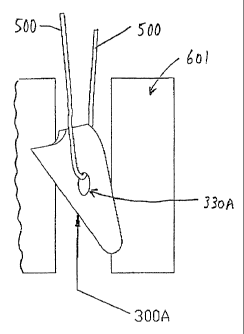
Next, the user uses installation too( 400 to drive suture anchor 300 into a
bore
hole. This is done by aligning suture anchor 300 with a bore hole 600 formed
in a bone
601 (Fig. 63) and thE~~~ pushing the suture anchor into the bone hole. As this
occurs,
the suture anchor's plow surface 316 will first tend to engage rim 603 of bore
hole 600,
causing the distal end of shaft tip 404 to flex as the suture anchor pivots to
enter the
bore hole. Further downward pressure on the installation tool's handle 410
causes the
distal end of the shaft tip to flex even further as the suture anchor's plow
surface 316
engages, and then rides along, wall 602 of the bore hole, with the suture
anchor's cam
surface 326 being slightly spaced from, or insignificantly in contact with,
the bore hole's
opposing wall 606 (see Fig. 64). Significantly, such flexing of the dista) end
of shaft tip
404 does not significantly undermine the attachment of suture anchor 300 to
installation too! 400, since only the distalmost portion of the shaft tip
(i.e., the threaded
portion 428) is actually secured to the suture anchor, with the shaft tip's
immediately-
proximal portion 432 being free to flex slightly within the suture anchor's
counterbore
340 without damaging the suture anchor. This is true even where suture anchor
300
may be formed out of a non-metallic material, e.g., a plastic or absorbable
material.
The user pushes suture anchor 300 downward into bore hole 600 anti! the
desired depth is reached. Such downward pressure keeps the suture anchor's
plow
surface 316 in engagement with the bore hole's wall 602. Preferably
installation toot
400 is sized so that nose 406 engages the top surface 604 of bone 601 when the
desired depth is reached.
Next, the user withdraws installation tool 400 from bore hole 600. As downward
pressure on installation tool 400 is released (to be replaced by opposite
upward
-pressure during tool withdrawal), the flexed shaft tip 404 tries to
straighten itself, ,
causing the suture anchor's sharp, well-defined biting edge 322 to press into
wall 602,
and causing the suture anchor to pivot slightly in the bore hole so that the
suture
CA 02224234 1997-12-09
WO 97/37595 PCT/US97/05925
-35-
anchor's cam surface 326 securely engages wall 606 of the bore hole. As the
user
retracts installation tool 400 from bore hole 600, rearward movement of
installation toot
400 causes progressively more distal portions of the suture anchor's cam
surface 326
to come into engagement with wall 606 of the bore hole. Since cam surface 326
is
arranged to cam the suture anchor laterally, such engagement of cam surface
326 with
bone wall 606 causes the anchor's sharp, well-defined biting edge 322 to be
driven
progressively further and further into wall 602 of the bore hole, until the
suture anchor's
abutment surface 312 rests against wall 606 (Fig. 65). As installation tool
400 is pulled
further back, the installation tool eventually breaks free from the lodged
suture anchor.
The installation tool is then withdrawn from the surgical site.
It should be appreciated that the presence of cam surface 326 significantly
enhances the ability of suture anchor 300 to set in bone 601, since the cam
surface
provides a force on the suture anchor's edge surface 322 which is
approximately
normal to the bore hole's wall 602. This force drives the suture anchor's edge
surface
322 into wall 602, ensuring that the suture anchor will be reliably set. This
is true even
where bone 601 is relatively hard (e.g., cortical bone) and the suture anchor
is made
out of a non-metallic material, e.g., plastic or a bioabsorbable material.
By changing the geometry of cam surface 326, the setting characteristics of
suture anchor 300 can be adjusted.
It should also be appreciated that the nature of the attachment of suture
anchor
300 to installation tool 400 is important. In particular, the suture anchor
must be
_ attached to the installation tool securely enough to cause the suture anchor
to turn in
the aforementioned camming action, yet release at the appropriate time so as
to leave
-the suture anchor in the bone as the installation fool is withdrawn. Thus it
is desirable
that the connection between suture anchor 300 and installation tool 400 be
well defined
at the time of manufacture, and remain intact up until the time that the
installation tool
breaks free from the anchor. In particular, it is important that this
connection not be
CA 02224234 1997-12-09
WO 97/37595 PCT/LTS97/05925
_3g_
undermined while the distal end of the installation tool flexes in the bore
hole. It has
been found that such a reliable connection can be established by providing the
distal
end of the shaft tip with a threaded portion 428 and an immediately-proximal
smooth
portion 432, and providing suture anchor 300 with a bore 338 and a counterbore
340,
where the shaft tip's threaded portion 428 makes a threaded engagement with
the
suture anchor within bore 338, but the shaft tip's immediately-proximal
portion 432 is
not secured to the suture anchor within counterbore 340. This permits the
necessary
flexing of the shaft tip to occur without undermining the connection between
the
installation tool and the suture anchor, even where the suture anchor is
formed out of a
non-metallic material, e.g., a plastic or absorbable material.
It is to be appreciated that as installation tool 400 separates from the
deployed
anchor 300, suture 500 can simultaneously slide along the inner surfaces of
the
shroud's channels 4G6, so as to permit the two members to separate. Depending
on
the length of suture 500 and the degree of separation imposed, suture 500 may
or may
not be fully removed from installation tool 400 during anchor deployment. To
the extent
that some of suture 500 remains attached to installation tool 400 after the
installation
tool has been withdrawn, the installation tool may be placed on a surgical
drape
adjacent to the surgical site until the suture 500 is to be completely removed
from the
installation tool. At that point the remaining suture may be pulled free of
the installation
tool.
For many procedures, retaining a portion of suture 500 in installation toot
400
until needed can be helpful for effective suture management. For example,
where an
arthroscopic procedure involves more than one suture anchor, the paired suture
ends
of several suture anchors might emanate from a single cannula opening. By way
of
example, an arthroscopic Bankhart procedure could involve four or even six
suture
lengths emanating from a single cannula opening. In such a situation,
retaining suture
lengths in the installation tool can help keep the suture lengths more easily
identifiable
for the surgeon.
CA 02224234 2004-11-26
-37-
Additionally, and/or alternatively, to help improve suture management, it may
be desired to mark various suture lengths. For exemple, each suture length
could
be distinctively color-coded, or distinctively pattern-coded. U.S. Patent No.
3,949,755 issued April 13, 1976 to Vauquois teaches the provision of
contrasting
shades on suture.
It is also possible to modify the suture anchor 300 described above.
For example, in the suture anchor 300 described above, the suture anchor's
through-hole 330 is preferably sized so as to slidably receive a single strand
of
suture. However, if desired, the suture anchor's through-hole 330 could be
sized so
as to slidably receive multiple stands of suture simultaneously. Of course, if
such a
construction were used, the shroud's channels 466 should be correspondingly
enlarged so as to accommodate the additional suture lengths provided, or some
other provision should be made to manage the additional suture ends present
with
such a construction.
Furthermore, with the suture anchor 300 described above, the suture
anchor's through-hole 330 is formed so as to have a substantially round
configuration. However, the suture anchor's through-hole can also be formed
with
an elliptical configuration, such as the through-hole 330A provided in the
suture
anchor 300A shown in Figs. 66-68. By forming the suture anchor's through-hole
with an elliptical configuration, stresses from the suture can be directed to
specific
portions of the suture anchor. In particular, these suture stresses can be
directed to
more robust portions of the suture anchor, thereby enabling the suture anchor
to
carry larger loads. This feature can be particularly useful where the suture
anchor is
formed out of a non-metallic material, e.g. a plastic or bioabsorbable
material. In
addition, by forming the suture ancho's through-hole with an elliptical
configuration,
subsequent proximal pulling of the suture can also help to further set the
suture
anchor with a desired orientation within the bone.
CA 02224234 1997-12-09
WO 97/37595 PCT/US97/05925
- 38 -
Also, with the suture anchor 300 described above, the suture anchor is
confiigured so as to have substantially planar side surfaces 320. As a result,
the
anchor must be farmed thin enough so that the suture lengths emanating from
the
anchor can pass alongside the suture anchor within the bore hole (see Fig.
69). This is
particularly true where it is desired to slide suture 500 relative to suture
anchor 300
when the suture anchor is set in a bone hole. However, in many situations it
can be
advantageous to form the suture anchor with the thickest possible
configuration, with
the sides of the suture anchor having a rounded outer surface, e.g., such as
the side
surtaces 3208 provided for the suture anchor 300B shown in Figs. 70 and 71. In
this
situation, a pathway 344 must be provided to permit suture 500 to pass from
the suture
anchor's through-hole 330 to the exterior of the bone. In this respect it
should also be
appreciated that inasmuch as the suture anchor must rotate within the bone,
the
pathways 344 should be formed with a configuration which broadens toward the
proximal end of the anchor, in the manner schematically illustrated in Fig.
70. Such a
construction permits the necessary anchor rotation to occur without causing
the suture
to come into abrasive engagement with the side walls of the bore hole.
Also, it is possible to omit the suture anchor's through-hole 330 and attach
the
suture directly to the anchor. Thus, for example, a suture anchor 300C is
shown in Fig.
72 where the suture 500 is molded or fused directly into the side 320C of the
body of
the anchor. Alternatively, and looking now at Fig. 73, the suture 500 could
enter the
proximal end surface 324D ofi a suture anchor 300D and then be internally
fused to the
interior of the suture anchor. This could be done with a single suture strand,
or with a
pair of independent suture strands, or with a looped suture strand where the
loop is
fused within the interior ofi the body.
It is also possible to attach a suture to the suture anchor by forming a
longitudinal hole through the suture anchor, passing the suture through that
longitudinal hole, and then knotting the suture at the distal end of the
suture anchor so
as to prevent the suture from being withdrawn proximally through the suture
anchor.
CA 02224234 1997-12-09
WO 97/37595 PCT/LTS97105925
-39-
Alternatively, an enlargement of some other sort could be positioned at the
distal end of
the suture to prevent suture withdrawal. Thus, for example, a suture anchor
300E is
shown in Fig. 74, wherein suture 500 passes through a pair of longitudinal
holes 346
opening on proximal end surtace 324D, and then are knotted at a knot 502 near
the
distal end of the anchor. If desired, a recess or cutaway 348 can be provided
near the
distal end of the anchor to accommodate knot 502. Alternatively, a single
suture strand
500 could be used, with the suture strand passing down one longitudinal hole
346 and
back up the other longitudinal hole, whereby the use of the knot 502 could be
eliminated.
Thus, the invention has been described with reference to the attached
drawings. It is
easily understood by one of ordinary skill in the art that changes may be made
to the
embodiments described herein without exceeding the spirit or scope of the
attached
claims.