Note: Descriptions are shown in the official language in which they were submitted.
CA 02224~12 1997-12-11
UNIVERSAL KINEMATIC IMAGING TABLE FOR RAPID POSITIONAL
CHANGES IN PATIENT CENTERING
BACKGROUND OF THE INVENTION
FIELD OF THE INVENTION
The present invention relates to a kinematic imaging table, and more
5 particularly, to a kinematic imaging table for rapid positional changes in patient
centering for use with a magnetic resonance imaging (MRI) machine, a
computed tomography (CT) imaging device, or other imagery systems including
x-ray units.
BACKGROUND AND SUMMARY OF THE INVENTION
The use of imagery systems like magnetic resonance imaging and
computed tomography imaging is well known in the medical field. A patient
going through such examinations typically must lie flat on an examination table
which is inserted into the imaging device. The patient is required to lie still for
15 extended periods of time in order to obtain accurate data from the imaging
device. For MRI devices, there are generally two types of coils which are used,
body coils and surface coils. Body coils are mounted around the gantry in the
body of the MRI device. Body coils generally suffer from the disadvantage that
they do not provide the desired signal-to-noise ratio and spatial resolution.
20 Surface coils provide superior spatial resolution and signal as compared to body
CA 02224~12 1997-12-11
coils. Surface coils are utilized by placing a posterior coil in the form of a
circular or rectangular plate which houses the coil on a table underneath a
patient. An anterior coil, also in the form of a circular or rectangular plate which
houses the coil is strapped to the patient's frontal region. The anterior coil is
5 disposed above the posterior coil. The posterior and anterior coils are provided
in the area of the body which is being imaged. Typically, when different areas
of the patient are to be examined, the coils used for creating the images must
be moved relative to the patient's body. The delay in moving the coils
contributes to the time necessar,v for obtaining the necessary images, thus,
10 contributing to the patient's discomfort and overall examination time.
Furthermore, in the imagery of, for example, blood vessels, a control material is
injected into the patient's blood stream. As the control material is circulated
through the blood vessels, it is necessary for the patient to be moved reiative to
the imaging coils in order to obtain an accurate image of the blood vessels.
15 Because the contrast is only effective for limited periods of time, the time
required for moving the surface coils is prohibitive for imaging blood vessels in
different anatomic locations.
In the case of an MRI device utilizing body coils, the bed on which the
patient is on can be moved relative to the body coils. However, the body coils
20 do not provide the desired spatial resolution and signal for obtaining clear
images. Furthermore, current table speeds are not sufficient for rapid positional
changes of patient centering. Therefore, x-ray angiography remains the
CA 02224~12 1997-12-11
predominant method of imaging blood vessels. However, x-ray angiography
suffers from the drawbacks that it is invasive upon the patient due to the fact that
it requires insertion of catheter devices into the patient's arteries or veins. These
catheters are subsequently injected with contrast which leads to vascular
opacification. Furthermore, x-ray angiography is expensive and time consuming.
Therefore, it is desirable in the art of radiology to provide an imaging device
which provides high resolution images and which permits rapid positional
changes in patient centering so that the time required for obtaining accurate
images of a patient may be reduced and so that movement of the patient relative
to the imaging coils is simplified.
Accordingly, the present invention provides a magnetic resonance imaging
device including an examination table movably supported on a track. A lower
surface coil is supported under the table, and an upper surface coil is supported
above the table. The table is movable relative to the upper and lower surface
1 5 coils.
According to another aspect of the present invention, an examination table
is provided for use with an imaging machine. The table includes a plafform
having a rolling track disposed on an upper surface thereof. An examination
table is movably mounted on the rolling track for supporting a patient thereon.
According to yet another aspect of the present invention, a method is
provided for rellorilLi"g an existing imaging device with an examination table
according to the present invention.
CA 02224~12 1997-12-11
The present invention provides a kinematic imaging table which provides
a kinematic imaging table which allows rapid positional change in the patient
centering in order to facilitate the imaging of blood vessels, in order to facilitate
easy moving of the patient relative to the imaging coils, and to reduce the timerequired for obtaining the necessary images for a medical imaging examination.
Further areas of applicability of the present invention will become apparent
from the detailed description provided hereinafter. It should be understood
however that the detailed description and specific examples, while indicating
preferred embodiments of the invention, are intended for purposes of illustration
only, since various changes and modifications within the spirit and scope of theinvention will become apparent to those skilled in the art from this detailed
description.
BRIEF DESCRIPTION OF THE DRAWINGS
The present invention will become more fully understood from the detailed
description and the accompanying drawings, wherein:
Figure 1 is a general schematic view of a CT and a MRI imaging device;
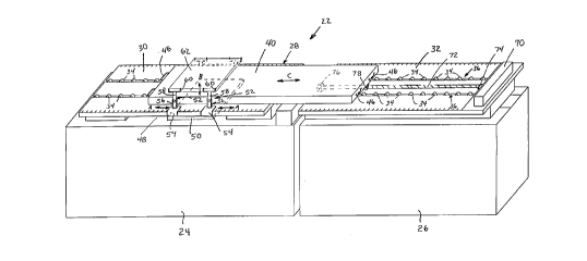
Figure 2 is a perspective view of the universal kinematic imaging table
according to the principles of the present invention;
Figure 3 is a perspective view of a plafform having a rolling track
according to the principles of the present invention;
Figure 4 is a sectional view taken along line 4-4 of Figure 3;
CA 02224~12 1997-12-11
Figure 5 is a perspective view of a second plafform section having a
rolling track disposed on an upper surface thereof;
Figure 6 is a perspective view of a movable table according to the
principles of the present invention; and
Figure 7 is an end view of the movable table shown in Figure 6.
DETAILED DESCRIPTION OF THE PREFERRED EMBODIMENTS
The present invention relates to a universal kinematic imaging table for
rapid positional changes in patient centering. In particular, the kinematic imaging
table is used in conjunction with a magnetic resonance imaging and/or computed
tomography imaging device 10. As shown in Figure 1, the imaging device 10
generally includes a computed tomograph gantry 12 or a magnetic resonance
imaging gantry 14. The computed tomography gantry and magnetic resonance
imaging gantry each include a tunnel 16 for receiving a patient therein. A
plafform 18 is provided for supporting an examination table 20 which is receivedin the tunnel 16 of the imaging device 10.
According to the principles of the present invention, a kinematic imaging
table is provided for rapid positional changes in patient centering, as shown inFigure 2.
V\llth reference to FIG. 2, the imaging table 20 of the present invention is
shown in combination with existing imaging and outer tables 24, 26, respectively.
The kinematic imaging table 22 of the present invention includes a base plafform
CA 02224~12 1997-12-11
28 including a first plafform portion 30 and a second plafform portion 32. The
first plafform portion 30 is disposed on the existing imaging table 24. The
second plafform portion 32 is provided on the existing outer table 26. Outer
table 26 may be provided with vertical adjustment capabilities for raising and
lowering second plafform portion 32. First and second plafform portions 30, 32
are provided with a plurality of rollers 34 arranged in a pair of columns 36.
Rollers 34 are supported by rods 38, as shown in FIG. 4. Rollers 34 and rods
38 are preferably made from a non-ferrous material such as plastic.
An examination table 40 is supported on the plurality of rollers 34.
Examination table 40 is provided with grooves 42 extending longitudinally along
a lower surface 44 thereof. Examination table 40 can be provided with handles
46 disposed on the ends or sides of the table 40. Examination table 40 is
preferably made from a non-ferrous material such as plastic or wood.
An area beneath the first plafform portion 30 is provided an opening which
defines a space 48 for receiving a lower or posterior surface coil 50 therein. The
posterior surface coil 50 is provided in the form of a rectangular plate which
contains the coils therein. A pair of movable support members 52 are disposed
on each side of the first plafform portion 30. The support members 52 include
C-clamps 54 which attach to the plafform 30 and are adjustable in the
longitudinal direction as shown by arrows A. The support member 52 further
includes a vertically-extending post 56 and an adjustable slide member 58
having a coil-supporting bracket 60 disposed at an upper portion thereof. The
- CA 02224~12 1997-12-11
slide members are adjustable relative to posts 56 as shown by arrows B. An
anterior or upper surface coil 62 is supported on the brackets 60 of support
members 52. The upper surface coil 62 is disposed directly above lower surface
coil 50.
In operation, the examination table 40 is disposed on the second plafform
portion 32 on outer table 26. A patient is placed on the examination table 40
and the table is rolled onto the first plafform portion 30 and positioned such that
the portion of the patient which is desired to be imaged is located between the
upper and lower coils 62, 50. During the imaging process, the patient can be
moved relative to the upper and lower coils 62, 50 by a technician who will use
the handles 46 for moving the table. Alternatively, a motor 70 can be utilized for
automatically adjusting the position of the examination table 40. Motor 70 is
provided with a forward and reverse operating mode and has an output shaft
attached to a screw device 72 which is supported by bearings 74, 76. Screw
device 72 engages a nut 78 attached to examination table 40. The rotation of
screw 72 causes nut 78 to travel along the axis of the screw 72, thereby moving
the examination table 40 in the longitudinal direction as indicated by arrows C.Reversal of the motor 70 causes the screw device 72 to drive in the opposite
direction, thereby causing the examination table 40 to move in the opposite
direction.
In the invention disclosed above, the examination table 40 and rollers 34
may be placed wider apart so that the posterior coil can be placed directly
CA 02224~12 1997-12-11
beneath the examination table between the columns 36 of rollers 34.
Furthermore, the rollers 34 may also be provided on the bottom of the
examination table 40 while the first and second plafform portions would then be
provided with a track for receiving the rollers. It should be understood that other
5 known track devices can be utilized for slidably supporting the examination table
on the base plafform.
The kinematic imaging table of the present invention allows for the use of
surface coils of an MRI machine for use in rapidly imaging different body
locations including blood vessels and could therefore replace the predominant
10 technique of x-ray angiography which is invasive, expensive and time
consuming. The kinematic imaging table can also be employed for rapid
positional changes of patient centering when conducting computed tomography
examinations and other imaging studies which utilize x-rays.
The invention being thus described, it will be obvious that the same may
15 be varied in many ways. Such variations are not to be regarded as a departure
from the spirit and scope of the invention, and all such modifications as would
be obvious to one skilled in the art are intended to be included within the scope
of the following claims.
