Note: Descriptions are shown in the official language in which they were submitted.
CA 02226476 l998-0l-07
WO 96/40021 PCTAU596~ 30
HIP OFFSET-INSET APPARATUS AND METHOD
TECHNICAL FIELD
The present invention relates to a method and a~aldtlls for f~r~ilit~tin~ a total
hip arthroplasty procedure in which both an acetabular cup prosthPeie and a femoral
implant prosthesis are in~t~ll~l, or a partial hip arthroplasty where one of thecomponents of an earlier arthroplasty are replaced. In particular, the invention relates
to a method and a~dLus for ~e~nring that the rçsnl~in~ leg length and hip offset of
the patient is optimi7ed.
RELATED APPLICATIONS
The present application is a con~iml~tion-in-part of US Serial No. 08/250,164,
filed May 27, 1994, which is a divisional application of US Serial No. 07/882,938,
filed May 14, 1992 and issued June 7, 1994 as U.S. Patent No. 5,318,571 for a
"Method and Apparatus for Simplifying Total Hip Arthroplasty".
BACKGROUND OF THE INVENTION
Total and repl~çmçnt arthroplasty procedures employing artificial acetabular
cup prostheses and femoral implants have been performed for a great many years.
The in5t~ tion of a replacement hip joint prosthesis involves surgically exposing and
dislocating the joint, cutting away the head and neck and a portion of the greater
trochanter in a femoral neck resection, and reaming the femoral canal to accept the
metallic stem of the femoral implant.
Femoral implants are available in a variety of lengths and cross-sectional
~limçn~ions to fit the shape of the reamed-out femoral canal. Such implants are also
provided with an angularly disposed femoral neck and spherical head that extend at
an angle to the stem to orient the replacement head in the acetabular cup prosthesis.
The acetabular cup prosthesis is itself installed in the patient's reamed-out
acetabulum. After both components are installed, the spherical head is inserted into
the cup and the muscles and tendons that were separated or severed in the surgical
exposure are reattached to hold the leg in place.
=
CA 02226476 l998-0l-07
WO 96/40021 PCTAUS96~ 3~9
-- 2 --
Numerous articles have been published describing and illustrating total hip
arthroplasty procedures employing a wide variety of total hip joint prosth~ses. For
example, the pllblic~tion entitled "Total Hip Arthroplasty Using a Univers~l Joint
Device," by Raymond G. Tronzo, M.D., published in 1970 by Richards
S M~nllf7~ctl~ring Conlpally, 1450 Brooks Road, Memphis, TPnn~se~ 38116, describes
such a procedure employing the products of the Richards M~mlf~cturing Company.
A further blocllul~ entitled "PERFECTA Femoral Prostheses With T-MATRIX
Acetabular Options--Surgical Protocol, " published in 1990 by Orthomet, Inc.,
MinnP~polis, Minnest ta, describes and illustrates the surgical procedure employed in
in~t~lling Orthomet products. Other m~mlf~-~tllrers publish similar instruction
m~m~ or protocols for their products.
A common problem that arises in completing the total hip arthroplasty
procedure involves the selection of the a~ liate length neck of the femoral implant
so that after the procedure is completed, the patient enjoys a normal or Pnh~ncecl leg
length. The dirr~ ces in leg length arise from the difference in sizes of the femoral
implants, the depth of insertion of the acetabular cup, and the length of the reducted
femoral neck. To change the length of a leg, surgeons often simply change the length
of the femoral neck that is used. Once the acetabular cup is in place, the cup itself
is rarely ch~nge~.
J. Edeen, et al., in "Clinical Significance of Leg-Length Inequality After TotalHip Arthroplasty" provides a summary of interviews and eY~min~tions of 68 patients
after total hip arthroplasty. Their findings confirm that there was a high rate of
ti~f?~ction among patients, particularly due to leg-length inequality. The authors
refer to various methods available to surgeons for eq~l~1i7in~ leg lengths
intraoperatively, but were unabIe to validate any particular method.
A variety of methods have been used to estimate the length of the leg upon
implanting a new femoral implant and acetabular cup. Some methods have involved
measuring the distance from the palpable iliac crest near the waist line to the greater
trochanter on the outside of the proximal femur, both of these markers are quitedistant from the true hip joint and only indirectly attempt to measure length.
Other methods described in the literature measure the distance from an anchor
installed percutaneously above the superior acetabulum to a marker on the exposed
CA 02226476 1998-01-07
WO 96/40021 PCTrUS~ 33~3
greater L.~ch~ ei. See, for example, S.T. Woolson, M.D., et. al., "A Method of
Inler~fdLive Limb Length Measurement in Total Hip Arthroplasty, " Clinical
Orthopaedics and Related Research, 1985, 194:207-210; W.H. Harris, M.D.,
"Revision Surgery for Failed Nonseptic Total Hip Arthroplasty, " Clinical
OrthopaedicsandRelatedResearch, 1975, 106:19-26; andN.M.J. McGee, F.R.C.S.,
et. al., "A Single Method of Obtaining Equal Leg Length in Total Hip Arthroplasty,"
Clinical Orthopaedics and Related Research, 1985, 194:269-270.
In an article by W.E. Knight, M.D. ("Accurate Determination of Leg Lengths
During Total Hip Repl~ em.-nt7" Clinical Orthopaedics and Related Research, 1977,
123:27-28), a tool is described for me~cllring the ~ t~nce between bone screws
placed in the exposed ilium about two inches above the margin of the acetabulum and
in the greater trochanter of the femur in line with the iliac screw in the coronal plane.
The tool is positioned laterally to the pins and the measurement is made parallel to
the femur, which is not necess~rily parallel to the weight-bearing axis.
These methods are flawed as they measure two ~iimencions, length (vertical
~iimçncion) plus lateralization (horizontal translation of the femur). The hip and leg
position must be virtually identic~l when pre-operative and post-operative
measurements are made to insure predictable results. In practice, the horizontal offset
and leg position have traditionally been difficult to replicate. Accordingly, better
surgical procedures for obt~uning exact leg length and hip offset are to be desired.
Copending application U.S. Serial No. 08/250,164, the disclosure of which
is incorporated herein by reference, describes a method and a~p~dLIls for use insurgical hip repl~çmt-nt procedures for ensuring that the replacement hip joint
colllLonents are properly sized so that the resulting length of the patient's leg is
correct. In the procedure, when the acetabulum and femur are exposed, one end ofa flexible measuring cable is attached to the superior eminence of the acetabulum and
the cable is extended inferiorly so that it is aligned with a specific point on the femur,
preferably the lesser trochanter.
The selected point on the femur is marked and the scaling or marking indicator
carried by the flexible cable is positioned in relation thereto. After this l~f~l~nce
~lict~nce between the fixed points on the acetabulum and the femur is determined, the
flexible cable is moved out of the way while its end remains fixed to the acetabulum.
CA 02226476 l998-0l-07
W O 9f~L-~-1 PCTAUS96/09309
Thereafter, the total hip arthroplasty procedure continues in the usual fashion
with the in~t~ tion of an acetabular cup prosthesic and the femur is plel,ar~d to
receive the femoral impl~nt After the femur is prepared, trial femor~l impl~ntc are
inst~ll~l to determine the proper fit with the femoral canal. The selection of the
S proper neck length to ensure the proper recl-lting leg length is f~rilit~tP~l by eYtPn~ling
the flexible cable ~tt~f~hPA at one end to the superior eminence of the acetabulum
toward the marked position of the femur so that the marked position is aligned with
the in~ tor fixed to the cable.
The art is still need of methods and articles for accurateIy r~lo. i.-g not onlynormal joint length, but also the center of hip rotation and femoral offset at the time
of hip repl~ PmPnt. This ability is nP~cc~ry in order to reduce problems such asdislocation-subluxation, and polyethylene wear that can lead to joint erosion and
loosening, as well as leg length inequality. In turn, the ability to reduce suchproblems will improve mechanical function and patient c~ticf~-tion.
SUMMARY OF THE I~VENTION
In one aspect, the present invention provides a method of reconstructive hip
surgery involving the replacement of a dysfunctional hip joint with a total hip
prosthesis, the method comprising the steps of:
(1) providing a hip length device for marking and measuring a pre-
repl~ement length distance between two fixed points along the weight bearing axis
of the joint;
(2) providing a femoral offset-inset apparatus for marking third and fourth
fixed points and for mP~cunng pre-replacement distances between these points and a
first reference point positioned in a medial ~limPncion with respect to the joint;
(3) surgically exposing the superior portion of the femur and the acetabular
region of the iliac bone;
(4) employing the hip length device to mark a first fixed point in the
acetabular region of the iliac bone and a second fixed point on the lateral portion of
the exposed femur, and measuring the pre-replacement length distance between thefirst and second points with the femur normally extended;
(5) employing the offset-inset apparatus to mark a third fixed point on the
CA 02226476 1998-01-07
W O 96/40021 PCT/U'~G~ 3D9
_ S _
medial portion of the exposed femur, and to provide a fixed reference point in amedial position with respect to the third fixed point, and me~ ring an offset ~ t~nee
between the fixed reference point and the third point with the hip in a reproducible,
rçstr~inç ~ position,
(6) p~lrolllling the surgical procedure of a hip dislocation and removal of
the femoral head in order to expose the surface of the acetabulum;
(7) employing the offset-inset appal~ s to identify a fourth fixed point on
the surface of the exposed acetabulum, and to measure an inset distance between the
fixed reference point and the fourth point with the hip in a reproducible, rçstr~ined
position;
(8) delell.li~ g a suitable proximal femoral width as the dirrelellce
between the measured inset and offset distances, and selecting a femoral component
with an a~pl~lia~ offset rlim~n~ion corresponding thereto;
(9) ~ rOl .--il~g the surgical procedure of implanting a trial acetabular
prosthesis and employing the offset-inset apparatus to measure the post-repl~eçment
inset distance;
(10) p~lror,--i-lg the surgical procedure of ~lepa-illg the femur and inserting
a trial femoral component;
(11) employing the offset-inset apparatus to recheck the offset ~ t~nce
between the third mark and the reference point to provide the desired post-
replacement ~limPn~ions; and
(12) employing the hip length device to recheck the distance between the
first and second ma-rks to provide desired post-replacement joint length.
In a ~!lerellc~d embodiment, the offset-inset apparatus shares one or more
components in common with the hip length device. In particular, the fixed ileac
marker of the hip length device is provided in the form of an extended post or other
suitable form. The post, in turn, is capable of serving as an anchor point for
establishing the fixed reference point of the offset-inset apparatus.
The ability to use a single fixed marker in both the hip length and offset/insetmeasurements greatly facilitates the ease and accuracy of the present method. This
ability also serves to alleviate unnecessary trauma to the body, by minimi7ing the
number and location of fixed markers.
CA 02226476 1998-01-07
WO 96/40021 PCT~US96/09309
In another aspect, the invention relates to an offset-inset a~aldlus, which may
be in kit form, for ~lr~l.l.ing the surgical procedure. The invention involves an
offsetinset a~ dLus which compricPs a first attachment ~tt~l~h~hle to the superior
eminPn~e of the acetabulum. The first ~tt~chment can be fixed directly to the bone,
but preferably is indirectly fixed by ~tt~hm~-.nt to a fixed point provided by the hip
length a~aldtus. The offset-inset a~hdL~s further involves a second attachment
point capable of being fixed to the femur at approximately the position of the greater
trochanter. Optionally, and preferably, the a~pdldL~s further includes a me~eurin~
device for mP~cllring and noting the ~ ct~nce between the reference point and either
the third or fourth fixed points.
BRIEF DESCRIPTION OF THE DRAWING
These and other advantages and features of the present invention will become
apL,alent from the following dPt~iled description of the preferred embodiments thereof
in conjunction with the Drawing in which:
Figure 1 is a posterior view of the right pelvic girdle and a portion of the
right leg bones;
Figure 2 is an illustration of a femoral implant of a type employing a sepaldL~
head adapted to fit in an acetabular cup of a total hip joint replacement prosthesis;
Figure 3 is an illustration of a first embodiment of a hip length d~JaldL~Is
modified of the type described in co-pending U.S. Application No. 08/250,164;
Figure 4 is an illustration of a first embodiment of an offset-inset apparatus
employed in the practice of the method of the present invention;
Figure 5 is an illustration of the use of the hip length apparatus of Figure 4
in initially mP~cllring the ~ ct~nce between points on the iliac bone and the femur in
a posterior surgical approach;
Figure 6 is an illustration of the use of the offset-inset apparatus of Figure 3in mP~cllTing the offset distance between a reference point and the greater trochanter
of the femur in a posterior surgical approach, showing the restrained position of the
foot with respect to the operating table;
Figure 7 is an illustration of the use of the offset-inset apparatus of Figure 3,
following hip dislocation, in initially me~cllring the inset distance between a reference
CA 02226476 1998-01-07
W O 96/40021 PCTAUS~6/~ 9
-- 7 --
point and the exposed acetabular surface;
Figure 8 is an illustration of the use of the offset-inset a~paralus of Figure 3,
following the implantation of an acetabular cup component, for rechecking the inset
distance in accordance with the method of the present invention practiced in a
posterior surgical approach; and
Figure 9 is an illustration of the use of the hip length appal~tus of Figure 4
in s~ .tinE a femoral implant of the a~plo~liate length in a posterior surgical
approach; and
Figure 10 is an illustration of the use of the offset-inset appa dlus of Figure
3 in selecting the d~l~liately sized femoral implant having the a~l~liate offsetdistance in accordance with the method of the present invention practiced in a
posterior surgical approach.
DETAILED DESCRIPTION
The method and apparatus of the present invention can be used to provide pre-
and corresponding post-operative measurements of joint length and joint offset-inset
dimensions, thereby substantially elimin~ting problems due to incorrect sizing of
components, and in turn, the need to reoperate.
Using the pleselltly described apparatus and method in the surgical field, pre-
repl~ ement length, center of rotation and femoral offset can be accurately measured.
The position of the calcar cut, the reaming of the acetabulum, and the selection of the
type of prosthesis in regard to head-neck length, femoral neck angle and built in
offset can be accurately guided by serial measurements during replacement. At the
conclusion of the procedure a stable hip of correct length, and having close to normal
anatomic geometry, can be expected. Improved function of the hip as well as lower
rates of dislocation, leg length disparity, polyethylene wear and revision surgery can
be expected. These devices can be expected to aid the joint manufacturers in
development of new prosthesis.
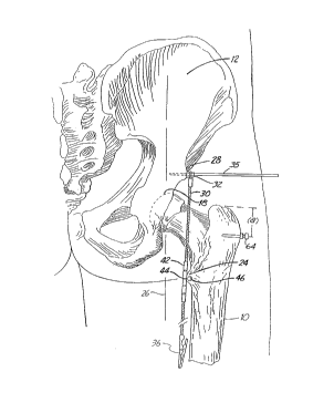
Turning now to the Drawing and first to Figure l, it illustrates a posterior
view of the right pelvic girdle and a portion of the right leg showing the normal
alignment of a human hip joint in relation to the knee joint. The femur 10, or thigh
bone, articulates at its proximal end with the iliac bone 12. Its distal end articulates
CA 02226476 1998-01-07
W O 96/40021 PCT~US96/~3~9
-- 8 --
with the tibia at the knee joint 14. The shaft of the femur bows m~Ai~lly so that it
approaches the femur of the opposite thigh. As a result of this convergence, the knee
joints are brought together to the body's line of gravity or weight supporting axis.
The proximal end of the femur 10 comprises the rounded head 16 that
articulates with the acetabulum 18 of the iliac bone 12. The neck 20 of the femur 10
is a constricted region distal to the head 16. A fairly common fracture in the elderly
occurs at the neck 20. Apparently the neck 20 becomes so weak that it fails to
support the body. The greater trochanter 22 and lesser troch~nt~r 24 are projections
on the femur 10 that serve as points of ~tt~chm-ont for some of the thigh and buttock
muscles.
Between the trochanters on the anterior surface is a narrow inLIocl~ r-~ ;c line.
Between the trochanters on the posterior surface is an intertrochanteric crest. The
shaft of the femur contains a rough vertical ridge on its posterior surface called the
linea aspera. This ridge serves for the ~tt~chment of several thigh muscles. Thedistal end of the femur is exp~nd~l and inchldec the medial condyle and the lateral
condyle. These articulate with the tibia and (with ~ttent1~nt 1i~mentc, etc.) from the
knee joint 14.
The weight of the body is borne by the normal pelvic girdle and lower
e,~ nlilies in conjunction with the associated muscles and lig~m~ntc in a weightsupporting axis extending generally through the superior aspect of the acetabulum 18
inferiorly alongside the femur and through the center of the knee joint 14 generally
as illustrated by broken line 26 in Figure 1. The weight supporting axis 26 passes
through or ~ t?nt to a line draw in between the superior eminence 28 of the
acetabulum and the lesser trochanter 24.
Through disease processes or injury, the hip joint may become less mobile,
painful or totally unusable, requiring surgical intervention and a total hip arthroplasty
employing a hip joint- prosthesis of the type described in the above-referenced
publi~tionc. A wide variety of prostheses are available from several manufacturers,
the prostheses typically including a metal acetabular cup for surgical replacement of
the natural acetabulum 18, a high density polyethylene cup liner positioned within the
cup prosthesis and a femoral impl~nt comprising a femoral stem and a femoral
ball-shaped head of the type illustrated in Figure 2.
CA 02226476 1998-01-07
W O 96/40021 PCT/U'~G~ 3~3
_ 9 _
Turning now to Figure 2, it illllctr~tPs a typical femoral impl~nt stem 31 and
femoral imrl~nt head 33 which are ~tt~-hed together to form the femoral impl~nt 29.
The femoral stem 31 is available in a number of cross-section shapes, sizes and
lengths having varying exterior contours, neck offsets and neck lengths. The femoral
S implant head 33 is usually a highly poliched, spherical metal ball provided in a
number of available ~ mP~tçrs and neck lengths and provided with a Morse taper
female receptacle for receiving the Morse tapered neck 35 of a stem 31. In practice,
manufacturers of total hip prostheses provide a selection of dirr~l~nt neck length
femoral implant stems 31 and femoral implant heads 33. Thus, it is possible to
customize the overall neck length as nP~ess~ry to replicate the proper leg length for
the individual patient.
As described above, upon completion of the operative procedure, it has often
been found that the overall leg length and/or offset ~limen~ion is incorrect, re~uiring
a reoperation, usually involving replacement of one or both of the components of the
femoral implant. The operative procedure can be simplified and the need to reoperate
subst~nti~lly Plimin~tç~ by providing a measurements of both joint length and the
offset-inset ~limpncions both prior to and following the implantation of trial
components.
To this end, Figure 3 illustrates a pleftll~d hip length device for m~rking and
mç~cllring a pre-replacement length distance between two fixed points along the
weight bearing axis of the joint. A suitable hip length device involves (a) a first
marker nail in the form of a post for providing fixed attachment to the superioreminence of the acetabulum, (b) a length of flexible cable ~tt~ch~hle at a fixed end
to the post, (c) one or more markers moveable along the length of the cable, and (d)
a second marker nail for marking a fixed position on the lesser trochanter.
In a pl~f~l.ed embodiment, the hip length device is of the type described in
co-pending application Serial No. 08/250,164. In a particularly ~-lertll~d
embodiment, the marker used to provide fixed attachment to the superior aspect of
the acetabulum is provided in the form of a post. The post can be of any form and
. 30 ~limçncions suitable to allow it to serve as the anchor point for establishing a
reference point medial to the greater trochanter.
Figure 3 illustrates a plerelled hip length apparatus 30 comprising length of
CA 02226476 l998-0l-07
WO 96/40021 PCTAUS96/09309
- 10 -
cable 34 and a bone nail 32, the form of an elong~tP~l tower, that are used to select
a properly sized femoral stem 31 and head 33 during the procedure and avoid
reoperation. The hip length d~ dLlls depicted in Figure 3 comprises a length of
stranded wire cable 34 extending between an alligator clip 36 and a loop 38 formed
S by bending the cable 34 back against itself and crimpin~ a crimp tube 40 around both
to form a loop. The loop 38 is formed after the bone nail 32 is fixed in the region
of the superior eminence 28 of acetabulum 18 as shown in Figure 4. The ~llig~t~rclip 36 is provided so that the free end of the cable 34 may be ~tt~ h~d to a surgical
drape to hold the apparatus 30 out of the surgical field when it is not used in the
manner depicted in Figures 5 and 6.
The hip length a~p~ s 30 also includes one or more slidable tubes (e.g., as
shown by reference number 42) having four calibration marks 44 spaced apart about
one cçntimt-t~r in the outer surface of the tube 42. The calibration tube 42 may be
slid back and forth on the length of stranded wire cable 34 so that one of the marks
4~ is aligned with a mark or ~tt~-~hment, e.g., another bone nail 46, made on the
femur 10 and crimped in place by a crimping tool.
In a pl~r~lled embodiment the tower portion of nail 32 is of sufficient length
and configuration to allow the rigid attachment of a removable rod ext~n-iing in a
subst~nti~lly perpendicular position. The rod, in turn, can be directed into a position
medial to the greater trochanter to provide a reference point for offset-inset
measurements.
Turning now to Figure 4, it illustrates a ~lerell~d offset-inset d~aldtlls 60 ofthe present invention, comprising a removable rod 62, a third bone nail 64.
Optionally, the apparatus depicted in Figure 4 further comprises a s~aldte me~ ring
device 66, e.g., in the form of a depth gauge, useful for determining the inset and
offset distances between the fixed reference point and the fourth nail and the exposed
acetabular cup, respectively. The depth gauge can include a barrel portion capable
of being grasped, together with a slidable distance probe, the movement of which t
corresponds with the appearance of distance or reference markings identifiable above
the barrel of the gauge.
A reference point can be formed in any suitable manner to provide a fixed,
reproducible reference point in a position medial to the femur. In a preferred
_
CA 02226476 1998-01-07
W O 96t40021 PCT~US96~30~
embodiment the reference point is formed from, and anchored to the nail used to
form a ffxed acetabular position for the hip length device.
The rod 62 is capable of being ~tt~rhp~ to the tower portion of bone nail 32
of the hip length device in order to form a fixed, reproducible reference point in a
position medial to the original position of the greater trochanter. In the embodiment
illustrated, for in~t~nce, rod 62 is formed of a longitu-lin~l arm portion 68 and a
perFen-lic~ r ~tt~chmpnt portion 70. As described below, ~tt~chmP-nt portion 70 can
be ~tt~hP~ to the tower portion of first bone nail 32 in any suitable manner, e.g., by
providing a female ~tt~chmpnt site for the tower portion.
The relative dimensions or shapes of the bone nail 32 and rod 62 are not
critical, nor is the means of ~tt~ hing the rod to the post, so long as together they
capable of reproducibly forming a fixed point in space that is medial to the original
position of the greater trochanter. The third bone nail 64 is capable of being fixed
in the region of the greater trochanter in order to form a fixed point for measurement
of the offset distance to the fixed reference point. The measuring device can also be
provided in any suitable form, e.g., as a depth gauge capable of being held beside the
third or fourth reference points to visually determine the ~ t~nce to the reference
point.
Referring to Figure 5, a surgical procedure is performed in order to expose
the superior portion of the femur and the acetabular region of the iliac bone. Prior
to surgery, and using pre-operative X-rays of the pelvis, the surgeon can initially
estim~te the me~ li7~ti~ n of the acetabulum caused by arthritic erosion of the medial
acetabular wall, or other causes. The surgeon can then estim~te the amount of hip
length lost by erosion of the articular cartilage, femoral head and acetabulum.
The method of the present invention will be described with respect to the
posterior approach, which Applicant has found to be a preferred approach due to the
technical ease in measuring the three variables of hip length, center of rotation and
femoral offset. In practice, the procedure may be used in an anterior surgical
approach as well. Selection of the anterior or the posterior approach is left to the
discretion of the surgeon. Those skilled in the art will appreciate the manner in
which the anterior or transtrochanteric approach can also be done with minor surgical
modifications to the procedure as well.
CA 02226476 1998-01-07
W O 96/40021 PCT~USg'/~309
The tensor fascia lata is ~ spctç~l and the hip external rotators are exposed.
In a preferred embodiment, the typical external rotator release is modified by also
re1P~ing the quadratus femoris as well as the usual rotator release. This allowsexposure of the lesser trochanter, which is crucial to allow accurate length
measurement. The lesser trochanter is exposed by extreme internal rotation of the
femur short of dislocation. A posterior hip capsulectomy is performed in routinefashion combined with a anterior capsulotomy and hemostasis is obtained.
A hip length device, of a modified type described in the co-pending U.S.
application, is then employed to measure a pre-operative length ~ t~nce Removal
of a Segmçnt of the glenoid labrum posterior superiorly allows clear vi~ li7~tion of
the superior articular surface of the acetabulum. A modified Taylor rekactor can be
placed under the gluteus medius and minimus muscle to allow exposure of the
sllpr~et~hular area posterior and superiorly in an area 3-4 cm proximal to the
acetabular rim.
The hip length device is used to mark a first fixed point in the acetabular
region of the iliac bone and a second fixed point on the lateral portion of the exposed
femur. Figure 5 illustrates the placement of the hip length apparatus 30 to effect a
calibrated measurement of distance along the weight supporting axis 26 between the
femur 10 and the iliac bone 12 before the head 16 is dislocated from the acetabulum
18.
It will be understood that for the sake of simplicity, the operating field,
inc~ ing the incision and the sep~r~tion of the muscles and lig~ments, are not
illustrated in the Figures. Assuming that those operative steps have been taken, the
proximal end of the femur 10 and the portion of the iliac bone 12 surrounding the
acetabulum 18 are exposed.
A first marker nail 32 is provided, preferably in the form of a cylindrical postwith an attached length cable and a depth stop. The first marker can be drilled or
otherwise impacted into the supraacetabular area, posterior-superiorly approximately
2 cm proximal to the roof of the acetabulum at the 11 o'clock or 1 o'clock positions
relative to the face of the acetabulum and dependent on which hip is being operated
on. ~2t-.c~lling that the reflected head of the rectus femoris is at the 12 o'clock
position superior to the acetabulum, care must be taken to place the tower on the
CA 02226476 1998-01-07
WO 96/40021 PCT/U~5GtO~309
vertical axis in both planes.
As shown in the Figure, the surgical nail 32 is driven into the region of the
superior eminence 28 of the acetabulum 18 about 2 centimeters above the acetabular
rim in the 11 o'clock position after the loop 38 in the cable 34 has been merh~nic~lly
~tt~ch~d to the shaft of the nail 32. Th~lc~r~el, the cable 34 is extended in the
direction of the weight supporting axis 26 bringing it alongside the lesser trochanter
24.
Then, as shown in Figure 5, the slidable calibration tube 42 is moved along
the length of cable 34 until one of its indicia 44 is aligned with a mark or nail 46 on
the lesser troch~ntt-r 24. The mark is typically a further surgical nail driven into the
bony protuberance of the lesser trochanter 24 since it is difficult to othenvise mark
the bone.
As shown in Figure 5, a second marker nail 46 (e.g. 1 cm in length) is
inserted into the posterior aspect of the lesser trochanter, being careful not to
penetrate the main femoral intramedullary canal. With the foot l~ ined in a footholder and the knee extended, a third marker nail 64 is implanted, for later use in
me~ ring hip offset distance. The third marker nail, approximately 1 cm in length,
is placed in the promin~nce of the greater trochanter approximately 5 cm (distance
"a") from the proximal end and once again in the vertical plane. Solid fixation in the
trochanter is confirmed.
After the indicia 44 and nail 46 are aligned, the tube 42 is crimped tightly
against the stranded wire cable 34 so that it cannot be moved or dislodged. With the
leg restrained in the foot holder, a retractor can be used to Vi.~ i7e the lesser
trochanter and the cable strung tightly from the tower to the marker on the lesser
trochanter, the sliding scale is crimped at the middle mark at the level of the lesser
trochanter marker nail. The selected indicia 44 is noted in the surgical record, as the
pre-replacement length distance, and the cable 34 is pivoted superiorly out of the
operating field.
The alligator clip 36 is ~tt~.'h~ to a surgical drape covering the patient's torso
in order to keep the apparatus 30 out of the surgical field to allow the surgeon to
proceed with offset-inset measurements. In particular, the surgeon will dislocate the
head 16 from the acetabulum 18 and proceed with the resection of the femur and the
CA 02226476 1998-01-07
WO96/40021 PCT~US96J'~
surgical placement of the acetabular cup prosthesis and the reaming of the femoral
canal to accept the trial femoral components.
As shown in Figure 6, a hip offset-inset device 62 of the present invention can
then be employed to use the third marker nail previously marked on the lateral
portion of the exposed femur.
In the course of me~c-lring pre- and post-operative inset and offset di~t~nces,
the patients foot is most preferably positioned in a fixed, reproducible position in
space, above the operating table. Fixing the foot position aids the accuracy of this
device, since factors such as variable leg position can significantly change the
mea~u~
In a pl~relled embodiment, a leg holding device is employed that compri~ec
a holding boot and an ~tt~hment apparatus for holding the boot in a fixed position
with respect to the surgical table. The leg is placed in the holding boot and the boot
is ~tt~ched to the leg holder in neutral position in regard to flexion-extension,
abduction-~ddllcti~ n and rotation. The boot can be easily removed from the leg
holder and is not attached to it during the procedure except at times measurements are
being made.
With the patient in the customary lateral position and m~int~ined by a standard
McGuire-type frame, a standard incision is made. Those skilled in the art will
llndt-r.~t~nd that the incision can be a lateral, anterolateral or posterolateral one,
depending on the surgeons preference and surgical approach.
In a pleft;ll~d embodiment, the fixed reference point is formed by placing a
removable rod portion 68 on the tower portion of nail 32 of the length device. The
rod portion can be attached to the nail in any suitable manner, for instance, byforming a perpendicular angle portion 70 that can be connected to the tower by the
use of a slip-fit coupling, a male-female threaded coupling, a serrated coupling, a
locking collar, and the like.
The respective lengths of the bridge portion 35 and angle portion 70 are not
critical, so long as the combined length once coupled is sufficient to allow the rod
portion 68 to extend a sufficient distance medial to the femur. The length of the
tower of nail 32 is preferably sufficient to terminate the tower about 3 cm lateral to
the lateral trochanteric surface. The rod portion 68 is of sufficient length so that the
CA 02226476 1998-01-07
W O 96/40021 PCT/U~ 53D~
- 15 -
depth gauge 66 can be laid aside its distal extension and onto the greater tro~h~ t;. ;c
nail 64, or preferably onto a point on the bone just above the marker nail.
With the depth gauge 66 contacting the bone above the greater troc~ c
nail marker 64 the length of the offset is measured. This length is recorded for later
S use.
Alternatively, means other than a calibrated depth probe can be employed for
d~~ g the offset ~lict~nce~. For in~t~nce, using a nail that is itself calibrated
along its tower portion, the rod can itself be moved laterally to a point where it
contacts the bone above the head of the greater troch~nteric nail. At that point, the
corresponding location of the rod along the tower can be noted and the rod
l~~ )oldlily removed. Later, the rod can be used to measure the inset distance, by
using a depth probe that extends down from the rod into the acetabulum. The
~nce can be determined by calibrations on either the probe or, if the rod is
moveable along the tower, on the tower itself.
Turning next to Figure 7, following the pre-operative offset measurement, a
further surgical procedure is performed to dislocate the hip and remove the femoral
head in order to expose the surface of the acetabulum. In the surgical procedure, the
foot is first removed from the foot holder, the hip dislocated and the neck divided at
the ~ o~lial~; level as e~ t~rl by the a~lopliate prosthetic head neck device,
usually approximately 2 cm proximal to the lesser trochanter. The head is removed
and the hip is internally rotated while retractors are placed anterior and posterior to
the acetabulum to adequately expose its anatomy.
Tht;l~;afLel-, the offset-inset apparatus 62 iS used to measure an inset distance
between the reference point formed by bridge 68 and a fourth point on the surface of
the exposed acetabulum with the hip in a reproducible, restrained position. With the
foot placed back in the foot holder, and bridge 68 positioned on the tower portion of
nail 32, the depth gauge 66iS dropped into the acetabulum to measure its depth.
The fourth fixed point can be visually identified by selecting an ~ upliate
- spot for the depth gauge to contact the medial acetabular wall. The fixed point can
be anterior, superior or posterior to the fossa ovalis, but preferably not in the depth
of the fossa. Measurement of the inset distance between rod 68 and the fourth fixed
point determines the preoperative articular joint depth of the hip. The inset
CA 02226476 1998-01-07
WO 96/40021 PCTAUS9~/~S3~9
- 16 -
measurement can also be recorded for later use.
The dirr~ ~nce between the inset and offset ~lict~nces can be used to determine
a suitable proximal femoral width (identified as distance "b" in Figure 6), and to then
select a femoral colllpollent with an a~ opliate offset ~limPnci~n coll~ ding to that
S width. The ~ulgeoll can then ~;lrc,l-l- the surgical procedure of impl~ntin~ a trial
acetabular prosthesis and employing the offset-inset al)pa d~us to measure the post-
replacement inset ~lict~nce
Turning now to Figure 8, it illustrates the pl~t~çment of an artificial acetabular
cup prosthesis 50 in the location of the acetabulum 18. As the acetabulum is the first
to be replaced, the ap~l~liate reaming depth will be determined by factors such as
the thicknPcc of the acètabular prosthesis and the amount of erosion of the medial wall
by the arthritis. If the opposite hip is normal, the çstim~tions will be more ~ cur~te.
The acetabulum is then reamed to the ap~lopliate depth, repeatedly cherking the inset
measurement as the reaming proceeds. In some cases with a gçneti~lly thin medialwall and a thicker acetabular prosthesis than the patients normal medial wall, the
center of rotation will of necçccity be lateralized a few millimeters. This can later
be factored in when pl~nning the femoral offset.
Once the acetabular position is restored to normal with the ~l.r~liaLe
reaming and thickn~sc of prosthçcic, the prosthesis is implanted with either cement
or bony ingrowth. If using cçment, care must be taken not to lateralize the cup with
a bolus of cement medial to the cup. Following cup fixation and liner insertion, the
acetabular ~lict~nc~e~ i.e., inset measurement, can be re-checked by repeating the steps
involved in its pre-operative measurement.
Following impl~nt~tion and chç~king of the acetabular component, the surgeon
then performs the surgical procedure of ~ a ing the femur and inserting a trial
femoral component and employing the offset-inset a~l,~dLlls to recheck the offset
distance between the third mark and the reference point on the bridge to provide the
desired post-repl~-çment ~limçncions.
At this point ~he surgeon has the critical measurements of pre operative hip
length, acetabular inset, femoral offset and proximal femoral width. Using the
preoperative pelvic X Ray, adjustments can be calculated to recreate normal length
and acetabular inset now changed by the erosive effect of arthritis. In the event that
CA 02226476 1998-01-07
WO 96/40021 PCT/U',''~33'9
- 17 -
it was determined prior to beginning the surgical procedure that the patient's leg
needed to be lengthened or shortened, then the lengthened or shortened amount may
be taken into account when the calibration mark 44 is noted. Similarly greater offset
may be desirable and can be obtained using the apparatus of the present invention.
S The proximal femur is reamed, irri~tt-A, flushed with epinephrine solution
and pl~lgg~ with a silastic plug to restrict cement in cases using cement fi~tion
Trial femoral prosthesis are selected to give correct intr~med~ ry fill, femoral offset
and hip length. Varying neck lengths, femoral offsets and femoral neck angles gives
the surgeon greater ability to approach the normal anatomy of the hip. With eachtrial of the femoral prosthesis, the hip is relocated, the foot holder is applied and the
length and femoral offset measurements are checked.
In the pl~:r~l~ed embodiment of illustrated in Figure 9, the femoral canal 52
is reamed out, a trial femoral implant 29 is inserted as shown in Figure 9 to test the
fit and the articulation of the leg. Figure 9 illustrates the insertion of a trial femoral
implant 29 comprising the femoral implant stem 31 and femoral implant head 33 inrelation to the acetabular cup prosthesis 50 and the femoral canal 52 outlined in the
resected femur 10.
Both the hip length apparatus 30 and offset-inset a~paldlus are employed in
the trial fitting stage to ensure that the selected neck length results in the desired leg
length and offset. As illustrated in Figure 9, once a trial femoral implant 29 is in
place, the stranded wire cable 34 is extended in the direction of the weight supporting
axis 26 to determine whether or not the calibration indicia 44 previously aligned to
the mark or nail 46 is again aligned. If the measurement in(lic~tes that the actual
position is superior or inferior to the desired calibration indicia 44, then the trial
femoral implant 29 is withdrawn and/or longer or shorter necked components are
substituted. The process is repeated until the desired calibration indicia is aligned as
closely as possible to the femoral mark or nail.
When the predetermined hip length and femoral offset are obtained with
selected prosthetic stem and head-neck trial components, final fixation of the
permanent prosthesis is performed. Final measurements of hip length and femoral
offset are recorded. The tower with the attached cable and the two marker nails are
removed. The hip is checked for stability and routine closure is performed. A post
CA 02226476 1998-01-07
WO 96/40021 PCT~US9~3~9
- 18 -
operative X Ray is obtained in recovery and critically analyzed by the sUl~ eon.After the ~r~lly sized components are in~t~ll~l, the surgical nail 32 and 46
(if used) as well as the cable 34 are removed and the incision is closed in the normal
manner. By use of the inventive ap~dld~us and method, the incid~nce of l~ldlion
required to correct for leg length errors is virtuaUy elimin~te~l. Obtaining the correct
offset will lower the risk of po~Lop~ldlional dislocation of the hip.
Critic~lly evaluating prosthetic hips at the time of impl~nt~til~n, or by later X-
ray evaluation, will reveal nine different geometric joint configurations that can be
used when con~id~ring length and offset. These configurations include
correct length with correct offset
correct length with increased offset
correct length with decreased offset
short length with correct offset
short length with increased offset
short length with decreased offset
increased length with normal offset
increased length with increased offset
increased length with decreased offset
In the course of m~nring the length of a repl~rçment hip, Applicant has
discovered a method and apparatus that reflects the fact that the nltim~te stability of
the joint, as well as the surface tension of the metal on polyethylene articulation, is
signific~ntly affected by the me li~li7~tion-lateralization of the center of rotation of the
hip as well as the offset of the shaft of the femur.
A variety of factors are known to me~ li7e the center of hip rotation,
inclu~ling arthritic erosion of the acetabulum, deep reaming by the surgeon, pelvic
and acetabular fractures, and small femoral prosthetic heads.
A number of other factors are considered to lateralize the center of hip
rotation, including inadequate acetabular reaming by the surgeon, overly thick
acetabular prosthesis, lateralization of the acetabular prosthesis by cement, and larger
femoral prosthetic heads.
Yet further factors are considered to medialize femoral offset, including
m~oAi~li7e~ acetabular prosthesis, increased femoral neck angle of the prosthesis
CA 02226476 1998-01-07
WO 96/40021 PCT~US~GI~~530
- 19 -
relative to the p~ti~nt~, excessive surgical removal of calcar-neck, excessively short
femoral neck of prosthesis selected, and small ~ mtoter femoral prosthetic head.Still other factors are considered to lateralize the femoral offset, including
lateralized acetabular prosthesis, decreased femoral neck angle of prosthesi~ relative
S to the patients natural neck angle, increased inset built into the femoral neck
prostheci~, inadequate surgical removal of calcar-neck, excessively long femoral neck
of prosthesis 5~ te~1, and larger femoral neck ~ meter.
Similarly, many factors can be i~lentifie~ as increasing the hip length,
inclu~ling excessive femoral neck length prosthesis selected, inadequate surgical
removal of calcar-neck, greater neck angle of femoral prosthesis than the patients own
neck angle, distal pl~ement of the acetabular prosthesis, and surgeons selection of
a long neck to help stabilize a loose feeling hip (usually due to inadequate offset).
Co~ ~ondingly~ factors that serve to decrease the hip length include shorter
femoral neck length prosthesis than the p~tient~ own neck length, excessive removal
of calcar-neck, loose eroding hip prosthesis, congenital dysphasia, arthritic erosion,
and previous fractures and growth disturbances.
Anatomic studies on cadavers reveal the variance in hip length between normal
individuals is 41 mm (over 1 and 1/2 inches), the variance in hip offset is 37 mm (1
and 1/2 inches), and the hip neck angle variance is 105 to 154 degrees (49 degrees).
The surgeon can modify length and offset during surgery by a variety of techniques,
however, the femoral neck angle is predetermined by most manufacturers at 135
degrees.
The term "measured hip" implies the surgeon measures and obtains correct hip
length, accurate center of rotation of the hip, and desired femoral offset, thereby
effectively restoring geometry of the hip as close as possible to the pre arthritic state.
It can be seen that a variety of factors interact to determine the replacement
length and femoral shaft offset-inset ~ict~nce. The accurate replacement of a patient's
anatomic variants correctly will improve the Illtim~te function of the joint and likely
affect surface tension which may result in reduced polyethylene debris and acetabular
wear.
CA 02226476 l998-0l-07
W O 96/40021 PCT/U~,G/'0~30
- 20 -
The invention has been described in detail with particular reference to the
plerelled embo-limP.nt.e thereof, but it will be understood that variations and
modific~tions can be effected within the spint and scope of the invention.