Note: Descriptions are shown in the official language in which they were submitted.
CA 02226899 1998-01-14
WO 97/07412 PCT/GB96/O1901
MAGNETIC RESONANCE IMAGING
This invention relates to an improved method and al)pa,dlu~ for im~ging samples,using either Nuclear Magnetic Resonance (NMR) techniques or Electron p~r~m~gnetic
Resonance techniques.
The principle of NMR was first developed in 1946, and the technique of field
gradients origin~ted in 1973. In early arrangements, resonance in a swept field was
detected either by a single coil resonator (bridge method) or a double coil resonator,
i.e. with two coils at right angles (induction method). Both methods are disclosed in
"High-resolution Nuclear Magnetic Resonance" by J. A. Pople et al., McGraw Hill, on
pages 52 and 54.
NMR is now widelv used for im~ging liquids and liquid-like m~t~ri~l such as
biological tissues, in which NMR spin-spin relaxation times T2 are in the millicecond
range. Such long relaxation times allow the use of pulsed Fourier llal~r~ NMR with
pulsed m~gnPtic field gradients, and good quality images with high spatial resolution can
be obtained in a short time. In solids, the NMR lines are much broader and T2 is very
much shorter. Nevertheless several techniques have been developed to provide images of
solids using NMR. The techniques include multi-pulse line n~ulowillg and Stray Field
Tmzlging, STRAFI, but such techniques are severely restricted by limitations of available
radio frequency (RF) power to produce sufficiently short pulses; the result is that the size
of sample which can be imaged is small, approximately 10 millimetres, as the power
increases as at least the cube of sample ~limencions. In addition. STRAFI requires the
sample to be tr~nc1~terl by small ~ t~n~os between NMR excitations, which is difficult with
large objects.
Magic angle spinning has also been used but requires the sample to be spun rapidly
within the magnet, which clearly imposes restrictions on sample size and geometry.
Another technique is the os~ tinp: gradient method, but this requires very strong gradients
to be generated, which again limits sample size as the DC gradient power requirement
increases as the fifth power of the coil dimensions.
CA 02226899 1998-01-14
W O 97/07412 PCT/GB96/019OI
In all of the above techniques for ims-ging solids, there is the further restriction that
they can be used only if the spin-spin relaxation time T2 is greater than about
1 0 microseconds.
In a dirr.,~Gll- approach, swept-field continuous wave (CW) NMR has been used to~lrO.l~ NMR spectroscopy of solids since 1949, see The Journal of Chemirz-l Physics,
Vol. 17, No. 10, October 1949, H. S. Gutowsky et al. Swept field pulsed NMR has also
been used for spectroscopy, with the mzlgn~tic field swept in step-wise manner, and low
intensity RF pulses applied to detect NMR resonance. In yet another spectroscopic
technique, a series of pulses of increasing or decreasing frequency has been applied at
constant mz~gnP.tic field; this technique of variable pulse frequency swept field NMR has
been used for wide line NMR spectroscopy.
In a different technology, electron par~m~gnetic resonance (EPR), a sample is
continuously irradiated with electromagnetic radiation, and an applied magnetic field is
swept past the likely resonances. The technique of continuous wave (CW) EPR has been
known since 1979 and has been developed more recently to provide images of free radicals
in biological systems, and for spatial loc~ ti- n of p~r~m~gn~tic centres in solids, as
disclosed by M. J. R. Hoch and A. R. Day in Solid State Coll,.llul.ications 1979, Vol. 30,
pages 21 1-213.
It is an object of the invention to provide a~paldLus for im~ging solids using NMR
techniques but without the previously-applicable limit~til)n~ of sample size and of spin-spin
relaxation time.
It is a further object of the invention to provide d~Jp~udLus for im~ging in vivo
samples using EPR technique which is less sensitive to sample movement than the
conventional reflection bridge methods.
According to the invention, d~pardlus for providing a magnetic resonance image of
a sample comprises:
means for providing a magnetic field having:-
a first, homogeneous, component,
a second, gradient, component which varies in strength across the sample, and
a third, swept, component which varies in strength with time,
CA 02226899 1998-01-14
WO 97/07412 PCT/GB96/01901
the second and third components being parallel to the first component and being of
substantially lower m~nit~
a birdcage resonator arranged within the m~nPtic field with its axis parallel to the
field;
means to connect a radio-frequency source to the resonator so as to generate a
magnetic field in a first resonance mode of the resonator, and
means to connect detection means to the resonator so as to detect any signal
received by the resonator in its second, orthogonal resonance mode.
Optionally the connections between the resonator and the source and the resonator
and the detection means are inductive cormections, but ~lt~rn~tively they may be capacitive
connections.
Optionally the means to provide the third component of the m~gnt?tic field
comprises means to supply a ramp signal to the ZO coils of a conventional supercor~ ting
NMR magnet.
The appaldlu~ may be arranged to operate by the technique of nuclear magnetic
resonance, or by the technique of electron paramagnetic resonance.
The invention will now be described by way of example only with reference to theaccompanying drawings, in which:-
Figure 1 is an end view and sçh~m~tic axial section of a solid sample in position
within a resonator and field modulation coils;
Figure 2 is a s~h~ m~tic end view of the sample, resonator and coils within the bore
of a m~gnet;
Figure 3 illustrates schem~tically an NMR ~p~dllls according to the invention,
Figure 4 illustrates the first derivative CW NMR spectra of the sample shown in
Figure l;
Figure 5 is the integrated CW NMR spectra of the sample shown in Figure 4, and
Figure 6 shows the first derivative and integrated spectrum obtained from the
d~p~alus illustrated in Figures 1, 2 and 3 in the absence of a sample.
In Figure 1 a sample 10, comprising two blocks 10A, 10B, is shown inside a
high-pass birdcage resonator 12. The resonator comprises a first cylindrical polymethyl
methacrylate (PMMA) former 14 carrying two conducting end rings 16 joined by eight
--3--
CA 02226899 1998-01-14
W O 97/07~12 PCT/GB96/01901
confl~lctin~ legs 18. Between the junctions of the end rings and each ~ c~nt pair of legs
is a variable capacitor 20, shown srhrm~tically.
Arranged coaxially around the resonator 12 is a second cylindrical PMMA
former 22 on the outer surface of which is a radio frequency shield 24 in the form of
a 25 ~m layer of copper foil. The former 22 carries magnetic field modulation coils 26 in
the form of a split solenoid; turns of e.g. 1 mm copper wire 28 with PVC in~ tion are
wound on the former; although only two sets of four turns are shown, in practice a greater
number, e.g. ten turns, would be used.
In Figure 2 the sample 10, birdcage resonator 12 and the former 22 for the fieldmodulation coils are shown positioned within the various coils 30 within the bore 32 of a
superconducting magnet 34. The coils 30, shown shaded, include the room te~ ldLu.~;
shim, gr:~-1i.snt and ZO coils of a conventional NMR m~gn~t The magnet provides an axial
magnetic field, shown as BH in Figure l.
Figure 2 also ill. l~ , in highly sch~rn~tic forrn, the inductive coupling loops La~
Lb, sitll~ti-fl between the resonator 12 and the radio-frequency shield 24 carried by the
former 22. Each loop is connected to a coaxial cable (not shown) for connection to the
circuit in Figure 3. Neither the loops nor the cable are shown in Figure 1 for reasons of
clarity, but in Figure 2 the direction of the field B la by the loop La is indicated.
In an ~ltern~tive arrangement (not illustrated), the resonator may be ç~p~ itively
coupled to the rest of the ~p;~dlus.
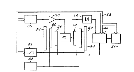
Figure 3 shows the electrical equipment used to provide a one--lim~n~ional imageofthesample 10.
An RF source 36 is connected through a power arnplifier 38 to the inductive
coupling loop La of the birdcage resonator 12, and the inductive coupling loop Lb of the
resonator is connPcte(l through a DC biassed diode detector 44 to a lock-in amplifier 46.
The amplifier 46 is connected to the field modulation coils 26 and to a co~ uL~l 56.
A DC power supply unit 48 is conn~cte-l to the field gradient coils 50 of the
magnet 34, and a field ramp controller or sweep generator 52 supplies the ZO field offset
coils 54 of the magnet 34; the magnet is not shown in this Figure.
The co~ uLel 56, through a bus 58, controls the amplifier 46, the RF source 36, and
the sweep generator 52.
--4--
CA 02226899 l998-0l-l4
W O 97/07412 PCTIGB96/01901
In operation, the RF source 36 ge~ s a co~ l amplitude sinewave at about
300 MHz; the signal is ~mplified by amplifier 38 and passes to the resonator 12, which in
its first resonance mode gener~t~s an RF magnetic field B la at the sample 10.
When the sample 10 resonates, the resonator 12 in its second resonance mode,
5 orthogonal to the first, receives a signal. This signal passes via the loop Lb to the
DC biassed diode detector 44; the output of thè detector is a DC level depending on the
amplitude of the resonance signal.
The field ramp controller 52 supplies a sawtooth waveform to the ZO coils 54; the
field gradient applied by DC unit 48 to the field gradient coils 50 can be set at a selected
10 fixed level.
In combination, the fields applied to the sample 10 are therefore:
a. the axial field BH provided by the magnet 22,
b. a gradient magnetic field parallel to BH and provided by the gradient coils 50,
c. a swept field, parallel to BH, provided by the ZO coils 54 of the magnet; and15 d. a constant field B la, perpendicular to field BH, provided by one resonance mode
of the birdcage resonator 12.
To record a spectrum the swept field is swept through an NM resonance of the
sarnple 10, either from below or from above resonance. At magnetic resonance, i.e. when
~I) = y Bo
20 when (1) is the RF angular frequency
y is the gyromagnetic ratio of the nucleus to be detected in the sample 10, and
Bo is the (total local) magnetic field strength,
the nuclear spins are disturbed from the Boltzmann population, and the nuclear
m:~gnf ti~tinn in the sample 10 starts to precess about the direction of BH at frequency h),
25 resulting in an RF signal being received by the resonator in its second mode of resonance.
To increase the s~ .iLivily, a field modulation technique is used, the lock-in
amplifier 46 generates a "le~.~..ce" sinewave at a frequency fm which is much less than
the R~ frequency of g~.~ Lol 36; the reference sinewave is applied to the field modulation
coils 26. When resonance is approached, the RF signal emitted by the resonator 12 t'nrough
30 the coupling loop Lb contains a component at frequency fm which remains on the output
of the diode detector 44 and is fed to the input of lock-in amplifier 46; amplifier 46
CA 02226899 1998-01-14
W O 97/07412 PCTIGB96/01901
contains a narrow-bandwidth filter, duLo~ ic~lly centred on fm7 so that it is capable of
recovering the mo~ ted signal from large out-of-band noise signals.
In a test arrangement a 7 Tesla magnet 34 with 125 millimetre free bore within its
shim/gradient coils 30 was used, and the field was swept by a value of 1.76 milli Tesla
5 over 64 seconds in a sawtooth w~vcrO-lll. The birdcage lesondLo~ 12 was excited at
300.015 MHz, and a m~gn~tic field mo~ ti~n of 15 micro Tesla was applied at 881 Hertz
through the field mo~ tion coils 26. A field v~ri~tinn of 1.76 milli Tesla is equivalent to
about 75 kHz proton frequency. The lock-in ~mplifier 46 had a time conet~nt
of 300 milliseconds.
The sample 10 had the form illustrated in Figure 1, it comprieed two blocks of
vulc~nieed rubber 10A, lOB both 1.5 cm thick and 3 cm long, spaced 3.5 cm apart. The
block 1 OA was of height 2.5 cm and the block 1 OB was of height 4 cm. The sample was
placed in the l~solldL~l 12 of diameter 75 mm and length 90 mm with a 120 mm diameter
copper shield 24.
The vulcanised rubber sample 10 had a T2 of about 1 millieecond, which is
relatively long in terms of the capabilities of the a~pdldLus, and which could have been
detected by known techniques, but was convenient for test purposes. The two rubber
blocks were scp~dl~d along the gradient direction, as shown.
Figure 4 shows the first derivative NMR signal S obtained from the sample with
magnetic field gradients Gz of 0, 3.6, 5.4 and 9.0 milli Tesla per metre applied by coils 50.
It can clearly be seen that with zero gradient, a single line is obtained, while with
the three non-zero gradients, two distinct lines are visible.
Figure 5 shows the integr~t~d signal Is. This was obtained by subtracting the zero
gradient signal from each of the non-zero gradients; p~-rc,l"lil,g a linear base line
correction; and pclrolll~illg integration. Such signal processing is conventional in EPR
systems. The Figure indicates two peaks corresponding to the blocks lOA, 10B, with a
spatial resolution increasing with the strength of the gradient. This can be c~le,~ te~l as the
ratio of the zero gradient spectral line width to the field gra~lient i,lLel.~iLy, and in the
c~c~;lllent the m;~x;---l---- spectral resolution was about 10 millimetres.
Figure 6 shows the spectrum with the sample 10 removed from the birdcage
resonator 12; both the signal and the base line-corrected integral are shown. The signal
--6--
CA 02226899 1998-01-14
W O 97107412 PCT/GB96/O1901
arises from the PMMA former 14 ~U~JpVl Lillg the birdcage resonator 12. Although the value
of the- RF m~gn~tic field BRF in the centre of the resonator 12 was set at a low value (about
2 micro Tesla) a~p~ liate to the relatively narrow proton resonance in rubber, the BRF
field was inevitably concc;llLldL~d close to the wires 16, 18 of the resonator, and was
5 sufficient to give a reasonably intense signal from the former 14. Glassy polymers such as
PMMA exhibit extremely short T2 values (about 40 microseconds) and are particularly
difficult to detect and image by conventional NMR techniques.
The pararneters of the ~paldLus were not optimised for the detection either of the
rubber NM resonance or for the PMMA resonance; particularly for the latter, a
10 considerably larger modulation field should ideally be used to achieve o~LilllUlll signal to
noise ratio.
In alternative app~hdlus, the diode detector 38 may be replaced by a mixer and a
c;r~lcllce arm. In a further ~ v~, use of a superheterodyne bridge configuration would
give better sensitivity at low RF power levels.
It will be appreciated that the results given above relate only to a one--limen~i~ n~l
projection of the test object 10. To obtain two-~limen~ional or three-dimensional images,
either the sarnple 10 can be rotated, or the field gradient can be rotated; either rotation can
supply projections at a sufficient number of orientations through the sample 10 to perrnit
signal analysis, for example by filtered back-projection or a similar technique, to
20 reconstruct an image of the sample. If deconvolution of the projection spectra with the
zero-gradient spectrum is required, as in Figure 2, this would be carried out prior to
back-projection. Such signal analysis techniques are conventional in NMR.
In the a~dLus described above, the continuous magnetic field gradient is
generated by use of the gradient coils of the magnet with DC power supplies. In an
25 :~ltem:~tive, the stray field of a supercnn~ ting magnet can be used, as in the STRAFI
technique, when the sample 10, resonator 12, field modulation coils 26, and field sweep
coils 54 would be placed near the edge of the magnet bore.
In the d~JaldLU~i described above, the magnetic field sweep is provided by ramping
the m~gn~tic field from the room-temperature ZO coil of the superconcl-l~ting magnet. In
30 an alternative, the field sweep may be genPr?~te~l by ramping the m~gnetic field of the
superconducting magnet itself.
--7--
CA 02226899 l998-0l-l4
W O 97/07412 PCT/GB96/01901
It is an advantage of swept field continuous wave NMR im~ging that it allows theuse of RF coils or resonators with extremely high Q-factors, because the detection circuitry
need have only a very narrow bandwidth, of the order of 1 Hertz. The use of high Q-factor
resonators h~ ase~ the signal to noise ratio. If such a high Q resonator is to be used, it is
5 advisable to incorporate ~llt-~m~ic frequency conkol circuiky to ensure that any frequency
drift of the resonator is followed by the RF source.
In an alternative app~ , not illll~tr~te~l, the supercon~lllcting magnet 34 is
replaced by a resistive magnet of much lower field, for example 0.01 Tesla for operation
at 300 MHz. All other parts of the ~ Lus are lmch~n~ed. The apl,~dlus can now be10 operated by the technique of electron pZ3r~m~n~tic l~so~ ce to provide EPR speckoscopy
and im~gin~