Note: Descriptions are shown in the official language in which they were submitted.
CA 02229620 1998-02-13
WO 97/18857 PCTrUS96/18680
,
8Y8TEN AND l~h~n~ FOR CONDITIONING
PELVIC NU~CULAT~RE U8ING AN
IMPLANTED MICRO~-.l.u~ATOR
R~c~grQl~nd of the Tnvention
The present invention relates to a system and
method for conditioning the pelvic musculature using one
or more implanted microstimulators, and more particularly
to a system and method for treating urinary incontinence
using a radio-frequency-controlled implanted
microstimulator.
Urinary incontinence is an enormous medical and
social problem, affecting 8% of people in industrialized
countries. Expenditures on its treatment are
conservatively estimated to be over 10 billion dollars.
Nevertheless, aggressive therapies for this condition are
used to treat only a small part of the affected
population, and much of the market is driven by the need
for absorptive products rather than state-of-the-art
devices.
Incontinence can be divided into at least three
major categories of pathophysiology:
1. Overflow incontinence, which arises from a
failure of the bladder to contract at all or
adequately for substantial emptying, so that
urine accumulates until it overflows. This
commonly occurs in spinal cord injury.
2. Stress incontinence, which arises from weakness
of the urethral sphincteric muscles, which are
unable to stop the forceful expulsion of urine
during transient increases in intra-abdominal
pressure such as during coughing and lifting.
This commonly occurs in women following
childbirth and/or menopause.
3. Urge incontinence, which arises from
spontaneous activity of the bladder and/or
sphincteric muscles producing a compelling
CA 02229620 1998-02-13
WO97/18857 PCT~US96/18680
feeling that the bladder needs to be emptied
even when it contains little urine. This
afflicts both men and women and probably arises
from a variety of poorly understood
dysfunctional reflexes.
It is has been observed over the past 50 years
or so that conditioning exercises of the pelvic
musculature can produce a reduction of stress
incontinence in a majority of women who undertake this
treatment. Pelvic conditioning therapies could
potentially assist a large proportion of the more than 6
million women affected by stress incontinence in the
United States alone. However, such exercises have not
proved easy for many women to perform by voluntary
contraction of the relevant muscles. Thus, electrical
stimulation has been used to force contraction of the
relevant muscles, and thereby achieve the desired muscle
conditioning exercises.
Prior art devices that provide electrical
stimulation to condition the pelvic musculature are based
on the use of transcutaneous electrical s~ tors which
exercise pelvic muscles using externally-controlled
regimes. While these prior art transcutaneous devices,
introduced into the vagina or anal canal, have proven to
be effective, they are often disliked by patients because
of the associated embarrassment and sanitary problems.
Further, in addition to stimulating the motor nerves,
transcutaneously applied electrical currents necessarily
stimulate the sensory nerves of the overlying s~in,
producing unpleasant sensations. It is thus apparent
that there is a need in the art for a system and/or
method for achieving the benefits of electrical
stimulation of the pelvic musculature without the
attendant problems associated with transcutaneous
devices.
CA 02229620 1998-02-13
W O97/18857 PCT~US96/18680
Stimulation of the bladder, sphincter and/or
adjacent pelvic musculature and cutaneous nerves may also
cause a reduction in the spontaneous sensations and
contractions associated with urge incontinence, which is
often combined with stress incontinence. The neural
mechanism is unclear, but is believed to relate to a
general property of neural reflect pathways that they can
drift into states of hypersensitivity or spasticity as a
result of various nonspecific insults and dysfunctional
patterns of usage. Electrical stimulation may give rise
to inhibitory neural activity that breaks the cycle of
spasticity during a particular bout of urge incontinence.
Repeated application of such stimulation may permit the
patient to resist inappropriate urges to empty the
bladder, thereby rebuilding normal bladder capacity and
reducing the frequency of sensation that the bladder
needs to be emptied. Repeated electrical stimulation may
also lead to a reduction in the hypersensitivity of the
neural pathways responsible for spontaneous sensations
and contractions and a concomitant reduction in the
incidence and/or severity of bouts of urge incontinence.
Hence, it is evident that improved methods and t~chn; ques
for electrically stimulating the bladder, sphincter
and/or adjacent pelvic musculature and cutaneous nerves
is socially and medically desirable, and would produce
enormous benefits for patients suffering from
incontinence, particularly stress incontinence and urge
incontinence.
In U.S. Patent No. 5,199,430, issued to Creasey
et al., there is disclosed an electrical assistive device
and a method for electrical stimulation that is used to
produce urination in patients who have lost voluntary
control of the bladder (such as the overflow incontinence
pattern described above).
CA 02229620 1998-02-13
W O 97/18857 PCT~US96/18680
In U.S. Patent No. 4,739,764, issued to Gleason
et al., a method is described for controlling bladder
emptying through the electrical stimulation of peripheral
nerves supplying the bladder wall and external urethral
sphincter. The method disclosed by Gleason et al.
includes two applications related to urinary dysfunction.
First, the method is suggested to assist bladder emptying
for patients who have lost the ability to urinate under
volitional control. Second, the method is suggested to
assist the patient who is incontinent by providing a
means to stimulate the sphincter muscles electrically.
Specifically, the method described stimulates identified
nerve bundles by using epineural electrodes, and by
providing moment-to-moment control of the urinary
sphincter at the time that continence is sensed to be
comp~ . This differs from the approach taken by
Applicants in the present application in that Applicants r
invention stimulates using small implantable stimulators
that are implanted in or near the pelvic floor muscles
for the purpose of providing conditioning stimulation on
a regular, ongoing schedule. Such ongoing conditioning
then results in continence via the natural voluntary
control available to the patient. In other words, while
the '764 patent addresses the continence problem by
attempting to control the urinary sphincter with
electrical stimulation, the present invention addresses
the continence problem by regularly conditioning the
pelvic muscles with electrical stimulation so as to
restore voluntary control to the patient.
~umm~ry of ~he Tnvent;on
The present invention addresses the above and
other-needs, particularly as such needs exist relative to
stress incontinence and urge incontinence, by providing a
system and method that uses one or more tiny implantable
CA 02229620 1998-02-13
W O 97/18857 PCTAUS96/18680
stimulators --termed "microstimulators"-- implanted in or
near certain pelvic structures so as to contact target
muscle tissue, e.g., pelvic floor muscles, and then
controls such microstimulators so that electrical
stimulation is provided to the target tissue in
accordance with a specified externally-controlled
exercise or conditioning regime. As the target muscle
tissue is conditioned in this manner, voluntary control
of such muscle tissue is returned to the patient.
In accordance with one aspect of the invention,
the microstimulators that form a key component of the
therapy system are hermetically encapsulated, leadless
electrical stimulators that are small enough (e.g., 2 mm
diameter by 13 mm length) to be injected percutaneously
(or otherwise implanted) into the patient's muscle
tissue. Thus, they are sufficiently small and innocuous
that they could be left in the patient permanently in the
unlikely event of a device failure. Furtherj
implantation, e.g., through a large hypodermic needle
under local anaesthesia could be performed as an out-
patient office procedure, avoiding the costs,
inconvenience and risk of morbidity that are normally
associated with major surgery.
In operation, the microstimulators used as part
of the present invention receive power and digital
addressing and command signals from an external
transmitter coil and control box. Advantageously, unlike
transcutaneous stimulators, the microstimulators are
unobtrusive and require no repeated insertions into body
orifices. By implanting the devices under the skin,
uncomfortable sensations associated with cutaneous
stimulation can be avoided. Further, the microstimulator
may be activated during work or leisure activities
without visible evidence to other individuals who may be
near the patient, e.g., in the same room as the patient,
CA 02229620 1998-02-13
W O 97/18857 PCT~US96/18680
thus increasing the convenience and acceptability of the
therapy.
In accordance with another aspect of the
invention, the implanted microstimulators are readily
controlled using an external control box, or control
unit, coupled to an external transmitting coil which
inductively couples (or otherwise transmits) progr~ in~
information to the implanted microstimulator(s). For
example, the transmitting coil may be placed in a cushion
on which the patient sits during treatment, or the
transmitting coil may be sewn into the patient's
clothing.
In accordance with an additional aspect of the
invention, the implantable microstimulators are implanted
in or near certain pelvic structures by a transvaginal or
transcutaneous approach, and are externally-controlled to
transmit the desired pattern of electrical stimulation
directly to adjacent motor and/or sensory nerve branches
without the inconvenience, discomfort and hygienic
problems associated with transcutaneous stimulators.
In accordance with yet another aspect of the
invention, the patient and/or medical personnel can
experiment with or test different st; l~tion regimes in
order to find the particular stimulation regime or
therapy pattern that is both comfortable and effective.
Rr;ef nes~;pt;on of ~he ~r~wings
The above and other aspects, features and
advantages of the present invention will be more apparent
from the following more particular description thereof,
presented in con3unction with the following drawings
wherein:
FIG. 1 is a cross-sectional view of one type of
microstimulator that may be used with the present
invention;
CA 02229620 1998-02-13
W O97/18857 PCT~US96/186~0
_ .
FIG. 2 is a block diagram illustrating the
transcutaneous transmission of power and information to
an implanted microstimulator;
FIG. 3 is a simplified embodiment of the
electrical circuit, including the electronic control
means, of a preferred implanted microstimulator;
FIG. 4A shows a transvaginal approach for the
implantation of a microstimulator, where the bladder neck
is depicted as if viewed from inside the pelvis, and
where the site of implantation of the microstimulator is
being identified by digital exploration;
FIG. 4B shows the transvaginal approach for
implantation of FIG. 4A, except that the site placement
is viewed as if it were sectioned parasagittally;
FIG. 5A is a block diagram that depicts the
various elements that comprise a stimulation system and
method which is made and practiced in accordance with the
present invention; and
FIG. 5B illustrates the types of coils that may
be used with the stimulation system in order to practice
the invention.
Corresponding reference characters indicate
corresponding components throughout the several views of
the drawings.
Det~;led Descr;pt;on of the Tnvent;on
The following description is of the best mode
presently contemplated for carrying out the invention.
This description is not to be taken in a limiting sense,
but is made merely for the purpose of describing the
general principles of the invention. The scope of the
invention should be determined with reference to the
claims.
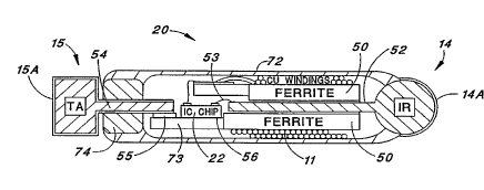
Referring first to FIG. 1, there is shown a
microstimulator 20 of one type that may be used with the
CA 02229620 1998-02-13
WO 97/18857 - PCT~US96/18680
present invention. The microstimulator 20 is typically
only about 10 to 15 mm in length, e.g., 13 mm, and
comprises a quartz, glass, or ceramic tube or capsule 72,
sealed at each end with a hermetic seal. For the
embodiment shown in FIG. 1, a first electrode 14
protrudes out from one end of the glass capsule 72, and a
second electrode 15 protrudes out from the other end of
the capsule 72. Other embo~ ts may have the two
electrodes 14 and 15 protruding out of the same end of
the capsule 72. Such electrodes 14 and 15 are made from
any suitable conductor, e.g., 0.025 to Q.150 mm diameter
platinum-iridium wire.
In one embodiment, the electrode 14 may be made
from iridium, and the electrode 15 may be made from
tantalum. An anodized layer 15A covers the tantalum
electrode 15, and an activated iridium layer 14A
envelopes the iridium electrode 14. The use of a
tantalum electrode 15 in combination with an iridium
electrode 14 in this manner provides, by its structure,
when immersed in body fluids, an electrolytic capacitor
20 having resistance 21. (See FIG. 3).
Inside of the glass capsule 72 is the
electronic circuitry associated with the microstimulator
20. In particular, in accordance with one embodiment,
the microstimulator 20 includes an integrated circuit
(IC) chip 22, a ferrite core 50, and a coil 11 wound
around the ferrite core 50. The IC chip 22 includes
several logic and other circuits, including memory
circuits.
All of the components and circuits within the
microstimulator 20 are interconnected in circuit
relationship so as to function as follows: (a) the coil
11 is inductively coupled to a modulated power signal
that is generated external to the glass capsule 22;
(b) the inductive coupling induces a modulated power
CA 02229620 1998-02-13
WO 97/18857 PCT~US96/18680
. . .
signal in the coil 11; (c) the induced modulated power
signal is rectified to provide operating power for the IC
chip Z2; (d) power from the rectified power signal
charges a storage capacitor (which may be internal to the
mi~rostimulaLor 20, ~r f~qrm~d by its el~ctrod~sj; (e~ Ihe
power signal is demodulated to extract an address word
therefrom; (f) the extracted address word is ~ red to
a preprogrammed microstimulator code stored in the
microstimulator; and (g) if the extracted address code
matches the preprogrammed microstimulator code, as
determined by logic circuits included within the IC chip
22, the capacitor is discharged through the two
electrodes 14 and 15 with an amplitude and pulse width
determined by the incoming data stream. In this manner,
then, the operation of the microstimulator, i.e., the
selective discharging of its storage capacitor, is
controlled through appropriate modulation of the power
signal.
Details associated with the design,
construction, and operation of different types of
microstimulators 20 that may be used with the present
invention are found in United States Patent Nos.
5,324,316; 5,405,367; and/or 5,312,439, all of which
patents are incorporated herein by reference.
FIG. 2 is a block diagram illustrating the
transcutaneous trAn~ ;~sion of power and information to
the implanted microstimulator 20, while FIG. 3 is a
simplified embodiment of the electrical circuit,
including the electronic control means, of one type of
implanted microstimulator. A thorough description of
FIGS. 2 and 3 is found in U.S. Patent No. 5,324,316, at
col. 4, 1ine 58, through col. 8, line 11, while
~ additional details associated with the construction and
operation of the microstimulator 20 are found throughout
_
CA 02229620 1998-02-13
WO 97/188~7 PCT~US96/18680
-- 10 --
.S. Patent No. 5,324,316, and the other patents cited
above.
With a working microstimulator 20 capable of
being independently addressed from an externally-
controlled transmitter to provide selective stimulationpulses between its two electrodes, it is possible using
ingenuity and creativity to fashion a wide variety of
stimulation systems and methods to fulfill various
patient needs. The present invention, for example,
utilizes one or more such microstimulators to
particularly address the problems associated with stress
incontinence and urge incontinence. In U.S. Patent
5,571,148, also incorporated herein by reference, another
application of utilizing a plurality of microstimulators
is disclosed.
It is thus seen that the microstimulator 20 as
described in the '316 patent lincluding all of its
variations as described therein and in the other cited
patents) represents an extremely versatile, and presently
available, "building block". The present invention is
directed to the application of the microstimulator
technology to accomplish the desensitization,
strengthening and/or general conditioning of the pelvic
muscles and reflex pathways involved in maintaining
continence, without encountering the limitations and
problems inherent in the previously used approaches.
As indicated above, microstimulators are
hermetically encapsulated, leadless electrical simulators
that are small enough (e.g., 2 mm diameter by 13 mm
length~ to be injected (or otherwise placed)
percutaneously into muscle tissue. The microstimulators
receive their power and digital addressing and ~- ~n~
signals from an external transmitter coil 102 driven by a
control box 100 (Fig. 2).
CA 02229620 1998-02-13
W O97/18857 PCTAUS96/18680
.
In accordance with the present invention, one
or more microstimulators 20, e.g., four microstimulators,
are implanted in or near certain pelvic structures by a
transvaginal or transcutaneous approach as suggested in,
e.g., FIGS. 4A and 4B. The control unit or box 100,
shown in block diagram form in FIG. 5, is used to
externally control the microstimulators 20. The control
unit is programmed to transmit the desired pattern of
electrical stimulation directly to adjacent motor and/or
sensory nerve branches without the inconvenience,
discomfort and hygienic problems associated with
transcutaneous stimulators.
The target tissue(s) in which the
microstimulator(s) 20 are implanted will vary according
to the specific pathophysiology of the patient. In most
patients with stress incontinence, the microstimulators
20 are placed bilaterally in the striated perineal
muscles that form a cuff around the anterior and lateral
aspects of the urethra, preferably near the entry points
of the muscle nerves. A single transmitting coil 102
(Fig. 5), e.g., placed in a cushion on which the patient
sits during treatment, provides the power and c~ ~nd
signals to all of the microstimulators 20 implanted in
the general perineal region. The muscles are stimulated,
i.e., conditioned, for 15-60 minutes (or other specified
time period) each day using intermittent trains of
electrical pulses sufficient in magnitude to result in
direct or indirect activation of muscle fibers. As a
result, the muscles and connective tissue are
strengthened so that they develop more force when they
contract around the urethra. Moreover, such conditioning
adds bulk to the periurethral tissues to buttress the
urethra.
In patients with urge incontinence, the
microstimulators 20 may be implanted near other pelvic
CA 02229620 1998-02-13
W O 97/18857 PCT~US96/18680
structures such as the bladder proper or under perineal
skin. The patient is then given control o~ the control
box or control unit 100 so that the patient initiates
stimulation whenever the sense of urgency occurs. While
clinical research will continue to establish the
diagnostic criteria that identifies the most ~o ;~ing
schedules and stimulation sites in various patients, the
technology, general region of application, and the
principles o~ treatment will be similar in all cases.
It should be noted that microstimulators offer
several advantages over transcutaneous stimulators that
are used increasingly for the treatment of stress
incontinence. Compared to transcutaneous stimulators,
implanted microstimulators are unobtrusive and require no
repeated insertions in to body orifices. Thus, by
implanting the devices under the skin, uncomfortable
sensations associated with cutaneous stimulation can be
avoided. Further, the microstimulators may be
selectively activated during work or leisure activities
without visible evidence to other individuals around the
patient, thus increasing the convenience and
acceptability o~ the therapy.
Further, microstimulators offer improvements
over bulkier implantable systems that have been
heretofore available ~or the stimulation of nerves and
muscles. In prior systems, a relatively large
receiver/power supply conveys electrical stimuli to the
excitable tissues via long wire leads attached to
electrodes that must be attached to or embedded in nerves
or muscles. Such implantations require invasive surgical
procedures that are both expensive and risky because of
post-surgical complications. The multi-component
implantable devices are also prone to failure because of
lead breakage or receiver malfunction, and must then be
remo~ed surgically. Microstimulators, on the other hand,
CA 02229620 1998-02-13
W O 97/18857 PCT~US96/18680
. - 13 -
are totally self-contained units without long leads.
They are sufficiently small and innocuous that they could
be left in the patient permanently in the unlikely event
of device failure. Because of their small size,
microstimulators may be implanted through a large
hypodermic needle 104 (Fig. 5) under local anaesthesia as
an out-patient office procedure, thereby avoiding the
costs, inconvenience and risk or morbidity that are
normally associated with major surgery.
A system for conditioning muscle or nerve
tissue of a patient with electrical stimuli in accordance
with the present invention comprises: (1) at least one
implantable microstimulator 20; (2) means for implanting
the at least one microstimulator 20 in the patient so
that it is in contact with target muscle or nerve tissue;
and (3) an external transmitter that includes circuit
components that allows it to generate a power and data
signal that it sends to the at least one microstimulator.
Because each microstimulator includes circuitry that
responds to the power and data signal sent from the
transmitter only when that particular microstimulator is
addressed by the power and data signal, and when so
addressed by providing an electrical stimulus of a
prescribed energy level, it is thus possible for the at
least one microstimulator to generate the prescribed
pattern of stimulation pulses as controlled by the
external transmitter.
The corresponding method for practicing the
invention, in broad terms, thus includes the steps of:
(a) implanting at least one implantable microstimulator
so as to be in contact with tissue that needs to be
stimulated (where the microstimulator includes electrical
circuitry responsive to an externally applied power and
data signal for generating a prescribed pattern of
stimulation pulses); and (b) externally generating the
CA 02229620 1998-02-13
W O 97/18857 PCTAJS96/18680
. - 14 -
, . .
power and data signal and transmitting it to the at least
one microstimulator.
A block diagram is shown in FIG. 5 that depicts
the various elements that comprise a stimulation system,
and which are used in practicing a method, in accordance
with the present invention. As seen in FIG. 5, such
system includes one or more microstimulators 20. Four to
six microstimulators are probably adequate for most
incontinence problems, but any number of microstimulators
can be used as needed. There must also, of course, be
some means for implanting the microstimulators at the
desired target tissue location. While any conventional
implantation ~chn; que could be employed, a preferred
approach is to use an insertion tool, such as a
hypodermic needle 104 for percutaneous implantation of
the microstimulator 20. In practice, a sharp trochar
with a plastic sheath may be used for this purpose. The
sharp trochar penetrates the skin to reach the targeted
site, and the microstimulator 20 is then pushed through
the sheath by a blunt plunger after the trochar is
removed.
Additionally, before the trochar is removed, a
conventional electrical stimulator 107 may be used to
apply stimulation via the trochar to the tissue at the
tip of the insertion tool in order to confirm that the
stimulation site is a correct location in the perineal
structures before the microstimulator is implanted
through the sheath. This is an optional step, but a
recor~cnded step, because it will help assure that a
correct stimulation site has been reached before actual
implantation of the microstimulator.
A key element of the invention is the
transmitting coil 102. The coil 102 may be contained
within a cushion or garment means to be applied to the
body in the vicinity of the implanted microstimulators.
CA 02229620 1998-02-13
W O 97/18857 PCTAUS96/18680
. - 15 -
_ . .
As illustrated in FIG. 5B, there are two types of coils.
A first type is a cylindrical coil 102a, and this is the
type of coil that is normally found within the
microstimulator. A second type of coil 102b is a flat,
or pancake, coil, and this is the type of coil that is
typically used for the transmitter coil 102. For good
inductive coupling between the two coils, it is preferred
that the cylindrical coil of the implanted
microstimulator be positioned a distance that is no
greater than the radius L of the flat or pancake coil
102b. In general, it is preferable to have the coil 102
be of r; ni um size to reduce energy output. Of course,
the size is governed by how many microstimulators are
implanted, and their relative location to each other.
Typical coil sizes for the coil 102 range from 15 cm to
about 50 cm in diameter.
The coil 102 is coupled to oscillator and
modulation circuitry 105, which circuitry is designed to
produce electrical current in the coil 102 that results
in a magnetic field which is coupled to the implanted
coils of the microstimulator(s), which induces a voltage
in the implanted coil. This induced voltage is the
mechanism through which power and data are transmitted to
the implanted microstimulators 20.
It should be noted that while inductive
coupling is the preferred mode of transmitting power and
data to the implant devices, other transmission
techniques and/or media could be used to provide the
requisite "link" between the implant device and the
external control box, e.g., optical power/signal
transmission, acoustic coupling, rf transmission, etc.
The oscillator/modulator 105 is driven by the
control unit or box 106, e.g., a microprocessor 114 and
EEPROM 116, and associated interface and signal
processing circuitry (amplifiers, filters, etc.) that is
CA 02229620 1998-02-13
W O 97/18857 PCT~US96/18680
- - 16 -
programmed to produce the desired sequence of stimulation
commands in response to a simple on/off switch activated
by, e.g., the patient. Such on/off switch is made
available to the patient through a suitable user
interface circuit 108.
The system also includes a means for fitting
the microstimulators 20 to a given patient. Such means
is best realized using a personal computer (PC) 110 that
has been programmed to allow a clinician to co- ~n~ the
control unit 106 to test the microstimulator function in
the patient, and with which the clinician can create and
transmit to the control unit the desired stimulation
pattern for muscle conditioning. The PC 110 may be any
suitable PC, e.g., a 386-, 486- or Pentium-based PC of
the type that i5 widely commercially available, having a
keyboard 111 and/or other appropriate user interface
devices (e.g., mouse, ~ouch sensitive screen). The
programming of such PC is straightforward (simply
defining a stimulation pattern, and then programming the
addresses of the individual microstimu~ators so that the
stimulation pattern is realized) and may readily be done
by a person of ordinary skill in the art given the
teachings provided herein.
Thus, it is seen that the fitting station
comprises a processor 110 having a user interface 111
that allows a user to temporarily select different
stimulus patterns and stimulus amplitudes. The selected
parameters are then formulated into appropriate co- ~n~
by the controller 106, which r_ ~nds are then sent to
the implanted microstimulator 20 through the coil 102.
In this manner, the selected stimulus pattern and
stimulus amplitude may be tested for a desired result
before finally selecting such stimulus pattern and
stimulus amplitude for long term use by the system.
CA 02229620 1998-02-13
W O 97/18857 PCT~US96118680
Thus, as part of the "fitting" process, the
clinician, in cooperation with the patient, may
experiment or run tests to determine the best stimulation
pattern for the patient based on the effectiveness and
com~ort to the patient. Ev~ry patient may ~hus likely
fashion a stimulation pattern that is unique to that
patient, and which can be altered, as required, as time
progresses and the conditioning of the muscle tissue
allows normal continence to occur.
The invention also optionally includes a test
well station or accessory 112 that allows the clinician
to identify the address and test the function of
microstimulators 20 immediately prior to implantation.
This can be done, e.g., by recording the stimulus
artifact produced by the microstimulator and capacitively
coupling this artifact into recording electrodes that do
not directly contact the microstimulator itself.
A DC wall box 118, if available in the area
where the invention is being used, may be used to power
the controller 106. Alternatively, commercially
available AC power (e.g., at llO Hz or 220 Hz) may power
the controller 106 through a conventional AC-to-DC
converter.
While the invention herein disclosed has been
described by means of specific embodiments and
applications thereof, numerous modifications and
variations could be made thereto by those skilled in the
art without departing from the scope of the invention set
forth in the claims.