Note: Descriptions are shown in the official language in which they were submitted.
CA 02229822 2003-06-13
INTER-VERTEBRAL IMPLANT
TECHNICAL FIELD
The invention relates to an intervertebral implant
having a frame-like cage, perforated cover and base faces,
two lateral surfaces and front and rear walls, where at
least one of the cover or base face has a plurality of
to perforations.
BACKGROUND ART
Intervertebral implants are used for the fusion of two
vertebral bodies, especially in the area of the lumbar
spine. One or two implants are used for each intervertebral
space.
Various types of such intervertebral implants are
already known from the prior art. However, all of these
have the disadvantage that they harbor the risk of the
implant sinking into the end plates of the affected
vertebrae. For example, an intervertebral implant in the
form of a ring or double ring open on top and bottom is
known from the U.S. Pat. No. 5,192,327 BRANTIGAN. Since
only the edge of the ring implant and at most also the
CA 02229822 2003-06-13
2
narrow connection web in the case of a double ring design
can act as bone contact surface, there is considerable risk
that the end plates of the thereby spaced-apart vertebral
bodies will sink in.
SUMMARY OF THE INVENTION
The invention is intended to remedy this. It is an
object of the invention to create an intervertebral implant
which can be inserted into the intervertebral space in.a
controlled manner, which has an optimal bone contact
surface, and, due to a number of perforations in the bone
contact surface, nevertheless promotes good ingrowth
behavior on the part of the bone.
This object is achieved by the use of an
intervertebral implant having a frame-like cage which is
essentially wedge shaped, encloses a cavity and has
perforated cover and base faces as bone contacting
surfaces, along with two lateral surfaces, and front and
rear walls. The cover and base faces diverge toward the
front wall, and at least one of those faces includes a
plurality of perforations whose total area makes up 40 to
550 of the total area of that face. The individual area of
an individual perforation is at most 200 of the area.
CA 02229822 2003-06-13
3
Also, the ratio of cavity volume to cage volume is
preferably in the range of 0.11 to 0.42.
This achieves the advant=age that, due to the large
bone contact surface of the cover and base faces, the
implant is prevented from sinking into the end plates of
the vertebral bodies.
However, at the same time a number of perforations in
the cover and/or base face allow the bone to grow in. The
perforations in the cover and/or base face are extremely
important for the bone to grow in, which causes the
adjoining vertebral bodies to fuse. Surprisingly, it has
appeared that the geometrical relationships of these
perforations have decisive significance for clinical
success. If the total area of these perforations is too
small, the bone cannot grow in to the required extent so
that fusion does not occur. On the other hand, if the
total area of these perforations is too large, the
remaining contact surface of the cover and base faces of
the implants relative to the end plates of the adjoining
vertebral bodies is too small, which results in excessive
contact forces between the implant and the end plates,
which again increases the risk that the implant will sink
into the end plates.
CA 02229822 2003-06-13
4
It has appeared that the total area of the
perforations in the cover and/or base face must lie in the
range of 40-55~ of the total area of the cover and/or base
face, in order to achieve'good clinical results. The total
area of the perforations in the cover and/or base face
preferably should be 43-51°s, typically 45-490 of the total
area of the cover and/or base face.
The dimensions of the individual perforations in the
cover and/or base face have also proven to be very
important for the degree of clinical success. If the area
of the individual perforations is too small, it becomes
more difficult for the bone to grow in, even though the
total perforation area may be considerable. On the other
hand, perforations in the cover and/or base face with too
great an average area also have a negative effect, because
they impair the uniform support of the end plate, thus
creating a risk of the implant locally sinking into the end
plate. It has appeared that the individual area of an
individual perforation may amount at most to 200 of the
total area of the cover and/or base face, in order to
achieve good clinical results. The individual area of an
individual perforation preferably should amount to 5-150,
typically 8-130 of the total area of the cover and/or base
face.
CA 02229822 2003-06-13
The diameter of the perforations preferably should be
at most 9.0 mm, typically at most 5.0 mm. The perforations
affixed in the edge region of_ the cover and/or base face
5 should on the average be smaller than the perforations
affixed in the central region of the cover and/or base
face, preferably with a gradual increase of diameter from
outside to inside. The result of this is that the
centrally affixed perforations permit the bone to grow in
l0 at the thinnest, and best suited, point of the end plate,
while on the other hand the peripheral part of the cover
and/or base face yields the best contact surface relative
to the more dense edge part of the bony end plate.
Finally, it has also appeared that the geometrical
relationships of the implant, which is designed as a hollow
body, are important for clinical success. In order to be
able to achieve good fusion of the adjoining vertebrae, it
is necessary to keep the ratio VH/VK between the volume of
the hollow space VH and the total volume VK of the cage in
a high range of 70-900. This guarantees that bone chips or
bone replacement materials can be introduced easily, which
offers the first optimal preconditions for fusion. The
ratio VH/VK between the volume of the hollow space VH and
the total volume VK of the cage preferably should lie in
the range of 75-85~, typically 78-82~.
CA 02229822 2003-06-13
6
In a preferred embodiment, with a three-dimensional
structure of the cover and base faces of the cage, high
positional stability of the implant is also achieved. The
three-dimensional structure can consist of teeth,
longitudinal grooves, or other suitable elevations or
depressions. The height of these structures should amount
to 0.5-2.0 mm, preferably 1.0-1.5 mm. For example, the
structure can consist of teeth, preferably in a regular
IO arrangement.
The three-dimensional structure can be a structured
hydroxylapatite coating. It is also possible to coat the
entire cage with hydroxylapatite or with another bioactive
material.
However, the three dimensional structure can also be a
structural coating consisting of titanium, titanium alloys,
or other physiologically compatible metals.
The cover and base faces preferably have a free edge
without structurization.
In another embodiment, the cover and base faces are
designed so as to bulge convex outward, so as to achieve
optimal matching to the geornetry of the end plates of the
adjoining end plates of the vertebral bodies.
In another embodiment, the lateral faces also have
perforations, whose total area should amount at most to 400
CA 02229822 2003-06-13
7
(typically at most to 300) and at least to 150 (typically
at least 20%) of the total area of the side faces. The
perforations in the side faces preferably are longitudinal
hole recesses.
The front wall can also have perforations, preferably
in the form of longitudinal :recesses.
In another embodiment, the front wall has means for
receiving an instrument, by means of which the cage can be
manipulated. The side faces also can have means for
receiving an instrument, by means of which the cage can be
manipulated.
In another embodiment of the invention, two
intervertebral implants are joined to form a combination
implant, with the two intervertebral implants being
integrally joined to one another at their missing lateral
faces. The combined front wall preferably has a
longitudinal hole recess.
The inventive implant has the following advantages
relative to the prior art:
a) secure against slipping;
b) improved x-ray transparency; due to the perforations in
the lateral faces, as well as in the front and rear wall,
the fusion behavior of the implant can easily be checked
radiologically, which is greatly hindered in the case of
CA 02229822 2003-06-13
g
implants according to the prior art, with closed lateral
faces;
c) compressibility of bone material which may be introduced
into the cage.
BRIEF DESCRIPTION OF THE DRAWINGS
The invention and further developments of the
invention are explained in more detail below by means of
the partially schematic representations of several
embodiments.
FIG. 1 shows a view, in perspective, of the inventive
IS implant.
FIG. 2 shows a longitudinal section through the
implant of FIG. 1.
FIG. 3 shows a cross section through the implant of
FIG. 1.
FIG. 4 shows a view, in perspective, of a variation of
an inventive implant.
FIG. 5 shows a top view of the implant of FIG. 4.
FIG. 6 shows a side view of the implant of FIG. 4.
FIG. 7 shows a view of the rear side of the implant of
FIG. 4.
CA 02229822 2003-06-13
9
FIG. 8 shows a top view of a modified implant in
accordance with FIG. 4.
DETAILED DESCRIPTION OF THE PREFERRED EMBODIMENTS
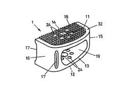
The intervertebral implant shown in FIGS. 1-3
essentially consists of a frame-like cage 1, closed at its
cover face 11 and base face 12 (except for perforations),
with two lateral faces 13 and 14, each of these having a
longitudinal hole 19, a front wall 16 which has two grooves
17, and a rear wall 15. The grooves 17 serve to receive a
manipulation instrument. The cage 1 is wedge-shaped, i.e.
with cover and base faces 11, 12 which diverge toward the
front wall 16.
In the design of FIGS. 1-3, the cover and base faces
11, 12 have a three-dimensional structure 18, preferably in
the form of pointed teeth in a regular arrangement, with a
height of about 1.75 mm, so as to improve the positional
stability of the implant. The cover and base faces 11, 12
have a free edge 32 without such a structure 1. The free
edge 32 reduces the risk of injury during and after the
operation.
CA 02229822 2003-06-13
The cover and base faces 1I, I2 have a plurality of
perforations 24, whose total area amounts to 400 of the
total area of the cover and base faces 11, 12. The
5 individual area of an individual perforation 24 amounts to
I5o of the total area of the cover and base faces 11, 12.
The ratio VH/VK between the volume VH of the hollow space
and the total volume VK of the cage 1 amounts to 0.22.
As FIG. 1 shows, the front wall 16 of the cage 1 has
10 two grooves I7 to receive an instrument, so that the cage I
can be inserted into the intervertebral space and can be
positioned there.
FIG. 4-8 show other embodiments of the invention.
Apart from the modifications described below, these have
15 the same features as the embodiment of FIGS. 1-3. The
inventive implant consists of a frame-like cage 1 with a
cover face 1I, a base face 12, two lateral faces 13 and I4,
each having a longitudinal hole 19, a front wall 16, having
an aperture 25, and a rear wall 25 having an aperture 26.
20 The aperture 25 has lateral grooves 27, which can accept a
suitable manipulation instrument. The longitudinal holes
19, which are positioned in the lateral faces 13, 14, also
have lateral grooves 2, which can accept a suitable
manipulation instrument.
CA 02229822 2003-06-13
The cage 1 is wedge-shaped, i.e. with cover and base
faces 11, 12 which diverge toward the front wall 16.
In the embodiment of FIGS. 4-7, the cover and base
faces 11, 12 have a plurality of perforations 24. The
total area of these perforations is 480 of the total area
of the cover and base faces 11, 12. The individual area of
an individual perforation 24 amounts to l00 of the total
area of the cover and base faces 11, 12. The ratio VH/VK
l0 between the volume VH of the hollow space 20 and the total
volume VK of the cage 1 amounts to 0.21.
The perforations 24 in the cover and base faces 11, 12
of the implant can be varied in many respects, within the
inventive range. For example, FIG. 8 shows a variation of
the perforations 24 in the cover and base faces 11, 12 of
the implant of FIG. 4. Here, the total area amounts to 50~
of the total area of the cover and base faces 11, 12. The
individual area of an individual perforation 24 amounts to
15s of the total area of the cover and base faces 11, 12.
The ratio VH/VK between the volume VH of the hollow space
20 and the total volume VK of the cage 1 is 0.22.
In all the embodiments, the cage 1 can be made of
titanium, titanium alloy, ceramic, or a biocompatible
plastic, e.g. polyethylene.
CA 02229822 2003-06-13
12
The clinical application will now be described in
detail below.
The cage 1 shown in FIG. 1 is filled with bone chips
(bone graft) or bone replacement material, possibly under
compression, through the lateral perforations 19 in the
form of longitudinal hole recesses. Then the filled cage 1
is pushed into the cleared intervertebral space with the
help of a distending instrument. A tool, which is inserted
into the two grooves 17 in the front wall 16 of the cage 1,
can here be used as a manipulator.
The cage 1 can be formed either as a semi-implant, as
shown in FIGS. 1-3, so that two implants must be inserted
into the intervertebral space, or else it is also possible
to form two semi-implants integrally, as shown in FIGS. 4-
7, so that only one implant must be inserted into the
intervertebral space.