Note: Descriptions are shown in the official language in which they were submitted.
CA 02230032 l998-02-20
W O 97/48339 PCTrUS97/10750
DEVICE AND METHOD FOR IMPROVE~
QUANTITATIVE CORONARY ART~RY ANALYSIS
Field of the Invention
The present invention relates generally to system for
improving the calibration of a cardiac analysis program and
more particularly to an improved system for performing
coronary artery analysis and more particularly to an
improved system using a novel arterial phantom having known
internal diameters in combination with an angiographic
catheter and an improved image quality phantom to increase
the accuracy of existing ~uantitative coronary artery
analysis programs.
Backqround of the Invention
During angiographic or other diagnostic procedures
which use X-Rays, a system similar to that shown generally
in Figure 1 is used. The system generally includes an X-
Ray tube 10 which emits X-Rays from a general point source.
The X-Ray tube is positioned under a table 12 on which the
patient or other object of interest is positioned. In
order to provide for X-Ray photography, cinegraphic
recording and/or viewing of the subject, and image
intensi~ier 14 is positioned above the subject.
The bottom surface of the image intensifier 14
includes a grid 16 having a plurality of narrowly spaced
strips thereon in order to attenuate scattered X-Rays so
CA 02230032 l998-02-20
W O 97/48339 PCTrUS97/10750
- 2 -
that only the X-~ays which pass through the subject
directly from the X-Ray tube. The top of the image
intensifier includes a TV or similar camera 18 to t
dynamically view the subject of the study. For example,
5 the heart or other organ of a patient may be viewed through
the camera to allow the physician the ensure the proper
positioning and observe the operation heart or other organ.
Additionally, the top portion of the image intensifier 14
also typically includes a recording medium 20 such as a
10 film camera or digital recording medium to record the study
for later review and analysis. The image intensifier
incorporates a phosphorous screen 22 and a series of
focusing coils 24 which tend to cause the X-Ray beams to be
directed toward a phosphorous output lens 26. The beam
15 from the output lens is split by a beam splitter 28 to
provide output to the TV camera 18 and recording medium 20.
Current systems which are used to analyze coronary
arteries during diagnostic angiographic procedures include
automatic analysis programs to calculate the dimensions of
20 the arteries of a patient. While these programs have been
used for many years, the programs suffer from certain
inaccuracies which result from distortions that occur
during the image acquisition process as well as limitations
in the sharpness of the pixels of the ac~uired image. For
25 example, various studies have shown that the accuracy of
the analysis programs decreases as the size of the artery
of interest decreases and it is the arteries having the
reduced sizes that are of particular interest to the
cardiologist.
CA 02230032 1998-02-20
W O 97/48339 PCTrUS97/10750
-- 3
One of the major sources of image distortion with the
current systems is geometric distortion. Geometric
distortion results in pincushion or barrel distortion of
the image and may result from the lenses and focusing coils
in the imaging system as well as passage of the X-Rays from
the conical or point source of the X-Ray tube to the
generally planar grid 16 and image intensifier 14. This
type of distortion may result in an image which is concave
~pincushion~ or convex (barrel) shaped near the edges of
the image. Attempts to overcome this type of distortion
include calibration of the imaging system when it is
installed using a platform phantom having a plurality of
lead lines. The lead lines are aligned in a grid shape
horizontally and vertically along the platform with a known
distance of 1 cm between each other. Although this
procedure provides the program with the ability to correct
for the calibrated geometric distortion of the system,
geometric distortion also arises as the components of the
system age or are replaced. Additionally, because there is
a strong desire to minimize the dose of X-Rays that the
patients are exposed to, the images include noise
distortion and the images of the lead lines lose their
sharpness around their edges. Additionally, the use of the
lead lines does not present an accurate depiction of the
absorption of the X-Rays for the organs of interest in a
patient because the lead lines distort the X-Rays of the
image system in different proportions than the organs of
interest of the patient. As a result of the foregoing, the
calibration analysis may include a certain amount of error
CA 02230032 l998-02-20
W O 97/48339 PCT~US97/10750
-- 4
which is then passed on to the calibrated images of the
analysis program. Despite these difficulties, it is still
desirable to provide an initial or partial correction for
geometric distortion.
A further approach to improving image ~uality and
analysis of the artery sizes involves the calibration of
the analysis program using the procedure catheter. In the
current approach, the outer diameter of the procedure
catheter is assigned as a known distance and the areas of
lo interest are then comparatively analyzed based on this
distance. Difficulties in this approach arise from the
lack of image sharpness inherent in the X-Ray type of
imaging system as well as from the many different
manufacturers and varieties of catheters which are
available today. Further complicating the attempts to
calibrate the analysis programs based on the catheter
diameter is the fact that the catheters are made of various
materials, each of which absorb and scatter the X-Rays
differently. Each of these difficulties is then
exacerbated by the magnification of the image for use in
the analysis program. Despite this, the use o~ a procedure
catheter to calibrate the analysis program is beneficial
because the procedure catheter is useful as a reference to
compare to the arteries because the absorption
characteristics of the X-Rays for the catheters and
arteries have greater similarities between each other than
the lead lines and arteries. Additionally, the similarity
in object size, dimension and object contrast between the
CA 02230032 1998-02-20
W 097/48339 PCT~US97/10750
- 5 -
procedure catheter and the nearby artery provides a useful
reference for identifying the walls of the arteries.
Based on the foregoing, there remains a need for
improved calibration or error correction devices and a
method of their use to improve the ~uality of existing
analysis programs for imaging systems.
Furthermore, there remains a need for a reliable and
consistent calibration or error correction system which may
be used to compare the relative differences between imaging
systems to allow the images to be analyzed by a common
analysis system without introducing additional errors.
Summary of the Invention
An advantage of the present invention is that it
provides a readily reproducible image system specific
correction which allows for the accurate comparison of the
procedure catheter and arterial cross-sections.
Another advantage of the present invention is that it
provides a reliable method to compensate for deviations in
the analysis program from the ideal regression curve caused
by image system distortion.
Yet another advantage of the resent invention is that
it permits calculated arterial diameters to relate directly
to known procedure catheter diameters.
Yet another advantage of the system of the present
invention is that it reduces the procedure induced errors
which occur during the initial calibration of the image
CA 02230032 l998-02-20
W O 97/48339 PCT~US97/10750
- 6 -
system and also increases the accuracy of the comparative
calculations between the procedure catheter and the artery.
The present invention includes improvements in the
imaging phantom, as well as improvements in the calculation
of the imaged catheter size and dimensions. The imaging
phantom of the present invention preferably includes a grid
pattern which is made of bronze balls having a diameter of
about 1 mm. The use of bronze balls rather than the
traditional lead lines is preferred because the absorption
characteristics of bronze more closely resembles the
absorption characteristics of the iodine based dyes which
are used during the imaging procedure. Additionally, the
smaller size is chosen to more closely approximate the size
of the arteries of interest.
The improvements in the calculation of the size of the
imaged procedure catheter and artery include the use of an
image ~uality phantom having a dye filled telescopic-shaped
interior that is compared directly to the imaged procedure
catheter so that the imaged dimension of the procedure
catheter may be compared directly to a variety of known
dimensions present in the imaged quality phantom. The body
of the image quality phantom is formed of a material which
approximates the absorption characteristics of the arteries
of the patient. The results of this comparison may then be
used to identify the absorption characteristics of the
procedure catheter and to correct the distortion and image
degradation present in the image provided to the analysis
program at each of the known dimensions of the image
~uality phantom through the use of a regression curve which
CA 02230032 1998-02-20
W O 97/48339 PCT~US97110750
-- 7
is applied during the final edge detection pass of the
analysis program to modify edge placement by the analysis
program. Initial studies indicate that this comparison
significantly increases the accuracy of the analysis
program, particularly for the smaller diameter measurements
of the artery.
As described more fully herein, the present invention
provides a system for overcoming many of the inherent
limitations of the current level of reliance on the
procedure catheter as a scaling device to compensate for
geometric magnification in current analysis programs.
Among the benefits of the present invention are the
abilities to measure, on an imaging system specific basis,
the overall regression curve for diameter response of the
analysis program and the ability to relate the calipered
diameter of a particular procedure catheter to the overall
regression curve of the analysis program. Additionally,
the present invention allows the user to compensate for
deviations from the ideal linear response diameter response
for the specific imaging system.
The method of the present invention generally includes
a radiographic phantom consisting of a series of
cylindrical model arterial segments spanning the range of
diameters encountered in coronary angiography (0.5 mm to
5.0 mm~. The segments of the phantom are filled with an
iodinated material of the concentration of standard iodine
contrast medium, are arranged coaxially, nd-to-end, in 5 mm
lengths and imbedded in a block of tissue equivalent
absorber. This phantom is imaged in-vitro along side a tip
CA 02230032 1998-02-20
WO 97/48339 PCTrUS97/10750
-- 8
of the particular procedure catheter which is to be used
clinically. The analysis program is applied to thee image
of the phantom and the procedure catheter tip at a sampling
density several times that employed clinically to ensure
precise sampling at each diameter. This method yields an
imaging system specific diameter regression curve over the
range of diameters represented and a ratio of the calipered
to detected procedure catheter diameters. The ratio is
applied as a scaler correction to the regression curve to
compensate ~or any difference between detected and
calipered procedure catheter diameters. The resulting
scaler-corrected regression data is used to derive
diameter-specific correction factors to linearize the
diameter response of the analysis program for a particular
imaging system and procedure catheter. Tables of these
corrected regression curves are stored and used in
subsequent clinical applications of the analysis program.
The result is improved linearity of diameter response as
well as increased precision of analysis program results
under varying imaging conditions using different imaging
systems.
Brief Description of the Drawinqs
Figure 1 is a schematic drawing of a typical imaging
system;
Figure 2 is a top view of the image quality phantom of
the present invention;
CA 02230032 1998-02-20
W 097/48339 PCT~US97/10750
g
Figure 3 is a top view of a prior art phantom as
disclosed in U.S. Patent No. 4,873,707;
_ Figure 4 is a top view of the arterial phantom of the
present invention;
. Figure 5 is a side view of the arterial phantom of the
present invention;
Figure 6 is an end view of the arterial phantom of the
present invention;
Flgure 7 is a top view of the arterial phantom of the
present invention placed on the table 12 of the imaging
system;
Figure 8 is a top view of the arterial phantom of the
present invention and a portion of the procedure catheter
placed on the table 12 of the imaging system;
Figures 9A and 9B are comparative plots with and
without the calibration method of the present invention,
respectively.
Detailed DescriPtion of the Present Invention
Although each of the individual devices are described
herein as being part of the overall system to improve the
accuracy of the analysis program, it is not believed to be
necessary that each of the devices and each step in the
method described below be present to provide significant
improvement in existing analysis programs.
As shown in Figure 2, the image quality phantom 30 of
the present invention includes a series of spaced apart
ball shaped members 32. These ball shaped members 32 are
CA 02230032 1998-02-20
W O 97/48339 PCT~US97/10750
-- 10 --
preferably made of bronze to closely approximate the
absorption characteristics of the imaging material. In
angiographic procedures, the imaging material is typically
an iodine based material such as the imaging material sold
as RENOGRAPHIN. The diameter of the ball shaped members 32
is preferably about 1 mm so that the each imaged ball
shaped member has a diameter which is similar to the
diameter of the arteries of interest. Additionally, the
centers of each ball shaped member 32 are spaced apart from
each other a constant distance such as 1 cm. The overall
diameter of the image quality phantom 30 is sufficient to
cover the entire image field of the image system when the
image quality phantom 30 is placed on the table 12 of the
image system. In the preferred form of the present
embodiment, the diameter is about 33.5 cm and has a
thickness of about one-eighth inch to provide the desired
X-Ray scatter. The preferred material is a polycarbonate
resin or acrylic material although other materials are
believed to be similarly suitable for the intended use of
the image quality phantom 30.
The image quality phantom 30 may be used during the
initial calibration of the image system or at anytime
thereafter to evaluate the performance of the image system.
Typically, the image quality phantom 30 will be used
whenever degradation of the image quality from the image
system is suspected. The evaluation report will usually
evaluate the sharpness, spatial linearity, brightness
uniformity and signal or noise of the images either
regionally, globally or both. The results o~ the
CA 02230032 l998-02-20
W 097/48339 PCTrUS97/1~750
evaluation will then be used to ad3ust the components of
the image system or to provide image correction in the
r event of image distortion of the types known as pin cushion
or barrel distortion.
A further improvement in the system of the present
invention includes an arterial phantom 34 of the type shown
in Figures 4-7.
Figure 3 is illustrative of a prior art phantom for
use in computer tomography. The arterial phantom 34 of the
10 present invention is preferably a generally elongate block
shaped member with a telescopic shaped recess 36 therein.
In the preferred form of the present embodiment, the
arterial phantom has a dimension of about 30 mm and a
height of about 20 mm. The arterial phantom 34 is
15 preferably made of a polycarbonate or acrylic material
which approximates the X-Ray scatter and distortion that
occurs during an X-Ray of the coronary arteries of a
patient. The recess 36 of the arterial phantom preferably
includes multiple decreasing diameter stepped cylindrical
20 surfaces and is filled with an angiographic imaging dye
such as RENOGRAPHIN. In the preferred embodiment, the
recess 36 includes ten different diameter surfaces each
having an identical length of about 5 mm. The diameter of
the largest surface is preferably about 5 mm and the
25 diameter of the smallest surface is preferably about
r 0~5 mm.
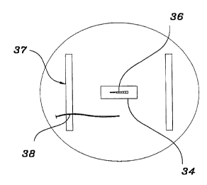
As shown in Figure 8, the procedure catheter 38 or a
catheter of the same type, size and manufacturer of the
catheter to be used during the procedure are placed on the
CA 02230032 1998-02-20
W O 97/48339 PCTrUS97/10750
- 12 -
table 12 of the imaging system to perform the image
calibration step of the present invention. In this
example, the table 12 may include adhesive type strips 37
to retain the procedure catheter 38 in the desired position
relative to the arterial phantom. The data received from
the in vitro imaging procedure catheter 38 and the arterial
phantom 34 is used to create a regression curve which is
developed from the arterial phantom plot over the range of
diameters represented. Additionally, the imaged procedure
catheter 38 and arterial phantom 34 are used to create a
ratio of calipered-to-detected catheter diameters based on
the known diameter of the catheter and the known diameter
of the various portions of the arterial phantom 34.
Thereafter, the tables of regression curves are stored and
used in subsequent clinical applications of the analysis
program. Finally, during the final pass of the analysis
program over the data from the image of interest, the
analysis program applies the regression correction data
from the regression curve to modify the edge placement of
the analysis program. As verification of this method, a
pair of standard analysis program plots using the same
arterial model before and after the application of the
present invention are shown in Figures 9A and 9B. The plot
of the diameter vs. the segment length of the arterial
phantom 34 shown in Figure 9A illustrates the nonlinear
response of the analysis program and the over estimation of
the measurements under 1 mm of a currently available
analysis program without use of the devices and method of
the present invention. The plot of the diameter vs. the
CA 02230032 1998-02-20
W 097/48339 PCTrUS97/10750
- 13 -
segment length of the arterial phantom 34 shown in Figure
9B illustrates the improvements to the linearity of
diameter response and the significantly increased accuracy
of measurements under 1 mm of the same currently available
analysis program using the arterial phantom 34 and the
method of the present invention.