Note: Descriptions are shown in the official language in which they were submitted.
CA 02230063 1998-02-20
W O 97/07744 PCT~US96/13570
OPEN HE~ICAL ORGANIC TISSUE ANCHOR
AND MET~OD ~F FACILITATING ~T.TNG
FI~LD OF lN V ~ llON
The present invention relates to tissue anchors
as well as to methods of promoting healing or repairing
hard or soft, living, organic tissue using an open helix
anchor.
R~R~ROUND OF THE lNv~ION
The present invention relates to an anchor (or
connector) which can be used to fasten organic tissue in
close proximity in order to afford the tissue the opportu-
nity to heal. The anchor of the present invention can be
used to anchor and clamp dense, regular and/or dense,
irregular connective tissue in place in relation to bone.
The anchor can also be used for cartilage transplants,
i.e., for holding cartilage in fixed relation to bone, and
can also be used in bone as a buttress, such as for
buttress plating techniques, or to fasten pieces of bone
together as a screw substitute.
As compared to the prior art, the anchor of the
present invention allows a method of holding together
organic tissue with minimal disruption to the biological
environment or to the tissue itself. For example, prior
art devices and methods customarily require a large hole
for insertion of the anchoring device, causing not only
structural damage to the implantation site, but also
inflicting further trauma to the biological site such as
generating heat, introducing further possibility for
infection, and destroying bone which may be needed to help
heal the repaired area. Such trauma is amplified in cases
where prior art devices malfunction during the implant
procedure. Hooks or screws can get stuck and further
obscure the operating site or require tedious removal.
The anchor of the present invention may be very
useful for applications such as anchoring ligaments or
tendons when performing soft tissue surgical reconstruc-
tion, ruptured tendons, or torn ligaments, in which the
CA 02230063 1998-02-20
W O 97/0774~ PCT~US96/13570
- 2 -
surgeon wants to reconstruct or repair connective tissue
with respect to the bone.
The anchoring device functions to hold together
the tissue (such as connective tissue to bone) for a
relatively limited time frame, e.g., six to twenty-six
weeks, during which time the biological system will heal.
The anchor of the present invention can be used
with advantage in many of the same applications in which
cancellous screws are used in addition to applications in
which traditional prior art anchoring techniques are
unsatisfactory. The anchor of the present invention is
far less invasive to implant than cancellous screws or
hook-style anchors, i.e., the implant has a minimized
mass, the insertion point is small relative to the size of
the implant, and the device involves minimal removal of
native tissue. In addition, the area of bone which is
needed to secure the present invention can be of poorer
quality than for prior art devices.
Additionally, the anchor of the present inven-
tion can be removed and minimally reangulated in order toutilize the same surgical site. Prior art devices require
a large hole (relative to implant size) to be drilled in
order to implant the device, and once the hole is contami-
nated by malfunction or misalignment of the device, it is
necessary to drill another hole far enough away to achieve
stability in a new location. Given the surgical context,
this is extremely inconvenient.
The anchor of the present invention can be used
in methods of ligament, tendon, or other tissue repair.
For example, the anchor can be used for a method involving
cartilage transplant and it can be used alone or in
conjunction with a plate for a method of buttressing bone
where the quality of bone may be questionable due to
trauma or degenerative disease. The anchor may be used in
methods of fixation involving connective tissue repair and
replacement and may be inserted using a plunge-handle or
CA 02230063 1998-02-20
W O 97/07744 PCT~US96/13570
-- 3
"T" handle inserter which utilizes longitudinal travel in
order to achieve rotational insertion.
Specifically, the anchor is used in ligament or
tendon repair in which a pilot hole, having a diameter
much smaller than the outer diameter of the helical
anchor, is drilled in the cortex of the bone. The angle
of implantation can be varied as necessary. The anchor is
subsequently mounted or loaded into the insertion tool,
threaded into the pilot hole, and screwed into the bone an
appropriate di5tance so that the anchor head can be
accessed but is not obtrusive. The ligament or tendon is
attached to the anchor, such as by suturing.
In addition, the anchor of the present invention
can be used to anchor plates and is particularly useful in
instances where the bone is of poor quality. In one
embodiment, a modular head is used. A particularly
desirable head has an internal hex slot to permit the
anchor to be implanted In addition, the head has a
transverse through slot to hold a suture. The head has a
low, rounded profile with a distal stem which fits inside
a ring of the helix and is laser-welded thereto.
8UMMARY OF THE lNv~NllON
The anchor in accordance with the invention
comprises an open helical structure which is a constant or
varied-diameter, elongate member, fiber, or filament
comprised of a relatively rigid, biocompatible material
such as a wire having a diameter which may vary optimally
from about 0.2 millimeters to about 5.0 millimeters. The
length of the anchor will depend upon the particular
application, but will range generally from about 3.0
millimeters to about 75.0 millimeters with the upper
ranges being useful for buttressing techniques. The outer
diameter of the helix will also vary in accordance with
the application, but it will range generally from about
1.5 millimeters to about 15.0 millimeters. A suitable
rate of slope for the helix is from about 0.5 to about 10
CA 02230063 1998-02-20
W O 97/07744 PCTAJS96/13570
turns per centimeter. The aspect ratio of the helix,
which as used herein means the ratio of the helix outer
diameter to the fiber diameter, is an important ratio in
order to achieve the proper stiffness to enable insertion
and to firmly seat in the bone; a suitable range is 3.5 to
4.5.
Advantageously, the anchor of the present
invention involves relatively simple, cost-effective
manufacturing processes. The present anchor is also less
intimidating to doctors and patients than prior art
devices and can be used with simple, straight-forward
instrumentation. Finally, since the device is relatively
noninvasive, several can advantageously be used together
in instances where more than one prior art device could
not be used. It is preferred, but not necessary, that the
helix has a constant circular diameter and a constant
slope (meaning the rate of turn per unit of longitudinal
length).
For its connective applications, the anchor
includes an attachment head at one end which is suitable
for securing the tissue or suture which is to be held.
For example, in the case of a filamentary anchor, the
anchor may have a hook, crossbar or eyelet. For applica-
tions in which the anchor secures rigid material such as
cartilage or a buttressing plate, the head may have a
surface which is designed to distribute the load evenly
over the rigid material.
In a second embodiment, the anchor will have a
modular head. For example, the helical anchoring portion
may terminate at the superficial end in a post that will
accommodate one of several head options. These head
options may include a button, clamp, clip, snap, or rivet.
At the other end, the anchor includes a cutting or self-
tapping point.
In accordance with another embodiment of the
invention, a buttressing system is provided which compris-
CA 02230063 1998-02-20
W O 97/07744 PCT~US96/13570
-- 5
es a plate having at least two through bores which are
each engaged by an open-helix anchor.
In accordance with a method of the present
invention, an anchoring site is surgically accessed, the
helical anchor is screwed into the anchoring site, and
connective tissue is secured to the attachment head of the
anchor.
In accordance with another method of the inven-
tion, a bone is buttressed by surgically accessing an
implant site, aligning a plate having at least one aper-
ture over the site, and securing the plate to the implant
site by inserting an open-helix anchor through the aper-
ture and into the implant site to anchor the plate with
respect to the implant site.
DESCRIPTION OF THE DRAWINGS
FIGURE 1 is an elevational view of the anchor
device showing the attachment head in side elevation;
FIGURE 2 is a top view taken of FIGURE 1;
FIGURE 3 is an elevational view, similar to
FIGURE 1, but showing the anchor rotated 90~ to the right
so that the attachment head is seen in an end view;
FIGURE 4 illustrates the pilot hole in the bone
prior to insertion of the anchor;
FIGURE 5 illustrates an anchor in place in the
cancellous portion of the bone with the attachment head
projecting above the surface of the bone in order to allow
attachment of the soft tissue to the anchor;
FIGURES 6 and 7 illustrate the tool which may be
used for inserting the anchor;
FIGURE 8 is a cross-section of a second embodi-
ment of the anchor having a modular head; and
FIGURE 9 is a top view of the head illustrating
the slot in phantom.
CA 02230063 1998-02-20
W O 97/07744 PCT~US96/13570
-- 6 --
DE~TT~n DESCRIPTION OF THE lNv~:N-lloN
In accordance with the invention, FIGURES 1-3
illustrates the anchoring device in accordance with the
invention enlarged to show the invention in detail gener-
ally at 10. The anchoring device lO comprises an open
helix 12 having a pointed insertion tip 14 at one end and
an attachment head 15 at the other end.
Preferably, the anchoring device is comprised of
a rigid, biocompatible material having a high-yield
strength such as stainless steel or titanium. The device
can also be made from a biodegradable material such as
polyglycolic acid ("PGA"), polylactic acid ("PLA"),
polydiaxone hydroxy apatite ("PDA"), and the like. For
example, the device lo may be made from surgical-grade
titanium or stainless steel wire having a wire diameter
ranging from about 0.4 millimeters to about 3.0 millime-
ters, and more specifically from about 0.5 millimeters to
about 2.0 millimeters, and most specifically from about
1.0 millimeters to about 2.0 millimeters. Optionally, the
helix diameter may be of variable cross-section ranging
from a smaller-diameter wire at the insertion tip to a
larger-diameter wire near the attachment head 15.
The "slope" of the helix is used herein to mean
the number of turns (i.e., one 360~ rotation) per unit
length and varies from about 0.5 turn per centimeter to
about 10 turns per centimeter, and more specifically from
about 0.5 turn to about 4 turns per centimeter, and most
specifically from about 1 to about 2 turns per centimeter.
The anchor generally comprises a length of helix suffi-
cient to achieve from 0.75 to 4 complete 360~ revolutions,or more specifically from about 1 to about 3 revolutions.
Accordingly the length of the anchor for most general
fastening or anchoring applications is from about 3 to
about 18 millimeters, and more specifically from about 4
to about i5 millimeters, and most specifically from about
8 to about 15 millimeters. For plating or buttressing
applications, the length of the anchor will generally
CA 02230063 1998-02-20
WO 97/07744 PCTrUS96/13570
-- 7
range from about 5 to about 75 millimeters, preferably
from about 5 to about 40 millimeters and most preferably
from about 10 to about 20 millimeters.
The overall outer diameter of the open helix
portion 12 of the anchoring device 10 ranges from about
1.5 to about 11 millimeters, and more specifically from
about 3 to about 9 millimeters, and most specifically from
about 5 to about 7 millimeters. The wire is generally
circular in cross-section, although it is envisioned that
it may be angular such as diamond-shaped or rhombohedral.
It is important that the anchor have an aspect
ratio of from about 3 to about 5, preferably from 3.5 to
4.5, and most preferably around 4. As used herein, aspect
ratio means the ratio of the helix outer diameter to the
wire diameter. If the ratio i5 too large, the device is
too rigid, whereas if the ratio is too small, the device
is overly flexible.
The attachment head 15 of the anchoring device
10 may vary according to the specific application. For
example, it may be desirable to include a broader compres-
sion area for direct attachment of connective or soft
tissue to bone, as compared to suture techniques involving
suturing or wiring the soft tissue in place with respect
to the anchoring device. Examples of attachment heads
suitable for suturing or wiring connective tissue include
crossbars, hooks and eyelets.
FIGURE 1 illustrates an attachment head 15
having a crossbar 17 which arches slightly above the last
helical turn and is attached such as by spot welding 18 at
the terminal end. It may be further preferable to include
an opening 19 or cannulation in the crossbar to allow for
cannulated surgical techniques (i.e., placement of the
anchor over a positioned wire which may be subsequently
removed). The opening may range in size from 0.5 millime-
ters to 1.5 millimeters depending on the application.
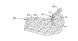
The device and method of the invention areillustrated in FIGURES 3-5. In particular, FIGURE 4
CA 02230063 1998-02-20
W O 97/07744 PCTAUS96/13570
- 8 -
illustrates a section of bone generally at 20 having a
cortex 22 and a cancellous portion 24. A pilot hole 25
has been drilled in the cortex 22 in order to ease inser-
tion of the anchoring device 10. A countersink hole 26
through the cortex is also illustrated.
FIGURE 5 illustrates the anchoring device 10 as
it has been partially implanted through the pilot hole 25
into the cancellous portion of the bone. In some instanc-
es where the cortex is particularly thin, a pilot hole may
be unnecessary. The soft tissue is attached to the
anchoring device when the device is in position such as by
suturing or wiring to the attachment head 15 of the
anchoring device 10.
FIGURES 6 and 7 illustrate an instrument which
can be used for the implantation of the anchor in accor-
dance with the present invention. Specifically, the
instrument includes a central shaft 30 having a T-shaped
handle 32 designed to allow the surgeon to easily grasp
the handle 32 and rotate the shaft 30 to screw the anchor
10 into the bone through the optional pilot hole. The
placement guide 34 includes a bottom surface 36 which can
rest against the cortical surface where the anchor 10 is
to be implanted. The guide 34 further includes an inter-
nal opening 38 having a diameter sufficient to receive the
top portion of the anchor 10. The guide 34 further
includes a bore 40 which provides a bearing surface for
the shaft 30. At its lower end, the shaft 30 includes a
head 42 having an internal slot 44 which receives the
crossbar of the anchor 10 to enable the surgeon to apply
torque to the anchor. The head 42 has an external diame-
ter which cooperates with the internal diameter of the
anchor 10. Optionally, the shaft 30 may also include a
longitudinal, internal opening to receive a guide wire to
allow for further cannulated surgical techniques
During use of the anchor of the present inven-
tion, the attachment location is approached with standard
surgical exposure. A pilot hole is drilled through the
CA 02230063 1998-02-20
W O 97/07744 PCT~US96/13570
near cortex only and a drill sleeve is used to protect
surrounding soft tissues. The anchoring device 10 is
inserted with an insertion tool such that the attachment
head 15 is left out of the bone. The angle of insertion
may be perpendicular to the bone surface or at a 45~
angle. A suture may be passed under the exposed crossbar
17 of the attachment head 15 once or twice, depending on
the surgeon's choice. The attachment tool is then used to
countersink the attachment head 15 below bone level. The
ligament or tendon is then sutured into place with a pre-
ferred suturing method such as Bunnell, whip, or modified
Kessler. The wound is subsequently closed and the proce-
dure is completed in standard fashion.
FIGURES 8 and 9 show a second embodiment of the
anchor 80 having a modular head 82 attached to a helix 84.
The helix 84 engages the bone as shown in the earlier
embodiments. This version rotates through 540~ (1~ full
rotations) and terminates at one end in a three-sided
point 86. At the other end, the helix 84 is formed into
a ring 88 to form a seat for the head 82. The ring 88 may
be a complete circle or less than a circle, so long as it
forms a good seat for the head 82. Preferably the ring 88
is the same diameter as the helix and the head 82 has the
same outer diameter as the ring in order to allow the head
to be countersunk into a plate or bone.
Preferably both the head 82 and helix 84 are
formed of implant-grade stainless steel (such as SS 22-13-
5). The head 82 has a low, rounded, top profile, project-
ing from about 0.02 to 0.2 inch, and preferably from 0.05
to 0.1 inch from the top surface of the helix ring 88.
The head 82 also includes an internal hex opening 90 to
receive an anchor driver. The head 82 also includes a
transverse through slot 92 shown in phantom in FIGURE 9.
The slot can be used to hold sutures in order to anchor
tendons or ligaments. On the opposite side, the head 82
includes a necked area or stem 94 which is a constant
CA 02230063 1998-02-20
WO 97/07744 PCT~US96/13570
-- 10 --
diameter cylinder welded or otherwise adhered along the
bottom edge to the ring 88.
EXAMPLE
Six samples of surgical-grade, stainless steel
bone anchors in accordance with the invention were placed
in a sample of artificial cancellous bone. Two samples
each had a total longitudinal length of about 20 millime-
ters. The other four samples each had total lengths of
about 13 millimeters. The outer diameter of all samples
was 5 millimeters and the wire diameter was 1.5 millime-
ters. Both long samples and two short samples had attach-
ment heads which were crossbars and were attached by
heliarc spot welding. The other short samples had cross-
bar attachment heads which were not welded.
Pullout tests were conducted using an MTS
instrument. Straight, longitudinal pull was applied to
the embedded anchors; this reproduced the least favorable
condition for pullout characteristics. The results are
shown in the table below. "Displacement" refers to
bending of the crossbar in the longitudinal direction.
TAB~E I
PLASTIC DEFORMATION
SHORT/NON-WELDED SHORT/WELDED LONG/WELD~D
Average 48 lbs. Average 52 lbs. with Average 58 lbs. with
with 2 millimeters 2.2 millimeters of 2.4 millimeters of
of displacement displacement displacement
All of the numbers represent desirable anchoring values.
While in accordance with the patent statutes the
best mode and preferred embodiment has been set forth, the
scope of the invention is not limited thereto, but rather
by the scope of the attached claims.