Note: Descriptions are shown in the official language in which they were submitted.
CA 02230967 1998-05-01
Improved Nucleic Acid Assays
Field of the Invention
The present invention relates to the detection of specific nucleic acid
sequences in a target
test sample.
In particular, the present inventioni relates to the automated detection of
specific nucleic
acid sequences v/hich are either unamplified or amplified nucleic acid
sequences (amplicons).
In addition, the present invention 1-elates to the use of automated
amplification, methods
and compositions for monitoring successful amplification, improved methods for
reducing the
lo chance for contamination, and the use of ianified reaction buffers and unit
dose aliquots of
reaction components for amplification.
Finally, the present invention also relates to unique constructs and methods
for the
conventional or automated detection of one, or more than one different nucleic
acid sequences in
a single assay.
The Backg~rounci of the Invention
The development of techniques for the nianipulation of nucleic acids, the
amplification of
such nucleic acids when necessary, and t?he subsequent detection of specific
sequences of nucleic
acids or amplicons has generated extremely sensitive and nucleic acid sequence
specific assays
fo r the diagnosis of disease and/or identification of pathogenic organisms in
a test sample.
Amplification of nucleic acids
When necessary, enzymatic amplification of nucleic acid sequences will enhance
the
ability to detect such nucleic acid sequer.ices. Generally, the currently
known amplification
schemes can be broadly grouped into two classes based on whether, the
enzymatic amplification
reactions are driven by continuous cycling of the temperature between the
denaturation
temperature, the primer annealing temperature, and the amplicon (product of
enzymatic
arnplification of nucleic acid) synthesis temperature, or whether the
temperature is kept constant
throughout the enzymatic amplification process (isothermal amplification).
Typical cycling
nucleic acid amplification technologies P,thermocycling) are polymerase chain
reaction (PCR),
McDonnell Boehnen
Hulbert & Berghoff
300 S. Wacker Drive
Cliicago, Illinois 60606
(312)913-0001 2
CA 02230967 2008-01-07
50621-4
and iizase chain reaction (LCR). Specific protocols for such reactions are
discussed in. for
example. Short Protocols in Molecular Biology, 22"` Edition, A Compendium of
Methods from
Current Protocols in iviolecular Biolow, (Eds. Ausubel et al., Tohn Wiiev &
Sons, New York.
1992) chapter 15. Reactions which are isothemial include: transcription-
mediated amplification
(TMA), nucleic acid sequence-based amplification (NASBA), and strand
displacement
amplification (SDA).
ti.S. Patent documents which discuss nucleic acid amplification include
4,683,195;
4,683,202; 5,130,238; 4,876,187; 5,030,557; 5,399,491; 5,409,818; 5,485,184;
5,409,818;
5,554,517; 5,437,990 and 5,554,516. It is well known that methods such as
those described in these
i c~ patents permit the amplification and detection of nucleic acids without
requiring cloning, and are
responsible for the most sensitive assays for nucleic acid sequences. However,
it is equally well
recognized that along with the sensitivity of detection possible with nucleic
acid amplification, the ease of
contamination by minute amounts of unwanted exoaenous nucleic acid sequences
is extremely
great. Contamination by unwanted exogenous DNA or RNA nucleic acids is equally
likely. The
15 utility of amplification reactions will be enhanced by methods to control
the introduction of
unwanted exogenous nucleic acids and other contaminants.
Prior to the discovery of thermostable enzymes, methods that used
therrnocycling were
made extremely difficult by the requirement for the addition of fresh enzyme
after each
denaturation step, since initially the elevated temperatures required for
denaturation also
20 inactivated the polymerases. Once thermostable enzymes were discovered,
cycling nucleic acid
amplification became a far more simplified procedure where the addition of
enzyme was only
needed at the beginning of the reaction. Thus reaction tubes did not need to
be opened and new
enzyme did not need to be added during the reaction, allowed for an
improvement in efficiency
and accuracy as the risk of contamination was reduced, and the cost of enzymes
was also reduced.
25 An example of a thermostable enzyme is the polymerase isolated from the
organism
Thermopliilus aquaticus.
In aeneral, %sothermal amplification can require the combined activity of
multiple enz'VnIe
activities for which no optimal thermostable variants have been described. The
initial step of an
amplification reaction will usually require denaturation of the nucleic acid
target, for example in
CA 02230967 1998-05-01
the TMA reaction, the initial denaturation step is usually _ 65 C, but can be
typically _ 95 C, and
is used when required to remove the secoridary structure of the target nucleic
acid.
The reaction mixture is then cooled to a lower temperature which allows for
primer
annealing, and is the optimal reaction temperature for the combined activities
of the amplification
enzymes. For example, in TMA the enzymes are generally a T7 RNA polymerase and
a reverse
trariscriptase (which includes endogenous RNase H activity). The temperature
of the reaction is
kept constant through out the subsequent -isothermal amplification cycle.
Because of the lack of suitable thermostable enzymes, some isothermal
amplifications
will generally require the addition of enzymes to the reaction mixture after
denaturation at high
temperature, and cool-down to a lower teinperature. This requirement is
inconvenient, and
requires the opening of the amplification reaction tube, which introduces a
major opportunity for
coritamination.
Thus, it would be most useful if such reactions could be more easily performed
with a
reduced risk of contamination by methods which would allow for integrated
denaturation and
amplification wiithout the need for manual enzyme transfer.
Amplification Buffer and Single Ts:eaction Aliquot of Reagents
Typical reaction protocols require the use of several different buffers,
tailored to optimize
the activity of the particular enzyme being used at certain steps in the
reaction, or for optimal
2o resuspension of'reaction components. For example, while a typical PCR 10x
amplification buffer
will contain 500inM KCI and 100mM Tris HC1, pH 8.4, the concentration of MgC12
will depend
upon the nucleic acid target sequence and primer set of interest. Reverse
transcription buffer (5x)
typically contains 400mM Tris-Cl, pH 8.2; 400mM KCl and 300 mM MgC1Z1 whereas
Murine
Maloney Leukemia Virus reverse transcriptase buffer (5x) typically contains
250mM Tris-Cl, pH
8.3; 375mM KC1; 50mM DTT (Dithiothreitol) and 15mM MgC1z.
While such reaction buffers can be prepared in bulk from stock chemicals, most
commercially available amplification products provide bulk packaged reagents
and specific
buffers for use with the amplification protocol. For example, commercially
available manual
arnplification assays for detection of clir-ically significant pathogens (for
example Gen-Probe Inc.
Chlamydia, an(i Mycobacterium tuberculosis detection assays) requires several
manual
McDonnell Boehnen
Hulbert & Berghoff
300 S. Wacker Drive
Chicago, Illinois 60606
(312)913-0001 4
CA 02230967 2008-01-07
50621-4
manipulations to perforrn the assay, including dilution of the test sample in
a sample dilution
buffer ( SDB). combination of the diiuted sample with amplification reaction
reagents such as
oliaonucleotides and specific oliQonucleotide promoteriprimers which have been
reconstituted in
an amplification reconstitution buffer (ARB). and finally, the addition to
this reaction mixture of
enzymes reconstituted in an enzvme dilution buffer (EDB).
The preparation and use of multiple buffers which requires multiple manual
additions to
the reaction mixture introduces a greater chance for contamination. It would
be most useful to
have a sinale unified buffer which could be used in all phases of an
amplification protocol. In
particular, with the commercially available TMA assays described above, the
requirement for
1 o three buffers greatly complicates automation of such a protocol.
Bulk packaging of the enzyme or other reaction components by manufacturers,
may
require reconstitution of the components in large quantities, and the use of
stock amounts of
multiple reagents, can be wasteful when less than the maximal number of
reactions are to be
carried out, as some of these components may be stable for only a short time.
This process of
15 reconstitution also requires multiple manipulations by the user of the
stock reagents, and
aliquoting of individual reaction amounts of reagents from stocks which
creates a major
opportunity for contamination.
Methods and compositions for the preparation of bulk quantities of preserved
proteins are
known, see for example, U.S. Patent 5,098,893; 4,762,857; 4,457,916;
4,891,319; 5,026,566 and
20 interr.ational Patent Publications WO 89/06542; WO 93/00806; WO 95/33488
and WO
89/00012. However, the use of pre-aliquoted and preserved reagent components
ui single reaction
quantities/dose is both very useful and economical. Single aliquots of enzyme
reagent avoids multiple use
of bulk reagents, reducing waste, and greatly reducing the chance of
contamination. Further, such single
reaction aliquots are nlost suitable for the automation of the reaction
process.
The requirement for m,any changes of buffer and the multiple addition of
reagents
compiicates the automation of such reactions. A single dose unit of reaction
buffer mixture. and a
unified combination buffer will both simplifi- automation of the process and
reduce the chance of
contamination.
CA 02230967 2008-01-07
50621-4
.qistomation of.N-ucleic Acid Detection witi7 or l~itliout.qnzplification
~ucleic acid probe assays. and combination arnplification probe assays can be
rapid.
sensitive. hi zhlv specific, and usually require precise handlin2 in order to
mini-nize
contamination with non-specific nucleic acids, and are thus prime candidates
for automation. As
-with conventional nucleic acid detection protocols. it is Qenerally required
to utilize a detection
probe oliQonucleotide sequence which is linked by some means to a detectable
sivnal ocneratinv
component. One possible probe detection system is described in U.S. Patent
4,581,33 In addition, automation of a nucleic acid detection system targeting
unamplified or
1o amplified nucleic acid, or a combined automated amplification/detection
system will generally be
adaptable to the use of nucleic acid capture oligonucleotides that are
attached to some form of
solid support system. Examples of such attachment and methods for attachment
of nucleic acid to
solid support are found in U.S. Patent 5,489,653 and 5,510,084.
15 Automation of amplification, detection, and a combination of amplification
and detection
is desirable to reduce the requirement of multiple user interactions with the
assay. Apparatus and
methods for optically analyzing test materials are described for example in
U.S. Patent 6,122,28=.
Automation is generally believed to be more economical, efficient,
reproducible and accurate for the
processing of clinical assays. Thus with the superior sensitivity and
specificity of nucleic acid detection
assays, the use of amplification of nucleic acid sequences, and automation at
one or more phases of an
assay protocol can enhance the utility of the assay protocol and its utility
in a clinical setting.
Advantage of Internal Control Sequences
25 Nucleic acid amplification is highly sensitive to reaction conditions, and
the failure to
amplify and/or detect any specific nucleic acid sequences in a sample may be
due to error in the
amplification process as much as being due to absence of desired target
sequence. Amplification
reactions are notoriously sensitive to reaction conditions and have generally
required includina
control reactions with known nucleic acid target and primers in separate
reaction vessels treated
3o at the same time. However, internal control sequences added into the test
reaction mixture would
6
CA 02230967 2008-01-07
50621-4
truly control for the success of the amplification process in the subject test
reaction miature
and would be most useful. U.S. Patent 5,457,027 teaches certain internal
control sequences
which are useful as an internal oligonucleotide standard in isothermal
aanplification reactions
for Mycobacteriunz tuberculosis.
i
- Howevcr it wouid b: extremely useful to have a general method of yeneratinL,
internal
control sequences, that would be useful as internal controls of the various
amplification
procedures, which are specincally tailored to be unaffected by the nucleic
acid sequences present
in the target organism, the host orzanism, or nucleic acids present in the
normal flora or in the
environment. Generally, such internal control sequences should not be
substantially similar to
anv nucleic acid sequences present in a clinical setting, including human,
pathogenic organism,
normal flora orEanisms, or environmental orcranisms which could interfere with
the amplification
and detection of the internal control sequences.
Detection of More than one Nucleic Acid Sequence in a Single Assay
In general, a sinale assay reaction for the detection of nucleic acid
sequences is limited to
the detection of a single target nucleic acid sequence. This single target
limitation increases costs
and time required to perform clinical diagnostic assays and verification
control reactions. The
detection of more than one nucleic acid sequence in a sample using a single
assav would greatly
enhance the efficiency of sample analysis and would be of a gxeat economic
benefit by reducing
costs, for example helping to reduce the need for multiple clinical assays.
Multiple analyte detection in a single assay has been applied to antibody
detection of
analyte as in for example Intemational Patent Publication number WO 89/00290
and WO
93/21346.
In addition to reducing cost. time required, the detection of more than one
nucleic acid
taraet sequence in a single assay would reduce the chance of erroneous
results. In particular
multiple detection would greatly enhance the utility and benefit using
internal control sequences
and allow for the rapid validation of negative results.
7
CA 02230967 1998-05-01
Summary of the Invention
The present invention comprises methods for the automated isothermal
amplification and
detection of a specific nucleic acid in a test sample to be tested comprising:
a) combining a test sample to be tested with a buffer, a mixture of free
nucleotides,
specific oligonucleotide primers, and optionally thermostable nucleic acid
polymerization
enzyme, in a first reaction vessel and placing the reaction vessel in an
automated apparatus such
that;
b) the automated apparatus heats the first reaction vessel to a temperature,
and for a time
sufficient to denature, if necessary, the nucleic acid in the sample to be
tested;
c) the automated apparatus cools the first reaction vessel to a temperature
such that
oligonucleotide primers can specifically anneal to the target nucleic acid;
d) the automated apparatus transfers the reaction mixture from the first
reaction vessel to a
second reaction vessel, and brings the reaction mixture in contact with
thermolabile nucleic acid
aniplification erzzyme;
e) the automated apparatus maintains the temperature of the second reaction
vessel at a
temperature which allows primer mediated amplification of the nucleic acid;
f) the automated apparatus contacts the amplified nucleic acid in the second
reaction
vessel with a capture nucleic acid specific for the nucleic acid am licon to
be tested such that
they form a specifically-bound nucleic acid-capture probe complex;
g) the automated apparatus optionally washes the specifically captured
amplified nucleic
acid such that non-specifically bound nucleic acid is washed away from the
specifically-bound
nucleic acid-capture probe complex;
h) the automated apparatus contacts the specifically-bound nucleic acid-
capture probe
complex with a labeled nucleic acid probe specific for the amplified nucleic
acid such that a
complex is fornned between the specifically amplified nucleic acid and the
labeled nucleic acid
probe;
i) the automated apparatus washes the specifically-bound nucleic acid-capture
probe-
labeled probe complex such that non-specifically bound labeled probe nucleic
acid is washed
away from the specifically bound complex;
McDonnell Boehnen
Hulbert & Berghoff
300 S. Wacker Drive
Chicago, Illinois 60606
(312)913-0001 8
CA 02230967 2008-01-07
50621-4
j) the automated apparatus contacts the specifically bound complex with a
solution
wherein an detection reaction between the labeled nucleic acid probe is
effected betu-een the
solution and the label attached to the nucleic acid such that a detectable si--
nal is aenerated from
the sample in proportion the amount of specifically-bound amplified nucleic
acid in the sample;
wherein the steps h, i., and j may occur sequentially or simultaneously;
k) the automated apparatus detects the signal and optionally displays a value
for the
signal, or optionally records a value for the signal,
As used herein, the terrn test sample includes samples taken from living
patients, from
non-living patients, from surfaces, gas, vacuum or liquids, from tissues,
bodily fluids, swabs from
1o body surfaces or cavities, and any similar source. The term buffer as used
here encompasses
suitable formulations of buffer which can support the effective activity of a
label, for example an
enzyme placed into such buffer when treated at the appropriate temperature for
activity and given
the proper enzymatic substrate and templates as needed. The term specific
oligonucleotide
nucleic acid primers means an oligonucleotide having a nucleic acid sequence
which is
15 substantially complementary to and will specifically hvbridize/anneal to a
target nucleic acid of
interest and may optionally contain a promoter sequence recognized by RNA
polymerase. The
term reaction vessel means a container i.n which a chemical reaction can be
performed and
preferably capable of withstanding temperatures of anywhere from about -80 C
to 100 C.
9
CA 02230967 2008-01-07
50621-4
The present invention further provides a method
for the detection of the presence or absence of a single
stranded or double stranded first nucleic acid in a sample,
by automated isothermal amplification of said first nucleic
acid in a dual chamber reaction vessel, wherein said dual
chamber reaction vessel comprises two reaction chambers, a
first and a second, which can be placed in fluid
communication with each other, whereby said fluid
communication can be controllably interrupted, said method
comprising: a) combining in said first reaction chamber: a
sample, said sample potentially containing said first
nucleic acid, reaction buffer, a mixture of free
nucleotides, a first and second specific oligonucleotide
primer, and placing said reaction vessel in an automated
apparatus such that; b) the automated apparatus heats the
first reaction chamber to a sufficient temperature, and for
a sufficient time to render any double stranded first
nucleic acid in the sample to be tested into sufficient
single stranded nucleic acid such that a hybridization
product can form, said hybridization product comprising said
first nucleic acid and at least one of said first and second
oligonucleotide primer; c) the automated apparatus then
cools the first reaction chamber to a sufficient temperature
such that said hybridization product forms, if said first
nucleic acid is present; d) the automated apparatus then
transfers the reaction mixture from the first reaction
chamber to said second reaction chamber via said
controllable fluid communication, such that the reaction
mixture is brought into contact with nucleic acid
polymerization enzyme; e) the automated apparatus maintains
the temperature of the second reaction chamber at a
sufficient temperature which allows for the specific
oligonucleotide primer mediated amplification of said first
nucleic acid, if present; f) the automated apparatus then
9a
CA 02230967 2008-01-07
50621-4
contacts any amplicon product from said first nucleic acid
in the second reaction chamber with a capture nucleic acid
specific for said amplicon product from said first nucleic
acid such that a specifically-bound nucleic acid-capture
probe hybridization complex can form; g) the automated
apparatus optionally washes the hybidization complex mixture
such that non-specifically bound nucleic acid is washed away
from the specifically-bound nucleic acid-capture probe
complex; h) the automated apparatus contacts the
specifically-bound nucleic acid-capture probe complex with a
labeled nucleic acid probe specific for said amplicon
product produced from said first nucleic acid such that a
specifically-bound nucleic acid-capture probe-labeled probe
complex can form; i) the automated apparatus optionally
washes the specifically-bound nucleic acid-capture probe-
labeled probe complex such that non-specifically bound
labeled probe nucleic acid is washed away from the
specifically-bound nucleic acid-capture probe-labeled probe
complex; j) and the automated apparatus detects the presence
or absence of said generated signal and optionally displays
a value for the signal, and optionally records a value for
the signal, wherein the automated apparatus contacts the
specifically-bound nucleic acid-capture probe-labeled probe
complex with a solution wherein a detectable signal is
generated if said amplicon product and first nucleic acid is
present, wherein the signal generated from the sample is
proportional to the amount of said first nucleic acid in the
sample; wherein each of steps h, i and j can be performed
sequentially or concurrently.
The present invention further provides a device
for the automated detection of a first target nucleic acid
and a second target nucleic acid, said apparatus comprising
a solid phase receptacle, wherein said receptacle comprises
9b
CA 02230967 2008-01-07
50621-4
a pipet-like device having a pipet-like tip and is coated
with a first capture nucleic acid which can form a specific
hybridization complex with said first nucleic acid, and a
second capture nucleic acid which can form a specific
hybridization complex with said second nucleic acid.
The present invention further provides a method
for the automated detection of the presence or absence of a
first target nucleic acid and a second target nucleic acid
in a sample, said method comprising: a) contacting said
sample with a solid phase receptacle, wherein said
receptacle comprises a pipet-like device having a pipet-like
tip and is coated with a first capture nucleic acid which
can form a specific hybridization complex with said first
nucleic acid, and a second capture nucleic acid which can
form a specific hybridization complex with said second
nucleic acid; b) allowing specific hybridization complex to
form if said nucleic acid is present; c) contacting said
solid phase receptacle hybridization complex with a first
detection nucleic acid, wherein said first detection nucleic
acid can form a specific hybridization detection complex
with said first nucleic acid, and is conjugated to a means
for generating a detectable signal selected from the group
consisting of enzyme, chromophore, chemiluminescent
compound, radioisotope, and fluorophore; d) allowing
specific detection complex to form, then generating said
detectable signal; e) detecting said signal if said first
nucleic acid is in said sample; f) contacting said solid
phase receptacle hybridization complex with a second
detection nucleic acid, wherein said second detection
nucleic acid can form a specific hybridization detection
complex with said second nucleic acid, and is conjugated to
a means for generating a detectable signal selected from the
group consisting of enzyme, chromophore, chemiluminescent
9c
CA 02230967 2008-01-07
50621-4
compound, radioisotope, and fluorophore; g) allowing
specific detection complex to form, then generating said
detectable signal; h) detecting said signal if said second
nucleic acid is in said sample; i) and wherein optionally,
between steps, said hybridization complex can be washed to
remove excess non-specifically bound nucleic acid; j)
wherein the absence of a detectable signal correlates with
the absence of said nucleic acid in said sample.
The instant invention further provides for the
method described above, wherein the reaction buffer is a
unified buffer and as such is suitable for denaturation
nucleic acids and annealing of nucleic acids, and is further
capable of sustaining the enzymatic activity of nucleic acid
polymerization and amplification enzyme. Further
encompassed by the invention is the method wherein the
nucleic acid amplification enzyme is administered in the
second reaction chamber as a single assay dose amount in a
lyophilized pellet, and the reaction chamber is sealed prior
to the amplification step.
The invention teaches an apparatus for the
automated detection of more than one nucleic acid target
sequences or amplicons comprising a solid phase receptacle
(SPR pipet-like device) coated with at least two distinct
zones of a capture nucleic acid oligonucleotide.
The invention teaches a method for the automated
detection of more than one nucleic acid target sequence
comprising contacting a solid phase receptacle (SPR
pipet-like device) coated
9d
CA 02230967 1998-05-01
with at least two distinct capture nucleic acid oligonucleotides in a single
or multiple zones to a
sample to be tested and detecting a signal(s) from specifically bound probe.
In one embodiment
of the invention, the SPR is coated with two distinct zones of capture nucleic
acid
oligonucleotides which are specific for different nucleic acid sequence
targets. In another
:5 embodiment of the invention, the SPR is coated with at least one capture
probe for a target
nucleic acid seqluence, and one capture probe for an amplification control
nucleic acid sequence
which when detected confirms that amplification did take place.
The present invention also compriises an internal amplification randomly
generated
positive control nucleic acid including the nucleic acid sequence of RICI and
a second internal
lo amplification positive control nucleic acid having the nucleic acid
sequence of RIC2.
The present invention also comprises internal amplification positive control
nucleic acids
having the nucleic acid sequence of CRIC-2, GRIC, MRIC, and HRIC.
The present invention further coniprises a method for generating an internal
amplification
positive control nucleic acid consisting of:
l5 generating random nucleic acid sequences of at least 10 nucleotides in
length, screening
said random nucleic acid sequence and selecting for specific functionality,
combining in tandem a
number of such functionally selected nucleic acid sequences, and screening the
combined nucleic
acid sequence and optionally selecting against formation of intra-strand
nucleic acid dimers, or
the formation of hairpin structures.
Brief Description of the Drawinas
Present:ly preferred embodiments of the invention will be described in
conjunction with
the appended drawings, wherein like reference numerals refer to like elements
in the various
views, and in which:
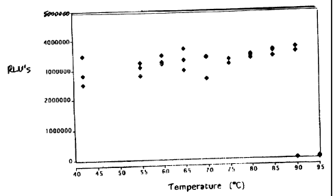
Figure 1 is a graph illustrating single dose reagent pellet temperature
stability;
Figure :2 illustrates the general TMA protocol;
Figure 3A is a schematic representation of a disposable dual chamber reaction
vessel and
the heating steps associated therewith ta perform a TMA reaction in accordance
with one possible
embodiment of the invention;
McDonnell Boehnen
Hulbert & Berghotf
300 S. Wacker Drive
Chicago, Illinois 60606
(312)913-0001 10
CA 02230967 1998-05-01
Figure 3B is a schematic represeritation of altelnative form of the invention
in which two
separate reaction chambers are combined to form a dual chamber reaction
vessel;
Figure 3C is a schematic representation of two alternative embodiments of a
dual chamber
reaction vessel that are snapped into place in a test strip for processing
with a solid phase
receptacle and optical equipment in accordance with a preferred embodiment of
the invention;
Figure 4 is a schematic representation of an alternative embodiment of a dual
chamber
reaction vessel formed from two separal:e chambers that are combined in a
manner to permit a
fluid sample in one chamber to be transferred to the other chamber, with the
combined dual
chamber vessel placed into a test strip such as illustrated in Figure 3C;
Figure 5 is a perspective view of a stand-alone amplification processing
station for the test
strips having the dual chamber reaction vessels in accordance with a presently
preferred form of
the invention;
Figure 6 is a perspective view of one of the amplification modules of Figure
4, as seen
from the rear of'the module;
Figure 7 is a perspective view of the front of the module of Figure 5;
Figure S. is another perspective view of the module of Figure 7;
Figure 9 is a detailed perspective view of a portion of the test strip holder
and 95 C
Peltier heating subsystems of the module of Figures 6-8;
Figure 1.0 is an isolated perspective view of the test strip holder of Figure
9, showing two
test strips installed in the test strip holder;
Figure 1.1 is a detailed perspective view of the test strip holder or tray of
Figure 7;
Figure 12 is a block diagram of the electronics of the amplification
processing station of
Figure 7;
Figure 13 is a diagram of the vacuum subsystem for the amplification
processing station
of Figure 6; anci
Figure 14 is a graph of the thermal cycle of the station of Figure 6.
Figure 15 illustrates a schematic of the operation of the multiplex VIDAS
detection.
Figure 16 illustrates the production of SPR with two distinct capture zones;
Figure 17 illustrates the VIDAS apparatus strip configuration for multiplex
detection;
McDonnell Boehnen
Hulbert & Berghoff
300 S. Wacker Drive
Chicago, Illinois 60606
(312)913-0001 1 1
CA 02230967 1998-05-01
Figure 18 illustrates and graphs the results of verification of the VIDAS
multiplex
protocol detectirig only Neisseria gonorrhoeae (NG) target;
Figure 19A is a graph showing the results when 1x10¹² CT targets were mixed
with 0,
1 x 10⁹, 1 x 10' , 1 x 10", or 1 x 10' z, NG targets, and detected with the
VIDAS instrument using the
multiplex protocol and SPRs coated with Chlamydia trachomatis (CT) capture
probes on the
bottom zone of the SPR, and NG capture probes on the top zone of the SPR.
Figure 19B illustrates the results when 1 x 10¹² NG targets was mixed with 0,
1 x 10⁹,
1 x 10¹⁰, l x 10", or l x 10'2, NG targets, and. detected with the VIDAS
instrument using the
multiplex protocol and SPR coated with CT capture probes on the bottom zone of
the SPR, and
1o NG capture probes on the top zone of the SPR.
Figure 2OA is a diagram showing detection of M.tb nucleic acid by VIDAS
apparatus
after amplification.
Figure 20B is a diagram showing detection of M.tb nucleic acid by VIDAS
apparatus.
Figure 21 is a diagram showing detection of M.tb nucleic acid by VIDAS
apparatus after
amplification.
Figure 22 is a diagram showing detection of M.tb nucleic acid by VIDAS
apparatus after
amplification using the binary/dual chamber protocol.
Figure 23 illustrates the results generated by the method described showing a
collection of
strings of nucleic acid sequences and screening for specific functional
parameters.
Figure 24 shows the nucleic acid sequence of Random Internal Control 1(RIC 1)
with the
possible oligonucleotide primers/probes for amplification and detection of the
control sequence.
Figure 25 shows an analysis of the possible secondary structural components of
the RIC1
sequence.
Figure 26 shows the nucleic acid sequence of Random Internal Control 2 (RIC2)
with the
possible oligonucleotide primers/probes for amplification and detection of the
control sequence.
Figure 27 shows an analysis of the possible secondary structural components of
the RIC2
sequence.
Figure 28 illustrates results from detection of RIC1 DNA, where the ran2l was
the
capture probe and ran33 was an enzyme-linked detector-probe, and shows that
amplification and
detection occurs under standard assay conditions.
McDonnell Boehnen
Hulbert & Berghoff
300 S. Wacker Drive
Chicago, Illinois 60606
(3 l2)913-0001 12
CA 02230967 2008-01-07
5062.1-4
Figure 29 shows that RICI RN'A, amplified by T1V1A and the chemically
activated siQnal
detected on a VIDAS instrument (bioMerieux Vitek, Inc.) using the enzyme-
linked detection
systern, has a limit of sensitivity of about 1000 molecules of RIC1 RNA
(,Aithout optimization of
conditions).
Fi.2ure 30 shows the nucleic acid sequence for internal control
oligonucleotides designed
for assavs for detecting: Chlamydia trachomatis (CT) identified as CRIC-2; for
Neissei-ia
conorrhoeae (NG) identified as GRIC; for Mvco%)acteriurn tuberculosis (MT)
identified as
MRIC; and internal control for HIV identified as HRIC.
1o Description of the Invention
The following examples are provided to better illustrate certain embodiments
of the
present invention without intending to limit the scope of the invention.
Exampie 1 Single Dose Reagents and Unified Buffer
The implementation of a TMA reaction (see U.S. Patent 5,437,990) on-line in
a VIDAS or off-line in a separate instrument (with detection occurring on a
VIDAS instrument) requires modification of the chemistry used to perform the
reaction manually.
First, bulk packaged reagents have been modified into single aliquot doses,
and second, the buffer
components of the reaction have been altered to form a single comprehensive
multifunctional
unified buffer solution.
Under the current manual technology, the reagents are prepared as lvophilized
"cakes" of
multiple-assay quantities. The amplification and enzyme reagents thus must be
reconstituted in
bulk and aliquoted for individual assays.
Thus the automated form of TMA on the VIDAS system improves on the above
manual
method by utilizing single dose pellets of lyophilized reaction components
that can be
resuspended in a single unified buffer which will support sample dilution,
denaturation of nucleic
acids, annealing of nucleic acids, and desired enzymatic activity.
13
CA 02230967 1998-05-01
A) Unified Buffer and Sin lge Dose Reagents
To test the feasibility of single dose amplification reagents, standard
Chlamydia TMA
Amplification artd Enzyme reagents (Gen-Probe Inc.), the bulk reagents were
reconstituted in
0.75 ml of water. 12.5 1 of either the water reconstituted amplification or
enzyme reagent (i.e. a
single dose aliquot) were aliquoted into microcentrifuge tubes. These tubes
were placed in a
vacuum centrifuge with low heat to remove water. The end result of this
procedure was
microcentrifuge tube containing a small, dry cake of either enzyme or
amplification reagent at the
bottom of the tube.
The combined Unified Buffer used in this example, consists of a combination of
standard
1o commercially available Gen-Probe Inc. Sample Dilution Buffer (SDB),
Amplification
Reconstitution Buffer (ARB), and Enzyrrie Dilution Buffer (EDB) in a 2:1:1
ratio. To each dried
amplification reagent microfuge tube was added 100 1 of the combined Unified
Buffer, and
positive control nucleic acid (+), and overlaid with 100 1 of silicone oil.
The tube was then
heated to 95 C for 10 minutes and then cooled to 42 C for 5 minutes. The 200 1
total volume
15 was then transferred to a tube containing the dried enzyme reagent. This
was then gently mixed to
resuspend the el--zyme reagent, and the solution was heated for one hour at 42
C.
Control reactions were prepared using Gen-Probe Control reagents which were
reconstituted in the normal 1.5m1 of AR13 or EDB according to instructions
provided in the Gen-
Probe kit. In each control reaction 25 1 of the reconstituted amplification
reagent was combined
2o with 50 1 of the SDB with the positive control nucleic acid (+). The
mixture was also heated to
95 C for 10 minutes and then cooled to 42 C for 5 minutes. To this was added
25 l of the
reconstituted enzyme reagent and incubated at 42 C for one hour. Negative
control had no
nucleic acid.
Both the test Unified Buffer (Unified) reactions and the standard Control
(Control)
25 reactions were then subjected to the Gen-Probe Inc. standard Hybridization
Protection Assay
(HPA) protocol. Briefly, 100 1 of a Chlamydia trachomatis specific nucleic
acid probe was
added to each tube and allowed to hybridize for 15 minutes at 60 C. Then 300 1
of Selection
Reagent was acided to each tube and the differential hydrolysis of hybridized
and unhybridized
probe was allowed to occur for 10 minutes. The tubes were then read in a Gen-
Probe Inc. Leader
McDonnell Boehnen
Hulbert & Berghoff
300 S. Wacker Drive
Chicago, Illinois 60606
(312)913-0001 14
CA 02230967 1998-05-01
50 luminometer and the resultant data recorded as Relative Light Units (RLU)
detected from the
label, as shown i.n Table 1 below. Data re.ported as RLU, standard C.
Trachomatis TMA/HPA
reaction.
'rABLE 1 Unified single dose aliquot of amplification and enzyme reagents
Control (+) Unified (+) Control (-) LJnified (-)
2,264,426 2,245,495 6,734 3,993
2,156,498 2,062,483 3,484 3,765
1,958,742 2,418,531 5,439 5,836
2,451,872 2,286,773
2,346,131 1,834,198
The data. in Table 1 demonstrates that comparable results are obtained when
using the
single dose aliquots of dried amplification and enzyme reagent. In addition,
the data shows that
the results were comparable using three separate buffers (ARB, EDB and SDB)
and one unified
combined buffer (SDB, ARB and EDB combined at a ratio of 2:1:1) to resuspend
the reagents
and run the reactions.
B) Pellil:ization of Single Dose R-agents
In order to simplify the single dose aliquoting of reagents, methods which
will allow for
pelletization of these reagents in single dose aliquots were used. Briefly,
reagent pellets (or
beads) can be niade by aliquoting an aqueous solution of the reagent of choice
(that has been
combined with an appropriate excipient, such as D(+) Trehalose (a-D-
Glucopyranosyl-(X-D-
glucopyranoside, purchased from Pfanstiehl Laboratories, Inc., Waukegan, IL)
into a cryogenic
fluid, and then using sublimation to remove the water from the pellet. Once
the reagent/trehalose
m mixture is aliquoted (drops) into the cryogenic fluid, it forms a spherical
frozen pellet. These
pellets are then placed in a lyophilizer where the frozen water molecules
sublimate during the
vacuum cycle. The result of this procedure is small, stable, non-flaking
reagent pellets which can
be dispensed ir.ito the appropriate packaging. Single dose aliquot pellets of
reagents which
contained RT, T7 and sugar were subjected to a wide range of temperatures to
examine pellet
stability. After being subject to a test ternperature for 10 minutes, the
pellets were then used for
McDonnell Boehnen
Hulbert & Berghoff
300 S. Wacker Drive
Cliicago, Illinois 60606
(312)913-0001 15
CA 02230967 1998-05-01
CT amplification. The results are graphed in Figure 1. The results show that
the single dose
reagent pellet remains stable even after to exposure, to high temperatures for
10 minutes.
The extraordinary stability of enzymes dried in trehalose has been previously
reported
(Colaco et al., 1992, Bio/Technology, 10, 1007) which has renewed interest in
research on long-
term stabilization of proteins has become a topic of interest (Franks, 1994,
Bio/Technology, 12,
253). The resultiing pellets of the amplification reagent and enzyme reagents
were tested by use in
C. Trachomatis TMA/HPA reactions.
The prepared amplification pellets were placed in a tube to which was added 75
l of a
mixture of ARB and SDB (mixed in a 1:2 ratio) with positive control nucleic
acid. This sample
was then heated to 95 C for 10 minutes and then cooled to 42 C for 5 minutes.
To this was added
25 1 of enzyme reagent, which had been reconstituted using standard Gen-Probe
Inc. procedure.
This mixture was allowed to incubate for one hour at 42 C. The reactions were
then analyzed by
the HPA procedure, as described above. 'The results of this test are reported
as RLU in Table 2,
and labeled AMP Pellets(+). As above, negative control reactions were run
without nucleic acid
(-).
The prepared enzyme pellets were tested by heating 100 1 of a combination of
SDB with
positive control nucleic acid, EDB, and the standard reconstituted
amplification reagent (in a
2:1:1 ratio) at 95 C for 10 minutes and then cooled to 42 C for 5 minutes. The
total volume of
the reaction mix was added to the prepar=ed enzyme pellet. After the pellet
was dissolved, the
:2o reaction was heated to 42 C for one hour and then subjected to HPA
analysis as above. The
results of this test are reported as RLU in Table 2 below, labeled Enzyme
Pellet (+). Control
reactions were prepared using standard (3en-Probe Inc. reagents following
standard procedure.
Data reported as RLU, standard C. Trachomatis TMA/HPA reaction.
TABLE 2 Single dose aliquot of pelleted amplification and enzyme reagents
Control (+) Amp Pellets Amp Pellets :Enzyme Enzyme
+ Pellets + Pellets
2,363,342 2,451,387 2,619 2,240,989 3,418
2,350,028 2.215,235 2,358 3,383,195 1,865
2,168,393 2.,136,645 3,421 2,596,041 2,649
2,412,876 2,375,541 2,247 2,342,288 1,653
McDonnell Boehnen
Hulbert & Berghoff
300 S. Wacker Drive
Chicago. Illinois 60606
(312)913-0001 16
CA 02230967 1998-05-01
The data in Table 2 demonstrates that there was no significant difference when
using the
standard Gen-Probe Inc. reagents, or the (iried, prepared, single dose
amplification reagent pellet,
or the enzyme reagent pellet. Thus the sinlgle dose aliquots of reagents are
suitable for use with a
:5 single unified buffer for application to automation using a VIDAS system.
Example 2 Automated Isothermal Amplification Using Thermolabile Enzymes
In order to automate the isothermal amplification assay reaction for use with
clinical assay
1o apparatus, such as a VIDAS instrument (bioMerieux Vitek, Inc.), a novel
dual-chamber reaction
vessel has been designed to implement thie use of the unified buffer and
single reaction aliquot
reagent pellets clescribed above in isothelmal amplification assay of test
samples which can be
further used in combination with a stand alone processing station.
15 A) Dual reaction chambers
The use of two chambers will facilitate keeping separate the heat stable
sample/amplification reagent (containing the specific primers and nucleotides)
from the heat
labile enzymatic components (i.e. RNA 1-everse transcriptase, RNA polymerase
RNase H).
Figure 3A is a schematic represe;ntation of a disposable dual chamber reaction
vessel 10
?0 and the heating steps associated therewith to perform a TMA reaction in
accordance with one
possible embodiment of the invention, Chamber A contains the amplification
mix, namely
nucleotides, primers, MgC12 and other salts and buffer components. Chamber B
contains the
amplification enzyme that catalyzes the amplification reaction, e.g., T7
and/or RT. After addition
of the targets (or patient sample) into chamber A, heat is applied to chamber
A to denature the
25 DNA nucleic acid targets and/or remove RNA secondary structure. The
temperature of chamber
A is then cooled down to allow primer annealing. Subsequently, the solution of
chamber A is
brought into contact with chamber B. Chambers A and B, now in fluid
communication with
each other, are then maintained at the optimum temperature for the
amplification reaction, e.g.,
42 degrees C. By spatially separating chamber A from chamber B, and applying
the heat for
McDonnell Boehnen
Hulbert & Berghoff
300 S. Wacker Drive
Chicago, Illinois 60606
(312)913-0001 17
CA 02230967 1998-05-01
denaturation to chamber A only, the thermolabile enzymes in chamber B are
protected from
inactivation during the denaturation step.
Figure 313 is a schematic represer.itation of an alternative form of the
invention in which
two separate reaction chambers 12 and 14 are combined to form a dual chamber
reaction vessel
10. Like the ernbodiment of Figure 3A, Chamber A is pre-loaded during a
manufacturing step
with an amplification mix, namely nucleotides, primers, MgC12 and other salts
and buffer
components. Chamber B is pre-loaded during manufacturing with the
amplification enzyme that
catalyzes the arriplification reaction, e.g... T7 and/or RT. Fluid sample is
then introduced into
chamber A. The targets are heated for denaturation to 95 C in chamber A. After
cooling
lo chamber A to 42 C, the solution in chamber A is brought into contact with
the enzymes in
chamber B to trigger the isothermal amplification reaction.
If the reaction vessel is designed such that, after having brought the
contents of chambers
A and B into contact, the amplification chamber does not allow any exchange of
materials with
the environmerit, a closed system arnplification is realized that minimizes
the risk of
1:5 contaminating the amplification reaction with heterologous targets or
amplification products from
previous reactions.
Figure 3C is a schematic representation of two alternative dual chamber
reaction vessels
and 10' thalt are snapped into place; in a test strip 19 for processing with a
solid phase
receptacle and optical equipment in acco;rdance with a preferred embodiment of
the invention. In
the embodiments of Figure 3, a unidirectional flow system is provided. The
sample is first
introduced into chamber A for heating to the denaturation temperature. Chamber
A contains the
dried amplification reagent mix 16. After cooling, the fluid is transferred to
chamber B containing
the dried enzynie 18 in the form of a pellet. Chamber B is maintained at 42 C
after the fluid
sample is introduced into Chamber B. The amplification reaction takes place in
Chamber B at
the optimum reaction temperature (e.g., 42 C). After the reaction is
completed, the test strip 19
is then processed in a machine such as the VIDAS instrument available from
bioM6rieux Vitek,
Inc., the assignee of the present invention. Persons of skill in the art are
familiar with the VIDAS
instrument.
The steps of heating and cooling; of chamber A could be performed prior to the
insertion
_30 of the dual chamber disposable reaction. vessel 10 or 10 ' into the test
strip 19, or, alternatively,
McDonnell Boehnen
Hulbert & Berghoff
300 S. Wacker Drive
Chicago, Illinois 60606
(312)913-0001 18
CA 02230967 1998-05-01
suitable heating elements could be placed adjacent to the left hand end 24 of
the test strip 19 in
order to provide the proper temperature control of the reaction chamber A. The
stand alone
amplification processing station of Figures 4-14, described below,
incorporates suitable heating
elements and coritrol systems to provide the proper temperature control for
the reaction vessel 10.
Figure 4 is a schematic representation of an alternative embodiment of a dual
chamber
reaction vessel 10 " formed from two separate interlocking vessels 10A and lOB
that are
combined in a manner to permit a fluid sample in one chamber to flow to the
other, with the
combined dual chamber vessel 10 " placed into a test strip 19 such as
described above in Figure
3C. The fluid sample is introduced irito chamber A, which contains the dried
amplification
reagent mix 16. Vessel A is then heated off-line to 95 degrees C, then cooled
to 42 degrees C.
The two vessels A and B are brought together by means of a conventional snap
fit between
complementary locking surfaces on the tube projection 26 on chamber B and the
recessed conduit
28 on chamber A. The mixing of the sample solution from chamber A with the
enzyme from
chamber B occurs since the two chambers are in fluid communication with each
other, as
indicated by the arrow 30. The sample can then be amplified in the combined
dual chamber
disposable reaction vessel 10 " off-line, or on-line by snapping the combined
disposable vessel 10
" into a modified VIDAS strip. The VIDAS instrument could perform the
detection of the
amplification reaction in known fashion.
B) Amn.lification Station
Figure 5 is a perspective view of a stand-alone amplification processing
system 200 for
the test strips 19 having the dual chamber reaction vessels in accordance with
a presently
preferred form of the invention. The system 200 consists of two identical
amplification stations
202 and 204, a power supply module 206, a control circuitry module 208, a
vacuum tank 210
and connectors 212 for the power supply module 206. The tank 210 has hoses 320
and 324 for
providing vacuum to amplification stations 202 and 204 and ultimately to a
plurality of vacuum
probes (one per strip) in the manner described above for facilitating transfer
of fluid from the first
chamber to the second chamber. The vacuum subsystem is described below in
conjunction with
Figure 14.
McDonnell Boehnen
Hulbert & Berghoff
300 S. Wacker Drive
Chicago, Illinois 60606
(312)913-0001 19
CA 02230967 1998-05-01
The amplification stations 202 and 204 each have a tray for receiving at least
one of the
strips and associated temperature control, vacuum and valve activation
subsystems for heating
the reaction wells of the strip to the proper temperatures, transferring fluid
from the first chamber
i.n the dual chamber reaction wells to the second chamber, and activating a
valve, such as a
thimble valve or preferably a ball valve, to open the fluid channel to allow
the fluid to flow
between the two chambers.
The stations 202 and 204 are designed as stand alone - amplification stations
for
performing the amplification reaction in an automated manner after the patient
or clinical sample
has been added to the first chamber of t:he dual chamber reaction vessel
described above. The
processing of the strips after the reaction is completed with an SPR takes
place in a separate
machine, such as the VIDAS instrument. Specifically, after the strips have
been placed in the
stations 202 anct 204 and the reaction rull in the stations, the strips are
removed from the stations
202 and 204 and placed into a VIDAS instrument for subsequent processing and
analysis in
known fashion.
The entire system 200 is under microprocessor control by an amplification
system
interface board (not shown in Figure 5). The control system is shown in block
diagram form in
Figure 12 and will be described later.
Referrin.g now to Figure 6, one of the amplification stations 202 is shown in
a perspective
view. The other amplification station is of identical design and construction.
Figure 7 is a
perspective view of the front of the module of Figure 6.
n
Referrir.Lg to these figures, the station includes a vacuum probe slide motor
222 and
vacuum probes slide cam wheel 246 that operate to slide a set of vacuum probes
244 (shown in
Figure 7) for the thimble valves up and down relative to a vacuum probes slide
246 to open the
thimble valves and apply vacuum so as to draw the fluid from the first chamber
of the reaction
vessel 10 to thie second chamber. The vacuum probes 244 reciprocate within
annular recesses
provided in the vacuum probes slide 246. Obviously, proper registry of the pin
structure and
vacuum probe 244 with corresponding structure in the test strip as installed
on the tray needs to
be observed.
McDonnell Boehnen
Hulbert & Berghoff
300 S. Wacker Drive
Chicago, Illinois 60606
(312)913-0001 20
CA 02230967 1998-05-01
The station includes side walls 228 and 230 that provide a frame for the
station 202.
Tray controller board 229 is mounted between the side walls 228 and 230. The
electronics
module for the station 202 is installed ol;l the tray controller board 229.
A set of tray thermal insulation covers 220 are part of a thermal subsystem
and are
provided to envelop a tray 240 (Figure 7) that receives one or more of the
test strips. The
insulation covers 220 help maintain the temperature of the tray 240 at the
proper temperatures.
The thermal subsystem also includes a 42 C Peltier heat sink 242, a portion of
which is
positioned adjacent to the second chamber in the dual chamber reaction vessel
in the test strip to
maintain that chamber at the proper temperature for the enzymatic
amplification reaction. A
zo 95 C heat sink 250 is provided for the front of the tray 240 for
maintaining the first chamber of
the reaction well in the test strip at the denaturation temperature.
Figure 8 is another perspective view of the module of Figure 7, showing the 95
C heat
sink 250 and a set of fins 252. Note that the 95 C heat sink 250 is positioned
to the front of and
slightly below the tray 240. The 42 C lieat sink 242 is positioned behind the
heat sink 250.
Figure 9 is a detailed perspective view of a portion of the tray 240 that
holds the test strips
(not shown) as seen from above. The tray 240 includes a front portion having a
base 254, a
plurality of discontinuous raised paralllel ridge structures 256 with recessed
slots 258 for
receiving the test strips. The base of the: front 254 of the tray 240 is in
contact with the 95 C heat
sink 250. The side walls of the parallel raised ridges 256 at positions 256A
and 256B are placed
as close as possible to the first and second chambers of the reaction vessel
10 of Figure 3A so as
to reduce thermal resistance. The base of the rear of the tray 240 is in
contact with a 42 C Peltier
heat sink, as best seen in Figure 8. The portion 256B of the raised ridge for
the rear of the tray is
physically isolated from portion 256A for the front of the tray, and portion
256B is in contact
with the 42 C heat sink so as to keep the second chamber of the reaction
vessel in the test strip at
the proper temperature.
Still referring to Figure 9, the vacuum probes 244 include a rubber gasket
260. When the
vacuum probes 244 are lowered by the vacuum probe motor 222 (Figure 6) the
gaskets 260 are
positioned on the upper surface of the test strip surrounding the vacuum port
in the dual chamber
reaction vessel so as to make a tight seal and permit vacuum to be drawn on
the second chamber.
McDonnell Boehnen
Hulbert & Berghoff
300 S. Wacker Drive
Chicago, Illinois 606(6
(312)913-0001 21
CA 02230967 1998-05-01
Figure 10 is an isolated perspective view of the test strip holder or tray 240
of Figure 9,
showing two test strips installed in the tray 240. The tray 240 has a
plurality of lanes or slots 241
receiving up to six test strips 19 for silnultaneous processing. Figure 10
shows the heat sinks
:242 and 250 for maintaining the respective portions of the tray 240 and
ridges 256 at the proper
temperature.
Figure 11 is a detailed perspective view of the test strip holder or tray 240
as seen from
below. The 95 C Peltier heat sink whiclh would be below front portion 254 has
been removed in
order to better illustrate the rear heat sink: 242 beneath the rear portion of
the tray 240.
Figure 12 is a block diagram of the electronics and control system of the
amplification
1o processing system of Figure 5. The control system is divided into two
boards 310 and 311,
section A 310 at the top of the diagram devoted to amplification module or
station 202 and the
other board 311 (section B) devoted to lthe other module 204. The two boards
310 and 311 are
identical and only the top section 310 will be discussed. The two boards 310
and 311 are
connected to an amplification station interface board 300.
The interface board 300 communicates with a stand alone personal computer 304
via a
high speed data bus 302. The personal computer 304 is a conventional IBM
compatible
computer with hard disk drive, video monitor, etc. In a preferred embodiment,
the stations 202
and 204 are uncler control by the interface board 300.
The board 310 for station 202 controls the front tray 240 which is maintained
at a
2o temperature of 95 C by two Peltier heat sink modules, a pair of fans and a
temperature sensor
incorporated into the front portion 254 of the tray 240. The back of the tray
is maintained at a
temperature of 42 C by two Peltier madules and a temperature sensor. The
movement of the
vacuum probes. 244 is controlled by the probes motor 222. Position sensors are
provided to
provide input signals to the tray controller board as to the position of the
vacuum probes 244.
The tray controller board 310 includes a set of drivers 312 for the active and
passive components
of the system vvhich receive data from t:he temperature and position sensors
and issue commands
to the active components, i.e., motors, fans, Peltier modules, etc. The
drivers are responsive to
commands from the amplification interface board 300. The interface board also
issues
commands to the vacuum pump for the vacuum subsystem, as shown.
McDonnell Boehnen
Hulbert & Berghoff
300 S. Wacker Drive
Chicago, Illinois 60606
(312)913-0001 22
CA 02230967 1998-05-01
Figure 13 is a diagram of the vacuum subsystem 320 for the amplification
processing
stations 202 and 204 of Figure 5. The subsystem includes a 1 liter plastic
vacuum tank 210
which is connected via an inlet line 322 to a vacuum pump 323 for generating a
vacuum in the
tank 210. A vacuum supply line 324 is provided for providing vacuum to a pair
of pinch solenoid
valves 224 (see Figure 6) via supply lines 324A and 324B. These vacuum supply
lines 324A and
324B supply vacuum to a manifold 22ti distributing the vacuum to the vacuum
probes 244.
Note the pointed tips 245 of the vacuum probes 244 for piercing the film or
membrane 64
covering the strip 19. The vacuum system 320 also includes a differential
pressure transducer
321 for monitoring the presence of vacuum in the tank 210. The transducer 321
supplies pressure
signals to the interface board 300 of Figw=e 12.
Figure 14 is a representative graph of the thermal cycle profile of the
station of Figure 5.
As indicated in line 400, after an initial ramp up 402 in the temperature
lasting less than a
ininute, a first temperature T1 is reached (e.g., a denaturation temperature)
which is maintained
for a predetermined time period, such as 5-10 minutes, at which time a
reaction occurs in the first
chamber of the reaction vessel. Thereafter, a ramp down of temperature as
indicated at 404
occurs and the temperature of the reaction solution in the first chamber of
the reaction vessel 10
cools to temperature T2. After a designated amount of time after cooling to
temperature T2, a
fluid transfer occurs in which the solutioii in the first chamber is conveyed
to the second chamber.
Temperature T2; is maintained for an appropriate amount of time for the
reaction of interest, such
2o as one hour. At time 406, the temperature is raised rapidly to a
temperature T3 of 65 C to stop
the amplification reaction. For a TMA reaction, it is important that the ramp
up time from time
406 to time 408 is brief, that is, less than 2 minutes and preferably less
than one minute.
Preferably, all the ramp up and ramp down of temperatures occur in less than a
minute.
Other elnbodiments of reaction vessels and amplification station components
are also
envisioned, and. such alternative embodiinents are encompassed in the present
disclosure.
Example 3 Automated VIDAS Test for Non-amplified and Amplified Detection of
Mycobacterium tuberculosis (M.tb)
Using the VIDAS instrument (bioMerieux Vitek, Inc.), modified to 42 C, we have
developed an iii-line simple rapid nucleic acid amplification and detection
assay for the clinical
McDonnell Boehnen
Hulbert & Berghoff
300 S. Wacker Drive
Chicago, Illinois 60606
(312)913-0001 23
CA 02230967 1998-05-01
laboratory for the detection of M.tb in test samples which can be completed in
a short time. The
entire assay is designed to take place on a single test strip, minimizing the
potential for target or
amplicon containination. The amplification based assay is capable of detection
of M.tb where the
sample contains only 5 cells similar to the sensitivity achieved by the Gen-
Probe commercial kit.
The amplification based assay utilizes isothermal transcription-mediated
amplification
(TMA) targeting unique sequences of rRNA, followed by hybridization and enzyme-
linked
fluorescent detection of nucleic acid probe (amplicon) in the VIDAS
instrument.
The amplification/detection assay can detect approximately lfg of M.tb rRNA,
or less
than one M.tb organism per test, and is s-pecific for all members of the M.tb
complex. Specific
w probes for the detection of M.tb can be found in C. Mabilat, 1994, J. Clin.
Microbiol. 32, 2707.
Standard smears for acid-fast bacilli are not always reliable as a diagnostic
tool, and even
when positive r.nay be a mycobateria other than M.tb. Currently, standard
methods for diagnosis
of tuberculosis requires culturing the slow-growing bacteria, and may take up
to 6 weeks or
longer. During this time, the patient is usually isolated. Initial results are
that this automated test
matches or exceeds the clinical sensitivity of the culture method, and offers
a highly sensitive
method to rapiclly (in less than three hours) detect M.tb in infected samples,
thereby aiding rapid
diagnosis, isolation and treatment.
A) Sample Preparation
A 450 1 volume of specimen is added to 50 l of specimen dilution buffer in a
lysing tube
containing glass beads, sonicated for 15 minutes at room temperature to lyse
organisms, heat
inactivated for 15 minutes at 95 C. Where required, isothermal amplification
was conducted as
per a commercially available manual assay kit (Gen-Probe Inc.) following the
kit instructions
using standard kit reagents. However, similar assays can be conducted using
the modified
components as described in the Examples above.
B) Dete=ction
In order for the automated detection assay to operate, the detection system
requires
hybridization of the target nucleic acid or amplicon to a specific capture
nucleic acid bound to a
solid support, (in the VIDAS system called a "solid phase receptacle" SPR
pipet-like devise), and
McDonnell Boehnen
Hulbert & Berghoff
300 S. Wacker Drive
Chicago, [llinois 60606
(312)913-0001 24
CA 02230967 1998-05-01
to a labeled detection probe nucleic acid i(for example where the label can be
alkaline
phosphatase, a chemiluminescent signal compound, or other reagent that will
allow for specific
detection of botmd probe).
In an automated system such as the VIDAS, after several wash steps to remove
unbound
probe, the SPR transfers the probe-target hybrid to an enzyme substrate,
whereby the detectable
signal is triggered from the bound probe and detected by the assay instrument.
In one
embodiment, the detection probe is conjugated to alkaline phosphatase, and
once placed in
contact with substrate of methyl umbelliferyl phosphate (MUMP), the substrate
is converted into
4-methyl umbel.liferone (4-MU) by the alkaline phosphatase. The 4-MU produces
fluorescence
which is measured and recorded by the standard VIDAS instrument as relative
fluorescence units
(RFU). When target nucleic acid is not present, no detection probe is bound,
and no substrate is
converted, thus no fluorescence is detected.
C) AnalZical sensitivity: Controls
1,5 Generally controls are prepared in a matrix of specimen dilution buffer
with positive
controls containing 5fg of M.tb rRNA, or the equivalent rRNA of approximately
1 M.tb cell.
Sensitivity of the automated probe assay can be determined by testing
dilutions of lysed M. tb
cells. The cell lysates can generally be plrepared with a 1 l loop of cells
(the assumption being
that there are approximately 1x10⁹ colony forming units (CFU) per l l loop-
full, based upon
n previous titration and CFU experiments). Dilutions of the M.tb lysates can
then be tested with the
automated probe assay.
Figure 20A is a graph showing detection of M.tb amplicons according to the Gen-
Probe
kit. Figure 20B is a graph showing detection of M.tb amplicons from the same
reactions as in
25 Figure 20A by the VIDAS instrument.
Figure 21 is a graph showing amplification and detection of M.tb nucleic acids
on the
modified VIDAS apparatus. Enzyme was used in liquid form and amplification was
performed
in-line with VIDAS assay instrument.
Figure 22 is a graph showing amplification and detection of M.tb nucleic acids
on the
30 modified VIDAS apparatus using the bi:riary/dual chamber disposable
reaction vessel. The
McDonnell Boehnen
Hulbert & Berghoff
300 S. Wacker Drive
Chicago, Illinois 60606
(312)913-0001 25
CA 02230967 1998-05-01
denaturation step was performed off-line of the VIDAS instrument,
amplification and amplicon
detection was performed in-line with VIDAS instrument.
Example 4 Automated VIDAS Test for Amplified Detection of Chlamydia
trachomatis (CT)
Using the VIDAS instrument (bioMerieux Vitek, Inc.), we have developed a
simple, fully
automated, highly specific assay for the rapid detection of Chlamydia
trachomatis (CT) from test
samples. The test utilizes isothermal TMA targeting unique sequences of the
rRNA followed by
hybridization and enzyme-linked fluorescence detection. The automated test
specifically detects
lo all the clinically important serovars of Chlamydia trachomatis (CT) from
urogenital specimens in
less than two hours. We obtained an analytical sensitivity of 0.5fg of rRNA,
or the equivalent of
approximately 1/10' of an elementary body of Chlamydia trachomatis (CT).
Agreement between
the automated test and Gen-Probe's Amplified CT test for two-hundred seven
(207) clinical
endocervical svvabs and urines showed complete agreement.
Chlamydia trachomatis (CT) infection is the leading cause of sexually
transmitted disease
in the United Sitates and Europe. It is currently estimated that about four
million new CT infection
occur each yeai= in the United States.
Chlamydia trachomatis (CT) is a. small obligate intracellular parasite that
causes
infections in both females and males, adults and newborns. The greatest
challenge to the control
:20 of CT infection is that as many as 75% of infected women and 50% of
infected men are
asymptomatic. This results in a large reservoir of unrecognized infected
individuals who can
transmit the CT infection. The rapid and simple detection of CT infection
would greatly assist
identification infected individuals.
A) Patient Specimens and sample preparation
Coded samples (n=207) were obtained from patients with symptoms consistent
with CT
infection. The cervical samples were collected with a Gen-Probe sample
collection kit containing
Gen-Probe transport medium; the urine samples were collected into standard
urine collection
devices. All samples were stored at 4 C.
McDonnell Boehnen
Hulbert & Berghoff
300 S. Wacker Drive
Chicago, Illinois 60606
(312)913-0001 26
CA 02230967 1998-05-01
Cervical. swabs were centrifuged at 425xg for 5 minutes to bring all liquid to
the bottom
of the tube. The swabs were then treated with 40 1 Gen-Probe Specimen
Preparation Reagent and
incubated at 60"C for 10 minutes. 20 l of the treated sample was then
pipetted into 400 l of
sample dilution buffer (SDB).
Two ml of each urine sample was warmed to 37 C for 10 minutes and microfuged
at
12,000xg for 5 minutes. The supernatant was discarded and 300 1 of sample
dilution buffer was
added to each specimen. All 15 serovars of CT were used for inclusive samples,
specimens were
quantified and 20 1 of specimens containing 4x 1 02 IFU/ml (inclusion forming
unit per ml) of
each serovar was added to 400 1 of SDB. A panel of exclusive urogenital
micororganisms was
to obtained and quantified and 20 1 of 2x10⁹/ml microorganisms were pipeted
into 400 l of SDB.
Positive contro:l containing 0.5fg rRNA or the equivalent of 0.1 CT elementary
body was diluted
in SDB.
B) Sample am_plification and VIDAS detection
Samples were amplified using the TMA protocol, and rRNA targets were
hybridized to
oligomer conjugated to AMVE copolymer and an oligomer conjugated to alkaline
phosphatase.
See for example U.S. Patent 5,489,653 and 5,510,084. As described above, the
solid phase
receptacle (SPR pipet-like devise) carries the bound hybrids through
successive wash steps and
finally into the substrate 4-MUP. The alkaline phosphatase converts the
substrate to fluorescent
4-MU, which is detected by the VIDAS assay machine and recorded as relative
fluorescence
units.
Table 2B below illustrates detection of CT by VIDAS automated assay following
amplification as RFV (RFV = RFU - Background RFU) against concentration of CT
rRNA.
Dilutions of C. trachomatis purified rRl`1A from 0 to 200 molecules were
amplified (n=3) and
detected in the VIDAS automated probe assay. Detection limit is 20 molecules
of purified rRNA.
McDonnell Boehnen
Hulbert & Berghoff
300 S. Wacker Drive
Chicago, Illinois 60606
(312)913-0001 27
CA 02230967 1998-05-01
TABLE 2B: CT Detection by VIDAS
rRNA Input Molecules VIDAS RFV
0 1
2 121
20 3260
200 8487
C) Analytical specificity and Results
Amplifications and detection were carried out in the presence of each of the
following
ATCC organisms with detections reported as RFV in Table 3 below.
TABLE 3 F-xclusivity panel for CT
Bacillus subtilis Branhamella Candida albicans Chlamydia Chlamydia
33 catarrhalis 26 pneumoniae psittaci
39 11
Escherichia coli Klebsiella Lactobacillus Neisseria Neisseria
11 pneumoniae acidophilus elongata lactamica
13 27 44 18
Neisseria Neisseria Propionibacterium Pseudomonas Staphylococcus
meningitidis-D meningitidis-Y acnes aeruginosa aureus
61 52 14 13 13
Streptococcus Streptococcus Streptococcus Yersinia Chlamydia
agalactiae bovis pneunzoniae enterolitica trachomatis
16 45 34 11 10673
Negative Control
12
10 Analytical specificity for Chlamydia serovars data reported as RFV is shown
in Table 4
below.
TABLE 4 Inclusivity Panel for CT
Serovar A Serovar B Serovar Ba Serovar C Serovar D
5421 7247 9626 8066 10849
Serovar E Serovar F Serovar G Serovar H Serovar I
4608 9916 1008.2 7769 9733
Serovar J Serovar K Serovar L1 Serovar L2 Serovar L3
9209 2423 10786 1812 5883
Positive Control Negative Control
3 775 9
McDonnell Boehnen
Hulbert & Berghoff
300 S. Wacker Drive
Chicago, Illinois 60606
(312)913-0001 28
CA 02230967 1998-05-01
Table 5 below illustrates the results of clinical cervical swab specimen
testing for CT
comparing resullts from the Gen-Probe manual AMP-CT assay and the VIDAS
automated probe
assay.
TABLE 5 Amplified Clinical Cervical Specimen Detection of CT
Gen-Probe rrianual AMP-CT assay
VIDAS off-line + -
automated probe + 35 0
assay - 0 85
Table 6 below illustrates the results of clinical urine specimen testing
comparing the
results of manual AMP-CT assay and the VIDAS automated probe assay.
TABLE 6 Amplified Clinical Urine Specimen detection of CT
Gen-Probe nianual AMP-CT assay
VIDAS off-line + -
automated probe + 25 0
assay - 0 62
Thus there was perfect agreement in assay results between the automated probe
assay
using the VID?.S instrument and the manual Gen-Probe AMP-CT assay.
:20 Example 5 Multiplex (Multiple Sequence) Nucleic Acid Detection
The value of diagnostic tests based on nucleic acid probes can be
substantially increased
through the detection of multiple different nucleic acid molecules, and the
use of internal positive
controls. An automated method has beeri devised for use with the VIDAS
instrument (bioMerieux
Vitek, Inc.) which can discretely detect at least two different nucleic acid
sequences in one assay
McDonnell Boehnen
Hulbert & Berghoff
300 S. Wacker Drive
Chicago, Illinois 60606
(312)913-0001 29
CA 02230967 1998-05-01
reaction, and is termed the Multiplex protocol. Thus a nucleic acid
amplification procedure, or a
processed test sarnple may be screened for more than one amplified nucleic
acid molecule in the
same assay. This method relies on the spatial separation of discrete nucleic
acid probes which can
specifically capture different target nucleic acid sequences (amplicons), on
the SPR pipet-like
devise of the VIDAS instrument. The SPR is a disposable pipet-like tip which
enables fluid
movements as well as acting as the solid support for affinity capture. The
multiplex capture by
SPR is demonst:rated using capture probes specific for Chlamydia trachomatis
(CT) and Neisseria
gonorrhoeae (NG).
Figure 15 illustrates a schematic of the operation of the multiplex VIDAS
detection. The
i.o SPR tips are coated in two distinct zones with oligonucleotide nucleic
acid sequences which are
used to specifically capture complementary nucleic acid sequences (amplicons)
with their
corresponding specific reporter probe or detector probe nucleic acids labeled
with alkaline
phosphatase (AKP). Following washes to remove unbound reporter probes, AKP
localized to the
SPR bottom is detected with the fluorescent substrate 4-MUP. The AKP is
stripped from the
bottom of the SPR with NaOH or other reagents which promote denaturation of
nucleic acid
hybrids or inaciitvates AKP activity. The enzyme reaction well is emptied,
washed, and re-filled
with fresh 4-MUP. To confirm removal of AKP from the bottom of the SPR, the
new substrate is
exposed to the bottom of the SPR and arly residual fluorescence is measured.
Finally, AKP-
reporter probe bound to the top of the SPR is detected by immersing the SPR in
the 4-MUP, and
:20 representing the presence of the second amplicons.
Figure 16 illustrates the production of SPR with two distinct capture zones.
The SPR is
inserted tip-first into a silicon plug, which are held in a rack. Differential
pressure is used to
uniformly draw a solution of a specific capture probe at about 1 g/ml,
conjugated to AMVE
copolymer, into all SPRs at one time. The amount of fluid drawn into each SPR,
and thus the size
of the zone, is controlled by regulating the amount of pressure in the system.
Attachment of the
conjugate to the SPR surface is achieveci by passive adsorption for several
hours at room
temperature. After washing, and drying, the SPRs are capped with a small
adhesive disc and
inserted into new racks in a tip-down orientation. The lower portion of the
SPR is then similarly
coated with a second capture probe conjugate. SPRs are stable when stored dry
at 4 C.
McDonnell Boehnen
Hulbert & Berghoff
300 S. Wacker Drive
Chicago, Illinois 60606
(312)913-0001 30
CA 02230967 1998-05-01
Figure 17 illustrates a preferred embodiment of the VIDAS apparatus strip
configuration
for Multiplex detection. The strip can be pre-filled with 200 l of AKP-probe
mix (about 1x101z
molecules of each probe) in hybridization buffer in well X1, 600 1 of wash
buffer in wells X3,
X4, X5, 600 1 of stripping reagent in wells X6 and X7, and 400 1 of AKP
substrate in X8 and
sealed with foil, A foil-sealed optical cuvette (XA) containing 300 1 of 4-MUP
is snapped into
the strip, and the strips are inserted into the VIDAS instrument at 37 C. The
Multiplex VIDAS
protocol is then executed using SPRs coated with two capture probes in
distinct zones.
The VII)AS Multiplex protocol can involve many steps. For example the
validation test
protocol contained thirteen (13) basic steps as follows:
1o l. Transfer of 203 1 target from X0 to AKP-probes in X1,
2. Hybridize and capture to the entire SPR,
3. Wash SIPR (316 l) twice with PBS/Tween (X3, X4),
4. 4-MUP to SPR bottom (89.6 l) in XA for 5.3 minutes then read signal,
5. 4-MUP to SPR bottom (89.641) in XA for 14.8 minutes then read signal
(optional),
1.5 6. Transfer used substrate from XA to X2 (5 x 67.1 l),
7. Strip AKP from SPR bottom (112.6 1) with NaOH (X7),
8. Wash XA with fresh NaOH (3 x 112.6 1; X6 to XA to X6),
9. Wash XA with PBS/Tween (3 x 112.6 l; X5 to XA to X5),
10. Transfer fresh 4-MUP from X8 to XA (6 x 48 1),
20 11. 4-MUP to SPR bottom (89.6 l) in XA for 10.7 minutes then read signal,
12. 4-MUP to SPR top (294 l) in XA for 5.5 minutes then read signal,
13. 4-MUP to SPR top (294 1) in XA for 15 minutes then read signal (optional).
Hybridization, substrate, wash and stripping steps can all involve multiple
cycles of
25 pipeting the respective solution into the SPR, holding the solution for a
defined period of time,
and pipeting the solution out of the SPR,. Hold times for hybridization,
substrate and washing or
stripping are 3.0, 0.5 and 0.17 minutes respectively. The fluorescence signal
is detected by the
apparatus. Total assay time for the research protocol was about 1.75 hours but
can be reduced to
about 75 minutes.
McDonnell Boehnen
Hulbert & Berghoff
300 S. Wacker Drive
Chicago, Illinois 60606
(312)913-0001 3 1
CA 02230967 1998-05-01
F'igure 18 illustrates and graphs the results of verification of the VIDAS
Multiplex
protocol executed as described above, except the SPR was homogeneously coated
with only a
single capture probe for Neisseria gonorr=hoeae (NG). The number of NG
oligonucleotide targets
in the test sample was varied from 0, 1x10¹⁰, or 1x10" molecules in the test
sample. The data
shown are averalges of replicate samples. The graph as illustrated is divided
into two parts; the left
and right halves show the results of two iluorescent measurements from the
lower and the upper
zones of the SPR, respectively. The measurements taken from the bottom zone
after stripping the
lower area of bound nucleic acid, and exposure for about 11 minutes in fresh 4-
MUP substrate
was approximately 46 RFU for all samples tested, and was equivalent to
background fluorescence
measured. This measurement is shown by the 0 time point in the center of the
graph. Thus the
graph illustrates two sequential sets of measurements of fluorescence from a
single SPR, the first
set of ineasurements being taken from the bottom half of the SPR (left half of
the graph), and a
second set of measurements taken from the top of the SPR (the right of the
graph). This
experiment validates that the multiplex protocol and zone coated SPR prcedure
yield essentially
1.5 idnetical results. As indicated by the fluoresecense intensities in the
left and right hand parts of
the graph, from the lower and upper portions of the SPR.
Figure 19 illustrates Multiplex detection of CT and NG oligonucleotide targets
at different
input amounts. Figure 19A is a graph showing the results when 1x10¹² CT
targets were mixed
with 0, 1 x 10⁹, 1 x 10' , 1 x 10", or 1 x 10' z, "NG targets, and detected
with the VIDAS instrument
using the Multiplex protocol and SPRs coated with CT capture probes on the
bottom zone of the
m
SPR, and NG capture probes on the top zone of the SPR. Figure 19B illustrates
the results when
1 x 10" 2 NG targets was mixed with 0, 1 x 10⁹, 1 x 10' , 1 x 10", or 1 x 10'
2, CT targets, and detected
with the VIDAS instrument using the Multiplex protocol and SPRs coated with CT
capture
probes on the bottom zone of the SPR, and NG capture probes on the top zone of
the SPR. The
25 data is graphed as above where the graph illustrates two sequential sets of
measurements of
fluorescence from a single SPR, the first. set of measurements being taken
from the bottom half of
the SPR (left half of the graph), Strippect and verified (the center of the
graph) and a second set of
measurements taken from the top of the SPR (the right of the graph) with
verification of stripping
of the SPR in t'he center of the graph. Irr.lportantly, this experiment shows
that the two zones of
30 the SPR act independently in the multiplex protocol, since high
fluorescence signals from one
McDonnell Boehnen
Hulbert & Berghoff
300 S. Wacker Drive
Chicago, Illinois 60606
(312)913-0001 32
CA 02230967 1998-05-01
zone do not interfere with signals produced for the second zone. This is
regardless of whether
these latter signals are high (lxl0`2), or low (1x10), or negative.
Table 7 below summarizes the data obtained by Multiplex VIDAS detection of CT
and
NG in a sample at various target levels, reported in RFUs.
TABLE 7 Detection of CT and NG targets in saunple
RFUSA noneB 1x10⁹ 1x10¹⁰ 1x10" 1x101lx10"
nonec 43 /40E 43/116 46/693 62/7116 174/11817 273/12136
1x10⁹ 189/41 246/118 169/773 220/5750 422/12522 399/11401
1x10¹⁰ 1736/41 2258/125 1937/734 1931/6639 2128/12390 2371/11180
1x10" 10339/48 9815/145 9858/760 9369/4571 9784/11825 10252/10312
1x10'2 12149/49 13520/148 12940/796 13593/4397 11239/11786 10158/9900
1x10" 11545/57 11713/121 10804/815 12805/5404 12305/12326 11416/10490
^ Data is reported in RFUs, after -5 minute exposure of 4-MUP to bound AKP-
probe
B Columns are data for that number of NG targets in sample
c Rows are the data for that number of CT targets in sample
The first value reported is RFU detected from the CT assay portion
E The second value reported is RFU detected fro;m the NG assay portion
Thus the Multiplex VIDAS protocol is clearly operative and enables the rapid
and discrete
detection of more than one different nucleic acid in a sample. This protocol,
and the SPR coating
can be manipulated in many formats to present coating zones of different
surface area with
different sized gaps between two or more detection zones. The SPR can be
coated with nucleic
acids which are designed to capture different regions of the same nucleic acid
sequence to detect,
for example, tn.incated gene expression, different alleles or alternatively
spliced genes. The SPR
can be coated to capture amplicons from internal control nucleic acid
molecules which can be
used to detect and confirm successful nucleic acid amplification reactions.
Thus the VIDAS
Multiplex protocol is a flexible method for detection of more than one nucleic
acid sequence in
the same saunplie, in a single assay, with or without amplification.
EXAMPLE 6 Internal Control Sequence and Method
McDonnell Boehnen
Hulbert & Berghoff
300 S. Wacker Drive
Chicago, Illinois 60606
(312)913-0001 33
CA 02230967 1998-05-01
The construction of internal control sequences composed of functional building
blocks of
sequences chosen by random generation of nucleic acid sequences for use as
amplification
reaction internal positive controls ideally requires that the control
sequences be specifically
designed to be used for the various nucleic acid amplification protocols
including but not limited
to PCR, LCR, TMA, NASBA, and SDA. The internal control nucleic acid sequence,
in
combination with the appropriate sequence specific oligonucleotide primers or
promoter-primers
will generate a positive amplification signal if the amplification reaction
was successfully
completed.
Ideally, the internal control nucleic acid is useful regardless of the nucleic
acid sequences
present in the target organism, the host organism, or nucleic acids present in
the normal flora or in
the environmer.it. Generally, the internal control sequences should not be
substantially similar to
any nucleic acid sequences present in a clinical setting, including human,
pathogenic organisms,
normal flora organisms, or environmental organisms which could interfere with
the amplification
and detection of the internal control sequences.
The internal control sequences of the instant invention are comprised of
functional blocks
of sequences cliosen from a list of randomly generated nucleic acid sequences.
The functional
blocks are segments which provide for a. special property needed to allow for
amplification,
capture, and detection of the amplification product. For example, in a TMA
reaction, the internal
control sequences are most useful when the functional blocks meet certain
functional
requirements of the amplification protocol, such as: a) a primer binding site
on the anti-sense
strand; b) a capture site; c) a detector probe binding site; d) a T7-promoter
containing primer
binding site on the sense strand. Each oi.'these functional elements has its
own particular
constraints, such as length, %G-C content, Tm, lack of homology to known
sequences, and
absence of secondary structural features (i.e. free from dimer fonmation or
hairpin structures)
which can be used to select the appropriate sequence. Thus randomly generated
functional blocks
of sequences can be screened for the desired functional properties before use
in constructing
internal control sequences.
In order to construct internal coiitrol sequences having the desired
properties comprising a
specified number of functional blocks and satisfying the desired constraints
within each block, a
random sequence generator was used to generate strings of numbers; each number
being limited
McDonnell Boehnen
Hulbert & Berghoff
300 S. Wacker Drive
Chicago, Illinois 60606
(312)913-0001 34
CA 02230967 1998-05-01
to the range from 0.000 to 4.000. The lerigth of the strings is flexible and
chosen based upon the
desired lengths of the functional blocks.
Each number in the string (i.e. nl, n2, n3, n4 ... nx where x is the length of
the string) was
then assigned a corresponding nucleotide as follows: guanosine (G) if 0< n<_
1; adenosine (A) if
1< n_ 2; thymidine (T) if 2< n<_ 3; anct cytosine (C) if 3< n_ 4. A large
collection of such
strings was produced and screened for those meeting the sequence and
structural requirements of
each functional block. Figure 23 illustrates the results generated by the
method described
showing a collection of strings of nucleir, acid sequences and screening for
specific functional
parameters. The internal control sequence can include DNA, RNA, modified
oligonucleotides, or
io any combination of nucleic acids, such that the illustrated sequences using
DNA nomenclature
can be readily adapted as desired to the appropriate nucleic acid.
Potential internal control (IC) sequences were then constructed by assembling
the
functional blocks (selected at random) in the proper order. Finally, the
assembled internal control
sequences were; then examined to insure that overall sequence and structural
constraints were
maintained. For example, in a TMA reaction, the internal control sequence
should not have
significant base-pairing potential betwee:n the two primer binding sites or
form stable 3' dimer
structures. Those internal control sequences which pass thorough these layers
of screening were
then physically produced using overlapping oligonucleotides and tested for
performance in actual
amplification/d.etection assays.
Although any one functional block may have some homology to sequences present
in a
clinical setting (a perfect match of a 21 nucleotide block is expected at a
random frequency of 1
in 4e 21 sequences or about 4 x 101z; generated sequences were screened
against the GenBank data
base) it is high:ly unlikely that all functional blocks will be found to have
substantial homology.
Since the internal control nucleic acid sequences are constructed of a group
of functional blocks
placed in tandem, the chance possibility that a natural nucleic acid sequence
will have an
identical string of nucleic acid sequence blocks in the same tandem
organization is remote.
Two specific intern.al control sequences have been constructed using the
method described
above. Random Internal Control 1(RICI) is shown in Figure 24 with the possible
oligonucleotide
primers/probes for amplification and detection of the control sequence. Figure
25 shows an
analysis of the possible secondary struciiure of the RIC1 molecule. RIC1 was
constructed using
McDonnell Boehnen
Hulbert & Berghoff
300 S. Wacker Drive
Chicago, Illinois 60606
(312)913-0001 35
CA 02230967 1998-05-01
randomly generated strings ranl6, ran 19, ran21 and ran33. The functional
blocks requiring primer
binding were met by ran 16 and ran 19, while the capture site was satisfied by
ran2l and the
detector probe binding site was met by ran33. The choice of a capture probe or
detection probe
sequence designation can be interchanged, as long as the appropriate linker
molecule is attached
to the appropriate probe, wherein a reporter probe oligonucleotide is linked
to a means for
generating a detectable signal, and the capture probe oligonucleotide is
linked to a means for
adhering the capture probe to an appropriate support. The probes and oligos
are described with
the understanding that in the case of doulble stranded DNA, the complementary
strand can be the
target or as appropriate can be converted for use as the strand for detection.
Thus in the
1.0 appropriate circumstance, one of ordinary skill in the art will be able to
modify the sequences as
disclosed to getierate alternative probes and primers which are suitable for
use in an equivalent
fashion as described herein.
Random Internal Control 2 (RIC2) is shown in Figure 26 with the possible
oligonucleotide primers/probes for ampl:ification and detection of the control
sequence. Figure 27
shows an analysis of the possible secondary structure of the RIC2 sequence.
Similarly to RIC1,
RIC2 was constructed using randomly generated strings ran27, ran32, ran39 and
ran5 1. Thus,
illustrating that it is also possible that the functional blocks requiring
primer binding, capture
probe binding, detector probe binding can be met by alternative random
sequences generated by
the method described above.
:20 Figure 28 illustrates results from detection of RIC1 DNA, where the ran21
was the capture probe
and ran33 was an enzyme-linked detectar-probe, and shows that detection occurs
under standard
assay conditior.is with expected fluorescence intensities. Figure 29 shows
that RIC 1 RNA,
amplified by TMA and detected on a VIDAS instrument (bioMerieux Vitek, Inc.)
using the
enzyme-linked detection system, has a limit of sensitivity of about 1000
molecules of RIC1 RNA
(without optimization of conditions). Siinilar analysis of RIC2 sequences was
performed and
found to be similar to RIC 1. It is significant that the amplification and
detection system of the
internal control functioned effectively under the conditions optimized for the
selected target.
As an alternative approach for Multiplex detection using internal controls
(IC), SPRs can
be homogeneously coated with a mixture of different capture nucleic acid
sequences in a single,
whole-SPR zone. For example, two capture nucleic acid sequences can be
combined in one zone,
McDonnell Boehnen
Hulbert & Berghoff
300 S. Wacker Drive
Chicago, Illinois 606C6
(312)913-0001 36
CA 02230967 1998-05-01
one specific for a target test sequence, and one specific for an internal
control sequence. Target
amplicons, if present, and internal contro:l amplicons are simultaneously
hybridized to the SPR,
amplicons. In the presence of labeled probe nucleic acid sequences specific
for the target test
nucleic acid sequence. Following washing, a first signal read is done to so
that the presence or
absence of label on the SPR is determined to ascertain the presence or absence
of the test target.
.A second hybriclization is then done (sequential hybridization) to the SPR
using a labeled specific
for the internal control. The SPR is washed to remove excess unbound detection
probe, and the
second label is measured to indicate the presence or absence of the internal
control. If the first
signal is negative, a positive signal from the IC second read confirms the
functionality of the
amplification/detection system. In this case, one can conclude that the test
target nucleic acid
sequence was truly absent or below detection (true negative). If the first
signal is positive, this
alone is enough to confirm functionality of the amplification and detection
system, and the
second signal is immaterial (positive result). If the first and second label
are the same, an additive
signal will result from the positive first read and the positive second IC
read. If both the first
signal is negative and the second IC signal is also negative, then the
amplification/detection
functionality fa:iled, which could be due to for example, sample interference
or mechanical
failure. In this case the test result is reported invalid (false negative) and
re-testing is
recommended. If the labels used are different then neither sequential
hybridization or sequential
detection steps would be necessary.
There is great interest in the use of internal controls, the underlying
rational being that "...
if the sample will not support the amplifiication of the internal control, it
is unlikely to support the
amplification of the target nucleic acid sequence." (NCCLS Document MM3-A,
Molecular
Diagnostic Methods for Infectious Diseases; Approved Guideline, p. 55, March
1995).
Using a sequential hybridization approach with multiple detector probes, it
has been
:25 possible to design protocols which allow for the discrete detection of
first signal read (ie. pure CT
signal) and an additive "mixed" second signal read (ie. additive CT and IC
signals; see Table 7A
below). This protocol will not need stripping. For example, Table 7A shows the
results when
different mixtures of CT and IC synthetic targets were first captured with
homogeneously coated
SPRs and first hybridized with the CT detector probe. After the first read,
hybridization was
performed with the IC detector probe, followed by a second read (same
substrate).
McDonnell Boehnen
Hulbert & Berghoff
300 S. Wacker Drive
Chicago, Illinois 60606
(312)913-0001 37
CA 02230967 1998-05-01
This type of protocol can also be used for a combined GC/CT/internal control
assay, if a
screening approach is allowed (no discrimination between GC and/or CT
positives during the
first read). GC and CT specific signals have to be resolved by running the CT
and GC specific
assays on screen positive samples (5-10% of cases, depending on prevalence)
SPRs would be
coated homogerleously with 3 capture probes (CT/GC/internal control).
Alternatively, the IC
could share a capture probe with either CT or GC.
TABLE 7A: Homogeneous Coated SPR Detection of multiple signals
Target CT 1s` Read IC 2nd Read Bkg. RFU
1010 CT 7077 8608 58
1010 IC 58 4110 56
1010 1C/CT 5594 8273 57
1010 1C/CT 5712 8317 57
no target 66 89 57
Thus internal control sequences described above are useful for application
with VIDAS
apparatus with coated SPR and the use of the Multiplex system to provide for
combined assay
detection of a nucleic acid and monitorinlg control for successful reaction.
EXAMPLE 7 Internal Control Sequence
Refinenient of the randomly generated internal control sequences will allow
for
optimization of'such internal control sequences for specific assay systems.
Following the
methods described above, internal control nucleic acid sequences have been
designed and
validated for use in various amplification and detection systems including an
internal control for a
Chlamydia trachomatis (CT) assay identified as CRIC-2; for a Neisseria
gonorrhoeae (NG) assay
identified as GRIC; and for Mycobacterium tuberculosis (MT) identified as
MRIC. An internal
control was generated for HIV assays identified as HRIC, wherein both the
capture probe
sequence and reporter probe sequence were derived from random sequence. The
sequence of the
McDonnell Boehnen
Hulbert & Berghoff
300 S. Wacker Drive
Chicago, Illinois 60606
(312)913-0001 38
CA 02230967 1998-05-01
internal control. and the corresponding target sequence are shown in Figure
30. In each of these
internal control sequences, the Random Sequence Probe #1082 can be used as the
reporter probe,
when suitably conjugated to a reporter molecule as described previously. In
the HIV internal
control, a capture oligonucleotide Random Sequence Probe #1081 has been
designed for use in
the capture of the control sequence, for il;nproved quantitation by
elimination of competition
between the target amplicons and IC amplicons for a common capture probe.
McDonnell Boehnen
Hulbert & Berghoff
300 S. Wacker Drive
Chicago, Illinois 60606
(312)913-0001 39
CA 02230967 1998-05-28
SEQUENCE LISTING
(1) GENERAL INFORMATION:
(i) APPLICANT: BIOMERIEUX VITEK, INC.
(ii) TITLE OF INVENTION: IMPROVED NUCLEIC ACID ASSAYS
(iii) NUMBER OF SEQUENCES: 18
(iv) CORRESPONDENCE ADDRESS:
(A) ADDRESSEE: SMART & BIGGAR
(B) STREET: P.O. BOX 2999, STATION D
(C) CITY: OTTAWA
(D) STATE: ONT
(E) COUNTRY: CANADA
(F) ZIP: K1P 5Y6
(v) COMPUTER READABLE FORM:
(A) MEDIUM TYPE: Floppy disk
(B) COMPUTER: IBM PC compatible
(C) OPERATING SYSTEM: PC-DOS/MS-DOS
(D) SOFTWARE: ASCII (text)
(vi) CURRENT APPLICATION DATA:
(A) APPLICATION NUMBER: CA
(B) FILING DATE: 01-MAY-1998
(C) CLASSIFICATION:
(vii) PRIOR APPLICATION DATA:
(A) APPLICATION NUMBER: US 08/850,171
(B) FILING DATE: 02-MAY-1997
(viii) ATTORNEY/AGENT INFORMATION:
(A) NAME: SMART & BIGGAR
(B) REGISTRATION NUMBER:
(C) REFERENCE/DOCKET NUMBER: 76909-79
(ix) TELECOMMUNICATION INFORMATION:
(A) TELEPHONE: (613)-232-2486
(B) TELEFAX: (613)-232-8440
(2) INFORMATION FOR SEQ ID NO:1:
- 40 -
76909-79
CA 02230967 1998-05-01
(i) SEQUENCE CHARACTERISTICS:
(A) LENGTH: 87 base pairs
(B) TYPE: nucleic acid
(C) STRANDEDNESS: both
(D) TOPOLOGY: unknown
(ii) MOLECULE TYPE: other nucleic acid
(A) DESCRIPTION: /desc = "random internal control 1"
(ix) FEATURE:
(A) NAME/KEY: misc feat:ure
(B) LOCATION: 4..24
(D) OTHER INFORMATION: /note= "RAN16 TMA primer"
(ix) FEATURE:
(A) NAME/KEY: misc feat:ure
(B) LOCATION: 46..66
(D) OTHER INFORMATION: /note= "RAN21 AMVE-probe, amino
link at 5' end"
( ix ) FEA'.CURE :
(A; NAME/KEY: misc feature
(B) LOCATION: 25..45
:!0 (D) OTHER INFORMATION: /note= "RAN33 AKP-probe, amino link
at the 5' end"
(ix) FEATURE:
(A) NAME/KEY: misc feature
(B) LOCATION: 1..87
(D) OTHER INFORMATION: /note= "RIC1 target"
(xi) SEQUENCE DESCRIPTION: SEQ ID NO:1:
GGGAGCGAAT GTTAGGGCAC ACTCATGGGT GAGCAAGTCT TTCTGTAAGG GCTGATGTCA 60
GGCGTATTGA CAAGCATGAC GACCAGA 87
(2) INFORMATION FOR SEQ ID NO:2:
(i) SEQ'JENCE CHARACTERISTICS:
(A) LENGTH: 48 base pairs
(B) TYPE: nucleic acid
(C) STRANDEDNESS: single
(D) TOPOLOGY: linear
(ii) MOLECULE TYPE: other nucleic acid
(A) DESCRIPTION: /desc = "Random internal control 1
detection oligo"
McDonnell Boehnen
Hulbert & Berghoff
300 S. Wacker Drive
Chicago, Illinois 60606
(312)913-0001 41
CA 02230967 1998-05-01
(xi) SEQtirENCE DESCRIPTION: SEQ ID NO:2:
CAATACGCCT GACATCAGCC CTTACAGAAA GACTTGCTCA CCCATGAG 48
(2) INFORMATION FOR SEQ ID NO:3:
(i) SEQLfENCE CHARACTERISTICS:
(A) LENGTH: 56 base pairs
(B) TYPE: nucleic acid
(C) STRANDEDNESS: single
(D) TOPOLOGY: linear
(ii) MOLECULE TYPE: other nucleic acid
(A) DESCRIPTION: /desc = "RIC1 top oligo
(ix) FEATURE:
(A) NAME/KEY: misc feat:ure
(B) LOCATION: 3..22
(D) OTHER INFORMATION: /note= "T3 Promoter"
(xi) SEQLJENCE DESCRIPTION: SEQ ID NO:3:
GCAATTAACC CTCACTAAAG GGAGCGAATG TTAGGGCACA TCATGGGTGA GCAGTC 56
(2) INFORMATION FOR SEQ ID NO:4:
( i ) SEQIJENCE CHARACTERISTICS:
(A) LENGTH: 64 base pairs
(B) TYPE: nucleic acid
(C) STRANDEDNESS: single
(D) TOPOLOGY: linear
(ii) MOLECULE TYPE: other nucleic acid
(A) DESCRIPTION: /desc = "RIC1 bottom oligo"
(xi) SEQIJENCE DESCRIPTION: SEQ ID NO:4:
TCTGGTCGTC ATGCTTGTCA ATACGCCTGA CATCAGCCCT TACAGAAAGA CTTGCTCACC 60
CATG 64
(2) INFORMATION FOR SEQ ID NO:5:
(i) SEQIJENCE CHARACTERISTICS:
(A) LENGTH: 48 base pairs
(B) TYPE: nucleic acid
(C) STRANDEDNESS: sing.le
(D) TOPOLOGY: linear
(ii) MOLECULE TYPE: other nucleic acid
(A) DESCRIPTION: /desc = "T7 promoter/RAN19 primer"
(ix) FEATURE:
(A) NAME/KEY: misc feature
McDonnell Boehnen
Hulbert & Berghoff
300 S. Wacker Drive
Chicago, Illinois 60606
(312)913-0001 42
CA 02230967 1998-05-01
(B) LOCATION: 28..48
(D) OTHER INFORMATION: /note= "RAN19 primer"
(xi) SEQUENCE DESCRIPTION: SEQ ID NO:5:
AATTTAATAC GACTCACTAT AGGGAGATCT GGTCGTCATG CTTGTCAA 48
(2) INFORMATION FOR SEQ ID NO:6:
(i) SEQUENCE CHARACTERISTICS:
(A) LENGTH: 96 base pairs
(B) TYPE: nucleic acid
(C) STRANDEDNESS: both
(D) TOPOLOGY: unknown
(ii) MOLECULE TYPE: other nucleic acid
(A) DESCRIPTION: /desc = "Random Internal Control 2"
(ix) FEATURE:
(A) NAME/KEY: misc feature
(B) LOCATION: 1..96
(D) OTHER INFORMATION: /note= "RIC2 target"
(ix) FEATURE:
(A) NAME/KEY: misc feat:ure
(B) LOCATION: 1..26
(D) OTHER INFORMATION: /note= "RAN51 TMA primer"
( ix ) FEA".."URE :
(A) NAME/KEY: misc feat:ure
(B; LOCATION: 28..48
(D) OTHER INFORMATION: /note= "RAN27 AMVE-probe, amino
link at 5' end"
(ix) FEA'f'URE :
(A) NAME/KEY: misc feature
(B) LOCATION: 49..69
(D) OTHER INFORMATION: /note= "RAN32 AKP-probe, amino link
at 5' end"
(xi) SEQIJENCE DESCRIPTION: SEQ ID NO:6:
CAGTAGAGGT AGGGGCTGCT AGGAGTATAA CAGAAGCCAG TGTACGGAAC GACTCAGCAC 60
GGCGAATACT TTGCTACCAG ACCTAGAGGA GTGCGT 96
(2) INFORMATION FOR SEQ ID NO:7:
( i ) SEQ'UENCE CHA.RACTERISTICS :
(A) LENGTH: 47 base pairs
(B) TYPE: nucleic acid
McDonnell Boehnen
Hulbert & Berghoff
300 S. Wacker Drive
Chicago, Illinois 60606
(312)913-0001 43
CA 02230967 1998-05-01
(C) STRANDEDNESS: single
(D) TOPOLOGY: linear
(ii) MOLECULE TYPE: other nucleic acid
(A) DESCRIPTION: /desc = "RIC2 detection oligo"
(xi) SEQ'JENCE DESCRIPTION: SEQ ID NO:7:
AAGTATTCGC CGTGCTGAGT CGTTCCGTAC ACTGGCTTCT GTTATAC 47
(2) INFORMATION FOR SEQ ID NO:8:
(i) SEQ'UENCE CHARACTERISTICS:
(A) LENGTH: 67 base pa.irs
(B) TYPE: nucleic acid
(C) STRANDEDNESS: single
(D) TOPOLOGY: linear
(ii) MOLECULE TYPE: other nucleic acid
(A) DESCRIPTION: /desc = "RIC2 Top oligo"
(ix) FEATURE:
(A) NAME/KEY: misc feature
(B) LOCATION: 3..22
(D) OTHER INFORMATION: /note= "T3 promoter"
(xi) SEQUENCE DESCRIPTION: SEQ ID NO:8:
GCAATTAACC CTCACTAAAG GGCAGTAGAG GTAGGGGCTG CTAGGAGTAT AACAGAAGCC 60
AGTGTAC 67
(2) INFORMAT'ION FOR SEQ ID NO:9:
(i) SEQUENCE CHARACTERISTICS:
(A.) LENGTH: 66 base pairs
(B) TYPE: nucleic acid
(C) STRANDEDNESS: single
(D) TOPOLOGY: linear
(ii) MOLECULE TYPE: other nucleic acid
(A) DESCRIPTION: /desc= "RIC2 bottom oligo"
(xi) SEQUENCE DESCRIPTION: SEQ ID NO:9:
ACGCACTCCT CTAGGTCTGG TAGCAAAGTA TTCGCCGTGC TGAGTCGTTC CGTACACTGG 60
CTTCTG 66
(2) INFORMA7.'ION FOR SEQ ID N0:10:
(i) SEQUENCE CHARACTERISTICS:
(A) LENGTH: 52 base pairs
(E3) TYPE: nucleic acici
(C) STRANDEDNESS: single
McDonnell Boehnen
Hulbert & Berghoff
300 S. Wacker Drive
Chicago, Illinois 60606
(312)913-0001 44
CA 02230967 1998-05-01
(D) TOPOLOGY: linear
(ii) MOLE;CULE TYPE: other nucleic acid
(A) DESCRIPTION: /desc = "T7 promoter/RAN39 primer"
( ix ) FEA'I'URE :
(A) NAME/KEY: misc feature
(B) LOCATION: 28..52
(D) OTHER INFORMATION: /note= "RAN39 primer"
(xi) SEQIJENCE DESCRIPTION: SEQ ID NO:10:
AATTTAATAC GACTCACTAT AGGGAGAACG CACTCCTCTA GGTCTGGTAG CA 52
(2) INFORMATION FOR SEQ ID NO:11:
( i ) SEQIJENCE CHARACTERISTICS:
(A) LENGTH: 111 base pairs
(B) TYPE: nucleic acid
(C) STRANDEDNESS: both
(D) TOPOLOGY: unknown
(ii) MOLIECULE TYPE: other nucleic acid
(A) DESCRIPTION: /desc = "CT internal control target"
(xi) SEQIJENCE DESCRIPTION: SEQ ID NO:11:
CGGAGUAAGU UAAGCACGCG GACGAUUGGA AGAGUCCGUA GAGCGAUGAG AACGGUUAGU 60
AGGCAAAUCC GCUAACAUAA GAUCAGGUCG CGAUCAAGGG GAAUCUUCGG G 111
(2) INFORMATION FOR SEQ ID NO:12:
(i) SEQIJENCE CHARACTERISTICS:
(A) LENGTH: 111 base pairs
(B) TYPE: nucleic acid
(C) STRANDEDNESS: both
(D) TOPOLOGY: unknown
(ii) MOLECULE TYPE: other nucleic acid
(A) DESCRIPTION: /desc = "CT internal control"
(ix) FEATURE:
:30 (A) NAME/KEY: misc feature
(B) LOCATION: 34..54
(D) OTHER INFORMATION: /note= "Random Seq Probe #1082
(reporter)"
(xi) SEQUENCE DESCRIPTION: S:EQ ID NO:12:
CGGAGUAAGU UAAGCACGCG GACGAUUGGA AGAAUGGGUG AGCAAGUCUU UCUGGUUAGU 60
AGGCAAAUCC GCUAACAUAA GAUCAGGUCG CGAUCAAGGG GAAUCUUCGG G 111
(2) INFORMATION FOR SEQ ID NO:13:
McDonnell Boehnen
Hulbert & Berghoff
300 S. Wacker Drive
Chicago, Illinois 60606
(312)913-0001 45
CA 02230967 1998-05-01
(i) SEQUENCE CHARACTERISTICS:
(A) LENGTH: 110 base pairs
(B) TYPE: nucleic acid
(C) STRANDEDNESS: both
(D) TOPOLOGY: unknown
(ii) MOLECULE TYPE: other nucleic acid
(A) DESCRIPTION: /desc = "NG internal control target"
(xi) SEQIJENCE DESCRIPTION: SEQ ID NO:13:
GGCGAGUGGC GAACGGGUGA GUAACAUAUC GGAACGUACC GGGUAGCGGG GGAUAACUGA 60
1.0 UCGAAAGAUC AGCUAAUACC GCAUACGUCU UGAGAGGGAA AGCAGGGGAC 110
(2) INFORMATION FOR SEQ ID NO:14::
(i) SEQUENCE CHARACTERISTICS:
(A) LENGTH: 110 base paiirs
(B) TYPE: nucleic acid
(C) STRANDEDNESS: both
(D) TOPOLOGY: unknown
(ii) MOLIECULE TYPE: other nucleic acid
(A) DESCRIPTION: /desc = "NG internal control"
(ix) FEATURE:
(A) NAME/KEY: misc feature
(B) LOCATION: 29..49
(D) OTHER INFORMATION: /note= "Random Seq Probe #1082
(Reporter)"
(xi) SEQUENCE DESCRIPTION: SEQ ID NO:14:
'25 GGCGAGUGGC GAACGGGUGA GUAACAUAAU GGGUGAGCAA GUCUUUCUGG GGAUAACUGA 60
UCGAAAGAUC AGCUAAUACC GCAUACGUCU UGAGAGGGAA AGCAGGGGAC 110
(2) INFORMATION FOR SEQ ID NO:15:
(i) SEQUENCE CHARACTERISTICS:
(A) LENGTH: 125 base pairs
(B) TYPE: nucleic acid
(C) STRANDEDNESS: double
(D) TOPOLOGY: unknown
(ii) MOLECULE TYPE: other nucleic acid
(A) DESCRIPTION: /desc = "MT internal control target"
(xi) SEQUENCE DESCRIPTION: SEQ ID NO:15:
GGGAUAAGCC UGGGAAACUG GGUCUAAUAC CGGAUAGGAC CACGGGAUGC AUGUCUUGUG 60
GUGGAAAGCG CUUUAGCGGU GUGGGAUGAC CCCGCGGCCU AUCAGCWGU UGGUGGGGUG 120
McDonnell Boehnen
Hulbert & Berghoff
300 S. Wacker Drive
Chicago, Illinois 60606
(312)913-0001 46
CA 02230967 1998-05-01
ACGGC 125
(2) INFORMATION FOR SEQ ID NO:16:
(i) SEQL''ENCE CHARACTERISTICS:
(A) LENGTH: 96 base pairs
(B) TYPE: nucleic acid
(C) STRANDEDNESS: double
(D) TOPOLOGY: unknown
(ii) MOLECULE TYPE: other nucleic acid
(A) DESCRIPTION: /desc = "MT internal control"
(ix) FEATURE:
(A) NAME/KEY: misc feature
(B) LOCATION: 54..74
(D) OTHER INFORMATION: /note= "Random Seq Probe #1082
(Reporter)"
(xi) SEQUENCE DESCRIPTION: SEQ ID NO:16:
GGGAUAAGCC UGGGAAACUG GGUCUAAUAC CGGAUAGGAC CACGGGAUGC AUGAUGGGUG 60
AGCAAGUCUU UCUGAGCTTG TTGGTGGGGT GACGGC 96
(2) INFORMATION FOR SEQ ID NO:17:
(i) SEQiJENCE CHARACTERISTICS:
(A) LENGTH: 109 base pairs
(B) TYPE: nucleic acid
(C) STRANDEDNESS: double
(D) TOPOLOGY: unknown
(ii) MOLECULE TYPE: other nucleic acid
(A) DESCRIPTION: /desc = "HIV internal control
target"
(xi) SEQUENCE DESCRIPTION: SEQ ID NO:17:
ACAGCAUACA AAUGGCAGUA UUCAUCCACA AUUUUAAAAG AAAAGGGGGG AUUGGGGGGU 60
ACAGUGCAGG GGAAAGAAUA GUAGACAUAA UAGCAACAGA CAUACAAAC 109
(2) INFORMAT:LON FOR SEQ ID NO:18:
(i) SEQIJENCE CHARACTERISTICS:
(A) LENGTH: 90 base pairs
(B) TYPE: nucleic acid
(C) STRANDEDNESS: double
(D) TOPOLOGY: unknown
(ii) MOLECULE TYPE: other nucleic acid
(A) DESCRIPTION: /desc = "HIV internal control"
McDonnell Boehnen
Hulbert & Berghoff
300 S. Wacker Drive
Chicago, Illinois 60606
(312)913-0001 47
CA 02230967 1998-05-01
(ix) FEATURE:
(A) NAME/KEY: misc feature
(B) LOCATION: 20..40
(D) OTHER INFORMATION: /note= "Random Sequence Probe #1082
(Reporter)"
( ix ) FEA'CURE :
(A) NAME/KEY: misc feature
(B) LOCATION: 41..61
(D) OTHER INFORMATION: /note= "Random Sequence Probe #1081
il0 (Capture) "
(xi) SEQUENCE DESCRIPTION: SEQ ID NO:1B:
ACAGCAUACA AAUGGCAGUA UGGGUGAGCA AGUCUUUCUG UAAGGGCUGA UGUCAGGCGU 60
AGUAGACAUA AUAGCAACAG ACAUACAAAC 90
McDonnell Boehnen
Hulbert & Berghoff
300 S. Wacker Drive
Chicago, Illinois 60606
(312)913-0001 48