Note: Descriptions are shown in the official language in which they were submitted.
CA 02231771 1998-03-11
WO 97110495 PCTlUS96/13791
SIMULTANEOUS DUAL EXCITATION/SINGLE EMISSION
FLUORESCENT SENSING METHOD FOR pH and t~C02
Technical Field
The present invention relates generally to
methods of using optical sensors for measuring
analytes in a sample. More particularly, the
invention relates to a novel ratiometric method of
measuring an analyte in a sample. The method is
useful for the measurement of pH and detection and
quantitation of gases such as carbon dioxide.
Backctround
Chemical sensors are generally known for use
in a wide variety of areas such as medicine,
scientific research, industrial applications and the
like. Fiber optic and electrochemical approaches are
generally known for use in situations where it is
desired to detect and/or measure the concentration of
a parameter at a remote location without requiring
- 25 electrical communication with the remote location.
Structures, properties, functions and operational
details of fiber optic chemical sensors are well known
and described, for example, in United States Patent
No. 4,577,109 to Hirschfeld, U.S. Patent No. 4,785,814
- 30 to Kane, and U.S. Patent No. 4,842,783 to Blaylock, as
well as Seitz, "Chemical Sensors Based on Fiber
Optics," Analytical Chemistry, Vol. 56, No. 1, January
1984, and Wolfbeis, Fiber Optic C emical Sensors and
Biosensors, Volumes I and II, CRC Press, Boca Raton,
35 Florida, 1991.
Publications such as these generally
illustrate that is it known to integrate a chemical
-1-
CA 02231771 1998-03-11
WO 97/10495 PCT/LTS96/13791
sensor with a fiber optic waveguide, an
electrochemical gas sensor or the like, in a manner
such that the chemical sensor will interact with the
analyte. This interaction results in a change in
optical properties, which change is probed and
detected through the fiber optic waveguide or the
like. These optical properties of chemical sensor
compositions typically involve changes in colors or in
color intensities. In these types of systems, it is
possible to detect particularly minute changes in the
parameter or parameters being monitored in order to
thereby provide especially sensitive remote monitoring
capabilities.
Chemical sensor compositions that are
incorporated at the distal end of fiber optic sensors
are often configured as membranes that are secured at
the distal tip end of the waveguide device or optrode.
Sensors of this general type are useful in measuring
gas concentrations such as carbon dioxide and oxygen,
monitoring the pH of a fluid, and the like. Ion
concentrations can also be detected, such as
potassium, sodium, calci:.n and metal ions.
A typical fiber optic sensor device
positions the sensor material at a generally distal
location with the assistance of one or more types of
support means. Support means must be such as to
permit interaction between the parameter-sensitive
indicator, e.g., a fluorescent dye or the like, and
the substance being subjected to monitoring,
measurement and/or detection. Known approaches in
this regard include the use of permeable membranes and
composites incorporating micro-encapsulation. ,
One problem with such intensity-based fiber
optic chemical sensors is that they are sensitive to
interfering effects such as temperature changes,
mechanical stresses applied to the fiber, vibration-
induced misalignment of optical components, and the
-2-
CA 02231771 2001-10-30
'WO 97/10495 PCT/~)S96/13791
like. These physical effects induce unwanted
intensity fluctuations in the output signal not
related to changes in the quantity of the analyte and
result in measurement errors.
A well-recognized problem with commonly used
parameteY-sensitive chemical indicators is that they
are photolabile. The radiant energy in light induces
photochemical reactions which hasten the decomposition
of the indicators and thereby abbreviate their useful
.LO lives. This photodecomposition results in a
coordinate signal decay commonly referred to as
photodrift, or simply drift.
Various approaches have been used to solve
the problem of photodrift. For example, some
.'L5 parameter-sensitive indicators have visible spectrum
with a portion that is sensitive to environmental
changes and a portion that shows either a total
environmental insensitivity (e. g., an isosbestic
point) or a relative insensitivity. This spectral
20 property can be used to advantage to compensate for
photodrift by ratioing the signal from the
environmentally sensitive portion of an inaicator~s
spectrum to that from the insensitive portion of the
spectrum. The ratio of the signals should be
:~5 invariant as the indicator molecule photodecomposes
and the absolute signal value decays. This principle
has been employed to ratio the signals obtained from a
fluorescent indicator when measuring pH. Wolfbeis,
supra, Vol. I, p..103.
,30 Another strategy for contending with the
problem of photodrift involves the incorporation of a
separate internal reference dye in the sensor. The
reference indicator is chosen to be environmentally
insensitive and to photodecompose at the same rate as
:35 the parameter-sensitive indicator. When an internal
reference dye is incorporated into an optical sensor,
the signal from the environmenta:Lly sensitive
3~
CA 02231771 1998-03-11
WO 97/10495 PCT/US96113791
indicator may be calibrated by comparison with the
signal from the reference dye. As a result of the
similarity of the decay rates of the indicator dye and
the reference dye, the ratio of the signals should be
invariant as the two dyes photodecompose.
In addition to the problem of photodrift,
photochemical reactions that are the result of
exposure to light ultimately engender the
decomposition of the organic dyes used as chemical
indicators. As an indicator decomposes, with a
concomitant decrease in signal intensity, the sensor
must be repeatedly calibrated. The use of a system
employing a method of ratioing signals from indicator
and reference dyes not only permits compensation for
photodrift but extends the intervals between which the
sensor needs to be recalibrated to operate with
accuracy and precision as well.
Calibration of the emission signal of the
indicator dye may be effected by ratioing it to that
of the reference dye. Thus, the indicator and
reference dyes may be irradiated with light of a
specific wavelength, more than one specific
wavelength, or a range of wavelengths, which may or
may not be the wavelength of maximum absorption. The
fluorescence emission may be measured at specific
wavelengths, which may or may not be the wavelength of
maximum emission intensity, or a range of wavelengths
in conjunction with specific light filtering devices.
By this procedure, the fluorescence emission of the
indicator dye may be discerned from that of the
reference dye. Expressing the emission of the
indicator dye as a fraction of the emission of the
reference dye yields a signal ratio that is sensitive
to the analyte of interest and less sensitive to the
effects of exposure to light (photodecomposition of
the signal, photodecomposition of the compound) than a
-4-
CA 02231771 1998-03-11
WO 97/10495 PCT/US96/13791
single indicator dye sensor composition, and a
prolonged useful life of the sensor.
U.S. Patent No. 4,792,689 to Peterson
describes an improved fiber optic sensor and a method
for correcting for variations in signal intensity in
fiber optic sensors. This approach, typically
referred to as "single excitation/dual emission" uses
a fiber optic sensor having two fluorescent indicator
dyes, one sensitive and one insensitive to the analyte
of interest. Two wavelengths of light are passed
through a single fiber optic sensor, thereby exciting
the sensitive and insensitive dyes, one of which
produces an anaiyte-sensitive fluorescence emission
and the other of which produces an analyte insensitive
emission. The dyes are chosen to simultaneously
fluoresce at different optical wavelengths; these
fluorescent emission signals are carried to the
detection electro-optics by a single fiber optic
waveguide. In this "common mode" method, all of the
physical phenomena presented occur simultaneously and
traverse the same optical pathway--both for the
delivery of optical energy to the sensing region and
for the capture of the resultant fluorescent signals.
' 25 At this point in his teachings Peterson
interjects a dispersive optical element, i.e., a
dichroic mirror, which spatially separates the sensing
and reference optical signals. Each of these signals
is routed to its respective, separate detector
circuitry, i.e., the common-mode optical pathway has
been interrupted at the last possible moment.
Ideally, the two signals would have been routed
simultaneously to the same optical detection circuitry
and independently.detected. In this manner, all
common-mode effects, even changes in the electronic
gain of the detector circuitry, would have been
-5-
CA 02231771 1998-03-11
WO 97/10495 PCT/US96/1379I
corrected for by ratioing the sensing and reference
signals.
Improvements on the Peterson method have
been described for "simultaneous common-mode" sensing
techniques of fiber optic chemical sensing. For
i
example, the so-called "time decay" method is a
"single excitation/single emission" method in which a
single fluorescent (or phosphorescent) dye species is
used to sense the presence of dissolved oxygen.
Typically, the emission signal is captured in the time
domain by a high speed analog-to-digital converter and
direct analysis (normally the determination of the 1/e
decay time) of the signal yields the oxygen
concentration. The results are independent of the
absolute intensity of the returning optical signal.
Although this technique is conceptually compelling
because no reference dye is needed, it has not been
readily commercializable for a variety of reasons, nor
can it be used in the area of pH sensing.
Other methods of correcting common-mode
effects include two general methods referred to as
"dual excitation/dual emission ratiometric sensing"
and "dual excitation/single emission sensing."
In the dual excitation/dual emission method,
two dye species are used in the sensing region of a
fiber optic sensor in a manner similar to that
described by Peterson. In contrast to Peterson, the
dye species have different absorption regions and they
fluoresce into different optical spectra. As with the
Peterson method, dual excitation/dual emission systems
separate the signals prior to detection and they have
separate optical detectors. Thus, the common-mode
optical pathway is disrupted, thereby introducing
noncommon-mode effects.
In a typical dual excitation/single emission
system, a single dye species is used which absorbs
optical energy at two different excitation wavelengths
-6-
CA 02231771 1998-03-11
WO 97/10495 PCT/CTS96/13791
and emits optical energy into the same spectral
region. This system has the advantage that, since the
resultant emission signal is the same color for both
excitation signals, the identical optical pathway,
S i.e., the same optical filters and detector system,
can be used for both signals. However, in this
system, the only way to distinguish the two signals is
to make the measurements at different times;
simultaneity is lost in the reference measurement.
Thus, for example, if the instrument or optical energy
source drifts between the sensing and reference
measurements, the ratio has been corrupted.
Thus, there is a need in the art for a
method which provides for simultaneous dual
excitation/single emission sensing of analytes which
corrects for all common-mode effects by ratioing
sensing and reference emission signals from an
environment-sensitive indicator species.
Disclosure of the Invention
Accordingly, it is a primary object of the
invention to address the above-mentioned needs in the
art by providing a novel ratiometric method of
quantitating an analyte in a sample.
It is another object of the invention to
address these needs by providing a novel method for
quantitating an analyte in a sample that involves the
use of a dual excitation/single emission ratiometric
technique.
It is another object of the invention to
provide an apparatus for quantitating an analyte in a
sample that incorporates a dual excitation/single
emission method.
Additional objects, advantages and novel
features of the invention will be set forth in part in
the description which follows, and in part will become
apparent to those skilled in the art upon examination
-7-
CA 02231771 1998-03-11
WO 97!10495 PCT/US96/13791
of the following, or may be learned by practice of the
invention.
In one aspect of the invention, a method for .
quantitating an analyte in a sample is provided that
involves providing an optical sensor having an
indicator species having an absorption or excitation
spectrum that includes a first region and a second
region such that the first and second regions do not
overlap substantially, and an emission spectrum that
1o is distinct from the absorption or excitation
spectrum, contacting the sample with the optical
sensor, simultaneously exciting the indicator species
using radiation of a first optical wavelength
corresponding to the first region, thereby producing a
first indicator emission signal, and radiation of a
second optical wavelength corresponding to the second
region, thereby producing a second indicator emission
signal, wherein the radiation of first and second
optical wavelengths are respectively transmitted at
first and second electrical frequencies, calculating
the apparent quantity of analyte present in the sample
from the first and second indicator emission signals,
and correcting the apparent quantity of analyte
present for variations resulting from external
factors, by determining the ratio of the first and
second indicator emission signals.
In another aspect of the invention, an
apparatus is provided that includes an optical sensor
having an indicator species with an absorption or
excitation spectrum that includes a first region and a
second region such that the first and second regions
do not overlap substantially, and an emission spectrum
that is distinct from the absorption or excitation
spectrum, a means for simultaneously generating
radiation of first and second optical wavelengths by
which the indicator species can be excited, a means
for modulating the first and second optical signals, a
_g_
CA 02231771 1998-03-11
WO 97/10495 PCT/US96/13791
means to detect the emission signals from the excited
indicator and a means to demodulate simultaneously the
emission signals.
Brief Description of the Fictures
In the course of this description, reference
will be made to the attached drawing, wherein:
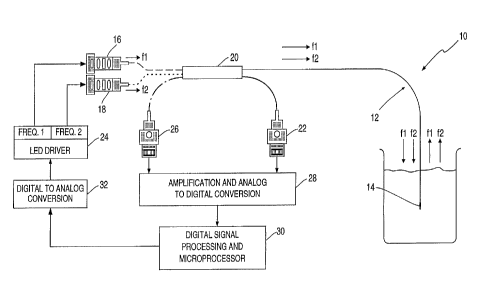
FIG. 1 is a schematic drawing of a system
for quantitating an analyte in a sample that involves
l0 the use of a simultaneous dual excitation/single
emission technique in accordance with the teachings of
the invention.
FIG. 2 is a graphical representation of a
comparison of arterial blood pH obtained using a
standard laboratory blood-gas analyzer with that
obtained using a paracorporeal fiber optic sensor
system and a simultaneous dual excitation/single
emission technique in accordance with the teachings of
the invention.
2o FIG. 3 is a graphical representation of a
comparison of arterial blood pCO2 obtained using a
standard laboratory blood-gas analyzer with that
obtained using a paracorporeal fiber optic sensor
system and a simultaneous dual excitation/single
emission technique in accordance with the teachings of
the invention.
Modes for Carrying Out the Invention
Before the present apparatus and methods for
- 3o quantitating an analyte in a sample are disclosed and
described, it is to be understood that this invention
is not limited to specific sensor formats, specific
indicator compositions, or specific excitation energy
sources as such, of course, may vary. It is also to
be understood that the terminology used herein is for
the purpose of describing particular embodiments only
and is not intended to be limiting.
_g_
CA 02231771 2001-10-30
WO 97/10495 PCT/US96/13791
It must. be noted that, as used in the
specification and the appended claims, the singular
forms "a," "an" and "the" include plural referents
unless the context clearly dictates otherwise. Thus,
for example, reference to "source of excitation
energy" includes more than one source of excitation
energy, reference to "an indicator material" includes
mixtures of suitable indicator materials, reference to
"an optical sensor" two or more such sensors, and the
like.
In describing and claiming the present
invention, the following terminology will be used in
accordance-with the definitions set out below.
The term "optical fiber means" is used
herein to refer to a single optical fiber or a bundle
of optical fibers. Suitable materials for optical
fibers will be outlined below.
The term "sample" as used herein refers to a
liquid or gaseous material which may be analyzed using
the presently disclosed sensors, either with respect
to a parameter such as pH, or with regard to the
presence or concentration of gases such as carbon
dioxide, or the like. Generally, "sample fluids"
analyzed using the sensors manufactured herein will 4e
physiological fluids such as blood.
The term "indicator" as in "indicator
composition's, "indicator material" or "indicator
component" refers t.o a species which has an optical
absorption or excitation spectrum that includes a
first region that is sensitive to the analyte of
interest in the sample undergoing analysis and a
second region that is insensitive to the analyte.
Preferably, the first and second regions do not
overlap substantially. By the phrase "do not overlap
substantially" is intended that the wavelength of peak
sensitivity to thE: analyte of interest of the first
region .is separated by preferably more than 20
-1~D-
CA 02231771 1998-03-11
WO 97/10495 PCT/US96/13791
nanometers from the wavelength of maximum
insensitivity to the analyte of the second region. In
addition, the indicator species has an emission
spectrum that is distinct from the absorption or
excitation spectrum and emits in a third spectral
region. The term "distinct" is used herein to signify
that the indicator species has an emission spectrum
that has a peak wavelength that is separated
preferably by more than 25 nanometers from both the
peak of the first region and the most insensitive
point of the second region.
For measuring pH, the indicator will
generally be a fluorescent dye or some other
fluorescent material which is pH-sensitive. For
35 carbon dioxide sensors, virtually any pH-sensitive
fluorescent or absorbent dye can be used, although
preferred indicators include fluorescein and
fluorescein derivatives such as carboxyfluorescein,
seminaphthorhodafluor, seminaphthofluorescein,
naphthofluorescein, hydroxypyrene trisulfonic acid,
dichlorofluorescein and the like. Particularly
preferred indicators are 8-hydroxypyrene-1,3,6-
trisulfonic acid ("HPTS") and fluorescein.
The term "isosbestic point" is used herein
to indicate a wavelength in the excitation or
absorption spectrum of an indicator material that is
insensitive to the changes in the analyte, to which
the indicator material is sensitive at other optical
wavelengths, i.e., the emission signal from the
indicator species when exposed to incident light at
the isosbestic point does not change with changing
- analyte concentration. Thus, for example, when an
indicator compound exists in two distinct species, the
interaction of an analyte in a sample with the
indicator compound may lead to the conversion of one
indicator species into the other. As this occurs, the
excitation, absorption or emission spectrum can change
-11-
CA 02231771 1998-03-11
WO 97/10495 PCT/LTS96/13791
such that one band of the spectrum may display an
increase in amplitude with increased analyte
concentration, while the amplitude of another band may
simultaneously decay. Certain bands of the spectrum
may be observed for which the amplitude does not
change in response to changing concentrations of the
analyte. Such analyte--insensitive regions of the
spectrum are referred to herein as isosbestic points.
The invention, together with additional
features and advantages thereof, may be best
understood by reference to the following description
taken in connection with the illustrative drawings.
With reference to FIG. 1 a system (10) is
generally provided for quantitating an analyte, for
example, pCO~ or pH in a sample. The system comprises
optical fiber means (12) that includes fluorescent dye
species (14) having a first region of its absorption
and/or excitation spectra which is analyte sensitive
and a second region of its absorption and/or
excitation spectra which is analyte insensitive. In
response to light corresponding to the first region
from first light source (16), e.g., blue light, and to
the second region from second light source (18), e.g.,
violet light, the dye species emits light energy,
e.g., fluoresces, into the same third spectral region,
e.g., green light. An optional optical coupler (20)
provides a means for combining the output of light
source (I6) and light source (18) to simultaneously
excite dye species {14) at two distinct regions of its
3o absorption or excitation spectrum. In addition,
optical coupler (2o) provides a means whereby a
reference signal may be routed to reference detector
(22). As shown in FIG. Z, light sources (16) and (18)
are light emitting diodes. ,
At the outset, an optical fiber means is
provided which serves to communicate optical signals
from a sample fluid to a detection means. The optical
-12-
CA 02231771 1998-03-11
WO 97/10495 PCT/US96113791
fiber means will typically comprise a single elongated
optical fiber, although it may comprise a bundle of
optical fibers associated in parallel.
Examples of suitable fiber substrate
materials include glass, plastic, glass/giass
composite and glass/plastic composite fiber
waveguides. A critical characteristic of optical
fibers is attenuation of the optical signal. Thus,
glasses which contain unacceptable levels of
1.0 transition-metal impurities when prepared from
naturally occurring materials lead to high absorption
losses. Silica fibers of acceptable quality can be
prepared from purified starting materials (e. g.,
silicon tetrachloride and germanium tetrachloride)
using conventional glass-melting techniques and
drawing into fibers.
Generally, although not necessarily, the
fiber will be provided with a cladding means. As will
be appreciated by those skilled in the art, the
cladding means serves to provide structural support
for an otherwise fragile fiber, and also provides a
coating which guides light conducted along the fiber.
In the present case, the cladding means typically
comprises a fluoropolymer such as polymeric
fluoroacrylate. However, the cladding means may also
be comprised of glass, or it may comprise polystyrene,
polyimide or any other suitable plastic material.
Preferably, the indicator species is a
single fluorescent or phosphorescent dye species
having an isosbestic point that can serve as the
second region of the excitation or absorption
spectrum. Alternatively, for an indicator species
that can exist simultaneously in two forms, e.g., acid
and base, the relative amounts of which depend on the
presence of an analyte. The excitation and emission
wavelengths used will then depend on the excitation or
absorption spectra of the two forms of the dye
.. -13-
CA 02231771 1998-03-11
WO 97/10495 PCT/US96/13791
species. For example, the acid and base forms of a
pH-sensitive dye species can be excited simultaneously
at independently modulated and distinct wavelengths
and the intensity of the emission can be measured at
the same optical wavelength for both excitations,
demodulated and processed to obtain a ratiometric
determination of the pH of the sample.
Indicator species may be provided on the
distal tip of the optical fiber means by any method
known in the art. One example of such a method is
found in U.S. Patent No. RE 31,879 to Liibbers et al.
which discloses a device wherein indicator material is
provided in solution form and separated from the
external environment by a membrane. An alternative
approach is to attach an indicator composition to the
tip of an optical fiber using a silanization technique
as described in, for example, U.S. Patent No.
5,354,825 to Klainer et al. Still another technique
involves direct bonding of photoactive polymers to the
tip of an optical fiber, as described in U.S. Patent
No. 5,354,825 to Klainer et al. Still another
approach involves the use of an inner adhesive layer
for affixing an indicator composition to the distal
end of a fiber optic sensor.
w 25 Briefly, this method involves the deposition
of a layer of a curable adhesive composition to the
tip of an optical fiber using a simple dip coating
procedure, partially or fully curing the adhesive
layer so provided using moisture, heat, ultraviolet
radiation or the like, coating the adhesive layer with
at least one outer layer of a curable indicator-
containing composition using a similar dip coating
technique used to provide the adhesive layer and
curing the outer.layer. The coated probe tip is
stored in a saline solution in order to hydrate the
fiber coating.
-14-
CA 02231771 1998-03-11
WO 97/10495 PCT/US96/13791
Yet another approach involves the use of a
C02-permeable end cap filled with a fluorescent
indicator and affixed to the distal tip of the optical
fiber means.
Briefly, this method involves prefilling a
C02-permeable silicone cap with a 3iquid solution
containing a C02 sensing dye. The prefilled cap is
applied over the tip of a fiber optic waveguide and
secured using a silicone adhesive that is deposited
onto the cap-fiber interface to secure the cap to the
fiber. The capped fiber is then suspended in a humid
environment to moisture-cure the silicone.
The source of light may be an incandescent
lamp, an arc or flash lamp, a solid state emitter, or
a laser. Preferably, the source of light is a light
emitting diode ("LED").
The output of light sources (16) and (18)
are simultaneously and independently amplitude
modulated by electronic means. As depicted in FIG. 1,
the output of light sources (16) and (i8) are
amplitude modulated at different electronic
frequencies, fl and f2 (indicated respectively by the
dashed (----) and dotted (~~~-) lines in FIG. 1), by
light source driver (24), which is exemplified in FIG.
-25 1 as an LED driver. The electronic frequencies are
selected such that they can be electronically
resolved. It is preferred that they differ by at
least 1 Hz and that they are not multiples of each
other, e.g., harmonics, or linear combinations
- 30 thereof. It is also preferred that the electronic
frequencies are not 60 Hz or multiples thereof.
Electronic modulation may be accomplished
using amplitude modulation schemes, at a constant
frequency, using current modulation (sinusoidal,
35 triangular, square-wave or the like), voltage
modulation or spatial filtering with optical shutters.
Alternatively, electronic modulation using frequency
-15-
CA 02231771 1998-03-11
WO 97/10495 PCT/US96/13791
modulation schemes, at constant amplitude, may be
accomplished using methods well known in the art
including acousto-optic modulation, electro-optic
modulation or non-linear crystals. In addition, the
optical signals from light sources (16) and (18) may
be modulated using phase modulation schemes, such as
electro-optical modulation typically employing
piezoelectric crystals. Frequency modulation and
phase modulation may be useful in conjunction with
coherent light sources while amplitude modulation
schemes may be used with coherent and/or incoherent
light sources. In one preferred embodiment, the
optical signals from light sources (16) and (is) are
modulated using amplitude modulation schemes, more
particularly amplitude modulation schemes employing
current modulation. The system may optionally include
a means to generate a lamp reference signal which may
be an optical coupler/beam splitter, the signal from
which is routed to an optional reference detector.
Electronic modulation of the optical signals
from light sources (16) and (i8) results in the total
returning emission signal from dye species (1~) being
composed of two distinct fluorescent components--a
component at electronic frequency f1 (the sensing
s~_gnal the amplitude of which is pH dependent) and a
second component at electronic frequency f2 (the
reference signal the amplitude of which is pH
insensitive). The two emission signals are routed
through optical coupler (Z0) and are presented
- 30 simultaneously to optical detector (Z6) (the total
returning emission signal is represented in FIG. 1 by
the line composed of alternating dots and dashes
(._._)),
The optical detector may be a solid state
detector or an array of such detectors, non-solid
state detectors, thermal detectors or the like.
Examples of solid state detectors include silicon
-16-
CA 02231771 2001-10-30
W(J 9?/IU495 ~m 1/VJYV/IJ/YI
detectors and arrays thereof. Examples of non-solid
state detectors include photomultiplier tubes
("PMTS"). Therma:L detectors include thermopiles and
bolometers.
The signal detected from dye species (14) by
optical detector (26) can be demodulated using any of
a variety of demodulation schemes well known in the
art. The scheme that can be used to demodulate the
signal depends on what scheme was used to modulate the
optical signals from light sources (16) and (18).
Thus, far. optical signals that have been
modulated using amplitude modulation schemes,
demodulation may be done by any method well known in
the arty including digital demodulation or analog
demodulation schemes. If the samples were frequency
or phase modulated, the signal detected from dye
species (14) can be demodulated by frequency or phase
demodulation schemes, respectively. Preferably,
amplitude modulated optical signals from dye species
(14) are received by optical detector (26), which
typically provides an analog output, amplified,
digitized by a high-speed analog-to-digital (A/D)
converter (28) and routed to a digital signal
processing (DSP) device (30). Here, spectral analysis
is performed on the digitized version of the detector
output by discrete F'ourier transform ("DFT")
techniques well known in the art. The net result is
the demodulation and separation of the two emission
signals into their respective amplitudes--the pH-
3o dependent sensing signal and its simultaneously
demodulated reference signal. These numerical results
are then available for subsequent post-detection
processing to quantify the analyte.
The DSF? device (30) also serves as a digital
microprocessor which, through digital-to-analog
converter (32), provides a signal to light source
-17-
CA 02231771 2001-10-30
WU y//IU~IyJ
Cl_ lluJyu~IJ~W
driver (29) to modulate the output of light sources
(16) and (18) .
This fiber-optic based fluorescent sensing
technique for pEi and/or pC02 has applications for the
measurement of pH and quantitation of dissolved gases
such as carbon dioxide in samples, e.g., for measuring
pH and pCO2 in aqueous samples. Given the general
remote sensing architecture of. the instrument/sensor
electro-optics, the technique is adaptable to any
to application that might require the remote monitox-ing
of an acid-bass: chemistry system.
In addition, the invention may be useful
when incorporated in paracorporeal blood gas
monitoring system such as disclosed in commonly-
assigned U.S. Patent No.
5,697,366 entitled "In Situ Calibration System for
Sensors Locattrd in a Physiologic Line," inventor
Kimball et al.., filed on January 27, 1995, and
described in Martin et al. (1994) Proc. biomed. Fiber
Optics Instrumentation 2131:426-436.
Briefly, the system includes fiber optic
sensors that are contained in a housing with standard
luer lock adapters that attach into an arterial
pressure line, allowing monitoring to occur
"paracorporeally"; patient blood is moved into the
line and housing, via care-giver draw, for discrete
measurements and returned to the patient upon
completion of the measurement.
It w ill be appreciated by those working in
the art that sensors fabricated using the presently
disclosed and claimed techniques may be used in a wide
variety of contexts, including measurement of carbon
dioxide or other gases, glucose determination,
measurement of potassium .ions, calcium ions, magnesium
-18-
CA 02231771 1998-03-11
WO 97/10495 PCT/US96113791
ions, and the like. Also, while the invention has
primarily been described in conjunction with the
measurement of analytes in blood, the sensors
fabricated using the present method may be used to
evaluate a wide range of parameters in any number of
sample types.
Thus, it is to be understood that while the
invention has been described in conjunction with
preferred specific embodiments thereof, the foregoing
description, as well as the examples which follow, are
intended to illustrate and not limit the scope of the
invention. Other aspects, advantages and
modifications within the scope of the invention will
be apparent to those skilled in the art to which the
invention pertains.
Examt~le 1
Use of Simultaneous Dual Excitation/Sinale Emission
Method to Measure pH and nC02
A paracorporeal fiber optic blood gas and pH
monitoring system employing the simultaneous dual
excitation/single emission technique and apparatus as
shown in FIG. 1 was used to measure arterial pC02 and
- 25 pH as described in Martin et al., supra.
Human clinical data obtained using the
paracorporeal device were compared with assay values
generated by standard laboratory pH/blood gas
techniques and analyzers, e.g., a Radiometer
Corporation Model ABL 500-#2 blood gas analyzer. The
arterial samples were split so that pH and pC02 were
_ measured by each technique using the same sample. All
procedures involving human subjects were approved by
the appropriate clinical site review committee.
The fiber optic sensors used in these
experiments for measuring arterial blood pH were
prepared containing fluorescein (Aldrich, Milwaukee,
-19-
CA 02231771 2001-10-30
WU y7/IU~iy~ f'c.:'l'/US96/13791
WI), while those sensors used for measuring pC02 were
prepared containing 8-hydroxypyrene-1,3,6-trisulfonic
acid, trisodium salt ("HPTS") (Molecular Probes,
Eugene, OR). The sample was alternately interrogated
using the pH and pC02 sensors as follows.
The indicator species in the fiber optic pH
sensor was simultaneously exposed to excitation light
centered at 488 nm and 442 nm, with an emission signal
from the indicator species monitored in the region
529.5 nm ~ 15.5 nm. The 488 nm and 442 nm signals
were respectively modulated at 37 Hz and 24 Hz. The
442 nm signal corresponds to a pH-insensitive region of
the fluorescein excitation spectrum. The optical
signals were modulated using an amplitude modulation
scheme using sinusoidal current modulation.
The indicator species in the fiber optic
pCO2 sensor was simultaneously exposed to excitation
light centered at: 442 nm and 415 nm, with an emission
signal from the indicator species monitored in the
region 529.5 ~ 15.5 nm. The 442 nm and 415 nm signals
were respective:lv modulated at 37 Hz and 24 Hz. The
415 nm signal corresponds to an isosbestic point of the
HPTS excitation spectrum. The optical signals were
modulated as described above.
The emission signals were detected using a
silicon detector, the analog output signal from which
was digitized and fed to a microprocessor where the
signals were demodulated.
Sensor precision, expressed as standard
deviation about the mean ("SD"), and sensor accuracy,
expressed as average difference from the values
obtained using standard blood-gas analyzers were
calculated using the data gathered from 10 independent
patient blood-gas measurements.
In 10 measurements of arterial blood pH, the
value obtained using the standard blood-gas analyzer
was 7.414. The value obtained using the paracorporeal
-20-
CA 02231771 2001-10-30
device employing the simultaneous dual
excitation/single emission technique and apparatus of
the invention was 7.375 (SD = 0.008). The average
difference between the ten values obtained using the
standard analyzer and the paracorporeal method was
0.039.
The arterial pC02 value obtained using the
standard blood-gas analyzer was 38.3 while that
obtained using the paracorporeal device was 40.6 (SD =
l0 4.00%) while the average difference was 5.9%.
Sensor performance was plotted for pH and
pCO2 in FIG. 2 and FIG. 3, respectively, which show
individual data points for blood pH and pC02
measurements, respectively, as well as the identity
lines calculated by linear regression analysis (pH: r2
- 0.906; pC02: r2 = 0.884).
These data demonstrate the accuracy and
precision of data collected from fiber optic sensors
using the simultaneous dual excitation/single emission
system of the invention.
30
-21-