Note: Descriptions are shown in the official language in which they were submitted.
CA 02232999 1998-03-24
W O 97/11651 1 PCTrUS96/15722
DESCRIPIION
FAS~NERS HAVING COORDINATED SELF-xh ~:KI~G
CONFORMING ME~ERS AND USES 1~ R~F
5 FIELD OF 'I'~; lNVENTlON
The present invention relates to f~tp-nprs having cooldina~d self-sePkin~
cnnforming members and uses thereof. The f~tenPrs may be used for ~nchoring a
prosthesis, or for c~ g or ~1ignin~ a stlucture within a hole, tube, or cavity. In
particular, hard tissue impl~nt f~tPnp-rs are envisioned for ~tt~c,hin~ a prosthP,si~ to bone
10 or cartilage. M~,tho~l~ of using said f~tPnP,rs are also provided.
BACKGROI~ND OF 'L~i~; INVENTION
Prior art references relate to structures adapted to expand the radial projection of
the device after the device has been inserted into a cavity. Three groups of prior art are
15 outlined by structural ~imil~riti~s in their PYp~n~ion char~tPri~tics. A fourth group relates
to other eYr~n~ion ch~r~-tPri~ s.
Axial contraction is used to produce radial expansion once a device has been
inserted into a cavity. Wigam (U.S. Patent 3,505,921) and Talan (IJ.S. Patent 4,309,136)
disclose construction f~,~tening devices. Fischer et al. (U.s. Patent 3,779,239) and
20 Rl-blenik (SU 1,386,182-A) relate to elongated f~ctenPrs that employ radial el~p~n~ion
el~,m~,nt~ at the distal end of the device. These f~te-nprs are intpnded to secure fractured
portions of bone tissue. Kuslich (U.S. Patent 5,059,193) relates to a spinal imrl~nt for
use between vertebrae. Tansey (U.S. Patent No. 4,681,590) relates to a structure having
metal strips secured between an upper plate and a nut. The nut is mounted on a screw
25 and cnn~tr~inPd against rotation so that rotating the screw reduces the axial s;;~ ion of
the nut and upper plate causing the metal strips to expand radially. Tansey also relates to
a femoral stem prosthesis.
Oblique contact has been used between moving element~ to expand the radial
projection of devices that have been inserted into a cavity. One common eY~mple of this
30 type of f~tPner is from Aginsky (U.S. Patent 4,091,806). In this mPrh~ni~m, a central
shaft is displaced relative to an outer concPntric shaft. The central shaft incllldçs a wedge
that obliquely contacts a longitll-lin~1ly slotted portion of the outer con~entric shaft. The
oblique contact tr~n~l~tes the a7cial force on the wedge into a radial force that expands the
outer concçntric shaft radially. Prior art having ml-ltiple P,lement~ actuate the çlP,ment~
CA 02232999 1998-03-24
W O 97/11651 2 PCTrUS96/15722
such that the elemPnts cannot be PYp~n~e~ in~3epPn~lpntly to adapt to the collloul~ of the
cavity in which they are placed.
Pivotal connPctinn~ have been used to expand the radial projection of devices once
they are inserted into a cavity. Prior art relates to çlçmPnt~ pivotally connPcte~ to the
S device that are contacted by an axially rli~l~r~P~hle PlP-m~ont The aYial force at a rli~t~nce
from the pivotal ct)nnectinn creates a torque that rotates the pivotally connected PlPmPnt~
into a new position that has a greater radial tli~mPtçr. Some of these references tlic~lose
m~h~ni~m~ that also use oblique contact to provide the nP~ç~,y torque. As çY~mrlçs of
this type of mPrh~ni~m, see ~vila (U.S. Patent 3,986,504), Davis (U.S. Patent
10 5,057,103), Dobelle (U.S. Patents 2,685,877 and 3,024,785), and Firer (SU 1,524,880A).
None of the references in this group relate to means for coordinated self-seeking
conforming of elem~nt~ Aginsky (U.S. Patents 4,204,531 and 4,227,518) relate to use of
pivotal connections in a different structure. A pivot point is movably mounted in a
longitudinal slot. The pivot point is pivotally coupled to two legs that are pivotally
15 coupled at their other ends to two sections of the outer sheath. When the outer sheath is
displaced axially, the pivot point is constrained by the slot and the section~ of the outer
sheath are rotated radially by the legs. This structure does not allow coordinated self-
seeking conforming ~t--~tion of the two outer sheath secti~n~.
Bolesky (U.S. Patent 4,275,717) and Street (U.S. Patent 3,216,414) relate to
20 elements that are biased to expand radially. These Çl~ment~ are e1~fi~-~lly conctr~ined by
a ring or cap that is axially ~lispl~ed once the device is inserted so that the biased
element~ can resume a radially expansive position. Erlich-DeGuemp (FR 2,387,638)relates to a device that uses the bone tissue surface to provide an oblique contact for radial
expansion. Muhlbayer (DT 1,075,793) relates to use of a rotating central shaft to
25 tr~n~l~te a band which has three pinned elem~-nt~ that are allowed to rotate radially. The
pinned ~lemPnt~ can rotate freely but are not driven by a me~h~ni~m and do not provide a
means to engage the three rotating element~ in a coordinated self-seeking conforming
manner.
A review of the product literature shows adaptations of merh~ni~m similar to
30 Livingston (U.S. Patents 2,699,774 and 2,490,364), Fisher (U.S. Patent 3,805,775) and
Flander (U.S. Patent 3,708,883). An Alta Modular Trauma System product (HowmpA
~utherford, New Jersey) uses a slotted sleeve, wedge shaped inner mandrel and
translation of the mandrel in the sleeve to increase radial ~ mçtçr. Other col"l.~nies are
CA 02232999 1998-03-24
W O 97/11651 3 PCTAUS96/15722
g~nl~r~11y introducin~ unicortical f~etrnrre (en~ çs only one bony cortex) conQ-iQ-ting of a
slotted PYtPrn~lly threaded hollow cylinder with a threaded inner mandrel that when
rotated e-r~ntlQ the radial di~m~ot~r of the outer cylinder. One eY~mp1P- of this type of
device is the Sargon Tmr1~nt system (Sargon I~nt~l~l;ses, Inc., Beverly Hills, CA).
Bone imp1~nte have been used to solve health care problems of orthopedic and
maxillofacial reconstruction, prostheeie fi~tit~n, drug delivery and fracture stabilization.
Heretofore, bone and cartilage (hard tissue) imp1~nte were f~etemPd with screw threads,
ihltelr~lcllce fits, uniformly exr~n~lin~ merh~nieme and ce~nPnt The majority of these
devices and ~eSoci~tpA techniques provide poor initial fixation, and following bone
10 formation around the device, provide good fixation but often for only a limited period of
time. Tmp1~nt removals are frequently pelrolllled following failure of the bone-imp1~nt
interf~re and clinical loosening of the device.
The principal cause for implant failure in hard tissue is the ~eçp~r~tinn of bone from
the surface of the implant. Bone resorption about an imp1~nt is indl1ced by micromotion
15 of the device relative to the surrounding hard tissue, adverse tissue reaction to the imp1~nt
m~tPri~1, or tissue necrosis due to drill heating and merh~nie~1 stress concentratione.
Micromotion is often due to poor initial stabilization of a threaded, inle.re,cllce fit or
cemçntP-d device.
Bone and cartilage are tissues with viscoelastic m~tPri~1 p ~t;,~ies. Their modulus
20 of e1~eticity and 111tim~te strength are much less than the metal and ceramic m~tPri~1e used
for hard tissue imp1~ntQ.. This miQm~tch in m~tçri~1 properties is a factor in device-tissue
interf~ 1 micromotion, intçrf~ce stress concentration and imp1~nt loosening. This
problem is compounded by bone's range of morphology and m~teri~1 pl~e.lies.
The bone organ contains two distinct types of bone tissue. Cortical bone is the
25 hard structural bone that forms the outer shell of the Q~P1~t~n C~ncpllous bone is
contained within cortical bone and makes up a .eignifi~nt portion of the volume of most
bones. Cancellous bone is porous, trabecular in structure, highly vascular, filled with
cellular elements and undergoes active remodeling (formation and reso,~Lion).
He.c~torolc~ bone implants placed transverse to the long axis of bone pÇnetr~t~d a
30 short segmPnt of the cortical bone shell and had a si~nifi~ ~nt portion of their surface
adjacent to c~nce11Ous bone. These devices relied on both the cortical and cancellous bone
for initial and long-term stability. Implant depen~en-e on porous low strength cancellous
bone and limited cortical bone contact causes poor device stability.
CA 02232999 1998-03-24
W O 97/11651 4 PCT~US96/15722
Heretofore bone impl~ntc that were placed along the long central a~is of bone were
desi~ned to occupy much of the c~ncPllnus bone space and contact the e-n-loste~l surface
(inner surface) of cortical bone. The irregular morphology of the en-loste~l surface caused
point-contacts and con~ e~ ,.ted loads between these devices and cortical bone. Initial
5 impl~nt fi~c~tinn was achieved by inLelL~ ce fit at these points of contact.
High ccn~-ç~ ted loads and stress-chiel~1ing btlween point-contacts can cause
resorption of bone and impl~nt loosening. Long-term impl~nt fixation is depen~ent on
cancellous bone growth around the surface of the impl~nt where it is not contacting the
endosteal surface of cortical bone.
Cements are commonly used to fill the void between the endosteal surface of
cortical bone and the implant. Though commonly used, cemPntc can cause adverse tissue
reactions and complicate the load char~- teri~tics of the bone-implant interface by adding a
third m~teri~l with its unique m~tPri~l properties. UncemPnted devices are beingintroduced for hip and knee prostheses. Some of these devices are contoured or require
the endosteal surface of the cortical bone to be machined to increase the implant-to-
cortical bone surface area. Contouring the device surface increases its cost and may
require patient im~gin~ and bone ch~pin~. M~ehining bone removes healthy tissue and
weakens the structural strength of the organ. Additionally, local heating of bone during
machining can cause its death.
Hand-tool reaming, press-fit inct~ tic)n and cemPnting require skill. These
procedures are sources of surgical variance and potential prostheses failure. The
combined issues of device design, device-bone intPrf~ee failure, micromotion, stress-
concentration, stress-chiel-ling m~teri~l ~r~lly differences and surgical variance limit the
useful lifetime of many prostheses to 5 to 20 years.
The present invention addresses these problems in the prior art and provides
devices having coold~lated self-seeking conforming members, where a known relationship
exists between the pressure exerted on the surrounding m~tPri~l and the mech~nicm
engagement torque or force, and having the ability to contour to an irregular defect.
SUMM~RY OF THE ~VENTION
The present invention provides f~ctPners having coordinated self-sePl~ing
conforming members for conforming to a cavity. An ~çh~tor mPch~nicm coo~dinates and
tr~ncl~tes applied force for the independent or dependent movement of each member. The
CA 02232999 1998-03-24
W O 97/11651 5 PCTAJS96/15722
process of conforming also aligns the f~t~n~--r, and thereby aligns that which is ~tt?~-hPd to
the f~tener. The present f~tPnPr~ are useful wherever im~ nts are desired, and
wherever ~ligning is needed. In particular, the f~tÇn~q-rs are useful for hard tissue
impl~nt~ for hllm~n~ and ~nim~l~, such as cartilage and bone impl~nt~; for anchoring
S prostheses; and for ~ligning devices as in in~1u~tri~l te~hnology.
An embodiment of the present invention provides a f~tener for imrl~nting into a
cavity, the f~t~n~r comrri~ing a body; a plurality of members movably connect~d to the
body, each member being independently movable for coordinated self-seel~in~ conforming
to the cavity; and an actuator mel~h~ni~m for coor 1in~tin~ and tr~n~l~ting applied force to
10 each member.
As used herein, a "f~t~ner" may also be referred to as an impl~nt; a "cavity" is an
enclosed space for receiving and hol~ling a structure and may be a hole, tube, cylinder,
well, hard tissue defect, or the like. A "hard tissue defect" is a defect in bone or cartilage
that may have been constructed surgically, by ~ei~nt or from disease. "Independ~ntly
15 movable for coordinated self-seeking conforming to a cavity" means that each member
responds to applied force in a coordinated manner until it engages a wall of the cavity and
a second member may continue to move until it also engages a wall of the cavity."Coordinated" means that all members move initially in response to applied force. "Self-
seeking" means that a member moves in response to an applied force until it engages a
20 wall of the cavity for independent movement. "Conforming" means that a memberengages a wall of the cavity. "Movably connected" means di~;Lly or indirectly
connected. A member may be a lever, a finger, a projection, a protrusion, a wing, a
shoe, or an ear, or the like.
In a plefelled embodiment, an interf~ res~u,c exists between each member and
25 the cavity when in use. In an even more plcr~lled embodiment where the cavity is a hard
tissue defect, a sllffic~ient int~rf~ l pl~s~ure exists between each member and the cavity
to cause hard tissue density, in particular, bone density, to increase when in use.
The actuator m~rh~ni~m may further comprise a locking mech~ni~m for locking the
actuator mechanism. The locking m~rh~ni~m may be selected from the group con~i~ting
30 of a cap, a jam nut, a taper, a transverse pin, a key, a spline, an abutment, and the like.
Where the cavity is a hard tissue defect, the f~tener as described herein may
further comprise a prosthesis connected to the f~tener. The prosthP~i~ may be a hard
tissue trauma fixation device or hardw~ selected from the group con~i~ting of a dental
CA 02232999 1998-03-24
W O 97/11651 6 PCT~US96/15722
impl~nt a spinal prosthesis, an intPrmPd~ ry rod, a knee prosthP~i~, a shoulder
prosthP~i~, a finger prosthPci~, a hip prosthP~ , a lelnpo~ mandibular joint, a dental
impl~nt with prosthetic tooth, a plate, a port, and the like.
A further embodiment of the present invention inl l~ldes a f~t~n~r as ~e~t~ril~ed
herein having a first member and a second member and wherein the ~-~nl~tor merh~ni~m
comrri~es a journal; and a first link and a second link, each link rotatingly and slidingly r
joined to the journal, the first link rotatingly joined to the first m~mhPr and the second
link rotatingly joined to the second member such that when the journal is moved, the first
link and the second link move and cause the first member and the second member to move
10 independently to conform to the cavity. The actuator mech~ni.~m may further comprise a
bearing having a bore for housing the journal, the journal further having an end movably
connected to the bearing so that when the bearing is moved, the journal moves through the
bore of the bearing.
The invention includes a f~ctPnPr as described herein having a first member and a
15 second member and wherein the actuator m~h~ni~m compri~es a journal; and a cam
slidingly and rotatingly connected to the journal, the cam further slidingly and rotatingly
joined to the first member and to the second member such that when the journal is moved,
the cam moves and causes the first member and the second member to move
independently to conform to the cavity. The ~t~tll~tor mech~ni~m may further comprise a
20 bearing having a bore for housing the journal, the journal further having an end movably
connected to the bearing so that when the bearing is moved, the journal moves through the
bore of the bearing.
Another embodiment is a f~tenp~r as herein described having a first member and asecond member and wherein the actuator mPf~h~ni.~m compri.~es a journal; a first link
25 having a first end and a second end, the first end pivotally connected to the journal; a
second link having a first end and a second end, the first end pivotally connectPd to the
second end of the first link, and the second end pivotally connected to the first member;
and a third link having a first end and a second end, the first end pivotally connected to
the second end of the first link, and the second end pivotally connected to the second
30 member, wherein when the journal is moved, the first link moves and acts on the second
link and the third link to cause the first member and the second member to move
independently to conform to the cavity. The actuator m~h~ni.~m may further comprise a
bearing having a bore for housing the journal, the journal further having an end movably
,
CA 02232999 1998-03-24
W O 97/1165~ 7 PCT~US96/15722
connecte~ to the bearing so that when the bearing is moved, the journal moves through the
bore of the bearing.
A further f~tener of the present invention as herein described has a first m~omher
and a second member and wherein the ~ct~tor m~h~ni~m compricçs a journal; a first
S link having a first end and a second end, the first end pivotally and slidingly connected to
the journal, and the second end pivotally connlo~tçd to the first member; and a second link
having a first end and a second end, the first end pivotally and slidingly connected to the
journal, and the second end pivotally conne~ted to the second member; wl.er~in when the
joumal is moved, the first link and the second link move to cause the first member and
10 the second member to move independently to conform to the cavity. The actuator
mçch~ni~m may further comprise a bearing having a bore for housing the journal, the
journal further having an end movably connected to the bearing so that when the bearing
is moved, the journal moves through the bore of the bearing.
The f~tçn~r as herein described having a first member and a second member is an
15 aspect of the invention wllc.eill the actuator me~h~ni.~m comprises a journal; and a cam
having a first slot and a second slot, the first slot slidingly and pivotally connected to the
journal, and the second slot slidingly and pivotally joined to the first member and to the
second member such that when the journal is moved, the cam moves and causes the first
member and the second member to move independently to conform to the cavity. The20 actuator mP~h~ni~m may further comprise a bearing having a bore for housing the journal,
the journal further having an end movably connected to the bearing so that when the
bearing is moved, the journal moves through the bore of the bearing.
The f~tçnPr as herein described having a first member and a second member, each
member having an arm, is an aspect of the invention, wherein the actuator mech~ni~m
25 comprises a shaft having a first end and a second end, and a component constr~in~d to be
in slidable contact with the second end of the shaft and in contact with the arm of each
member such that when the shaft is moved, the component moves in contact with the arms
and causes the first member and the second member to move in~1epçnd~ntly to conform to
the cavity. In a preferred embodiment, the shaft decreases in rli~mtott-r at the second end.
A further aspect of the invention is a f~ct-oner as described herein having a first
member and a second member, each member having an angled surface, and wherein the
single ~ t--~tor m~h~nicm comprises a shaft having a first end and a second end; and a
component constrained by the angled surface of each member to be in ~ ble contact
-
CA 02232999 1998-03-24
W O 97/11651 8 PCTAUS96/15722
with the second end of the shaft such that when the shaft is moved, the cc,~ nent moves
in contact with the members and causes the first mPmhçr and the second member to move
indepPndPntly to conform to the cavity. In this emb~imPnt each member is movablylimited by the body such that the cG...~,onent is csnt~inPIl within the body by the angled
S surface of each member. In a l)rer~l~d embo~imPnt, the shaft decreases in ~ mpt~r at
the second end.
A further embodiment of the invention is a f~ctPnPr for im~ nhn~ into a cavity,
the f~ctPnçr comprising a body comprising a movable structure; a plurality of members
rotatingly connected to the movable structure, each member being independently movable
with respect to the body for coordinated self-seeking conforming to the cavity; and an
actuator mech~ni~m within the movable structure for coor lin~ting and fr~ncl~ting applied
force to conform independently each member to the cavity, the actuator merh~nicmcomprising a journal rotatingly and slidingly connected to each member such that when
the journal is moved through the bore of the body, each member moves symmPtriç~lly
lS with respect to the movable structure and moves independently with respect to the body to
conform to the cavity.
An embodiment of the invention is a f~ctPnPr for impl~nting into a cavity, the
f~ctPn~r comprising a body; a plurality of members movably connected to the body, each
member being depPn~lPntly movable for coordinated self-seeking conforming to the cavity;
and an actuator mech~nicm for coor lin~ting and tr~ncl~ting applied force to each member.
"Dependently movable" means that the members move at the same time, when one
member engages a wall of the cavity, a second member is not able to move further. For
dependent movement, "self-seeking" means that movement of members is determined by a
first member Png~ging a wall of the cavity. Further members do not continue to move.
A further embodiment is a f~ctener having dependent movement as herein
described having a first member and a second member and wherein the actuator
me~h~nicm comprises a journal rotatingly and slidingly joined to the first member and to
the second member such that when the journal is moved, the first member and the second
member move dependently to conform to the cavity.
A further f~ctenPr having dependent movement has a first mPmber and a second
member and wherein the actuator mech~nicm compricPs a journal; a first link having a
first end and a second end, the first end pivotally connectP~ to the journal, and the second
end pivotally connected to the first member; and a second link having a first end and a
CA 02232999 1998-03-24
W O 97/11651 9 PCTAJS96/15722
second end, the first end pivotally connPctP~ to the journal, and the second end pivotally
connected to the second member; wherein when the journal is moved, the first link and
the second link move to cause the first member and the second member to move
depen~lently to conform to the cavity.
S A further f~tener having dependent movement has a first member and a second
member, each member having an arm, wherein the actuator mP~h~ni~m comprises a shaft
having a first end and a second end, the second end being in slidable contact with the arm
of each member such that when the shaft is moved, the first member and the second
member move dependently to conform to the cavity.
A further f~ctenPr having dependent movement has a first member and a second
member, each member having an angled surface, and wherein the actuator mPrh~ni.~m
comprises a shaft having a first end and a second end, the second end being in slidable
contact with the angled surface of each member such that when the shaft is moved, the
first member and the second member move dependently to conform to the cavity.
In each of the f~tPnt-r emborlim~nt~ having dependent movement, the actuator
mech~nism may further comprise a bearing having a bore for housing the journal, the
journal further having an end movably connected to the bearing so that when the bearing
is moved, the journal moves through the bore of the bearing.
A further embodiment of the invention is a f~tenPr for seCurin~ a structure to a20 cavity where the structure is configured to fit to an outer surface of the cavity. The
f~tenPr comprises a body having a first bore and a slot opening onto the first bore; a
cylinder within the first bore, the cylinder having a driving mP~h~ni~m; a member within
the slot, the member movably conne~tP~ to the body and acted on by the driving
mechanism for conforming to the cavity; and wherein when a force is applied to the
25 cylinder, the driving mech~ni~m causes the member to move to conro-lll to the cavity,
thereby securing the structure to the cavity. The driving me~h~ni~m may compri~e an
actuator, the member may comprise a lobe, and in this case, the ~ctu~tor meshes with the
lobe of the member. In a ~lefellc~d embodiment, the driving mech~ni~m comprises a
worm gear, the member comprises a plurality of gear teeth, and the worm gear meshes
30 with the gear teeth of the member. The f~tpn~r may further comprise a locking means
for locking the cylinder within the bore. The locking means may be selected from the
group con~ tin~ of a retainer or a jam nut. The structure may be a prosthesis and may be
selPct~P~ from the group of prostheses described herein.
CA 02232999 1998-03-24
W O 97/11651 lo PCT~US96/15722
M~teri~l~ suitable for f~hric~tion of a f~tPnPr of the present invention may be a
m~tPri~l that is at least bioco~ a~ible for the length of intended use, and has s~lfficiPnt
structural strength. A bioco",p~ihle m~tPri~l is s~PlPctP~ from the group con~i~ting of a
metal, a cPrAmic, a polymer, and a combination thereof. Where the f~tenPr is formed of
5 a metal, the metal is tit~nil~m~ ni~llll alloy, st~inlP~ steel, chlollliuln cobalt, ch,u~lliulll
cobalt alloy, or the like. Where the f~tPnPr is formed of a cPr~mic, the cP~mic is silica
glass, alumina, c~lcium phosphate, c~lcil-m carbonate, or the like. Where the f~tPnPr is
formed of a polymer, the polymer is delrin, nylon, polyester, polymethylmPth~-,rylate,
polyethylene, or the like.
Use of the f~tenPrs of the present invention for impl~nting into a cavity and
~ligning the f~tenP-r within the cavity is an aspect of the invention. The use compri~Ps
placing the f~tener into the cavity and applying force to conform each member of the
plurality of members to the cavity thereby ~ ning the f~ctener with the cavity. The
cavity may be related to industry, or a hard tissue defect such as a bone or cartilage
15 defect.
Use of the f~tenPrs of the present invention, where the cavity is a hard tissue
defect of an animal, for f~tening a prosthesis to the hard tissue defect is an aspect of the
invention. The use comprises placing the f~tPner into the hard tissue defect of the
animal; applying force to conform each member of the plurality of members to the cavity;
20 and ~tt~hing the prosthesis to the f~tPnPr. Preferably, the animal is a human. In a
;rell~d embodiment, a s~fflcient interfacial pl~ iUl'e exists between each member and
the cavity to cause hard tissue density to increase when in use.
Use of the f~teners of the present invention for f~tPning airplane skin at a cavity
site is an aspect of the present invention. The use comprises placing the f~ctener into a
25 cavity of airplane skin; and applying force to conform each member of the plurality of
members to the cavity thereby f~tPning the airplane skin.
Use of the f~tPnPrs of the present invention for ~ ning a part to an object in
automated production is a further aspect of the present invention. The use compri~P~s
placing the f~tener onto the part; and applying force to the f~tPner to conform each
30 member of the plurality of members to align the part to the object. In a ~,~re"ed
embodiment, the object is a robot or part thereof.
Several ~i~nific~nt advantages are achieved by the present invention. The f~teners
of the present invention, when implanted in bone tissue, for example, provide unique and
CA 02232999 1998-03-24
W O 97/11651 11 PCTrUS96/15722
novel designs that ~llh~illl;7~ micromotion, optimize the stress distribution to bone, allow
m~tPri~1 property Ill~ hi~ through active m~,h~ni~m~, assist in cP~.~e- ;n~ the device,
present forces to the bone to stimul~tp it to beco-lle more dense, minimi7~ or e~
surgical variance and allow it to be tightPned, if loose. Applied force to the f~tPnPr may
S be from a tool ~tt~hed to the f~tener. The tool may push on the achl~t~r mPrh~ni~m to
cause the members to move.
The effi~iency of the f~tPning device of the present invention is im~roved
conci~lPr~hly by the provision of its design and its oper~tion~l colll~alibility with bone.
Further advantages include independent movement of the ç~r~n~inn members for certain
10 embo-limçnt~, conformation of the imrl~nt-to-bone defect, application of a known bone-to-
implant intPrf~ci~l ~rGSSU1G~ capability to reengage or r~ Png~ge the mP~h~ni~m to refasten
or remove the impl~nt drawing of the implant into the defect by the action of the
expansion components, compression of small irreg~ ritiPs along walls of the hole or
defect by the e~cr~n~1in components to increase contact area, high implant surface area
15 against the ~u~ wlding m~tPri~l, control of impl~nt ~lignmPnt or CÇ~ ;ng within the
defect, and firm initial fixation. All of these advantages are accompli~hP~l without
bending any implant component. This minimi7es the likelihood of coll.~onent failure
through residual stress in the device and fatigue loading. These advantages are .cignific~nt
when co---~ared to press-fit, screw thread and uniformly e~rp~n-ling f~tenP-rs~
Following long-st~n-ling patent law convention, the terms "a" and "an" mean "oneor more" when used in this application, including the claims.
BRIEF DESCRIPIION OF l~E DRAVVINGS
Further objects and advantages of this invention will become a~ alGn~ from
25 cc)n~ er~tiQn of the drawings and ensuing description of the ~lGrGlled embo-lim~nt~.
Fig. la and Fig. lb are cross-sectional plane views of a fastener having a linkage-
member mech~ni~m in bone and configured as a bone contacting dental impl~nt
Fig. 2 is a cross-sectional plane view of a f~te-n~r having a c~mmP~-member
mP~h~ni~m configured and shown as a femoral co~ onent of a hip prosthesis.
Fig. 3 is a cross-sectional plane view of a f~tPner having a three-linkage
mech~ni~m configured as the mandibular component of a temporal-mandibular joint.Fig. 4 is a cross-sectinn~l plane view of a f~t~nPr with scissoring members and a
two slot cam mec-h~ni~m
CA 02232999 1998-03-24
W O 97/116~1 12 PCTAUS96/15722
Fig. S is a cross-secfion~l plane view of a f~t~n~r with s~icc~rinp members, andsphere and shaft actuator mech~nicm configured as a bone-cont~-ting dental imrl~nt with
prosthetic tooth.
Fig. 6a and Fig. 6b provide a cross-sectit)n~1 plane view of a f~cten~r with pinned
5 members, and sphere and shaft ~ctu~tor m~rh~nicm configured as a dental impl~nt The
dental imrl~nt is shown in bone with in-phase (Fig. 6b) and out-of-phase (Fig. 6a)
member expansion.
Fig. 7a and Fig. 7b provide a cross-sectional plane view of a f~ctçner with a
member merh~nicm in a spherical rotating housing configured in a plate that allows
10 rotation. The plate is held to the bone surface by the f~ctt~ner.
Fig. 8a and Fig. 8b provide a cross-sectional plane view of a f~ct~ner having a
conformal m~ch~nicm including members with worm gears and pinions. A cross-sectional
plane view m~gnified to illl-sfr~tP the member's worm gear and pinion is shown in Fig.
8b.
Fig. 9a and Fig. 9b provide merh~nic~l drawings of the impl~nt f~ct~ner used in
Example 1. Fig. 9a shows the f~ctçn~r ready for insertion in an extraction site. Fig. 9b
shows the impl~nt with its members partially engaged and op~,~LLh~g out-of-phase from one
another.
Fig. 10 shows the implant f~ctçn~rs of the present invention reduced to a simplified
20 model having the following parameters: A, Torque; B, Actuator (Power Screw); C, Cam;
D, Fixed Axis of Rotation; E, Members; cYl, Index member; o~2, Reference Member; ~B,
Cam angle.
Fig. 11 provides an equation for calculating interf~ci~l pre~ule.
Fig. 12 provides a graph of torque to ~,~s~ure mllltiplier (Z) versus member
25 nriçnt~tion; reference member angle (deg) (Y), and index member angle (deg) (X).
Fig. 13 provides a plot of ples~ult; vs. torque as a function of ~l~SSUl~ multiplier:
a, 10 Ncm; b, 20 Ncm; c, 32 Ncm; d, 300 mm Hg; ~, maximum multiplier; ~,
minimum multiplier.
Fig. 14 provides a plot of the force versus ~licpl~em~nt curve for the f~ctçn.~r Of
30 Example 1 with the members ~iicçn~ged (closed). Maxi~l~ul~l strength - 13.75 lb. Total
energy - 1.76 in-lb.
CA 02232999 1998-03-24
W O 97/11651 13 PCTAUS96/15722
Fig. 15 provides a plot of the force versus ~ r~ mPnt curve for the f~tenP of
FY~mI~le 1 with the members 75% engaged (3/4 open). ~i...l.... strength - 34 lb. Total
energy- 12.21 in-lb.
Fig. 16 provides a plot of the force versus ~1icrl~çmPnt curve for the Nobell h~Pn~osseous 20 mm X 3.5 mm screw thread imrl~nt Two tests were pelro~ ed to
determine repeatability of the methl d M~Xilllll--l strength - 28 lb. Total energy - 0.66
in-lb.
Fig. 17 provides a histogram of combined m~Yimllm strength (h~t~hP~ bar,\\\\\\)
and total energy (h~t~hed bar, l//////) for different designs and configur~tion~ of the
f~tPnPr of the present invention as col~ d to the Nobelphall~la implant: la and lb,
Example 1 f~tener, Member 3/4 Open; 2a and 2b, F.~mrle 1 f~tenPr, Member Closed;3a and 3b, Nobelpharma imrl~nt
Fig. 18 provides results from dual subtraction radiographs of a member f~tenPr
showing filling of the extraction site void between the members and below the imrl~nt and
an increase in bone density adjacent to the members due to forces exerted by the members
on the surrounding bone. Increases in bone mineral density is seen in light regions and
decreases in dark regions.
Fig. 19 provides a photograph of the member imr~l~nt f~tenPr specimen showing
dense bone adjacent to the foot of the members.
Fig. 20 shows a f~tP-nPr embodiment configured as a dental imr~l~nt Starting
from left is the f~tPnPr with one member closed and the second partially open
demonstrating independent action of the members. The abutment and screw are shown
separately above and to the right of the f~tener. At center left is the f~tenPr with
abutment ~tt~ hP~ The members are fully PYr~n~lP~ and opened equally. At center right
is the f~ten~r with the members together and at an angle to the body showing the ability
of the members to adapt to angled defects. The healing cap is ~tt~-'hP~ and a se~,i.te
healing cap and screw are shown above and to the right. At the far right is a fully
exp~nded f~tPner with prosthetic human tooth.
Fig. 21 shows a f~tener configured as a femoral neck compression f~tPner and
plate. Starting at the upper right is the plate colllp-ession and fixation screw, below it are
the plate and an embodiment of the eYr~n-ling conformal co--l ?onent. Below and ~rlj~cPnt
to the dog femur is the assembled femoral co~llpression system.
CA 02232999 1998-03-24
W O 97/11651 14 PCTAJS96/15722
Fig. 22 shows a cross-se~tion~l plane view of a f~tPnPr similar to that of Fig. 1
configured for dep~nd~nt movement of its mPmhers. The first link and second link 20 of
Fig. 1 have been removed to convert the art l~tor mP~h~ni~m to depPn~ nt m.,v~ en~.
Fig. 23 shows a cross-se~ti~ n~l plane view of a f~tlonPr inrlllfling an ~etu~tor
5 mtorh~ni~m using two links running in an ~t-~tor slot to allow coor~lina~d self-seeking
indepen~lPnt eYr~n~inll of its m~ml~rS
Fig. 24 shows a cross-section~l plane view of the f~t~nPr of Fig. 23 where the slot
has been changed to be a press-fit pin thus allowing the cooldinaled self-seeking
dependent ~oYp~n~ion of its m~,mh~,rs,
Fig. 25 shows a cross-sectional plane view of a f~ten~r where the sphere 420 of
Fig. S has been removed; in the present embo~1im~nt, the members are movable in a
cooldhlated self-seeking dependent f~hion
Fig. 26 shows a cross-sectional plane view of a f~tener as shown in Fig. 3
modified such that the member ~ri~nt~tion with respect to the ~h~tnr is reversed so as to
allow the members to swing out from the top of the body.
DETAILED DESCRIPIION OF THE PREEli,RRF,l) EMBOD~IENTS
In a pler~lled embodiment, the present invention provides a f~tening method and
device for bone so de~ nPd as to impart a co~ r~hly i~ ro~ed çffici~ncy under
subst~nti~lly all ~tt~hmt-nt con~lition~. An underst~n~ling of the orthopaedic problem;
bone biology, physiology, and anatomy; and mt~h~ni~l çn~in~ring and m~t~ri~lc science
have led to the development of novel devices that are nonobvious because of the diverse
t~rhni~l exFeriPnce required to combine this knowledge into a solution to this long
studied and difficult m~li,-~l problem.
Studies of bone biology and physiology have ~emc)n~tr~t~d the sensitivity of bone
cells to m-o~h~nical loads. FY~e~ive loads cause tissue death and resorption, modest loads
cause increases in density and strength as cles~rihed by Wolf's Law, while physiologically
low skeletal loads cause bone resorption, porosity and we~k~,ning (as in microgravity-
induced osteoporosis). Orthopaedic practice commonly requires f~ten~ors for use in
regular (drill hole) or irregular shaped (fracture) bone dçfect~. Prior art tç~hnology is
suboptimal with breakage and loosening a common problem.
In the present invention, m~t~ri~l plu,L~lLies of static and fatigue strength,
biocompatibility and toxicity have been combined with the m~-~,h~nic~ ui~lllents of the
CA 02232999 1998-03-24
W O 97/11651 15 PCT~US96/15722
f~tener to complemPnt the physiology and biology of bone and meet the general needs of
orthopaedic m~Aicine for prosth~ses and impl~nt~ that are located on, within or ~ln~ugl
bone or cartilage.
The f~t~onPr allows controlled e-p~n~inn of its m~mbi~rs, the adaptation of
col-,ponents of the device to the surrounding tissue, and the ability to determine the bone-
to-imrl~ntinterf~ci~l pl~s~ure. ~mpl~nteYr~n~ion iS driven by the ~ulgeon. The
eYr~n~ing col,lpollents, most commonly con~i~ting of pinned rotating members, may
operate independently or deFendently. Once eYr~n~ling cûlllponents are contacting bone,
the in~t~ tion force or torque can be used to COlll.~u~t; the bone-to-impl~nt intPrf~ri~l
10 ~f~S~u~
The m.-~h~nir~l load exerted by the members on the margins of the bone defect issuch that the force applied by the members is within a range that will not fracture bone or
cause it to det~rior~te, and is suffici~ntly great so as to firmly fasten the structure and
stim~ te bone to become more dense.
Several embo-liment~ of the ~etll~tor m~rh~ni~m are provided herein and are
inten~e~ not to limit the device design but to broaden its scope by in~lllrling examples of
m~h~nism~ with means to f~ ilit~tP the present invention. In particular, a bearing is an
optional aspect of the f~teners of the present invention. Different ~ctu~t~ r mP~h~ni~m~
allow for either independent or dependent muvt;..-ent of members, as described herein.
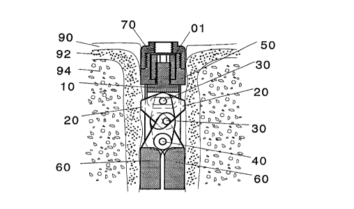
Fastener Having a Linkage-Member Actuator Mechanism: An embodiment of the
f~tener of the present invention (Fig. la, Fig. lb) in~ludes an assembly having a single
intern~l actuator me~h~ni~m in~ ling a bearing (01) with int~rn~lly threaded bore, an
externally threaded journal (10) having a transverse slot ~ nt to one end, and two links
(20) that when joined with pins (30) to the slot in the journal (10) and members (60) and
constrained to rotate about a second pin (40) within a lower body (50) together are
capable of causing members (60) to move indeF~n~lently of one another so as to engage,
conform, press upon, and be retr~ct~hle from, surrounding m~tlori~l The bearing (01) is
constrained within the upper body (70) and lower body (50) so as to contain the
mech~nism and resist fixation forces applied to the members (60).
The embodiment of Fig. la and Fig. lb is shown in a drilled hole in a m~n~lihle
with gingival tissue (90), compact bone (92) and c-~n~ellous bone (94) surrounding the
implanted f~t~n~or. In a drilled hole the members (60) expand equally to contact the
surrounding compact bone. In a tooth extraction site where the defect is irregular in its
CA 02232999 1998-03-24
W O 97/11651 16 PCT/US96/15722
shape, the members (60) operate in~lPpPn-l.qntly and conform to the defect as shown in
Fig. lb.
In the operation of the present embodimP-nt, when the be~ring (01) is "tightPnP~"
by rotating about its central axis within the bore of the lower housing (50), the journal
S (10) is forced to move out of the bore of the bearing (01). Tr~n~l~ti- l of the journal (10)
causes the linkages (20) to cause the members to rotate about pin (40) and engage the
surrounding bone (92). If one of the mPmbPrs (60) contacts surrounding bone (92) then
the linkages (20) begin to slide and rotate about pin (30) thus allowing the member (60)
which was not in contact with bone (92) to continue its rotation about pin (40) until it
10 conforms to and contacts bone (92). Once both members (60) contact bone (92) then
liti~n~l rotation of the bearing (01) loads the journal (10) causing a predictable force to
be applied by the members (60) to the surrounding bone (92).
Fastener Having a Cammed-Member Actuator Mechanism: A further embodiment
of the actuator mech~ni~m (Fig. 2) is shown configured as a femoral prosthesis placed in
15 the med~ ry co"~palL"lent of the femur (194). The mP~h~ni~m inClu(l~ps a bearing (100)
having an intern~lly threaded bore, eYtPrn~lly threaded journal (110) having a journal pin
(115) near one end, and a cam with slot (120) that together are capable of c~ in~ each of
two members (130) to move indepen~1Pntly of one another so as to engage, conform and
press upon and, if ~ eng~ed, retract from surrounding cancellous bone (190) or cortical
20 bone (192).
This embodiment uses an upper housing (140) and lower housing (150) to contain
the bearing (100) and support the movement of the members (130). The members (130)
are pivotally connP~te~l to the lower half of the housing (150) with pins (160) and to the
cam (120) with a second set of pins (170). The cam (120) is further ccnnPcted to the
25 journal (110) by a journal pin (115).
This embodiment is combined with a femoral ball (106) and carrier (102) to
complete the prosthesis. To ensure se;ulily of the mPrh~nicm a jam nut (180) is provided
to lock the mP~h~ni~m so as to prevent rotation of the bearing (100).
In the operation of the present embodiment, when the bearing (100) is "tightPnP11,l' ~
30 the journal (110) is forced to move out of the bore of the bearing (100). The cam (120) is
linearly moved by its pin (115) connPction with the journal (110) causing the members
(130) to be rotated about the pivotal c-nnPr*cns (160) with the housing (150). When a
first member (130) cont~ t~ a contour of the bone tissue cavity in its rotation, the cam
CA 02232999 1998-03-24
W O 97/11651 17 PCTrUS96/157Z2
(120) tilts and slides to allow conhnued rot tion of the second mPmhPr (130) as well as
contin-1e~ linear movement of the journal (110). Once both members (130) contact the
colltuul~ of the bone tissue cavity, ~ 1itinn~l downw~d force on the journal (110) is
~ b~l~nced by the resistive force caused by contact of the bone (192). A means of locking
S the mtorh~nism can be achieved by a cap (180) that is fixed to the housing to contact and
resist the rotation of the bearing (100).
Fastener Having a Three-Lin~age A~n/~~ter Mechanism: A further embodiment
of a f~tener of the present invention uses an intern~l mP~h~ni~m con~i~ting of a bearing
(200), externally threaded journal (210), and three linkages (220, 223 and 226) to operate
10 the members (Fig. 3).
This embodiment uses a housing to contain the mP~h~ni~m and support the
movement of the conforming portion of the device. In this embodiment the housingincl~ldes an upper (240) and lower half (250) with members (230) pivotally connected to
the lower half of the housing (250) with pins (260).
The upper (240) and lower (250) housing have a bore. In this bore a bearing (200)
with an intern~lly threaded bore engages an Pytp-rn~lly threaded journal (210) which is
pivotally coupled to one of the linkages (220) with a pin (263). This linkage is pivotally
coupled with a pin (266) to each of two linkages (223 and 226) which are pivotally
coupled at their second end to each of the members (230) with pins (270). Tr~n~l~tinn of
20 the journal (210) causes independent eYp~n~ion of the members (230).
In the operation of the present embo~im~nt, when the bearing (200) is '~ti~ht~n
the journal (210) is forced to move out of the bore of the bearing (200) causing the
linkage (220) that is pivotally connected to the journal to move. The tr~n~l~ti( nal and
rotational movement of this linkage (220) causes the other two linkages (223 and 226) to
25 move. Movement of the three linkages (220, 223 and 226) together cause the members
(230) to move independently into the surrounding tissue.
Fastener having Journal-Double Linkage Actuator Mechanism: In Fig. 23 is shown
an upper body (1240), lower body (1250), journal or ~ch-~tor (1210), cap (1001), member
(1230), links (1223 and 1226), and pins (1260), (1265) and (1270). The actuator pin
30 (1265) is slidingly and rotatingly conne~ted to links (1223 and 1226). This embodiment is
similar to that of Fig. 3, however, it is more simply made and is configured forindependent self-seeking movement of the members (1060). When in use, the bearing
(1200) is turned, forcing actuator (1210) out of its bore which causes actuator pin (1265)
CA 02232999 1998-03-24
W O 97/11651 18 PCT~US96/157Z2
to rotatingly slide in the ~tll~tnr slot causing links (1226) and (1223) to act on mPml~er~
(1230) so as to cause them to rotate about pin (1260).
Fastener Having a Two-Slot Cam Actuator Mechanism: A further f~tP-nP-~ uses a
housing to contain the mP~h~ni~m and support the movement of the conforming portion of
S the device. As with the other embo~1imPnt~ the housing cl n~i~t~ of an upper (340) and
lower half (350) with members (330) pivotally connP~ted to the lower half of the housing
(350) with a single pin (360) that is centrally located.
The upper housing (340) and lower housing (350) have a bore. In this bore a
bearing (300) with an intern~lly threaded bore engages an PYtPrn~lly threaded journal
10 (310) which is coupled to a cam (320) having a plurality of slots with a pin (313) that is
further coupled to each of the members (330) with pins (370). The cam (320) shows a
different configuration than previous embo~liment~ having a pivotal sliding connection to
the journal pin (313) and a second pivotal sliding connection to the two members (330)
with pins (370). This cam (320) configuration is applicable to other embo-liment~ and in
15 contrast to the prerelled first and second embo-limPnt~ the common pivot pin (360) of the
member provides greater initial leverage in its unpyr~n~le l configuration.
In the operation of the present embodiment, when the bearing (300) is "tight~Pnt~A,"
the journal (310) is forced to move out of the bore of the bearing (300). The cam (320) is
linearly moved by its connection with the journal (310) and the members (330) are rotated
20 outward about the pivotal connection (360) with the housing (350). When one member
(330) contacts a contour of the bone tissue cavity in its rotation, the cam (320) tilts to
allow continued rotation of the other member (330) as well as continl~e~l linear movement
of the journal (310). Once both members (330) contact the contours of the bone tissue
cavity, additional downward force applied by the journal (310) is b~l~nced by the resistive
25 force of the bone tissue contacts.
Fastener Having a Sphere-Shaft Actuator Mechanism with Scissoring Members: A
further embodiment uses a sphere (420) to actuate members (430). This embodiment uses
a one-piece housing (440) to contain the mPrh~ni~m and support the movement of the
conforming portion of the device. This housing has members (430) pivotally connected
30 with a single pin (460) that is centrally located in the housing (440).
The housing (440) has a bore with intern~l threads over a portion of its length and
an intern~l taper that expands to the level of the pin (460) which is the pivot connection
between the members (430) and the housing (440). In this bore an eYtern~lly threaded
CA 02232999 1998-03-24
W O 97/11651 19 PCT~US96/15722
shaft (400) engages the int~rn~l threads. This shaft (400) cont~ctC a sphere (420) that is
conctr~ined by the housing (440) and the upper arms (410) of the m~mherC (430). This
sphere (420) rides bc;Lw~n the upper arms (410) of the m-omhers (430). The shaft (400),
- sphere (420) and members (430) act logc~ to indepenrlPntly expand the two members
5 (430) from the ce~ , of the housing (440). The shaft (400) can be replaced with an
unthreaded member and used in a housing (440) with an unthreaded bore allowing an
impl~nt hoklinp instrument to push on this member and tr~ncl~te the ball (420). An
abutment (470) locks the shaft (400) and provides a support for cement (490) andprosthetic tooth (480).
In the operation of the present embo~lim~nt when the shaft (400) is turned, the
sphere (420) is forced to move out of the bore of the housing (440). The force on the
sphere (420) is transferred through the arrns (410) of the members (430) to produce
rotation of the members (430) about the central pin (460). When one member (430)contacts a contoul of the bone tissue cavity in its rotation, the sphere (420) slides on the
15 stationary arm (410) as it continuec rotation of the noncont~-~-ting member (430). Once
both members (430) contact the cont~ul~ of the bone tissue cavity, ~ lition~1 dow~lw~d
force applied by the shaft (400) is balanced by the resistive force of the bone tissue
contacts.
Fastener Having a Sphere-Shaft Actuator Mechanism with Pinned Members: A
20 further embodiment uses a sphere (510) to cause two members (530) to swing away from
the housing (540). This embodiment uses a one piece housing (540) to contain themech~nicm and support the movement of the conforming portion of the device. Thishousing has membOEs (530) each pivotally connecte~ with pins (560).
The housing (540) has a bore with intl-rn~l threads. In this bore an eyt~rn~1ly
25 threade~d sha~t (500) engages the int~rn~l threads. This shaft (500) contacts a sphere (510)
that is constrained by the opposing faces of the members (530). This sphere (510) rides
between members (530). The shaft (500), sphere (510) and members (530) act together to
independently expand the two members (530) from the cçnt~orline of the housing (540).
An abutment (570) locks the shaft (500) and provides a support for cement (590) and
30 prosthetic tooth (580). The impl~nt is shown in a tooth eYtr~t~tinn site surrounded with
gingival tissue (592), compact bone (594) and c~ncçllous bone (596).
In the operation of the present embodiment, when the shaft (500) is turned, the
sphere (510) is forced to move out of the bore of the housing (540). The force on the
CA 02232999 1998-03-24
WO 97/11651 20 PCTrUS96/15722
sphere (510) is transferred through the sphere (510) to the mPmber~ (530) to produce
rotation of the members (530) about the pins (560). When one member (530) cor t~ct~ a
contour of the bone tissue cavity in its rotation, the sphere (510) slides on the st~tion~ry
member (530) as it continl-çs rotation of the nQncont~etin~ member (530). Once both
members (530) contact the contuul~ of the bone tissue cavitv, ~ hon~l dowllw~d force
applied by the shaft (500) is b~l~nse~l by the resistive force of the bone tissue contacts.
The rotation of the members (530) is limited by contact between the housing (540) and
members (530). This range is limited so that the sphere (510) cannot be pushed past the
members (530) and out of the mPrh~ni~m.
0 Fastener with Mechanism in a Spherical Rotating Housing Conrigured in a
Structure: A further f~tçner can be used to secure a structure, for example a bone plate
(650), to the surface of a bone (692). This embodiment uses a spherical body (620)
having two members (630). The spherical body (620) is pivotally connPcted within the
plate (650) with a cap (640) having a bore with a spherical internal surface which supports
15 the movement of the members (630). Together the movement of the members (630) and
the pivoting of the sphere (620) between the plate (650) and cap (640) allows the members
(630) to independently conform to a body cavity.
The spherical body (620) has a bore. Within this bore is a bearing (600) with anintçrn~lly threaded bore. This bearing (600) is held within the bore of the sphere (620)
20 with a locknut (680) having a bore. In the bore of the bearing (600) an eYttorn~lly
threaded journal (610) engages the intPrn~l threads. This journal (610) is pivotally and
slidingly connected to members (630) with pins (670). The members (630) are pivotally
connected to the spherical body (620) with pins (660) and act together to Sym mçtriç~lly
expand from the cent~rlint-- in relationship to the ~rhPric~l body (620). The rotation of the
25 spheric~l body (620) combined with the symmPtric~l eYp~n~ion of the members (630) with
respect to the spherical body (620) allows independent eYr~ncion of the members (630)
with respect to the plate (650) and conformation to the bone (692).
In the operation of the present embodiment, when the bearing (600) is turned, the
journal (610) is forced to move out of the bore of the spherical body (620). The30 movement of the journal (610) causes the members (630) to rotate about pins (660).
When one member (630) contacts a conlour of the bone tissue cavity in its rotation, the
spheric~l body (620) rotates in the plate (650) to allow the noncont~cting member (630) to
continue to move. Once both members (630) contact the conluul~ of the bone tissue
CA 02232999 1998-03-24
W O 97/11651 21 PCT~US96/lS722
cavity, additional dow~w~d force applied by the journal (610) is b~l~nr~ by the resistive
force of the bone tissue contacts.
Fastener with Worm Gear and Pinion Mechanism Configured in a Structure: A
- further embodiment can be used to secure a body (700), for eY~mrlP.~ a plate, port or
S other structure, to the surface of a bone (780). This embodiment uses a gearedmPrh~ni~m con~i~ting of a drive cylinder (710) having a worm gear, memhPr (730) having
gear teeth (720), locking nut (750), member pin (760) and retainer (770). Two or more
geared mP~h~ni~m~ can be used with a body (700).
The members (730) are pivotally connectP~ to the body (700) with pins (760). The10 geared teeth (720) of the members (730) protrude into se~ tP bores cut through the body
(700) at its periphery. A drive cylinder (710) is located in each bore allowing a worm
gear cut on the drive cylinder (710) to mesh with the gear teeth (720) of the members
(730). The drive cylinder is retained and locked into the bore with retainer (770) and
locknut (750). The ability to actuate each member indepen~lPntly allows the members
lS (730) of the implant to conform to a body cavity and hold the port (700) in place. In the
design of this embodiment the separate drive cylinders (710) can be replaced by a
common drive cylinder so as to rotate each member (730) ~imlllt~nPously.
In the operation of the present embodiment, prior to placing the locknut (750),
when the drive cylinder (710) is turned, the worm gear drives the gear teeth (720) and
20 causes the member (730) to rotate. When one member (730) contacts a collloul of the
bone tissue cavity in its rotation the noncont~cting members (730) can be actuated to
rotate using the drive cylinder. Once both members (730) contact the contours of the
bone tissue cavity, additional rotational torque applied by the drive cylinder (710) is
b~l~nced by the resistive force of the bone tissue contacts. Once engaged the locknut
25 (750) is placed to stop any further rotation of the drive cylinder (710) or member (730).
Fastener having Journal-Member Actuator Mechanism for Dependent MoYement of
Members: In Fig. 22 is shown an upper body (1070), lower body (1050), journal oractuator (1010), jam nut (1001), member (1060), and pins (1030) and (1040). Thisembodiment is similar to that of Fig. 1, however, it is configured for dependent30 movement of the members (1060). When the actuator (1010) is pushed through the lower
body (1050), pins (1030) slidingly rotate in the actuator (1010) slot c~-lcing members
(1060) to precess about pin (1040) in a dependent and symmetrical manner.
CA 02232999 l998-03-24
W O 97/11651 22 PCTAUS96/15722
Fastener having Journal-Double Linkage A~n~fer Mechanism for Dependent
Movement of Members: In Fig. 24, the f~tçn~r of Fig. 23 has been modified to change
the journal or ~rtl-~t--r slot to a pin hole to receive pin (2265). This causes a rotational
connection between links (2223) and (2226) that causes dependent symmetric~l movement
5 of members (2230) about pin (2260). When in use, actuator (2210)is pushed through the
lower body (2250) causing the actuator pin (2265) to act on links (2226) and (2223) SO as
to cause members (2230) to rotate about pin (2260).
Fastener having Shaft-Member Arm A~tu~7f~r Mechanism for Dependent Movement
of Members: In Fig. 25, the f~tPnPr of Fig. 5 has been reconfigurPA for dependent
10 movement of members (1430) by removing sphere (420) of Fig. 5, and reshaping arms
(1410) at the point of sliding contact with the ~c~tll~tor(l400)
Fastener having Members Movable from the Top rather than the Bottom of the
Device: In Fig. 26, the f~tçner of Fig. 3 has been reconfigured to demonstrate that any
of the f~teners provided herein could be configured so that members (3230) pivot to
15 swing out from the top instead of the bottom of the device.
A method of implanting a f~tPnPr of the present invention into a hard tissue defect
includes the following steps: i) ob~ainillg an animal in need of a f~tenP-r of the present
invention, ii) determining size of an already eXi~tin~ hard tissue defect, or constructing a
hard tissue defect and de~ ining its size, iii) selecting a f~tener having a size to fit in
20 the defect and taking into account considerations of defect geometry, whether the members
will contact the walls of the defect or reach through the defect so as to extend into a
cavity, and the relationship between intPrf~ lC;~lllC; and ~(-tll~ti-n force and the
strength of the critical colllponents, iv) placing the f~tçnPr into the hard tissue defect, v)
applying force to the f~tenPr using a tool that applies force sufficient to secure the device
25 and stimul~te bone to become more dense. The method may optionally include vi) placing
a healing cap over the f~etPner, or placing a prosthesis onto the f~tenPr.
A method for de~llllh~ing intçrf~ci~l pl~ iUle exerted on bone ~ulloullding an
implanted f~tçnçr of the present invention inclllcles the steps of: i) applying force to the
impl~nted f~tener using a tool where the force can be measured, ii) mP~nring the extent
30 of journal translation, iii) me~urin~ index member angle, iv) mP~uring reference
member angle, v) de.termining interf~ l ples~ul~ from the equation provided in Fig. 11
for the device of Fig. 2. Similar equations can be derived for the other m~h~ni~m
CA 02232999 1998-03-24
W O 97/11651 23 PCTrUS96/15722
configur~tion~ in light of the te~rhings of the present ~ c1os1~re. In the case of dependPnt
movement, the index and reference m~mhPr angles are equal.
F~tçnPr~ of the present invention may be m~(~hinPd and ~emb1P~ by one of skill
in the art in light of the tç~rhings of the present ~ closllre~ Methods of use of a f~ten~r
5 of the present invention are also known to one of skill in the art in light of the te~rhing~
of the present disclosure.
Even though the invention has been described with a certain degree of
particularity, it is evident that many ~1tPrn~tives, mo-1ifir~tion~, and variations will be
a~L,~c~nt to those skilled in the art in light of the roregoing ~i~r1Os11re. Accordingly, it is
10 intPncle l that all such ~ltPrn~tives, mo~1ific~tiQns, and variations which fall within the spirit
and the scope of the invention be embraced by the defined claims.
The following example is included to demonstrate a ~"erel,~d embodiment of the
invention. It should be ap~l~ialed by those of skill in the art that the techniques
disclosed in the PY~mp1e ,~,Gsent techniques discovered by the inven~or to function well
15 in the practice of the invention, and thus can be considered to constitute pref~ d modes
for its pr~ctice~ However, those of skill in the art should, in light of the present
disclosure, a~r~iate that many changes can be made in the specific embo~limPnt~ which
are disclosed and still obtain a like or similar result without departing from the spirit and
scope of the invention.
F.Ys-mrl~ 1
In vivo Dental T...~ F&~t~ and Evaluation Thereof
The present PY~mp1e provides an ev~ tion of an entlos~P~us (bone contz~cting)
dental imp1~nt of the present invention. Devices were de~ignPA to be placed through the
25 gingiva into a tooth extraction site, expand, conform to the defect and become
immç~ tP1y stable.
The design used the c~mmPd linkage system to indeFçn-lPntly expand a pair of
members (Fig. 9a, Fig. 9b and Fig. 2; the reference n~1mer~1~ for Fig. 9a are: 810,
- healing cap; 820, actuator nut; 830, upper body; 840, actuator mandrel; 850, lower body;
30 860, ~ tll~tor-cam pin; 870, cam; 880, member). The design used two cylinders that are
press-fit together to hold the bearing and joumal, and to form the body of the f~tP-nPr.
Two pinned members rotate about two tabs that protrude from the lower face of the body.
The members are pinned with the journal in the cam slot, allowing the journal and
member pins to slide indeFçn~lçntly. The upper end of the journal is threaded into the
CA 02232999 1998-03-24
W O 97/116S1 24 PCT~US96/15722
actu~tcr bearing. Rotation of the bearing by applying force to the in~t~ tion instrument
causes the journal to move dow"w~L,d and engage the members through the cam. Thef~tPnP-rs may further include a healing cap driver, an ~-t. I~tor driver, or anti-rotation
spanner.
S The methods used to evaluate the f~tPners of the present invention addressed three
broad topics: enginP~ring design, Pn~inP~ring evaluation, and in vivo and explant
ev~ tion.
FnginP~ring design for the c~mmeA-member mech~ni~m involved the determination
of the a~l~liate size and shape for the experimPnt~l animal, its range of member motion
10 and expansion, its extent of out-of-phase member operation, component inte~rclcnce~ the
strength of its critical components, a method to lock the mP~h~ni~m, and its bone-imrl~nt
intPrf~ci~l pressure as a function of mPrh~ni~m engagement torque.
Requirements for size, shape and extent of expansion of both implant designs were
determined through the evaluation of fresh dog mandibles provided through the
15 expçriment~l surgery teaching program at the University of Texas Health Science Center,
(San Antonio, Texas). Mandibles were cut in the mesial-distal and buccal-lingual planes.
The mesial root of the first molar was selected as the first site for investig~tion. This
selection provided size and shape information for imrl~nt design. The strength
characteristic of the necropsy specimpn~ provided an çstim~tP of the extent of e~p~n~ion
20 required for the two impl~nt ~lesi~nc
The extent of member motion, out-of-phase operation and expansion for the
cammed-member implant required the det~ile~ design of all co,-lpollents. The body size
and shape, mech~ni~m's pin diameters, cam slot and overall length, member arm length,
~ct~l~tor-translation ~ t~nce~ and actuator-nut rotation versus translation relationship all
25 effect the operation of the me~h~ni~m
A co...~uLel design ~im~ tion for the member impl~nt was developed using
MATHCADn' to account for and understand the large number of design variables. This
simulation allows the selection of all design v~ri~hles, solved a system of equations,
computed the operational range of the mech~ni~m, and provided a graphical layout of the
30 mech~ni~m to check for coll-pollent inLe,relcllce. This ~imlll~tion allows rapid design
~c~ec~ment of operational parameters and is well suited to support the resizing and
optimi7~tion of the member impl~nt system.
CA 02232999 1998-03-24
W O 97/11651 25 PCTAJS96/157Z2
Critical mP~h~ni~m co~ ollent strength was ~ rc,l.l-ed using col.venlinn~l m~chinP
design theory. The critical co---~onents were ~iel~ d to be the ~t~tor-cam pin,
actuator journal bearing and nut, and cam (Fig. 9a, Fig. 9b, and Fig. 2).
- Actuator-cam pin: The standard method of ~ e .. ;.-;.. g the m~ximllm force
S ~uppolL~ble by a clevis-pin arrangement as found in the present impl~nt f~teners is the
double lap shear equation (Shigley, J.E., Mechanical Engineering Design, 3rd ed.,
Holman, J.P., ed., McGraw-Hill Book Co., 1977).
Given values of 60k psi Ill~ illllllll shear (Ti-64) and a pin cross-section~l area of
1.26 X 10⁻³ in2, the calculated m~ximllm force the pin will support before shear yielding
10 occurs is 150.8 lb. A safety factor of 2 should be applied, reducing the m~ximllm
expected s~ ct~in~hle load to 75.4 lb. This equates to 8.22 in-lb of torque applied to the
actuator (Table 1).
A second, more conservative method of analysis is to consider the pin as a beam
with constrained ends and a point load applied at the center. For such a beam, the force
15 required to initiate yielding is governed by the bending moment at the center of the pin
radius and cross-se~tion~l geometry. Using this method, maximum s--~t~in~hle force is
calculated to be 55 lb. This equates to 5.99 in-lb of torque applied to the actuator (Table
1).
Actuator journal bearing: Failure of a power screw type actuator journal can occur
20 in one of two different manners, co,~ Gs~ion yielding of the entire screw or stripping of
the threads. Thread recomm~-n-l~ti-~ns assume that the screw will fail just before the
threads strip. The minimllm thread engagement length n~ceC~ y to prevent stripping of
the threads is ~nm~l to be 0.07 in. When the members are deployed in the ori~nt~tion
of m~ximnm screw travel, the lG~ thread engagement length is 0.1165 in. With a
25 co~ lGs~i~te yield strength of 120k psi (Ti-64), the screw would be able to with.~t~nd a
co~ cssi\~e load of 996 lb. This equates to 109 in-lb of torque applied to the ~chl~tor
(Table 1).
C~n7: The upper section of the cam with a thicknP~ of 0.035 inches was mod
as a simply ~u~ulLed beam with a moment applied at each end. For such a beam, the
30 force required to initiate yielding is governed by the moment produced at the center of the
beam, 1/2 the height of the beam and the second moment of inertia. For the upper and
weaker section of the CAM, the maximum sllst~in~hle force is calculated to be 52.8 lb.
This equates to 5.75 in-lb of torque applied to the bearing. The lower section of the cam
CA 02232999 l998-03-24
W O 97/11651 26 PCT~US96/15722
with a thicknP~ of 0.06 inches was modPled as a simply ~u~o-led beam. The m~cimllm
sl-st~in~hle force for the lower beam is calculated to be 114 lb. This equates to 12.41 in-
lb of torque applied to the bearing (Table 1).
S Table 1: M;1Xi~hle forces for critical colllponents.
ComponentM~imllm Force (lb)
Cam
Upper Beam 52.8
Lower Beam 114.0
Re~ring and journal 996.0
Actuator-cam pin
Double Lap Shear Method 75.4
Beam Method 55.0
Using the beam method, the cam pin, and the upper portion of the cam can be
considered the critical members and equivalent, given the assumptions used for the
analysis. A su~t~in~hle force of 52.8 lb equates to 5.75 in-lb of torque applied to the
20 journal. The Vident IMPAC torque wrench chosen to tighten the member impl~nt
f~tener can deliver a m~imllm of 2.83 in-lb (32 Ncm) of torque. This allows a safety
factor of 2.0 for the merh~ni~m.
MP~h~ni~m locking and bacterial sealing is accomrli~hPA through inte.relc;nce ofthe healing cap 810, 280 or prosthesis abutment 470, 570 and the actl-~tor nut 820. The
cap 810, 280 or abutment 470, 570, when in~t~lled, jam against the actuator nut 820
causing it to bind against the intern~l aspects of the merh~nicm body. This creates three
metal-to-metal bacterial seals. One is b~lween the cap and the body, a second bt;~ween the
actuator nut and the intern~l face of the cap, and the third between the lip of the actuator
nut and the intern~l lip of the body. These three seal faces minimi7e the likelihood of
30 b~et~pri~l migr~tion through the implant mP~h~ni~m.
Bone-impl~nt int~rf~ jUlC; iS a function of the torque applied to the bearing
nut during the pl~-Pm~nt of the impl~nt (Fig. 10). The L.res~uie applied to the impl~nt-
bone intPrf~ee can be detPrmined by PY~min~tion of the mPrh~ni~m The design used in
this study allowed the imrl~nt members to rotate from -10~ to 57.5~ (positive is out and
CA 02232999 1998-03-24
W O 97/11651 27 PCTAUS96/15722
negative is in towards t'ne c~ P). This m~rh~ni~m allows the members to operate
indepenclçnt1y and 37.5 degrees out-of-phase from one another. The inte~rfarial ~JlC~ UÇC
applied to the bone intt~ can be measured by relating the force applied by the power
- screw (bearing and journal) to the cam and members.
S In opP~ation, turning the bearing applies a force to the cam which, in turn, causes
the members to rotate OuLw~L~d. It is col~vcnient for the purpose of this analysis to treat
this as two se~ c- functions. The act~tor screw can be analyzed as a power screwturning against a load and the cam and members as a simple three bar merh~ni~m
The position of the cam and second member are computed if the position of one
member is lcnown. This known position is equivalent to bone contact of one member
while the journal is translat~d and the second member is still moving. The rotation of the
cam (~ Cam-Angle), given a restrain~d-member angle (~-index), and extent of journal
tran~l~tion (AX) is colllpuled as shown in Fig. 11. The analysis of the power screw is a
straightforward application of the equation given in "M~rhanic~l F.ngin~ring Design",
Shigley and Mi~chk~, (as cited herein) and shown in Fig. 11, with known values
particular to the present implant fa~tent-rs. Using simple geometric princir~l~ and an
underst~n-lin~ of the forces applied to the members the intçrfaçial ~,lG~,~ure value can be
obtained.
The equation of Fig. 11 has three degrees-of-freedom: torque, index member
angle, and reference member angle. To generate the torque-pressure multiplier surface,
the reference member is swept through its full range of motion, then the index member is
advanced 1~ and again the reference member is swept. This algorithm is continued for
the range of the index member. The result is the torque-~lc~,~wc mll1tiplier surface shown
in Fig. 12. A new surface is created for each new value of applied torque and each
design (size) of the implant. The surface in Fig. 18 lc~rcs~ an applied torque of 1 in-
lb.
From the graph of Fig. 18, the ori~ntation colres~onding to the maximum (index
angle = 0~, reference angle = 16~) and minimllm (index angle = 34~, reference angle =
55~) multiplying effect can be found. To calculate the ~JlG~i~iUl'C (pSi) on the reference
member, the mllltiplier value associated with a particular set of member angles is
mllltipli~d by the torque applied to the implant. Taking these ...in;...~.... and ma~imllm
torque-to-~es~ulc mll1tiplic~ticn values as bollnl1~ries and varying the torque from 0 to 9
CA 02232999 1998-03-24
W O 97/11651 28 PCTAUS96/15722
in-lb (imrl~nt hinge pins fail at 6 in-lb to 8.22 in-lb), an envelope of imrl~nt intPrf~
p.~ssule as a function of torque can be plotted as shown in Fig. 13.
IMPAC torque wrench (Vident) values of 10, 20 and 32 Ncm and an oscillometric
method blood plGSsult monitor cuff release plGS:!iUl~, of 300 mm~Ig are shown in Fig. 13
5 for reference. If the imrl~nt will fail at the journal-cam pin at 8.22 in-lb applied torque, a
safety factor of over three exists for a bearing torque of 32 Ncm.
Instruments for manipulating the imrl~nt f~tenPr includPA an anti-rotation wrench,
a bearing driver, or a healing cap driver. The anti-rotation wrench is a two-pin sp~nn~r.
The pins of the sp~nn~r engage two holes on the upper face of the body of the impl~nt
10 The sp~nnPr can be f~tPnPA to the body of the implant with the healing cap to f~ilit~te
h~nllling of the implant during pl~cçmPnt The bearing driver has a male toroid shaped
end. This end fits the female socket in the bearing. Rotation of the driver turns the
bearing and operates the mPch~ni.~m The healing cap driver is a toroid shaped socket
driver used to place and remove the healing cap.
Me~h~nic~l testing of the pull-out reci~t~nce of dental impl~nt~ was performed for
the c~mmed member, wire-cage and collventional Nobelpharma implant designs. The
bone cont~ting portion of each of these imrl~nt designs was embedded in p~r~ffin and
pull-in tension to determine the intrin~ic rçci~t~nce of the impl~nt to being pulled from
bone. The wirecage and c~mmçd-member implants were tested in a dosed and e~rp~ndPd
20 configuration. The Nobelpharma 3.5 mm X 20 mm imrl~nt was sPlPcte~i for co~ )alison
testing because it is the largest and most clinic~lly succe~ful çn~lo~P~us imrl~nt on the
m~rkP.t
Instron model 1127 with a 1000 lb load cell and analog control system was used
for these tests. The imrl~nt was tested in two configurations, with member closed and
25 member 3/4 open. The Nobelpha,ll.a impl~nt was tested as delivered. Load (lb) -
deflection (in) curves dP~mon~tr~tP11 that more work (area under the curve) was required to
remove the impl~nt f~teners of the present invention in colll~ison to the Nobelpharma
implant (Fig. 14, Fig. 15, and Fig. 16).
The m~ximllm value of the pull-out strength was taken from the graphs. The area
30 under the force-displ~çmPnt curves were measured. This area is a measure of the work
(energy) required to extract the impl~nt from wax. Work and pull-out yield strength for
the three different implants were colll~ed in a histogram given in Fig. 17.
CA 02232999 1998-03-24
W O 97/11651 29 PCTAUS96/15722
All of the impl~nt configur~tion~ produced co~ ble ~ xi~ - .. strength values
(cros~h~trhP~1 bar). Conv~ely, the total energy (solid bar) required to completely
remove the impl~nt from the wax was ~i~nifit~ntly greater in the systems of the present
~ invention. This suggests that the imrl~nt f~te-n~r~ of the present invention l~e rul~l with
S greater mP~h~nic~l toughnPss, resisting pull-out even after failure of the imrl~nt-m~tPri~
intPrf~e has been initi~t~1
In vivo ev~ tinn~ were ~e ro~--led under AAALAC Protocol #92077-11-01-B2 by
placing two impl~nt~ into mandibular first molar extraction sites in six healLwoll~l-free
dogs following tooth extraction. Teeth were sectinn~d and extracted. Gingiva was closed
10 around the implant leaving the healing cap exposed. Implant sites were swabbed with
PERIDEX~ during the twice weekly evaluation for implant mobility, gingival
infl~mm~tinn and bleeding.
Clinir~l and radiûgraphic evaluations were ~;lrol---ed pre- and immP~ tply post-impl~nt~tion as well as at 2, 5, 8 and 12 weeks post-impl~nt~tinn and periodically for one
15 year. Soft tissue adaptation to the device was documPntP~d photographically. Tmr~l~nt
position and bone density and morphology were determined radiographically. Stability
was ~se~P~l subjectively. At the end of the 12-week period the me~h~ni~m of the
c~mmçd-member implant was actuated and found to be tight.
Qll~ntit~tive digital radiographic image analysis was used to ~et~-rminp~ the bony
20 changes in and around the impl~nt as described by Rllttim~nn et al. ("Automated
estim~tinn of lesion size", SPIE, Application of Optical Instr~mentation in Medicine XIII
535:325-330, 1985). A stent was used to hold the x-ray calibratiûn wedge and film. The
stent had a calibration wedge imbedded on the occlusal surface which was made of a
bone-~im~ ting m~tPri~l which was 25 mm long, 10 mm high and had a ramp of 0-Smm25 thicknp~. The reference wedge was made from a bone-equivalent epoxy resin (Model
450 Bone, Radiation MeasuremPntc, Inc., Mi~ltllPton, WI).
The processed radiographs were converted to llip;iti7P,d images in 640x480x8 bits
format with pixel size of 60 micrometers. ~se~mPnt of the alveolar bone changes were
performed with a qll~ntit~tive digital x-ray image subtraction technique. On the monitor
30 image of the radiograph, an area of interest (AOI) was drawn between the members of the
c~mmç~-member implant within the region of the wire cage and ~ çnt to the body of
the impl~nt~ with trackball driven mouse.
CA 02232999 1998-03-24
W O 97/11651 30 PCTAUS96/15722
The images were then subtracted and co~p~cd to the calibration wedge which
provided an estim~tto of the bone mass changes around the imrl~nt and in the apic,al defect
between the members.
Morphometric measurements of the alveolar bone were made using the same
5 standardized radiographs that were made for the ~lu~-t~ e image analysis. Digitized
radiographic images m~gnified eight-fold on the co~yu~er monitor with a pixel size of 60
micrometers were analyzed using RADWORKS~ sor~w~c for ~ t~n~ and area
measurements (Dove, RADWORKSsY, San Antonio, TX). The vertical alveolar bone
change was measured from impl~nt collar to the alveolar bone crest at the mesial and
10 distal impl~nt sides. The area of the crestal bone change between the adjacent teeth and
the implant was measured by outlining the crestal bone level in the b~elin~, radiograph
and follow-up radiographs and calculating the area difference between these outlines.
An oral photograph of an implanted f~tener healing cap showed good soft-tissue
appro~cim~tion and closure at 8 weeks following imrl~nt pl~çmP,nt
The results of this study ~emon~tr~t~d that increases in bone miner~l density could
be measured adjacent to and between the members at the point of int~rf~ JlC5~iUl'e. On
average, decreases in bone mineral density were noted at the crestal portion of the
imrl~nt.
Measurements of changes in bone morphology and density of the bone surrounding
20 the member impl~nt were made. Linear measurements of crestal bone height changes
were taken relative to the top of the impl~nt Bone min~,r~l density measurements were
taken at the mesial and distal border of the upper, mid and lower (at the member int~,rf~e
with bone) portion of the body and between the members at the apex of the extraction site.
The subtracted image showed increases in bone mineral density in light regions and
25 decreases in dark regions (Fig. 18). The members were demon~tr~t~ to have increased
bone mineral density adjacent to the members and at the apex of the extraction site. This
increase in bone density inllic~t~d that load applied by the mçeh~nicm was physiologic and
stim~ t~d bone to form with higher density and strength.
Explant and histologic evaluations were pelrol,--ed on three dogs l~ in~ at 8
30 weeks post-imrl~nt pl~ m~nt and at one year. The left and right sides of the mandibles
were separated at the sinphysis and fixed in FORMALIN~ for four weeks. Prior to
embedding, the mandibles were sectioned in the mesial-distal plane to reveal the imrl~nt
embedded in bone.
CA 02232999 1998-03-24
W O 97/11651 31 PCTAUS96/157Z2
Four princip~ e~m~nt~ were made from gross and sc~nning electron
microscope (SEM) eY~min~tion~. First, the imrl~nt was stable in bone. Second, bone
healed around and within the impl~nt in the eYtr~ tion site defect. Third, force exerted on
~ the sull. wlding bone caused it to become more dense. Fourth, there was direct impl~nt-
to-bone contact (Fig. 19).
Clinic~l and radiographic evaluations p~ ed over a period of one year showed
eYcçll~nt soft tissue healing and increases in bone min~r~l density in and around the
implant. Subsequent histologic evaluations confirmed the biocompatibility of the devices
and the ability of bone to subst~nti~lly fill the extraction site defect and heal around the
imr~l~nt
In summary, the princir~l observations from this study were: 1) the designs werefeasible for use in extraction sites and surgically ~ d osseous defects; 2) small
f~tener mech~nism~ could be built with snfficient strength for application in bone; 3) the
impl~nt~ and instruments supplem~-nt~d conventional oral surgical practice and may reduce
the sensitivity of im~ nt outcome to variation in technique; 4) soft-tissue healed around
the crestal portion of the impl~nt~; 5) bone healed throughout the defect and within the
implant mech~ni~m; 6) the impl~nt~ were biocollli~alible and stable in bone following
placement and after one year; 7) dual-subtraction techniques were sensitive to changes in
bone mineral density adjacent to and within the m~h~ni~m of the implants; and 8) the
pressure exerted by the member implant increased the density of bone at the impl~nt
interface.
~Y~mp'~ 2
Procthet;~ Teeth
Development of the f~t~-n~r of Example 1 led to the construction of a prosthetictooth and its testing as a dental imr~l~nt The impl~nt f~tençr structure, healing cap with
screw, and abutment with screw have been built and ev~ ted Prosthetic teeth have been
formed and ~tt~ hçd to the structure using the abutment. This embodiment as a dental
implant is shown in Fig. 20.
This embodiment is currently implanted in dogs. A nine-month follow-up
eY~min~tion showed that bone surrounding the impl~nt is increasing. None of the devices
have failed and the prosthetic teeth are easily placed into position.
CA 02232999 1998-03-24
W O 97/11651 32 PCTAJS96/15722
FY~ nP'e 3
Applir~tinnc to Orthop~ c
Further uses of the f~tPnPrs of the present invention apply to orthop~PAics. A
femoral neek compression f~tçner and plate have been fabrieated and are being evaluated
5 in an animal model (Fig. 21). The e~p~n~lin~ and conformal aspects of this system are
expeeted to provide ~ignific~nt benefit to p~tient~ with femoral neek fraetures. These
fractures often occur spontaneously in elderly osteoporotie p~tiPnt~. These p~tient~ have
poor bone quality and col,ve~,l;on~l femoral neck co,l,L,-cssion systems, which most often
use course serew threads, pull loose due to the poor bone quality. Reports intlic~t~P that
10 the failure rate of conventional co",plcs~ion systems is between 40% and 70% (Olerud et
al., "Internal Fixation of Femoral Neek Fractures: Two Methods Compared", J. Bone
Joint Surg [Br] 73-B: 16-9, 1991).
The impl~nt f~teners of the present invention contrast with eonventional
teehnology in that the eonformal and çlrr~n~ling portion of the subjeet invention provide an
15 exeellent interloek with bone and aet to pull the fraeture site together so as to f~cilit~tç
eo~ cssion aeross the fraeture site and stimulate healing. An additional advantage is that
the eonformal features of the device minimi7P the effeets of surgical teehnique with
respect to the angle of the hole drilled that is required to be placed into the femoral neek
and ball prior to insertion of the device.
FY~mpl~ 4
Applications to ~n~lnctry
The f~tenPrs of the present invention may be used as anchoring, cPntçring, or
ning devices in areas of tç-chnnll gies nP~ling sueh ~nchorin~, ee~le, ;,-~, or ~ ning,
~5 espeeially where such activity is within an irregular eavity, or where irreg~ rities exist in
an intçrn~l surface. Sueh areas of te~hnt~l~ gy inelude water and oil well teehnology,
welding teehnology, aircraft applieations and in automated production, for e~mple.
Fasteners of the present invention may be used for cçntPring~ holtling and/or
~lipning various tubular or cylin~lrie~l elPmPnt~ for various purposes, sueh as end-
30 finiching, attaehment of other members of various shapes and functions, or centçring aholding or cl~mping mP~h~ni~m with respeet to a tubular member. One problem in
welding tubular members together is getting the clamping and guiding structure in proper
position for holding the parts while forming the weld, espeeially where the tubular
member has a curved seetion. Fasteners of the present invention may be useful for
CA 02232999 1998-03-24
W O 97/11651 33 PCTAJS96/15722
~ligninp and hc)l~ling such parts, or as a means for guiding a welding unit, especially for
automatic or sPmi~--tomatic welding.
4lipnmPnt of a cl~mrinf~ or hol-ling m~rh~ni~m in a proper ori~nt~tion with respect
to a plane end surface of a pipe or other tube is a further use of f~t~n~rs of the present
invention. Further, holding or ~u~o~ g a special appa.~lus for positioning, holclinE
and/or welding br~k~t~, arms, special connPctors, and the like onto or near the ends of
tubular members particularly where ~CUr~t5 positioning and precision hol~linE are
e-c~enti~l iS a use of the present invention.
F~tençrs of the present invention are useful in oil field applications involving10 fishing tools, for eY~mrle. In situations of broken or twisted-off drill pipe in a well bore,
the downhole pipe must be recovered or the well is lost. The f~tenin~ and ~ligning
capabilities of the present f~ten~rs allow the capture and withdrawal of the downhole pipe
from the well bore. The f~t~ner would be fixed to the end of a wireline and lowered into
the well bore to the level of the broken end of the downhole pipe. The f~tener members
15 would be placed in the bore of the downhole pipe. Once engaged, the f~tener would be
e~p~nde~ and would lock to the walls of the bore of the pipe, while simlllt~n~ously
~ligninp the downhole pipe with the wireline. The f~tening and ~lignin~ allow a direct
and secure pull by the wireline on the downhole pipe so as to lift and allow recovery of
the downhole pipe.
Fasteners of the present invention have further utility in aircraft applications where
two plates overlap and require joining. In today's fleet of aircraft, mt~h~niç~l fatigue is
causing the riveted joints of the aircraft's ~ ,.. skin to fail. The failure can be
avoided by re~lrilling the rivet hole to remove any cracks and replacing the rivet.
Reriveting of the skin can be difficult and expensive. The present invention provides a
25 unique advantage for rejoining the skin panels. Once the old rivet is removed and the
hole pl~L)a,ed, a f~tener of the present invention can be inserted in the hole and e~p~n-le~
so as to compress the overlapping ~lllmimlm skins and form a new and strong union
between the skins and airframe.
In a further use of the f~ten~rs of the present invention, various col"~onents
30 require ~ ning in automated production. These production processes vary from assembly
to part ~liEnmpnt while running through a conveyor system. In order for wheels,
hto~.lliEht buckets, and other objects to be placed on the frame or body of an automobile,
for example, they must be retrieved from stock, held, transported and placed on the
CA 02232999 1998-03-24
W O 97/11651 34 PCT~US96/15722
automobile. These processes require an adaptive hokling and ~lignin~ device. F~t~nPr~
of the present invention would autom~tie~lly fasten to a wheel rim and align the rim with
the arm of a robot used to place the rim on the axle, for eY~mple. This hckling and
~lignment would allow the robot to aulo.l.~ ly know the orient~tic-n of the wheel so that
S it could repeatedly place a wheel on an axle.
All of the devices and methods disclosed and ~ l~imP~l herein can be made and
eleecuted without undue experim~-nt~tion in light of the present disclosure. While the
devices and methods of this invention have been described in terms of l~ref~ d
10 embc-liment~, it will be a~ar~nt to those of skill in the art that variations may be applied
to the device, methods and in the steps or in the sequence of steps of the method
described herein without departing from the concept, spirit and scope of the invention.
All similar substitutes and motlifi~tion~ apl~al~nt to those skilled in the art are dep-me~l to
be within the spirit, scope and concept of the invention as defined by the appended claims.