Note: Descriptions are shown in the official language in which they were submitted.
M
a
CA 02233501 1998-03-30
1
TITLE
Application of nanoparticles based on hydrophilic polymers as pharmaceutical
forms_
DESCRIPTION
Application of nanoparticles based on hydruphilic polymers as pharmaceutical
forms for the administration of bioactive molecules.
The major constituents of these nanoparticles are two hydrophilic polymers:
chitosan, which has a positive charge; and poly(oxyethylene), which has a non-
ionic
character. The active ingredient, which may be also a major constituent of
these
nanoparticles, is an antigenic or therapeutic macromolecule (peptide, protein,
ollgonucieotidc, RNA, DNA...). 'fhe electrical charge of these colloidal
particles can vary,
depending on the ratio of the two hydrophilic polymers, from a highly positive
value to a
near zero value. The size of the nanoparticles can be modulated as well, from
few
nanometers to a few microns, by adequately selecting the preparation
conditions.
Chitosan is a natural cationic polymer produced by deacetilation of the
polysaccharide chitin which is obtained from crustacean shells. Chitvsan is
available in the
market in a variety of forms (with different molecular weights and degrees of
deacetilation
and, also, in the form of chitosan base or chitosan salt: e.g..
hydrochlorhydrate, glutamate,
lactate).
Poly(oxyethylene) or polyethylene oxide) (PEO) is a synthetic non-ionic
polymer.
PEO and its block copolymers with polypropylene oxide) (PPO) are available in
the
market with different molecular weights and various ratios of ethylene oxide
to propylene
oxide groups. These block copolymers, especially the one containing 80%
ethylene oxide,
have been extensively used is the preparation of parenteral colloidal drug
carriers because
of their tack o* toxicity.
Bioactive macromolecules can be associated with these nanoparticles to
different
extents depending on the composition of the nanoparticles (on the ratio of the
two main
hydrophilic polymers and on the physicochemical characteristics of the
macromolecule
which is associated).
The incorporation of bioactive macromolecules within the nanoparticles can be
achieved by a very simple and mild procedure which is particularly effective
for preserving
the stability of the macromolecules.
The formation of the nanoparticles occurs spontaneously due to the
simultaneous
precipitation of chitosan and the bioactive macromolecule caused by the
incorporation of a
molecule with a basic character, i.e. sodium tripolyphosphate (counter anion).
This process
can be also considered as a process of ionic gelation or ionic eroslinking of
chitosan with
the counter anion. In this method, the utilizaxion of organic solvents,
extreme pH
conditions or auxiliary substances of toxic nature are avoided .
fe L /~ 7z - -ZL90408'- '00 'Q 1300W 3lf~ivl0 = 04 : fl L = 86-~O-OZ
h
J
CA 02233501 1998-03-30
2
The association of bioactive macromolecules with the nanoparticles occurs by a
combined mechanism which may involve ionic and non-ionic interactions between
the
bioactive macromolecule and chitosan and a physical entrapment process_ The
ionic
interaction hetween chitos~n zad uagativoly oharged polymors has been
prcviovsly
described as the main mechanism involved in the formation of microcapsules by
complex
coacervation (T. Takahashi, K_ Talcayama, Y. Machida and N. Nagai, Chitosan-
Alginate
complex coacervate capsules: effects of calcium chloride, plasticizers and
polyelectrolites
on mechanical stability, Biotechnology Progress, 4, 76-81, 1988) and of
polyion complexes
(M.M. Da,ly and D. Kixoor, Characteristics of polyion complexes of claitosan
with sodium
alginate and sodium polyacrylate, Int. J. Pharm. 61, 35-41, 1990). However,
the association
of bioactive macromolecules to nanoparticles made of chitosan or chitosan-PEO,
according
to an ionic interaction mechanism, has not.yet been described. In addition,
the originality
here relies in the fact that the incorporation of the bioactive macromolecule
into the
nanopartieles occurs upon the incorporation of an ionic crosslinldng agent
such as sodium
tripolyphosphate.
The current interest of hydrophilic nanoparticles is clearly illustrated by
the
growing amount of literature in this field. In this respect, it is worthwhile
to mention
several papers describing various methods of preparation of nanoparticles made
of natural
hydrophilic polymers and macromolecules (W. Lin, A.G.A_ Coombes, M.C. Garnett,
M.C.
Davies, E. Stacht, S.S. Davis and L. Illum_, Preparation of sterically
stabilized human
serum albumin nanospheres using a novel dextrano-MPEG crosslinking agent,
Pharm.
Res., 11, 1588-1592, 1994), (H_J. Watzke and C. Dieschbourg, Novel silica-
biopolymer
nanocomposites: the silica sol-gel process in biopolymer organogels , Adv.
Colloid.
Interface Sci., 50, i-14, 1994), (M. I2ajaonarivony, C. Vauthier, G. Courrage,
F. Puisiex
and P. Couvreur, Development of a new drug carzier from alginate, J. Pharm.
Sci., 82, 912-
917, 1993). However, the application of these nanoparticles for the
association and
delivery of high molecular weight active compounds such as peptides, proteins,
antigens
and oligonucleotides has not been described thus far. This could be partially
due to the fact
that most of the procedures described until now for the preparation of
nanoparticles involve
the use of organic solvents andlor covalent crosslinking agents as well as
drastic conditions
such as high temperatures or emulsification processes, which are extremely
harmful for
bioactive macromolecules. On the other hand, it has recently also been
proposed the use of
amphiphilic synthetic nanopaxticles made of copolymers of lactic acid and PEO,
for the
delivery of macromolecules (P. Quellec, R. Gref, P. Calvo, M.J. AIonso and E.
Dellacherie, Encapsulation of a model protein and a hydrophobic drug into long-
circulating
biodegradable nanospheres, Proceed. Intern. Symp. Control. Rel. Bioact.
Mater., 23, 815-
816 1996). Once again, however, the main limitation of these nanoparticles is
the necessity
of using organic solvents and emulsif cation processes for their preparation.
t /b 7z Zt~Ob08 ~. -oo ~a i~oow a>.rav~o : ov : w : es-so-az
J
CA 02233501 1998-03-30
3
Despite the important efforts which have been dedicated over the last years to
the
formulation of macromolecules, nothing has been reported so far dealing with
the
application of chitosan or chitosan-PEO nanopartieles for the association and
delivery of
bioaotivo maoromoloouloo with thorapautio or uxiniuaologiGal imtcrc~t. Tlac
lricrzua.Livu vl
chitosan nanoparticles without using harm~'ul crosslinking agents such as
aldehydes has not
been yet reported either.
The new pharmaceutical composition described in this patent, based on the
association of bioactive macromolecules to hydrophilic nanoparticles,
overcomes problems
previously encountered in the formulation of macromolecules. As indicated
before, the
maiW nQrediente of the nnnonartiolan nrc~ twn hydmr,h;lir. hc,Iymrrs: dvtosam
or claitvwaaa
salts and PEO or the block copolymers of poly(oxyethylene)-poly(oxypropylene)
(PEO-
PPO). The presence of PEO or PEO-PPO is not a requisite for the formation of
the
nanoparticles; however, the incorporation of these polymers in the system
makes it more
versatile since they affect the physicochemical properties of the
nanoparticles such as the
1 S particles's size and zero potential, as well as their release behavior and
increase their
biocompatibility. The chitosan: PEO ratio can vary enormously, reaching a
value of 1:50.
The association efficiency of the bioactiva macromolecules to the
nanoparticles can mach
values as high as 100%.
The nanoparticles covered in this invention, which are intended for the
association
and delivery of bioactivc macromolecules, offer numerous advantages over other
types of
nanoparticles previously described in the literature. These advantages rely
not only in their
preparation conditions but also from the point of view of their application
for the
administration of macromolecules by various routes. The most important
benefits include:
(1) the procedure for the incorporation of the bioactive macromolecule to the
nanoparticles
is instantaneous and does not require the use of ingredients which could be
toxic for
humans such as organic solvents, oils and aldehydic crosslanking agents; (2)
the
physicochemical properties of the nanoparticles, more specifically, their
size, hydrophilic
surface and surface charge, can be modulated by simply adjusting the ratio of
CS and PEO;
(3) these nanoparticles have an extraordinary capacity for the association of
active
ingredients of high molecular weight (macromolecules) and (4) they can deliver
the
associated active ingredient at different rates.
With respect to the routes by which these new nanopa.rticles can be
administered to
the human organism, it is convenient to distinguish between those which
involve the
contact of the nanoparticles with an epithelial or mucosal surface such as the
buccal, oral,
topical, transdcrmal, nasal, pulmonar, ocular and vaginal routes, and the
parenteral routes
which involve the injection of the coloidaI particles. In the first case
(epithelial, mucosal
routes), the contact of the particles with the epithelium or the mucosa which
are negatively
charged can be favored by providing the nanoparticles with a high positive
surface charge.
In the second case (parenteral routes), especially following intravenous
administration,
s ~ is a zLSO~oe _ -oo ~ i3oow a>,av~o ~ ov : s ,. c as-so-sz
h
CA 02233501 1998-03-30
4
these nanoparticles offer the possibility of modulating the biodistribution of
the active
molecules associated with them.
The nanoparticles covered in this invention are presented as colloidal
suspensions
in an external aqueous medium in which other ingredients i.e., cryprotective
preservatives,
viscosizers, salts..., could eventually be incorporated.
In the Context Of the present invention, the fictive ingredient (synonymous
with a
bioactive macromolecule) is the ingredient for which the formulation is
designed and,
therefore, the ingredient that will have a particular effect following its
administration to an
organism. The effect could be to prevent palliate or treat a disease and also
to improve the
physical appearance (delivery of cosmetic agents_..).
The pharmaceutical systems described here are characterized in that they have
a
size smaller than 1 N.m (nanoparticles) and a great capacity for the
association of bioactive
macromolecules. The size of the nanoparticles is mainly dependent on the
chitosan
concentration in the nanoparticles formation medium. Thus, for a very low
chitosan
aqueous concentration (lower than 0.01%) or a very high chitosan aqueous
concentration
(higher than 0.5%), am aqueous gel solution or a suspension of microparticles
(larger than 1
arm) is formed respectively. In addition, the size of the particles can be
also modulated by
incorporating PEO or PEO-PPO in the nanoparticles formation medium. As an
example,
results presented in Table 1 show the important augmentation in the
nanoparticle size
(from 275 nm to 685 nm) caused by the incorporation of increasing amounts of
PEO-PPE
in the medium (the chitosan/PEO-PPO ratio varied from 1/0 up to 1/50). Results
in Table 1
also show that the incorporation of PEO-PPO to the nanoparticles led to a
significant
reduction in their zeta potential values.
The great capacity of the nanoparticles for the association of bioactive
macronzolcculcs Izas Lccu acmoms~rW eQ for several proteins (bovine serum
albumin,
insulin, tetanus toxoid, diphtheria toxoid) and oligonucleotides. Using bovine
serum
albumin (BSA) as a model therapeutic protein, it was shown that its
association e~ciency
(percent of macromolecule incorporated with respect to the amount of
macromolecule to ~be
incorporated) to the nanoparticles was very high and influenced by the BSA
concentration
and the presence of PEO-PPO in the nonoporticlcs- formation medium (Table 2).
The size
and zeta potential of the nanoparticles were not affected by the incorporation
of BSA into
the nanoparticles. On the other hand, it was observed that the stage at which
the BSA was
incorporated into the process has a remarkable effect on its association
efficiency to the
nanoparticles_ Results in Table 3 reveal that a maximum association efficiency
was
achieved when the BSA was dissolved in the sodium tripolyphosphate aqueous
solution
and then added to the chitosan aqueous solution. In contrast, a minimum
incorporation
efficiency was obtazned when the BSA was incorporated after the sodium
tripolyphosphate,
in other words, once the nanoparticles were foizned. Finally, it was also
observed that the
pH of the nanoparticles formation medium has an important role in the
incorporation
8 L iG sx ZL90b08 : '00 '8 1300W 3liiivl0= 04 : fl L '- 86-~O-flZ
CA 02233501 2006-05-02
efficiency of the protein into the nanoparticles. Results in table 4 indicate
that the
higher the pH, the more important was the percentage of BSA incorporated into
the
nanoparticles. Results of the incorporation of tetanus and diphtheria toxoids
into
chitosan nanoparticles are presented in Table 5. These data provide evidence
that the
5 toxoids can be efficiently incorporated into the chitosan nanoparticles.
Another interesting feature of the chitosan nanoparticles described here is
that
they can deliver the macromolecule incorporated into them for extended periods
of
time. Furthermore, it was found that it is possible to modulate the release of
the
active ingredient from the nanoparticles by adjusting its loading and also by
the
presence of PEO-PPO in the nanoparticles.
According to an aspect of the invention, there is provided a pharmaceutical
composition for the administration of bioactive macromolecules comprising
nanoparticles having a size less than 1 micrometer made of hydrophilic
polymers,
wherein the main ingredients are an aminopolyssaccharide selected from the
group
consisting of chitosan or chitosan salt, a crosslinking agent with a basic
character, and
the bioactive macromolecule; and wherein the chitosan is crosslinked and
precipitated
with the crosslinking agent with a basic character.
According to another aspect of the invention, there is provided a process for
the preparation of nanoparticles having a size less than 1 micrometer and
comprising
chitosan and a bioactive macromolecule, comprising forming said nanoparticles
in an
aqueous medium by crosslinkage and precipitation of chitosan with a
crosslinking
agent of a basic character and simultaneously or subsequently contacting said
nanoparticles with said bioactive macromolecule.
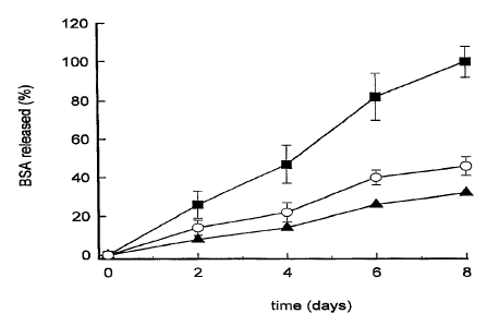
Figure 1 displays the percentages of BSA released "in vitro" from
nanoparticles made of different chitosan/PEO-PPO ratios. 1/0 (~), 1/5 (~) and
1/25
(O), following their incubation at 37°C for different time periods.
Figure 2 displays the percentages of BSA released "in vitro" from
nanoparticles containing different BSA loadings (amount of BSA entrapped in
100
mg of nanoparticles), 41 % (O), 25% (~) and 20% ( ~ ), following their
incubation at
37°C for different time periods.
CA 02233501 2004-06-15
5a
Results depicted in figure 1 indicate that the presence of PEO-PPO in the
nanoparticles significantly increases the BSA in vitro release rate. Results
showed in
figure 2 indicate that the higher the loading, the faster the release rate is.
In summary, this invention covers a new pharmaceutical composition which
can be used for the delivery of bioactive macromolecules following their
administration by different routes: topic. oral, nasal, pulinonary, vaginal,
ocular,
subcutaneous, intramuscular and intravenous.
Some examples of the composition and preparation of various formulations of
nanoparticles are described below.
Example 1
Association of BSA (bovine serum albumin) to chitosan nanoparticles. The
composition of the formulation (nanoparticles suspension) in % (w/w) was as
follows:
0
Chitosan base. . .. . .. . . .. .. . .. . .. .. .... . .. . .. 0.14 /o
Sodium tripolyphosphate.................Ø02
0
BSA.......................................... 0.0 14/0
0
Water. . .. . .. . .. . .. . .. . .. .. . . .. . .. . . . .. .. .up to 1 00 /o
Chitosan was dissolved at the concentration of 0.2% (w/v) in 25 ml of 0.05M
acetic acid solution. 'The pH of the solution was adjusted to pH 5Ø Then, 5
mg of
RCA zzrac
CA 02233501 1998-03-30
6
dissolved in the chitosan solution_ Finally, 10 ml of a sodium
tripolyphosphate aqueous
solution (0.1%, w/v) were added to the chitosan aqueous solution containing
the BSA and
the system was maintained under magnetic stirring for 30 min, after the
spontaneous
formation of the nanoparticlcs.
The size, zcta potential and BSA association efficiency for this formulation
were:
402 nm, 46 mV and 100% respectively.
Example 2
Association of BSA (boviine: serum albumin) to chitosanlPEO-PPO (1/5)
nanoparticles. The composition of the formulation in % (w/w) was as follows:
Chitosan base ----------- ------ 0.14
PEO-PPO _______~______~_________~__ 0.70
Sodium tripolyphosphate --------------- 0.02
BSA _______________________________________ 0.014%
Water ________________~_______________ up to 100%
Nanoparticles were prepared as described in example 1 with the exception that
PEO-PPO was dissolved in the chitosan solution prior to the incorporation of
BSA and the
pH of the chitosan solution was adjusted to pH 4Ø
The size, zeta potential and BSA association efficiency for this formulation
were:
519 nm, 44 mV and 78.2% respectively.
Example 3
Association of BSA (bovine serum albumin) to chitosan/PEO-PPO (1/25)
nanoparticles. The composition of the formulation in % (w/w) was as follows:
Chitosan base _________________~_________ 0.14
PEO_PPO ________________________________ 3.50
Sodium tripolyphosphate -_-_----------- 0.02
BSA -________________________~_________~_ p.014%
Water _______~_~______~_~___~ up to 100%
Nanoparticles were prepared as described. in example 1 with the exception that
PEO_PPO was dissolved at the concentration indicated above in the chitosan
solution prior
to the incorporation of BSA and the pH of the chitosan solution was adjusted
to pH 4.
The size, zeta. potential and BSA association efficiency for this formulation
were:
741 nm, 34 mV and 45.9 %, respectively.
s t ~s xt ZLHOV08 : -off 3i l3aow al~av'I~ = Ob : g y ~. g6-~O-GZ
CA 02233501 1998-03-30
7
Example 4
Accnriatinn of trtamic tnsrnir~ tn rhitncan nannharticha_ The compoaitioa of
+.1~
formulation in % (w/w) was as follows:
Chitosan base --------------- 0.14
Tetanus toxoid --------- 0.014
Sodium tripolyphosphate -------------- 0.02
Water ----------- ------- .up to 100%
Natwparticles were prepared as described in example 1 except for adding
tetanus
toxoid instead of BSA to the chitosan solution at the concentration indicated
above.
The size, zeta potential and tetanus toxoid association efficiency for this
formulation were: 24~ nm, 35 mV and 53 %, respectively.
Example 5
Association of diphtheria toxoid to chitosan nanoparticles. The composition of
the
formulation in % (w/w) was as follows:
Chitosan base -------------- ----- 0.14
Diphtheria toxoid -------------------- 0.007
Sodium tripolyphosphate ------------ 0.02
Water -____________________________ up to 1 00
Nanoparticles were prepared as described in example 1 but adding diphtheria
toxoid
instead of BSA to the chitosan solution at the concentration indicated above.
The size, the zeta potential and the tetanus toxoid association efficiency for
this
formulation were: 245 nm, 36 mV and 55 %, respectively_
s~ is ss zcnovos: -oo 'a iaoow a~av~o= ow-c~~.es-~o-flz
CA 02233501 1998-03-30
8
Table 1:
Mean values of particle size and zeta potential of nanoparticles composed of
different chitosan/PEO-PPO ratios.
ChitosanlPEO-PPO Size* Zeta potential ~'
(w/w) (nm) (mV)
1/0 275 ~ 17 44 f 1
1 /2.5 ' 283 ~ 11 41 t 2
1/S 300 ~ 14 40 ~ 1
1/25 430 t 20 28 t 1
1/50 d85 t 27 18 ~ 1
* Determined by Photon Correlation Spectroscopy
# Determined by Laser Doppler Anemometry
Table 2:
Particle size, zeta potential and association of chitosan nanoparticles
efficicncy
containing different chitosan/B5A ratios.
Chitosan/BSA Size * Zcta potcntiai
Association
(w/w) (nm) (mV) e~ciency
(%)
10/ 1 402 f 24 45 ~ 1 80 t 3
+/J. 339 ~ 3~ 45 l. 1 ~45 -L'
2/1 375 t 26 45 ~ 1 26 ~ I
1/I 368 1 72 46 ~ 2 2T t 2
* Determined by Photon Correlation Spectroscopy
# Determincd by Laser Doppler Anemometry
m lo v ss zt~ovos' -o~ ~a i.~oow aNav~~= or = o ~ .~ ss-so-cz
CA 02233501 1998-03-30
h
9
Table 3:
Association efficiency of bovine serum albumin (BSA) to chitosan
nanoparticles as a function of the stage at which BSA was incorporated and the
theoretical chitosan/BSA ratio.
ChitosanBSA (w/w) BSA association e~ciency (%)
13~A + nanoparticles BSA + chitosan BSA t TPP
10/1 80.413.2 10011.2
2/1 I0.8f3 26.80.7 45.23.9
1/1 21.612.0 41.812.0
Table 4:
Association efficiency of bovine serum albumin. (BSA) to chitosan
nanoparticles as a function of the pH of the chitosan solution and the
thcorctical chitosan/BSA ratio.
Chitosan/ BSA (w/w) BSA association efficiency (%)
pH 3 pH 4 pH 5
10/1 66.87.2 80.413.2 9I.713.6
2/I 25.7 ~ 1.4 26.8 ~ 0.7 39.1 t 2.4
1/1 19.4 ~ 3_6 21.612.0 35.515.1
S t / t t 3F ZLrv0408 = - '00 '8 1300W 3YIki f10= Ob : fl L '- 86-~O-flZ
CA 02233501 1998-03-30
Table 5.
Association e~ ciency of tetanus
and diphtheria
toxoids to
chitosan
nanoparticles_
5 Toxoid Chitosan/Toxoid% Association
Tetanus 1/0,06 56.7 t 2.7
Diphtheria 1/0,12 53,3 t 4.2
Diphtheria 1 /O, I2 55,1 t 5-5
10
s t ~z t xs Zc570voa'- 'off '8 134ow 311av1~= ov=9 ~ : gs-EO-CZ