Note: Descriptions are shown in the official language in which they were submitted.
CA 02233614 1998-03-31
WO 97/13838 PCT/US96/16517
1
ISOLATION OF CELLULAR MATERIAL UNDER
MICROSCOPIC VISUALIZATION
Technical Field
The present invention relates to methods and devices
for the molecular analysis of cellular samples. More
particularly, the present invention relates to methods
and devices for the microdissection and molecular
analysis of cellular samples which may be used in
combination with a number of different techizologies that
allow for analysis of proteins, such as enzymes, and mRNA
and DNA from substantially pure populations or
subpopulations of particular cell types. The present
invention further relates to libraries made from the
cellular material directly extracted in the method of the
invention.
Background Art
Many diseases are now understood at the molecular
and genetic level. Analysis of such molecules is
important for disease diagnosis and prognosis. Previous
methods for direct extraction of cellular tissue material
from a tissue sample are limited because the extraction
reflects only the average content of disease associated
markers. In reality, tissues are very heterogeneous, and
the most diagnostic portions of the tissue may be
confined to a few hundred cells or less in a lesion.
Normal tissue samples contain a variety of cell
types surrounding and adjacent to the pre-invasive and
invasive tumor cells. A region of the tumor tissue
subject to biopsy and diagnosis as small as 1.0 mm can
contain normal epithelium, pre-invasive stages of
carcinoma, in-situ carcinoma, invasive carcinoma, and
inflammatory areas. Consequently, routine scraping and
CA 02233614 1998-03-31
WO 97/13838 PCT/US96/16517
2
cutting methods will gather all of these 'types of cells,
and hence, loss of an_allele will be masked by presence
of a normal copy of the allele in the contaminating non-
malignant cells. Existing methods for cutting away or
masking a portion of tissue do not have the needed
resolution. Hence the analysis of genetic results by =
those previous methods are always plagued by
contaminating alleles from normal cells, undesired cells
or vascular cells.
The molecular study of human tumors is currently
limited by the techniques and model systems available for
their characterization. Studies to quantitatively or
qualitatively asses proteins or nucleic acid expression
in human tumor cells are compromised by the diverse cell
populations present in bulk tumor specimens. Histologic
fields of invasive tumor typically show a number of cell
types including tumor cells, stromal cells, endothelial
cells, normal epithelial cells and inflammatory cells.
Since the tumor cells are often a relatively small
percentage of the total cell population it is difficult
to interpret the significance of net protein or nucleic
acid alterations in these specimens.
The processes of tumor invasion and metastasis
depend upon increased proteolytic activity of invading
tumor cells. Matrix metalloproteinases, cathepsins B, D,
and L, and plasminogen activator have been implicated in
the metastatic cascade. Cathepsin D has been suggested
to be an independent marker of prognosis in breast
cancer. Several lines of correlation evidence support
the concept that proteases are important in tumor
invasion including: increased protease activity and/or
altered subcellular distribution of proteases in highly
metastatic tumor cell lines, increased protease
expression in invasive human tumors as determined by both
immunohistochemistry and assays of tumor tissue
homogenates, and increased protease mRNA levels in human
CA 02233614 1998-03-31
WO 97/13838 PCT/US96/16517
3
tumors. All of these techniques have generated important
information regarding protease expression in human
tumors, however, they have not provided definitive
evidence that proteases are up-regulated in specific
regions where tumor invasion is occurring.
Studies of human tumor cells in culture do not
account for the complex interactions of the tumor cells
with host cells and extracellular matrix, and how they
may regulate tumor cell protease productivity or
activation. Immunohistochemical staining allows one to
examine enzyme distribution in regions of tumor invasion,
however, results vary with tissue fixation and antibody-
antigen affinity, and provide only a semi-quantitative
assessment of protein levels. Furthermore, quantitative
interpretation of staining results is complicated by the
variability of staining patterns within tissue sections,
subjective evaluation of staining intensity, and the
difficulty in interpreting the significance of stromal
staining. In addition, many antibodies utilized in the
study of proteases do not differentiate pro-enzyme from
active enzyme species. Assays of enzyme or mRNA levels
from homogenates of human tumors does not account for
either the mixed population of cells within the
specimens, or the concomitant pathophysiologic processes
which may be occur in the tissue
Human tumors accumulate genetic abnormalities as
they develop from a single transformed cell to invasive
and metastatic carcinoma. Identification and
characterization of the genes which are mutated, lost or
abnormally regulated can provide important insights for
cancer diagnosis, prognosis, and therapy. Furthermore,
identification of such genetic lesions may facilitate
early diagnosis by definitive identification of
premalignant lesions so they can be treated before they
progress to invasive cancer.
CA 02233614 1998-03-31
WO 97/13838 PCT/ITS96/16517
4
A general dictum of cancer progression states that
cells can be transformed after acquiring two separate
alterations in a tumor suppressor gene. Subsequent
tumors progress stepwise from dysplastic lesions in situ,
to invasive and metastatic neoplasms. In situ carcinomas
are frequently observed arising in association with a
spectrum of epithelial hyperplasias and larger invasive
tumors are often associated with regions of carcinoma in
situ at the tumor periphery.
Pathologists have historically interpreted a side-
by-side association of atypical hyperplasia, in-situ
carcinoma, and invasive tumors as evidence of a cause and
effect relationship among the entities. However, little
direct evidence existed previously which supports this
model.
Prior methods of study have not allowed
investigators to specifically examine genetic alterations
in pre-invasive lesions. Even the most sophisticated
genetic testing techniques to date have been of limited
value because the input DNA, RNA or proteins to be
analyzed are not derived from pure cell populations
exhibiting the disease morphology. Several methods have
been reported for tissue microdissection to address this
problem, including gross dissection of frozen tissue
blocks to enrich for specific cell populations,
irradiation of manually ink stained sections to destroy
unwanted genetic material, touch preparations of frozen
tissue specimens and microdissection with manual tools.
These methods, however, are not sufficiently precise and
efficient for routine research or high throughput
clinical molecular diagnostic applications. Manual
microdissection, for example, has good precision but is
time consuming, labor intensive, requires a high degree of manual dexterity,
and is not generally suitable for
the ordinary technologist.
CA 02233614 2004-03-01
The present invention provides a novel improved
means to specifically examine genetic alterations in pre-
invasive lesions of common epithelial tumors such as
breast and prostate carcinoma. In particular, the
5 present invention permits the microsampling of as few as
one cell, with RNA and DNA extraction of the sampled
cell. This method has been demonstrated to be extremely
sensitive and to surpass previous and current
technologies by more than two orders of magnitude. It
has allowed the sensitive detection of loss of
heterozygosity in early pre-invasive lesions being a
gateway to the discovery of, for example, new genetic
loci on chromosome 11 for breast cancer and a new genetic
loci on chromosome 8 for prostate carcinoma.
The practice of the invention further permits the
construction of genetic libraries from the extracted
material. Thus, libraries from predetermined cells of
interest, particularly abnormal cells, may be constructed
and compared to libraries made from close-by, or
adjacent, other cells, such as normal cells. Such
libraries may be used, for example, to compare one or
more specific genetic loci, the expression of one or more
RNAs, particularly mRNAs, to isolate and/or clone one or
more specific nucleic acid, and the like.
CA 02233614 2004-03-01
6
Disclosure of the Invention
Various embodiments of this invention provide a method
of direct extraction of cellular material from a tissue
sample which comprises: providing a tissue sample;
contacting said tissue sample with a selectively activatable
transfer surface which can be activated to provide selective
regions thereof with adhesive characteristics; identifying
at least one portion of said tissue sample which is to be
extracted; selectively activating a region of said transfer
surface which corresponds to and is in contact with said at
least one portion of said tissue sample so that said
activated region of said transfer surface adheres to said at
least one portion of said tissue sample; and separating said
transfer surface from said tissue sample while maintaining
adhesion between said activated region of said transfer
surface and said at least one portion of said tissue sample
so that said at least one portion of said tissue sample is
extracted from a remaining portion of said tissue sample.
The adhesive characteristics may comprise chemical adhesive
properties or electrostatic adhesive properties. The
activating may comprise applying electromagnetic energy or
laser-derived energy. The contacting may occur before or
after activating.
Various embodiments of this invention provide a method
of direct dissection of cellular material from a tissue
sample which comprises: providing a tissue sample;
providing a selectively activatable transfer surface
separate from and independent of the tissue sample, the
transfer surface upon activation having adhesive
characteristics to the tissue sample; juxtaposing the tissue
sample and the transfer surface; identifying at least one
portion of the tissue sample which is to be dissected;
CA 02233614 2004-03-01
6a
selectively activating at least one region on the transfer
surface so that the at least one activated region of the
transfer surface can adhere to the at least one identified
portion of the tissue sample; contacting the tissue sample
with the at least one region of the transfer surface to
adhere the at least one portion of the tissue sample which
is to be dissected to the at least one region of the
transfer surface; and, separating the transfer surface from
the tissue sample while maintaining adhesion between the at
least one region of the transfer surface and the at least
one portion of the tissue sample so that the at least one
portion of the tissue sample is dissected from a remaining
portion of the tissue sample.
CA 02233614 2004-03-01
7
Brief Description of Drawincrs
The present invention will be described with
reference to the attached drawings which are given by way
of non-limiting examples only, in which:
Figure 1 is a functional system diagram depicting
how a tissue sample is microscopically imaged, displayed
on a display monitor, and how a region of the imaged
sample is selected and identified for subsequent
microdissection and analysis.
Figures 2a-2c are a series of functional system
diagrams which depict how a zone of tissue sample is
extracted from the slide-mounted tissue sample according
to one embodiment of the present invention.
Figure 3 is a schematic illustration of an
alternative device for extracting sample zones from the
slide-mounted tissue sample.
Figures 4a and 4b are schematic diagrams of a manual
extraction tool manipulator which can be used together
with the extraction device of Fig. 3 according to the
present invention.
Figure 5 is a functional system diagram which shows
how a zone of sample tissue can be directed to an
appropriate analysis protocol.
Figure 6a and 6b show the expression of MMP-2 in ten
invasive colon carcinoma cases (Fig. 6a) and in five
cases of invasive breast carcinoma (Fig. 6b) as compared
to normal colonic mucosa from the same patients.
Figure 7 shows SSCP analysis of MMP-2 activation
site.
CA 02233614 1998-03-31
WO 97/13838 8 PCT/US96/16517
Figures 8a - 8d are schematic illustrations of the
sequential steps of an adhesive transfer method according
to one embodiment of the present invention.
Figure 9 schematically depicts the laser capture
microdissection technique.
Best Mode for Carryina out the Invention
The present invention is directed to a method of
analyzing cellular material on a molecular or genetic
level which involves: visualizing a field of cells in a
tissue sample under a microscope, contacting an
identified area with a surface which simultaneously
dissolves, extracts and/or retains a cellular material of
interest, and transferring the cellular material of
interest to a suitable analysis system. The present
invention is particularly applicable to the analysis of
local tissue polypeptides proteins, such as enzymes and
antigens, and DNA, RNA, particularly mRNA, lipids,
carbohydrates, and other biological molecules and
assemblies thereof.
According to one embodiment, the present invention
is directed to adhesive transfer methods which involve
microscopic visualization and transfer of cellular
material to a procurement or transfer surface.
The present invention is also directed to a fully
automated system whereby a tissue can be visualized, for
example, on a screen, so that a precise field of cells of
interest can be identified, for example, by a variety of
labels, histochemical stains, antibodies, etc.,
circumscribed or there location otherwise demarcated, and
then be extracted and analyzed, either manually or
automatically, or by a combination of the two.
Figure 1 is a functional system diagram which shows
how a tissue sample is microscopically imaged, displayed
on a display monitor, and how a region of the imaged
sample is selected and identified for subsequent
CA 02233614 1998-03-31
WO 97/13838 PCTIUS96/16517
9
microdissection and analysis. As depicted in Fig. 1, a
tissue sample 1 is provided on a surface, such as a glass
slide 2, for microscopic examination and imaging. The
sample tissue 1 can be fixed on the glass slide 2
according to any conventional method, including
attachment to the glass slide 2 with an agarose gel,
fixing the tissue sample in paraffin, etc.
The glass slide 2 having the sample tissue 1
mounted thereon is placed on the stage of a microscope.
The microscope, generally indicated by reference numeral
3, receives an image of the tissue sample 1. An imaging
device, such as a video camera, (not shown) is connected
to the microscope 3. The imaging device receives the
image of the sample tissue 1 from the microscope 3 and
displays the image of the tissue sample on an imaging
display device, such as display monitor 4.
The image of the sample tissue 1 is limited to the
"field" of the microscope 3 for any given image. As
indicated iri Fig. 1, the field of the sample tissue image
may include several zones, "A", "B", "C", and "D" of
different types of cells which can be optically
distinguished by utilizing a suitable dye(s), labeled
molecules such as antibodies or fragments thereof, to
stain or otherwise differentiate the predetermined cells
of interest in the tissue sample. For exemplary
purposes, Figs. 1 and 2a-2c assume that zone "B" is the
zone of cellular material of interest. The image on the
display monitor 4 is used by the operator to select and
identify one or more zones of the tissue sample 1 which
are of interest. According to one embodiment of the
present invention, after the zone(s) of interest are
selected and identified, the operator manually
manipulates a device to extract the identified zone(s)
from the glass slide 2. The identification of the cells
of interest may also be done automatically through image
analysis software. The extracted zone(s) of sample
CA 02233614 1998-03-31
WO 97/13838 PCTIUS96/16517
material may include an analysis sample. Otherwise, the
identified and extracted zone (s) can include zones which
are to discarded and the remaining zone(s) which are
retained on the glass slide 2, can be later analyzed.
5 In addition to manual operation which is discussed
in more detail below, it is possible, according to
another embodiment of the present invention, to utilize
the image on the display monitor 4 to select and identify
a sample zone(s) whose relative position is determined
10 utilizing a computer which receives a digitized signal of
the image from the video camera (or microscope), and
which receives a reference position of the stage of the
microscope 3 upon which the sample is held. Such
positioning detection and recognition systems are
conventional in the art and can be readily applied to
automate the sample preparation method of the present
invention.
In this automated embodiment of the invention, the
computer which performs the positioning detection and
recognizing can also be used to control the movement of
the devices discussed below that are used to extract
tissue zones, thus automating the sample removal. In
addition, the image of the sample can be electronically
scanned to automatically identify zones having a
predetermined feature, such as a relevant degree of
staining, using known techniques and devices. Thus, in
a preferred embodiment, a computer could be used to
select and identify zones of interest and the relative
position of such zones, for manipulating a device to
remove such zones in a completely automated manner.
Figures 2a-2c are a series of functional system
diagrams which show how a zone of tissue sample 1 is
extracted from the slide-mounted tissue sample 1
according to one embodiment of the present invention. It
is to be understood that the steps depicted in Figs. 2a-
2c could be either preformed manually by an operator or
CA 02233614 1998-03-31
WO 97/13838 PCT/US96/16517
11
by a computer utilizing conventional positioning and
control methods, e.g. computer controlled robotics.
The embodiment of the invention depicted in Figs.
2a-2c utilize a contact probe 5 which has an
adhesive/extraction reagent 6 on the tip thereof. A
suitable adhesive/extraction reagent can include a
mixture of piccolyte and xylene. In Fig. 2a the contact
probe 5 is positioned either manually or by computer
control so as to be above and aligned with the sample
zone ("B") to be extracted. As can be readily understood
from Fig. 2a, the surface area of the contact probe tip
(and adhesive/extraction reagent) needs to be about equal
to, and no greater than, the surface area of the zone to
be extracted. Otherwise, excessive removal of adjacent
tissue zones will occur. Manufacture of probe tips of
the required size is well within the capabilities of
those skilled in the art.
Once the tip of the contact probe 5 is aligned with
the sample zone ("B") to be extracted, the contact probe
5 is lowered so that the adhesive/extraction reagent 6 on
the tip thereof contacts the sample zone (Fig. 2b). Of
course, depending on the specifics of the apparatus, the
probe 5 is raised or otherwise moved into contact with
the sample zone of cells of interest.
The adhesive/extraction reagent 6 is selected to
readily adhere to the sample zone. Once the
adhesive/extraction reagent 6 on the tip of the contact
probe 5 contacts the sample zone (Fig. 2b) and the sample
zone becomes adhered thereto, the contact probe 5 can be
retracted from the contact position (illustrated in Fig.
2b) and moved as shown in Fig. 2c. Since the relative
adhesive force of the adhesive/extraction reagent is
greater than the adhesive force used to mount the sample
on the glass slide, the contact probe 5 pulls the sample
zone "B" from the glass slide when withdrawn or
retracted.
CA 02233614 2004-03-01
REPLACEMENT PAGE
WO 97/13838 PCT/US96/16517
12
According to one embodiment of the present
invention, a glass pipette was used as the contact probe
5. In this embodiment, the tip of the glass pipette was
coated with a solution of piccolyte (568 g/1) and xylene 5 (437.5 g/1) by
dipping the tip of the glass pipette in a
piccolyte/xylene solution.
In addition to removing the sample zone from the
glass slide 2, the contact probe 5 can be used to
transfer the extracted sample zone to an analysis
container 7 as indicated in Fig. 2c or to any other
location, such as a waste container, a culture media,
etc. In a preferred embodiment, the contract probe 5 is
used to transfer the extracted sample zone to the sample
receiving stage of an automated clinical analyzer which
is designed to preform a desired analysis of the sample
zone. It thus should be understood that the present
invention can provide a fully automated method and system
for identifying sample zones on a sample on a surface
such as a slide, removing sample zones-of interest from
the surface-mounted sample, and transporting the
extracted sample zones to an automated analyzer which can
perform automated analysis of the extracted sample zones.
Such analysis can include, for example, analysis of
cellular DNA, RNA, proteins, polypeptides, lipids,
carbohydrates, and combinations and aggregates thereof.
In Fig. 2c the extracted sample zone is depicted as
being dispensed in a container 7 which, for example, can
be a test tube or similar container in which analysis on
the extracted sample zone can be initiated or performed.
As depicted in Fig. 2c, a reagent solution 8 which
removes all or a desired component of the extracted
sample zone from the contact probe tip can be placed in
the container 7 before the extracted sample zone is
deposited therein. For example, in the case of DNA
analysis, a solution of Tris (50 mM, pH8.5), EDTA (1mM),
TweerY 20 (0.5%-), and proteinase K (0.2 mg/mL) can be
CA 02233614 1998-03-31
WO 97/13838 PCT/US96/16517
13
used. This solution extracts the sample zone from the
tip of the contact probe 5 and dissolves the tissue
material for analysis purposes.
In addition to the contact probe depicted in Figs.
2a-2c, a hollow suction probe could also be used to
extract sample zones from the slide-mounted tissue sample
1. Such a suction probe could be provided with sharp
annular tip by which sample zones could be punched out
and extracted by suction forces.
Figure 3 is a schematic illustration of an
alternative device for extracting sample zones from the
slide-mounted tissue sample 1. The extraction device 9
shown in Fig. 3 includes a cutting blade 10 and a
grasping arm 11. The grasping arm 11 can be moved in an
opposed manner with respect to the cutting blade 10. The
grasping arm 11 is shown in its open position in Fig. 3.
The grasping arm 11 is movable between the illustrated
open position to a closed position in which the tip of
the grasping arm 11 contacts the cutting blade 10. The
movement of the grasping arm 11 can be controlled by a
cable and pulley system in which grasping arm 11 is
caused to pivot at its base by applying tension to a
cable which passes through a pulley located at the base
of the grasping arm 11. The tension on the cable can be
applied by actuating a lever or depressing a button 12 on
the device which applied tension to the cable in a known
manner. Such actuating mechanical structures are known
in the art of gripping devices.
In operating the device of Fig. 3, the cutting blade
10, which is at an obtuse with respect to the central
axis of the device can cut out and scoop up a portion of
a tissue sample by placing the cutting blade 10 on one
edge of a portion of the tissue sample to be extracted
and then moving the grasping arm 11 into the closed
position. As the grasping arm 11 comes into contact with
the tissue sample, it draws the cutting blade 10 into the
CA 02233614 2004-03-01
14
sample and presses a portion of the sample toward the
cutting blade 10 thereby causing a portion of the sample
contacted between the cutting blade 10 and the grasping
arm 11 to be cut out and scooped up from the sample.
In a further, alternative embodiment of the device
of Fig. 3, the movement of the grasping arm 11 can be
effected by a toothed gear instead of a pulley and a
cooperating toothed rod in place of a cable. Additional
such mechanical structures are known in the art of
gripping devices.
Figures 4a and 4b are schematic diagrams of a manual
extraction tool manipulator which can be used together
with the extraction device of Fig. 3 according to the
present invention. In Fig. 4a the extraction tool
manipulator is depicted as having a base 13 equipped with
a clamping means 14 for removable attaching the device to
a brace or support portion of the stage of a microscope
(see Fig. 4b). The clamping mechanism includes a
clamping plate 15 that is secured to a threaded shaft 16
which passes through a threaded bore 17 in a lower
portion of the base 13. A tightening knob 18 is provided
on the end of the threaded shaft 16. Turning the
tightening knob 18 causes the clamping plate 15 to move
with respect to an upper portion 19 of the base 13.
Thus, the extraction tool manipulator can be clamped to
a portion of the stage of a microscope 20 as depicted in
Fig. 4b by positioning a brace or support portion 21 of
the stage of the microscope 20 between the clamping plate
15 and the upper portion 19 of the base 13 and turning
knob 18 to tighten the clamping plate 15 against the
brace or support portion 21 of the stage of the
microscope 20.
The extraction tool manipulator includes a tool
holder 22 having a through-bore 23 therein for receiving
the shaft of an extraction tool. Ideally, the tool
holder 22 should allow for damped fore and aft movement
CA 02233614 1998-03-31
WO 97/13838 PCT/1JS96/16517
of the extraction tool. Therefore, according to a
preferred embodiment, the through-bore 23 of the tool
holder 22 contains a bushing which can be adjustably
tightened against the tool shaft by tool locking screw
5 24.
The tool holder 22 is supported by support shaft 25
which is connected at opposite ends by substantially 3600
damped swivels 26 and 27 to the tool holder 22 and the
base 13. The length of the support shaft 25 between the
10 360 damped swivels 26 and 27 is adjustable. The
adjustment of the independent 360 damped swivels 26 and
27 together with the adjustable length of the support
shaft 25 and the position of the tool shaft within
through-bore 23, allows a high degree of movement of the
15 extraction tool with respect to a slide-mounted sample
positioned on the stage of the microscope. Therefore, an
operator can manipulate an extraction tool held by the
extraction tool manipulator and remove selected tissue
zones from a slide-mounted tissue sample with a high
degree of precision.
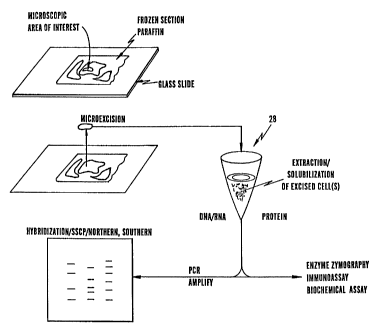
Figure 5 is a functional system diagram which shows
how a zone of sample tissue can be directed to an
appropriate analysis protocol. As depicted in Fig. 5, a
microextraction of a zone of tissue sample can be taken
from a slide-mounted tissue sample 1 as discussed above
and transferred to a sample preparation stage 28 in which
the cells of interest can be extracted and collected for
analysis. Excised cells may also be solubilized at this
stage. If these cells contain, or are suspected to
contain, one or more DNA or RNA of interest, the
extracted sample may subjected to polymerase chain
reaction (PCR) amplification, followed by, for example,
hybridization, strand conformational polymorphism, and
southern and northern blotting, sequencing, etc. as
desired. Of course, other techniques for analysis of DNA
CA 02233614 1998-03-31
WO 97/13838 PCT/US96/16517
16
and RNA are known to those skilled in the art and
encompassed by the spirit and scope of the invention.
If the extracted cells contain, or are suspected to
contain proteins or polypeptides of interest, the
extracted sample can be subjected to enzyme zymography,
for example using one or more labeled substrates, an
immunoassay utilizing, for example, labeled antibodies or
functional fragments thereof, a biochemical assay, and
the like.
Selective extraction or microdissection of frozen
tissue sections according to the present invention allows
for recovery and analysis of both active enzymes and
mRNA. Additionally, the DNA recovered from these
sections is in the native condition and can be used for
studies such as DNA fingerprinting. Microdissection of
paraffin embedded tissues according to the present
invention allows for PCR amplification of DNA, for
example, from pure cell populations representing less
than one high powered field, or a-single layer of
epithelial cells lining cystic spaces.
For general preparation of samples for frozen
section microdissection according to the present
invention microdissection slides can be prepared by
placing 1%- agarose on a standard histology slide and
cover slipping. After a short period of time, e.g.,
about 5 minutes the cover slip is removed leaving a thin
gel on the slide. A small frozen tissue section, e.g.
about 25 micron thick, is placed on the agarose gel and
briefly stained with eosin. The tissue may also be
treated with agents to denature or otherwise inhibit
RNase depending on the subsequent extraction method.
Under direct microscopic visualization the specific cell
population or sub-population of interest is procured from
the tissue section utilizing the techniques discussed
above.
CA 02233614 2004-03-01
REPLACEMENT PAGE
WO 97/13838 PCT/US96/16517
17
For enzyme analysis the procured tissue specimen can
be placed in an appropriate buffer depending on the
enzyme of interest, as known to the person skilled in the
art. The enzyme levels can be measured by several
methods including zymography and the use of specific
substrates, including fluorometric, colorometric and
radioactive substrates. The precise levels of enzyme
expression in a specific, predefined cell population can
be thus determined and, where desired, compared to that
of another, independently isolated sample from the tissue
sample.
For mRNA analysis the tissue specimen can be placed
on agarose and treated with agents to denature or
otherwise inhibit RNase, if desired. The procured tissue
specimen is immediately frozen in liquid nitrogen. The
tissue can be used immediately or stored at -70 C for
several months. The mRNA can be extracted using, for
example, column chromatography on oligo-dT (Micro-
FastTrac~mRNA Isolation Kit, Invitrogen Co.). The
recovered mRNA of the pure cell populations can also be
amplified and investigated using polymerase chain
reaction (PCR) technology, such as, for example, by RT-
PCR as known to those skilled in the art.
For DNA analysis the tissue specimen can be placed
in a single step extraction buffer solution of 50 mM
Tris, pH 8.5, 1mM EDTA, 0.5% Tween 20, and 0.2 mg/ml
proteinase K, incubated for four hours at about 37 C,
followed by ten minutes incubation at about 95 C. The
recovered DNA can also be amplified and analyzed using
PCR technology in combination with analysis techniques,
such as blotting, sequencing, etc., known in the art. If
native DNA is required for DNA fingerprinting analysis,
the proteinase K can be added after DNase in the
fingerprinting protocol.
For paraffin section microdissection routine
formalin fixed, paraffin embedded tissue sections are
CA 02233614 2004-03-01
18
microdissected after de-paraffinization and brief
staining with eosin. Tissue sections are visualized by
direct microscopy and cell populations or subpopulations
of interest are procured using a modified glass pipette
with the adhesive coated tip discussed above. Tissue
specimens as small as one cell can be procured with this
method. The specificity of dissection represents a
significant improvement over currently known techniques.
For DNA analysis of paraffin embedded tissue, the
glass pipette with the dissected tissue specimen is
placed in a single step extraction buffer solution of 50
mM Tris, pH 8.5, 1mM EDTA, 0.5% Tween 20, and 0.2 mg/ml
proteinase K, which removes the tissue from the pipette
tip. The sample is incubated, depending on sample size,
from two to twenty-four hours at about 37 C, followed by
a ten minute incubation at about 95 C. The glass pipette
tip can then be sterilized and reused, although this is
not generally recommended in the case of PCR-based
analysi-s due to the potential amplification of cross-
contaminating materials.
In another embodiment of the invention, one or more
cells of interest are isolated via laser capture
microdissection as exemplified hereinbelow. The
principle operation of laser capture microdissection is
depicted in Figure 9 (without depiction of the
microscope). In this method of the invention, a tissue
sample specimen is mounted on a support, as before, and
a transparent or translucent film or tape (the transfer
film) is placed on top of the tissue sample specimen.
The tissue sample is then examined microscopically for
predetermined target cells, such as abnormal cells (or
control cells for comparison). As before, the cells may
be stained with dye ( s), immunologically, etc. to identify
and/or differentiate the predetermined cells of interest
in the sample. The predetermined cells of interest are
next made to coincide with a target point, wherein
CA 02233614 1998-03-31
WO 97/13838 PCT/US96/16517
19
electromagnetic radiation may be focused. This
coincidence may be accomplished, for example, by x-y-z
translation of either the specimen or the target point.
For example, the target point may coincide with the
center of the imaging field and the microscope stage
translated such that the predetermined cells of interest
are brought to this target point. One or more focused
pulses of energy (e.g., electromagnetic energy in the
form of light from an infraredlaser, thermal energy,
etc.) is directed at the film overlying the target.
Sufficient energy is directed to the target point so as
to preferentially heat or otherwise alter the adhesive
characteristics of the film or tape covering the
predetermined cells of interest at the target point. In
this way, the film or tape is made selectively adhesive
at the specific target in the sample by optical
activation in precisely predefined locations.
It is preferred that lasers are used in the present
invention for providing electromagnetic energy to the
target spot. This is because lasers are high brightness
light sources of intense, collimated light that can be
readily and efficiently focused to small regions on a
given surface. By using a laser focus onto the optical
center of the field of view of an optical microscope,
activation energy can be supplied focally to a target
region of the film or tape lying on top of the tissue
sample. Moreover, the timing and duration of lasers are
readily controlled, such that a controlled amount of
energy can be directed to the target spot. Additionally,
a laser beam can be focused to spots as small as the
diffraction limit of the wavelength used and thus permit
selective adhesion to targets as small as one micron.
Thus, the spots can be small enough to select a
homogenous cluster of cells, an individual cell, or even
a portion of a cell.
CA 02233614 1998-03-31
WO 97/13838 PCT/IJS96/16517
When sufficient energy from the focused pulse of
radiation is absorbed to provide activation of the film
surface which is in contact with the predetermined cells
of interest in the tissue sample, an adhesive bond is
5 formed between the film or tape and the specifically
targeted cells. As long as the focal bond strength
formed between the film and the targeted tissue is
greater than the bond strength for the targeted tissue
for the underlying substrate (e.g., microscope slide),
10 the targeted tissue can be procured upon the removal of
the film. If the region of the film not activated form
weaker bonds with the untargeted regions of the sample
tissue slice than the strength of the particles (e.g.,
intercellular) within the tissue sample and those
15 intercellular bonds are weaker than the particle
(cellular) bond to the underlying substrate, and if in
turn the bonds between the tissue and the underlying
substrate are weaker than the bonds formed between the
activated film and the targeted tissue, then the targeted
20 tissue will be selectively attached to the film or tape
and can be selectively removed when the film is peeled or
otherwise removed from the tissue slide.
The size of the tissue transferred, depending on the
needs of the operator, can be varied by changing the
diameter of the laser beam and pulse durations. Highly
reproducible transfers in the 60 to 700 m diameter range
are easily attainable for procurement of small (100 m to
1 mm) lesions without the encroachment of adjacent, non-
neoplastic cells. In most basic and clinical research
studies, procurement of several hundred to several
thousand cells is necessary to provide sufficient genetic
material for reliable amplification and statistically
meaningful analysis. However, since laser beams can be
focused to less than a one cell diameter, transfers of
targeted single cells or even parts thereof is thought
possible under the practice of the invention.
CA 02233614 1998-08-31
WO 97/13838 PCT/US96/16517
21
Thermoplastic polymer films are widely used as heat
and pressure activated adhesives for bonding surfaces.
Most of these polymer films without added pigments are
transparent or translucent to visible light used in
conventional light microscopy. These films are, however,
intrinsically strongly absorptive in specific regions of
the electromagnetic spectrum (e.g., in regions of the
infrared associated with strong molecular vibration modes
such as 3000, 1800, 1400-960 cm-'). It is also possible
to add infrared absorbing dyes to the thermoplastic films
to provide strong absorption at other specific infrared
wavelengths without altering their transparency to
visible light. Such dyes are preferably IR absorbing
dyes, such as the metalonaphthalocyanines,
naphthalocyanines and cyanine dyes. If the focused pulse
of electromagnetic radiation (e.g., laser) is delivered
at wavelengths that are strongly absorbed by the film,
then the film may be rapidly efficiently focally heated.
Indicators may also be included, either in the
selectively adhesive transfer film or in a separate layer
or layers, to define the location of the optical
activation. Such indicators include thermochromic dyes,
dye precursors which combine upon melting to form a color
for visible or instrumental identification, and dyes
which are converted to color by other effects of optical
absorption. Suitable indicators also include physical
effects, such as the appearance or disappearance of
translucency or opacity upon optical exposure or upon
heating.
While wishing not to be bound by theory, it is
thought that when such thermoplastic films are heated to
near or at the melting point they flow and conform to an
adjacent surface (in this case, the targeted tissue
sample), forming a strong surface bond. This bond is
thought to occur without actual chemical cross-linking to
the tissue sample. Such strong bonds are formed most
CA 02233614 1998-03-31
WO 97/13838 PCT/iJS96/16517
22
reliably when pressure is applied to force the flow of
the "melt" into tight conformity with the sample
surfaces. However, by using smooth films applied in
close apposition to the tissue and delivering appropriate
pulse parameters to selected composition thermoplastic
f ilms , one can reliably focally heat the film to peak
temperatures associated with high film fluidity for a
sufficient period of time to form adequately strong bonds
between the film and tissue_for highly reproducible focal
microtransfer to occur. Moreover, by using a pulsed
infrared.laser source to activate the focal bond of the
targeted tissue to the film, the targeted tissue is
cluantitatively procured (virtually complete microtransfer
to the film) without chemical modification, while
preserving focal tissue morphology and allowing unaltered
microscopic observation prior to, during, and following
the microtransfer.
An optional additional step may also be used in
laser capture microdissection to improve the bonding
between the cells of interest and the activated polymer
film and decrease the bonding of the tissue of interest
to the substrate, so that the selected cells can be more
easily removed. The slide (i) can be chosen to be a
material that has an inherent lower affinity for the
tissue sample than the polymer film, (ii) can be pre-
treated with an agent that reduces this affinity, or
(iii) enclosed in a material compatible with the meltable
polymer film. For example, a glass slide with tissue on
it can be treated following the usual slide preparation
procedure by dipping in a 3%~ aqueous glycerol solution
followed by drying. Alternatively, the tissue can be
enclosed in a polymeric material which will form a strong
bond with the meltable film and be sufficiently water-
soluble to allow the tissue sample to be retrieved in the
analysis step. The enclosure of the tissue in such a
material can be done by a coating technique such as
CA 02233614 1998-03-31
WO 97/13838 PCT/US96/16517
23
application of the polymer in solution or placing the
material in film form on the tissue and melting it. The
enclosure may also be in the form of a coating on the hot
melt film, to enhance the bond of the meltable film to
the tissue and decrease the bond to the slide. It is
further possible to treat the surface/tissue combination
with a material which enhances the hond strength between
the tissue and the targetted surface, decrease the bond
strength between the tissue and the slide, and allow
reliable removable of the tissue/melted polymer from the
slide.
Any wavelength of electromagnetic energy can be used
under the practice of the invention provided that
suitable materials are used. In particular, it is
important that the transfer film absorb sufficient energy
(or contains one or more dyes that absorb sufficient
energy) at the chosen wavelength to melt or nearly melt
the thermoplastic polymer in the targeted region. For
thermoplastic materials such as ethylene vinyl acetate a
wavelength of about 3 to about 10 micrometers is
preferred as these materials intrinsically absorb in this
range. The power of the laser used is generally in the
range of from about 1 mW to about 200 mW, preferably from
about 10 mW to 100 mW, depending on the size of the
target (i.e, increasing power with increasing target
size).
It is also preferred that the wavelengths for laser
activation and film absorption be chosen outside the
normal range used for microscopic imaging. Reproducible
microtransfer of tissue can be obtained using a variety
of infrared wavelengths from a tunable carbon dioxide
laser (9.6- ll m).
The transfer film may be any that is selectively
activatable by electromagnetic or thermal energy, and is
preferably transparent or translucent to the
visualization wavelength. This selectively activatable
CA 02233614 1998-03-31
WO 97/13838 PCT/US96/16517
24
transfer film may be made of, for example, a wide variety
of thermoplastic materials, such as ethylene vinyl
acetate, polyurethanes, polyvinyl acetates, and the like.
In one embodiment of the invention, the selectively
activatable transfer film is a film of a polymerizable
substance, for example an electromagnetically-activated
polymerizable substance which polymerizes upon exposure
to electromagnetic energy.
Specific other selectively activatable materials
found useful in the practice of the invention are:
thermal sensitive adhesives and waxes, such as Precision
Coatings product #HAL-2 180C; thermally-a.ctivated hot
glues and sealants, such as those from Ban Fastening
Systems (Brooklyn, NY); ultraviolet sensitive or curing
optical adhesives, such as ThorLabs, Inc. product NO60-
NOA81; and thermal or optical emulsions, such as
silkscreen coated emulsion B6, high mesh, powdered,
reconstituted lelt fixit emulsion (Riso Kagaku Corp.).
The adhesive film described above may be self-
supporting or laminated with a support film.
Additionally, the support film may be made of a material
that does not absorb the electromagnetic energy so
strongly as to interfere substantially with the
activation of the thermoplastic polymer. The support
preferably absorbs weakly, if at all, at the activation
wavelength and at the visualization wavelength. The
activatable film, on the other hand, preferably absorbs
weakly, if at all, at the visualization wavelength but
strongly at the activation wavelength. The support
should also be unaffected by the resulting thermal
transients occurring during the activation.
Use of a microscope slide and transparent tape are
preferred in the practice of one embodiment of the
invention. Observation of the slide with a microscope
allows the pathologist or other microscopist to select
one or more spots on the tissue sample and expose them to
CA 02233614 1998-03-31
WO 97/13838 PCTlUS96/16517
the (invisible) infrared energy from any appropriate
optical system. The film becomes adhesive at the
selected one or more spots and the tissue at those spots
readily adheres to the polymer film, which can then be
5 removed from the slide to retain the tissue samples with
the film. The selected samples can then be removed from
the film by techniques appropriate to the subsequent
analysis desired, particularly molecular analysis such as
of gene mutations/deletions, altered gene expression as
10 measured by mRNA and/or protein concentrations, changes
in enzymatic activity, and the like. As more than one
sample of cells of interest can be obtained from a single
sample slice, normal cells may also be procured from the
same tissue, molecularly analyzed, and the analysis of
15 the cells of interest with the normal cells compared for
useful diagnosis and prognosis information.
The laser microdissection system can be
advantageously used with a variety of sample
preparations, such as stained thin sections of tissue or
20 stained cytology specimens of intact cells. In this
case, the transferred regions can be clearly identified
in a microscope by the focally transferred stained
material on an otherwise transparent film.
Alternatively, the film and tissue slide can be indexed
25 to an x-y coordinate system to give a specific slide
location to each transferred point which may then be
automatically recorded.
The microtransferred tissue may then be collected
from the film, for example, by punching the precisely
recorded spots directly into the desired reaction or
extraction vessels (e.g., by automatic x-y translation)
or by placing the whole film into a reaction vessel.
The molecular analysis of the extracted cellular
material, for example by RT-PCR, in one embodiment of the
invention requires localizing the small objects (e.g., 50
m spots of tissue) adhering to the substrate and
CA 02233614 1998-03-31
WO 97/13838 PCT/US96/16517
26
collecting them into an analysis chamber, for example,
punched out into a vial. Using an x-y encoding of the
position of each target site and automated translating
allow the area(s) to be punched to also be automated
using the same coordinates, so long as the support film
is not deformed or stretched in the application,
activation and removal steps of the process. Thus, the
sample collecting process is also amenable to automation.
Alternatively, one can ensure that the target sites are
at known positions on the transfer substrate. For
example, multiple small pieces of adhesive transfer film
can be selectively applied to those locations on the
tissue which correspond to sites to be extracted, rather
than applying a single large piece of substrate film to
the entire specimen. Exemplary of such a scheme is small
disks of adhesive film are applied at a fixed repeat
distance on a continuous polyester film, preferably a
strong and not easily stretched sheet, to provide a
linear array of separately activatable target sites.
After each target zone is identified, the next unused
small adhesive spot in the linear array is locally
applied at a fixed separation to this region by a small
pressure plate or an air jet.- The substrate/tissue slide
as a unit is then micropositioned under microscope
viewing to target specific cells (determined by the laser
spot diameter) within the target zone (i.e., the diameter
of the adhesive spots which is greater than the laser
spot diameter). Where the substrate film and the
mechanism applying these small pieces of film is located
with respect to a stationary reference (e.g., the
microscope objective) then it can be arranged that the
adherent tissue spots will always be known locations on
the substrate film (e.g., in the center of a narrow strip
if adhesive film at equally spaced distances). Targeting
of like cells within the target zone is accomplished by
microtranslating the sample between sequential laser
CA 02233614 1998-03-31
WO 97/13838 PCT/US96/16517
27
pulses. Thus, the operator positions the slide in the
microscope so that the tissue of predetermined interest
is at the center of the field, a pressure plate or other
means, for example an air jet, can then apposition the
adhesive film spot to the tissue such that the
predetermined cells/objects of interest are within the
known location on the support film. The adhesive is
activated, for example, by the IR laser pulses, and the
pressure plate is then released. The film is then
separated from the specimen slide and advanced so that a
fresh portion of film can be used at the next specimen
location. The process allows all transfers to occur on
an ordered series (numbered array) of spots with a fixed
spatial separation, as the film is not distorted in the
process. The size of the adhesive film spot determines
the size of the target zone within which selection of the
objects/cells ar made for that one transfer (array
number) in this embodiment of the invention. The target
zone size is determined by the selection of the
particular film (i.e., the geometry of ordered arrays),
but can be increased or decreased, for example with
parallel rows of spots of different sizes on the same
support film.
Examples of adhesive transfer films are such as in
the form of small pieces with or without a carrier
substrate or a tape with isolated, preferably equally-
spaced, portions of adhesive film. Use of a tape in this
regard provides ease of transferring the new portions of
film into an actuation region, and removal of the
activated film to a collection or storage means. Removal
of the adhesive film from the tissue can be accomplished
by tensioning the tape while holding the support (e.g.,
slide) in place. This is accomplished after the pressure
plate is removed in embodiments of the invention
employing a pressure plate. This may in some instances
require that the adhesive film is strongly bonded to a
CA 02233614 1998-03-31
WO 97/13838 PCT/US96/16517
28
substrate which will not deform in tension and a
mechanical mechanism for the tape transport which
maintains registration of the tape during the tensioning
and pressure plate actuation processes (e.g., symmetrical
deflections in the lateral tape transport direction).
In another embodiment of the invention that is
further simplified, the collection process is performed
by cutting off (rather than punching out) the desired
portions of adhesive films. This simplified embodiment
eliminates the requirement for close tolerances between
a punch and die. Alternatively, other means can be used
to separate the tissue/adhesive film from the rest of the
tape. Such means will be obvious to those skilled in the
art based on the disclosure made herein and include
direct peeling of the tape, focally dissolving either the
adhesive tape or its bond to a substrate film, excising
the spots with a hot wire knife (which is self-
sterilizing to eliminate contamination between
specimens).
An additional feature of the invention is directed
to the identification of the samples. The small portions
of film can have minute identifying marks (e.g., bar
codes) attached to them which would be seen under the
microscope and can be recorded along with a video image
of the specimen. A practical way in which this is
accomplished is to place the identifying marks on a
mechanically strong substrate adjacent to each discrete
spot of adhesive on the tape configuration mentioned
above. An additional use of these identification marks
is to control the advance of the tape for each new
specimen. A sensor in the microscope or analysis of the
various specimens determines when the mark is in, for
example, the center of the field as the tape is being
advanced and then turn off the tape transport.
Alternatively, a mechanical drive (e.g., with sprockets)
can be used to advance the tape a fixed known amount.
CA 02233614 1998-03-31
WO 97/13838 PCT/US96/16517
29
As a preferred embodiment of the invention, equally
spaced adhesive film spots along with minute adjacent bar
code identifiers are centrally placed on an thin (e.g. 1
mm wide, mechanically strong backing tape, for example,
about 0.002 inch thick mylar, which is preferably
supplied in a sterile cassette having a leader. The
cassette and a stepper motor takeup drive are attached to
the housing of an inverted microscope (or its stationary
stage if the slide is to be moved by hand), so that the
center of the tape is aligned with the center of the
microscope objective (and field) and the tape is above
the level of the specimen. The leader is attached to a
spool on the drive shaft of a stepper motor and wound up
enough so that the tape is firmly attached to the takeup
motor and the first adhesive spot is nominally in
position. A solenoid actuated pressure plate pushes down
on the tape so that it is close enough to the specimen
that a sensor in the microscope (video signal) can see
the identifier marks before the tape is firmly attached
to the specimen so that the tape can be advanced to its
exact final position. The tape is then advanced to its
final position and the pressure place is pushed firmly
against the film. An IR laser is activated, bonding the
selected tissue to the adhesive film and shortly
afterwards the pressure plate is released. The pressure
plate solenoid, which also holds a fixture with two
prongs which lie between the film and the stage is then
temporarily actuated in the upwards direction so that the
two prongs pull the tape off of the tissue. A next
specimen is optionally selected by the operator and the
pressure place is activated in its partial down position
so that the sensor can detect the identification mark and
the process is repeated as desired. After the final
specimen has been transferred, the motor advances the
tape further and then the takeup spool and the cassette
are removed and attached to the motor shafts of a
CA 02233614 1998-03-31
WO 97/13838 PCT/US96/16517
collection mechanism mounted on the stage. This
mechanism uses a hot wired knife to cut the adhesive
spot/adherent tissue away from the rest of the tape and
employs an air jet to separate the two (if necessary) and
5 deposit the sample into either a vial or a 96 well
microtitre plate. The known location of the adhesive
spots along the length on the tape can be used to
properly position the first spot and advance the tape to
subsequent positions. A computer-generated bar code
10 correlated with the film label is attached to the vial or
microtitre place for traceability. The bar code is
stored in a computer data entry of the microtransfer
sample (e.g., images, patient number, specimen number,
etc.) as well as being recorded directly in the image of
15 the target immediately after laser activation.
Laser capture microdissection (LCM) has many
advantages over the prior art techniques: LCM (1) is
simple to perform, (2) in its simplest form requires no
moving parts, (3) requires no manual microsection
20 dexterity or manipulations and, most importantly, (4)
transfers are a one-step procedure. Moreover, the tissue
transferred to the film retains its morphology,
permitting microscopic verification of the specificity of
the captured material prior to molecular analysis. A
25 further advantage of the present invention is that the
use of sterile, disposable film for transfer minimizes
any potential contamination problems, which is especially
important in analyses employing PCR technology. As yet
another advantage of the present invention, the capture
30 films can be activated with small amounts of energy, such
that small, inexpensive low power lasers (< 50 mW) that
can be attached to standard microscopes are sufficient to
provide complete transfer.
As an example of the benefits of the present
invention, an individual glomerulus can be procured from
a kidney tissue section sample in under ten seconds, and
CA 02233614 1998-03-31
WO 97/13838 PCTIUS96/16517
31
hundreds of glomeruli can be isolated by a single
operator in one hour with minimal effort. One skilled in
the art appreciates that such speed and efficiency cannot
be approached by conventional microdissection methods.
It should also be appreciated that laser capture
microdissection is not limited to use on biological
samples. Indeed, the techniques described herein may be
used for the sorting/removal of any object that need be
discriminated from other objects in a microscopic field.
For example, micromachined objects can be readily,
rapidly and efficiently sorted under the practice of the
invention. It should further be appreciated that the
practice of the invention is not strictly limited to the
use of electromagnetic energy as any energy source that
provides for a specific, localized melting of the
thermoplastic transfer film will operate in the
invention. A heat source, for example from an electrical
circuit may be desirable when the region to be
transferred issufficiently large as in, for example, a
relatively homogeneous tissue sample on the order of 1
millimeter in size. Also useful as sources of selective
energy are electrically heated radient heaters, irons or
pencil heating probes, flashbulb generated energy (when
used, for example, in conjunction with one or more
precision masks, focused xenon lamps, as obtainable from
ILC Technology, Inc. (Sunnyvale, CA), and the like, as
will be apparent to those skilled in the art based upon
the disclosure herein.
Features and characteristics of the present
invention will be illustrated by the following examples
to which the present invention is not to be considered
limited. In the examples and throughout percentages are
by weight unless otherwise indicated.
The following examples were performed in an attempt
to establish if the present invention could be used to
more specifically study protease distribution during
CA 02233614 1998-03-31
WO 97/13838 32 PCT/US96/16517
human tumor invasion. Levels of MMP-2 and cathepsin B in
fields of invasive breast and colon carcinoma were
measured to assess if the enzymes in these regions were
quantitatively increased as compared to matched numbers
of normal cells from the same patient.
In the following examples, normal and tumor samples
of colon and breast tissue from surgical resections were
maintained in a frozen condition (-70 C) until analysis.
Tissue section of invasive breast and colon carcinoma
were selected based upon histologic evaluation. For the
tumor sections histologic fields of tissue which
contained invasive tumor and stroma were selected, but
riot normal epithelium or significant numbers of
inflammatory cells. The control sections of normal
tissue contained epithelium and a thin section of
underlying stroma. The proportion of epithelial and
stromal tissue was similar for both normal and tumor
sections.
In the examples microdissection slides were prepared
by covering standard histology slides with 200
microliters of warm agarose (lO and over laying a cover
slip. After five minutes the coverslip was removed
leaving a thin bed of agarose on the slide. Twenty
micron thick frozen sections were prepared in a cryostat
and placed on the agarose gel- The tissue was briefly
dipped in eosin. Optimum microdissection was achieved by
starting at the edge of each section and systematically
dissecting and separating histologic fields of interest
with the microdissecting device of Fig. 3. Areas of
interest were retained on the slide for subsequent
analysis. The DNA content of the specimens was
determined by spectrophotometric measurement at 260 nm.
The DNA content of each sample was proportional to the
number of cells counted in each histologic section.
cDNA (and DNA) libraries of microdissected tigsue
sections are also provided for by the present invention
CA 02233614 1998-03-31
WO 97/13838 PCT/US96/16517
33
as well as methods of making such libraries. Such
wlibraries are useful, inter alia, in facilitating the
identification of transcripts specifically expressed in
cells of distinct histological origin and tumorigenic
stage.
Example 1
In this example, samples of normal and tumor tissue
matched for cell number were analyzed from each subj ect .
Levels of MMP-2 were determined by zymography and
quantified using an Arcus scanner. Results were
statistically analyzed using the students t-test.
Cathepsin B levels were determined as Vmax against the
substrate Z-Arg-Arg-NHMec.
The results of this example are set forth in Table
1 below which lists the cathepsin B activity in matched
pairs of invasive colon carcinoma/normal epithelium, and
invasive breast carcinoma/normal epithelium. Activity
measurement are expressed as V,,,ax, nmol/min x mg DNA.
Cathepsin B activity was increased an average of 2.3 fold
in the colon tumors (p<0.005), and 6.9 fold in the breast
tumors (p=0.077).
CA 02233614 1998-03-31
WO 97/13838 PCTYUS96/16517
34
TABLE 1
CATHEPSIN B ACTIVITY IN INVASIVE HUMAN
COLON CARCINOMA
SAMPLE NORMAL TUMOR TUMOR/NORMAL
1 1.38 4.75 3.4
2 1.89 2.25 1.2
3 1.98 6.32 3.2
4 0.49 1.88 3.8
5 0.44 0.72 1.6
6 1.03 1.92 1.9
7 0.47 1.35 2.9
8 0.19 0.33 1.7
9 1.07 0.90 0.8
10 0.33 0.88 2.7
Average 0.93 2.13 2.3
CATHEPSIN B ACTIVITY IN INVASIVE
HUMAN BREAST CARCINOMA
SAMPLE NORMAL TUMOR TNMOR/NORMAL
1 0.63 3.02 4.8
2 0.51 10.08 19.8
3 0.61 4.43 7.3
4 2.21 2.38 1.1
5 2.06 3.72 1.8
Average 1.20 4.73 6.9
As can be seen from Table 1, all five breast tumors
and nine of the ten colon tumors showed increased
activity of cathepsin B as compared to matched numbers of
normal cells from the same patient (Table 1) . Increased
activity in the colon tumors ranged from 1996 to 283%,
with an average increase in tumors of greater than two
fold. The increase of cathepsin B activity was more
pronounced in breast tumors with an average increase of
slightly less than seven fold.
CA 02233614 2004-03-01
Example 2
In this example, polymerase chain reaction (PCR)
analysis was preformed. On the basis of previously
reported cDNA sequences of 72 kDa type IV collagenase,
5 sense and antisense oligonucleotide primers were
synthesized for amplification of the enzyme activation
site (M. Onisto et al, "Reverse Transcription-Polymerase
Chain Reaction. Phenotyping of Metalloprote3nases and
Inhibitors in Tumor Matrix Invasion", Diagn. Mol.
10 Pathol, 2(2):74-80, 1993). The paired oligonucleotide
sequences were: 5' - CAA TAC CTG AAC ACC TTC TA (SEQ ID NO:1),
3' - CTG TAT GTG ATC TGG TTC TTG (SEQ ID NO:2). Labeled PCR for
Single Strand Conformation Polymorphism (SSCP) was obtained by
combining the following in a 10 microliter reaction:'1
15 microliter lOX PCR buffer (100 mM Tris-HCL, pH 8.3; 500
mM KC1; 15 mM MgC12; 0.1a w/v gelatin); 1 microliter of
DNA extraction buffer; 50 pmol of each primer; 20 nmol
each of dCTP, dGTP, dTTT, and dATP; 0.2 microliter
[32P] dCTP (6000 Ci/mmol) ; and 0.1 unit Taq DNA
20 polymerase. The amplification reaction was carried out
for 30 cycles at 95 C for 30 s, 60 C for 30 s, and 72 C
for 30 s.
Figure 6a shows the expression of MMP-2 in ten
invasive colon carcinoma cases as compared to normal
25 colonic mucosa from the same patients. The bar graphs
show increases of approximately three fold in the 72 kDa
pro-form of the enzyme (p<0.001) and ten fold in the 62
kDa active form of the enzyme (p<0.001).
Figure 6b shows the expression of MMP-2 in five
30 cases of invasive breast carcinoma. The bar graphs show
an appropriate increase of three fold in the 72kDa pro-
form of the enzyme (p<0.05) and ten fold in the 62 kDa
active form of the enzyme (p<0.05).
The 72 kDa pro-type IV collagenase and 62 kDa active
35 form of the enzyme were increased in all ten colon tumors
and all five breast tumors as compared to normal tissue
CA 02233614 1998-03-31
WO 97/13838 36 PCT/US96/16517
from the same patient. The increase was greater in the
62 kDa active form of the enzyme which was elevated an
average of ten-fold in both the colon and breast tumors
as compared to normal control tissue. The 72 kDa pro-
enzyme levels were increased an average of three fold in
both tumor types. For both breast and colon tumors the
increase in the 62 kDa active enzyme was more variable
than that of the pro-enzyme. Elevations in the 62 kDa
active enzyme in tumors ranged from 3 to 20 fold while
increases in the 72 kDa pro-enzyme were consistently in
the 2 to 5 fold range_ These results are similar to the
recent findings of Davis et al ("Activity of Type IV
Collagenases in Benign and Malignant Breast Disease", Br.
J. Cancer, 67:1126-1131, 1993) in their analysis of human
breast tumors. These authors performed zymogram analysis
of tissue sections from human breast cancer patients.
These analyses demonstrated that the fraction of total
MMP-2 present as the 62 kDa activated form was
statistically elevated in malignant disease, and a high
proportion of this active enzyme species was detected in
higher grade tumors. The present invention extends this
analysis by comparing and quantitating both 72 kDa and 62
kDa forms of the enzyme in specific regions of invasive
tumor and matched normal control epithelium from the same
patient.
Example 3
In this example, strand conformation polymorphism
(SSCP) analysis was preformed. Labeled amplified DNA was
mixed with an equal volume of formamide loading dye (95%~
formamide; 20 mM EDTA; 0.05%- bromophenol blue, and 0.05%-
xylene cyanol). The samples were denatured for 5 min at
95 C and loaded onto a gel consisting of 6%- acrylamide
(49:1 acrylamide:bis), 5% glycerol, and 0.6X TBE.
Samples were electrophoresed at 8W at room temperature
overnight. Gels were transferred to 3 mm Whatman paper,
CA 02233614 1998-03-31
WO 97/13838 PCT/US96/16517
37
dried and autoradiography was performed with Kodak X-OMAT
film.
Figure 7 shows SSCP analysis of MMP-2 activation
site. The figure shows representative cases of normal
colon is mucosa compared to invasive colon carcinoma, and
normal breast tissue compared to invasive breast
carcinoma. No difference is observed between the normal
and tumor specimens. The two band in each lane represent
single and double forms of DNA. Similar results were
obtained for ten colon carcinomas and four breast
carcinomas.
To assess if increased tumor levels of activated
MMP-2 are due to a mutation in the enzyme, PCR was used
to amplify DNA sequence coding for the activation site of
gelatinase A from the colon and breast tumors. The
activation site is located 10 kDa from the N-terminus of
the enzyme and contains the site of cleavage which
converts the 72 kDa pro-enzyme into the 62 kDa active
species. Amplification and analysis of this region by
PCR and SSCP showed no detectable mutations in any of the
ten colon tumors or four breast tumors studied. These
results suggest that increased levels of active enzyme in
invasive tumors is most likely due to a tumor associated
activating species. The sensitivity of PCR amplification
of DNA from microdissected frozen tissue sections was
determined to be less that one high power field. Similar
to the amplification of DNA, amplification of mRNA from
small cell populations was preformed according to the
present invention using reverse PCR.
A previous study indicated that MMP-2 is up-
regulated in human colon carcinoma. However, recently
several studies using in situ hybridization analysis
report that the MRNA level of MMP-2 in human colon
carcinoma is increased in the stromal cells as opposed to
the tumor cells. In order to address this possibility
frozen tissue sections were microdissected to measure
CA 02233614 1998-03-31
WO 97/13838 PCT/YJS96/16517
38
enzyme levels of MMP-2 in separate tumor and stromal cell
populations. From a single high power field sufficient
tissue was recovered to quantitate enzyme levels by
zymography. Studies of invasive tumor cells and adjacent
stroma from three cases indicate that 72 kDa pro-MMP-2
and active 62 kDa form are associated with both tumor
cell and stromal cell populations. Preliminary data
suggest that the highest enzyme levels are at the tumor-
stromal interface.
According to a preferred embodiment, the present
invention is directed to adhesive transfer methods which
involve microscopic visualization and transfer of
cellular material to a procurement or transfer surface.
According to the general procedure, an adhesive
surface is placed in contact with the surface of the
cells or tissue and the adhesive force binds the cellular
material of interest to the adhesive surface. The
adhesive surface which can be the tip of a tool or needle
is used to procure the material and transfer it to a
liquid analysis reaction mixture. Examples of adhesive
surfaces include adhesive coatings on the tip of the
tool, or the use of electrostatic forces between the tip
and the surface of the cellular material.
As described in detail below, the isolation and
transfer methods of the present invention can involve a
specialized continuous activatable adhesive layer or
surface which is applied to the cellular material over an
area larger than the area selected for microscopic
procurement. The adhesive function of the subsection of
the surface in contact with the area selected for
procurement is activated by electromagnetic or radiation
means. According to a preferred embodiment a laser or
other electromagnetic radiation source is used to
activate the adhesive forces between the cellular
material and the activatable adhesive layer or surface.
This allows for accurate generation of adhesive forces
CA 02233614 1998-03-31
WO 97/13838 PCT/US96/16517
39
only inthe precise microscopic area selected. Suitable
lasers are those providing a wavelength that is absorbed,
preferably strongly absorbed by the film. Such lasers
include CO2 lasers, laser diodes, tunable single
frequency Ti:sapphire lasers, and diode-pumped NdYAG.
Lasers having wavelength outputs from ultraviolet to
infrared can be used according to the present invention.
In addition to lasers, it is possible to activate
adhesive layer utilizing electrically heated radiation
heaters or heated probes, focused or masked non-laser
light sources such as flashbulbs, xenon lamps, etc.
Figures 8a - 8d are schematic illustrations of the
sequential steps of an adhesive transfer method according
to one embodiment of the present invention.
As depicted in Fig. 8a, the adhesive transfer method
utilizes a transfer surface 30 which includes a backing
layer 31 and an activatable adhesive layer 32. In
procedures which utilize laser activation of the adhesive
layer, the backing layer 31 is preferably transparent,
e.g. made of a transparent polymer, glass, or similar
material. The activatable adhesive layer 32 can be an
emulsion layer, a coated film, or a separate impregnated
web fixed to the backing layer. Examples of materials
from which the adhesive layer 32 can be make include
thermal sensitive adhesives and waxes (e.g., #HAL-2 180C
from Precision Coatings), hot glues and sealants
(available from Bay Fastening Systems, Brooklyn, NY),
ultraviolet sensitive or curing optical adhesives (e.g.,
N060-NOA81, ThorLabs Inc.), and thermal or optical
emulsions (e.g., silkscreen coated emulsion B6 Hi Mesh,
Riso Kagaku Corp.)
The backing layer 31 provides physical support for
the adhesive surface, and thus can be integrated
physically into the activatable adhesive surface.
The activatable adhesive layer 32 is characterized
by its ability to be stimulated (activated) by
CA 02233614 1998-03-31
WO 97/13838 PCT/US96/16517
electromagnetic radiation so as to become locally
adherent to the tissue. For purposes of selectively
activating the activatable adhesive layer 32 one or more
chemical components can incorporated into the layer,
5 which chemical components cause selective absorbance of
electromagnetic energy. Preferably, such chemical
components are IR-absorbable dyes suitable for use in
conjunction with, for example, laser diodes.
As depicted in Fig. 8a, the transfer surface 30 is
10 initially positioned over a cellular material sample 33
which can be a microtome section or cell smear which is
supported on a support member 34 which can be a
microscopic slide. In the case of a tissue microtome,
routine procedures can be used to provide paraffin
15 embedded, formalin-fixed tissue samples.
As shown in Fig. 8b, the transfer surface 30 is
brought into contact with the cellular material sample
33. It is noted that the activatable adhesive layer 32
preferably has a larger area than the subregion of
20 cellular material sample which is subsequently selected
for procurement.
The transfer surface 30 can be fixed to the cellular
material sample support 33 by clips, guides, tape,
standard adhesives, or similar convenient means. The
25 transfer surface 30 can also contain a label region 35
(see phantom lines in Fig. 8b) to write information such
as the patient's identification code or a test
designation.
After the transfer surface 30 is brought into
30 contact with the cellular material sample 33, the
cellular material sample is viewed by standard low or
high power microscopy to locate the region of interest
"A". This region can range in size to an area smaller
than a single cell (less than 10 microns) , to a few
35 cells, to a whole field of cells or tissue. When the
area of interest "A" is identified, the precise region of
CA 02233614 1998-03-31
WO 97/13838 PCT/US96/16517
41
the activatable adhesive layer 32 which is immediately
above region "A" is activated by a beam of
electromagnetic energy 36, e.g. a laser beam, sa depicted
in Fig. 8c.
Application of the electromagnetic energy 36 causes
the region of the activatable adhesive layer 32 which is
immediately above region "A" to adhere to region "A".
Although Figs. 8c and 8d depict a single region of
interest "A", it is to be understood that multiple,
discontinuous regions of interest could be selected and
procured by appropriate aiming and application of the
electromagnetic energy.
As depicted in Fig. 8d, after one or more regions of
interest are identified and the corresponding region(s)
of the activatable adhesive layer 32 is activated by a
beam of electromagnetic energy 36, the transfer surface
30 is detached from the cellular material sample support
34. As shown, the removed transfer surface 30 carries
with it only the precise cellular material from the
region of interest "A", which is pulled away from the
remaining cellular material sample
As mentioned above, a single transfer surface can be
used to remove a plurality of areas of interest from a
single cellular material sample. The transfer surface 30
carrying the procured cellular material can be treated
with suitable reagents to analyze the constituents of the
transferred material. This can be accomplished by
submerging the transfer surface 30, to which the procured
cellular material is adhered, in a suitable reagent
solution. Alternatively, one or more of the procured
cellular material regions can be removed from the
transfer surface 30, or portions of the transfer surface
30 to which the procured cellular material are adhered
can be punched out of the transfer surface 30 and
analyzed separately.
CA 02233614 1998-03-31
WO 97/13838 PCT/US96/16517
42
In the following Examples 4 and 5, the following
sample procurement method was followed. 5-10 micron
sections of formalin-fixed, paraffin-embedded tissue or
froze tissue were prepared on a glass slide according to
conventional surgical pathology protocol. The paraffin
sections were deparaffinized with xylene (x2), 95%-
ethanol (x2), 50%- ethanol (x2), distilled water (x2), and
air dried. Frozen or paraffin sections were stained
briefly in eosin (1k eosin in 80%- ethanol) and air dried.
An adjacent hematoxylin and eosin section was used
to assess the tissue section for optimal areas of
microdissection, i.e., localization of specific small
cell populations of interest, exclusion of regions which
contain significant inflammation; etc.
Microdissection of selected populations of cells was
performed under direct light microscope visualization.
A sterile 30 gage needle was used as the transfer
surface. Electrostatic interaction between the needle
and cellular material provided the adherence needed to
remove selected populations of cells. It was determined
that pure cell populations of as little as 5 cells could
be procured. In addition it was found possible to
procure cells arranged as a single cell layer, i.e.,
normal epithelium, epithelial lining of cystic lesions,
etc.
Example 4
Human prostate cancer has been proposed to progress
through an in situ tumor phase called prostatic
intraepithelial neoplasia (PIN) prior to evolving into
overtly invasive cancer. PIN lesions are frequently
found in association with prostate carcinoma, and
histologically the cells in PION foci have several
features similar to those of invasive prostate cancer
cells. Previous reports have shown that PIN lesions are
CA 02233614 1998-03-31
WO 97/13838 PCT/US96/16517
43
frequently aneuploid. However the precise relationship
between PIN and invasive carcinoma has remained unclear.
In this Example, frozen normal and tumor prostate
samples from 100 patients treated with transurethral
prostatectomy or radial prostatomy were collected. Of
these, 30 cases which contained clearly invasive cancer
as well as at least one focus of identifiable PIN were
selected for study during this Example. Fourteen of the
set cases contained more than one focus of PIN. The
histopathology of the tumors was variable and included
well differentiated, moderately differentiated and poorly
differentiated. PIN lesions were both low and high
grade.
Microdissection of selected populations of normal
epithelial cells, cells from PIN lesions, and invasive
tumor cells from frozen tissue sections was performed
under direct light microscopic visualization utilizing
the method discussed above. Specific cells of interest
were microdissected and procured from unstained 8 m
frozen sections. In each case, normal epithelium, PIN
cells, and invasive tumor cells from the same patient
were analyzed.
Procured cells were immediately resuspended in a 20
ml solution containing 10 mM Tris-HCL, pH 8.0, 100 mM
ethylenediamine tetraacetic acid (EDTA), lo Tween 20, 0.1
mg/ml proteinase K, and incubated overnight at 37 C. The
mixture was boiled for 5 minutes to inactivate the
proteinase K and 0.5-2%- of this solution was used for
polymerase chain reaction (PCR) analysis.
The oligonucleotide primers D8S136, D8S137, and NEFL
were used to locate chromosome 8p12-21. Reactions with
D8S137 and NEFL were performed in an MJ Research thermal
cycler as follows: 2 minutes at 950 C, followed by 40
cycles of: 950 C for 30 seconds, 620 C for 30 seconds,
720 C for 30 seconds, followed by a final 2 minute
incubation at 720 C.
CA 02233614 1998-03-31
WO 97/13838 1'CT/US96/16517
44
Reactions with D8S136 were cycled as follows: 2
minutes at 950 C, followed by 40 cycles of: 950 C for
30 seconds, 550 C for 30 seconds, 720 C for 30 seconds,
followed by a final 2 minute incubation at 720 C.
PCR was performed in 12.5 ml reactions with 200 mM
dNTP, 0. 8 mM primers, 2 fcl of alpha [32P] dCTP (NEN), and
1 unit of Taq polymerase. Labeled amplified DNA was
mixed with an equal volume of formamide loading dye (95%-
formamide; 20mM EDTA; 0.05% bromophenol blue, and 0.05
xylene cyanol ) .
The samples were denatured for 5 min at 950 C and
loaded into a gel consisting of 75~ acrylamide (49:1
acrylamide:bis), 5.7 M urea, 32% formamide, and 0.089 M
Tris, 0.089 M borate. 0.002 M EDTA (iX TBE). Samples
were electrophoresed at 95 Watts for 2-4 hours. Gels
were transferred to 3mM Whatman paper, and
autoradiography was performed with Kodak X-OMAT film.
The criterion for loss of heterozygosity (LOH) was
complete, or near complete absence of' one allele as
determined by visualization. Cases with LOH showed two
alleles in the normal epithelium control and one allele
in the tumor or PIN all with similar intensities. Cases
with complete or near complete loss (i.e., very faint
band) of one allele in tumor or PIN were considered
positive for LOH at that marker.
The present inventive method was used to
microdissect cells from tissue sections to study loss of
heterozygosity on chromosome 8p12-21 in patients with
both prostatic carcinoma and adjacent foci of PIN.
Tissue microdissection was conducted on 30 patients with
concomitant PIN and invasive prostate cancer. In each
case normal epithelium, invasive prostate cancer and at
least one focus of PIN from the same patient were
examined. In 14 cases multiple foci of PIN were
examined. In all cases each individual PIN lesion and
corresponding invasive tumor were selectively
CA 02233614 1998-03-31
WO 97/13838 4 5 PCT/US96/16517
microdissected from of adjacent stroma, normal epithelium
and inflammatory cells. Essentially pure populations of
cells of interest were procured.
LOH on chromosome 8p12-21 occurred in at least one
PIN lesion in 26 of 29 (89.6%) informative cases.
Fourteen of the cases contained more than one PIN lesion.
Eleven of these cases showed different allele loss
patterns among the PIN lesions, including lose of
opposite alleles. In total, 8p12-21 LOH was seen in
63.6% (35/55) of PION lesions studied. Allelic loss of
chromosome 8p12-21 was seen in invasive tumors in 28 of
29 (96.5%) patients. In contrast with the success
associated with the adhesive transfer technique of the
present invention, the use of a scraping dissection
technique produced an LOH of less than 15%. This
indicates the sensitivity of the adhesive transfer of the
present invention is much greater than conventional
techniques.
Example 5
Nascent in situ breast carcinomas are frequently
observed arising in association with a spectrum of
epithelial hyperplasias and invasive carcinoma.
Pathologists have historically interpreted the common
association of atypical hyperplasia, in situ carcinoma
and invasive carcinomas as evidence for a relationship
among the entities.
The polymorphic DNA marker used in this Example was
PYGM located on chromosome 11q13. Reactions were cycled
in a thermal cycler as follows: 94 C for 1.5 min., 55 C
for 1 min., 72 C for 1 min. for a total of 35 cycles.
PCR was performed in 10 l volumes and contained 1 l lOX
PCR buffer (100 mM Tris-HC1, pH 8.3; 500 mM KC1; 15 mM
MgC12; 0.1% w/v gelatin; 2 l of DNA extraction buffer,
50 pM of each primer; 20 nM each of dCTP, dGTP, dTTP, and
dATP; 0.2 l [32P]dCTP (6000 Cl/mM); and 0 .1 unit Taq DNA
CA 02233614 1998-03-31
WO 97/13838 PCT/US96/16517
46
polymerase. Labeled amplified DNA was mixed with an
equal volume of formamide loading dye (95% formamide; 20
mM EDTA; 0.05%- bromophenol blue; and 0.05%- xylene
cyanol). The samples were denatured for 5 min. at 95 C
and loaded into a gel consisting of 6%- acrylamide (49:1
acrylamide:bis). Samples were electrophoresed ar 1800
volts for 2-4 hours. Gels were transferred to 3mM
Whatman paper, dried and autoradiography was performed
with Kodak X-OMAT film. The criterion for LOH from the
microdissected in situ and invasive breast samples was
complete absent of an allele.
Using the adhesive transfer technique of the present
invention, cells were microdissected from normal
epithelium, in situ carcinoma and invasive carcinoma from
8 m thick formalin fixed deparaffinized sections from
individual biopsies. Allelic loss of chromosome 11q13
was found in 69% of human breast carcinoma cases studied
(n = 105). The allelic loss was observed in both the in
situ and invasive components of the tumors. In all cases
(26/28) where in situ and invasive cancer was present in
the same section, the identical allele was lost in the
in situ and the invasive carcinoma. This provides
molecular support for the long held hypothesis that in
situ breast cancer is a precursor to invasive cancer.
In order to finely map the LOH locus on chromosome
11q13, Genome center provided a series of SSCP probes
mapped to the relevant region of chromosome 11. The
initial LOH area was determined to be bracketed by the
proximal marker PYGM, and by the distal marker INT-2. A
subset of 20 of the 105 cases exhibited LOH of either
INT-2 or PYGM, but not both. Using these special cases,
a series of intervening markers were used to map the
smallest overlapping region between INT-2 and PYGM which
shows LOH. It has been possible to pinpoint the LOH zone
to a region encompassed by only one or two YAG or Cosmid
CA 02233614 1998-03-31
WO 97/13838 PCT/US96/16517
47
clones at a location which overlaps with the MEN-i
(Multiple Endocrine Neoplasia type 1) locus.
Sequencing gel analysis was performed on PCR-
amplified DNA isolated form human tissue microdissected
by the method depicted in Figs. 8a - 8b. The results
indicated that microdissection of frozen tissue sections
allows for more specific analysis of cell populations
within human tumors than by conventional techniques. The
microdissection technique of the present invention may be
used in combination with a number of different
technologies that allow for analysis of enzymes, mRNA and
DNA from pure populations or subpopulations of particular
cell types. This simple technique may have utility in
characterizing protease distribution during human tumor
invasion, precisely determining protease expression in
tumor and/or stromal cell populations as an indicator of
tumor aggressiveness, and monitoring the effectiveness of
anti-protease therapeutic agents in inhibiting protease
activity at the tumor-stromal interface. In addition,
combination of this microdissection technique with PCR,
RT-PCR, differential display and SSCP may identify
genetic alterations in specific subpopulations of tumor
or stromal cell that would not be evident in
heterogeneous human tumor samples.
Example 6
Standard 6 m sections from formalin or alcohol
fixed, paraffin embedded archival tissue samples were
prepared on non-coated glass slides. Sections were de-
paraffinized, stained with hematoxylin and eosin, treated
with 3% glycerol in water for 1 minute and air dried
prior to Laser Capture Microdissection (LCM). Fresh
tissue, when used, was snap frozen immediately after
surgery at -70 C. 6 m cryostat sections were prepared
on standard glass on standard glass histology slides.
Tissue section were fixed in formalin or alcohol, and
CA 02233614 2004-03-01
REPLACEMENT PAGE
WO 97/13838 PCT/US96/16517
48
stained with hematoxylin and eosin (Lerner Laboratories,
Pittsburgh, PA). Sections were dehydrated in graded
alcohols and air-dried for 5 minutes prior to LCM
transfer.
For LCM transfer, 10o m thick, flat films were made
by spreading a molten ethylene vinyl acetate (Adhesive
Technologies, Hampton, NJ) onto smooth siliconized or
polytetrafluoroethylene surfaces. The optically
transparent, thin films were placed on top of tissue
sections, and the tissue/film sandwich was viewed in an
inverted microscope (Olympus Model CK2, Tokyo) at 100X
magnification (10X objective). A pulsed carbon dioxide
laser beam was introduced via a small front-surface
mirror coaxial with the condenser optical path so as to
irradiate the upper surface of the EVA film. The carbon
dioxide laser (either Apollo Company Model 580, Los
Angeles, CA or California Laser Company Model LS150, San
Marcos, CA) provided the individual pulses of adjustable
pulse length and power. A ZnSe lens focused the laser
beam to an adjustable spot size onto the target specimen.
For 150 mm diameter transfer spots, 25 to 30 mW was
delivered to the film during a 600 msec pulse. For
smaller or larger spots, the power was decreased or
increased approximately in proportion to the diameter of
the laser spot focused on the target area. The
absorption coefficient of the EVA film measured both by
FT-IR spectrometry and direct transmission was about 200
cm'1 at a laser wavelength of 10.6 m. Since greater than
90t of the laser energy was absorbed within the
thermoplastic film, little direct heating of t~-a tissue
specimen occurred. The glass slide provided a large heat
sink which served to confine the full thickness,
transient focally molten plastic wets the targeted tissue.
After cooling and recrystallization, the film formed a
local surface bond to the targeted tissue stronger than
the adhesion forces of the tissue to the slide. The film
CA 02233614 1998-03-31
WO 97/13838 PCT/US96/16517
49
and targeted cells were removed from the tissue specimen,
resulting in focal microtranfer of the targeted tissue to
the film surface.
For polymerase chain reaction, the tissue film and
adherent cells were immediately resuspended in 40 l of
a solution containing 10 mM Tris-HC1 (pH 8.0), 1 mM EDTA,
1% Tween 20, and 0.1 mg/ml proteinase K, and incubated
overnight at 37 C. The mixture was then boiled for 10
minutes to inactivate the proteinase K. The tubes were
briefly spun (1000 rpm, 1 minute) to remove the film, and
0.5 l of the supernatant was used for PCR. For the most
efficient transfer recovery, the transfer film is
initially applied to the tissue section as a circular
disk of about 0.5 cm diameter. After LCM transfer the
disk placed into a well in a 96 well microtitre plate
containing 40 l extraction buffer.
For polymorphic DNA studies, oligonucleotides for
the loci D8SI36 and D8S33 located on chromosome 8q,
DI7S855-located.on chromosome 17q21, D11S449 located on
chromosome llql3, D9S17I on chromosome 9p, specific
primers for exon 2 of the VHL gene, and specific primers
for Mycobacterium tuberculosis were used (Research
Genetics, Huntsville, AL). All PCR reactions included
incorporation of 32P-dCTP for visualization of the PCR
product, with the exception of the amplification of M.
tuberculosis which was visualized by ethidium bromide
staining.
For reverse transcription-polymerase chain reaction
(RT-PCR), total RNA was extracted from the tissue sample
after LCM using a modification of a published RNA
microisolation protocol (Stratagene, La Jolla CA).
Volumes were proportionally adjusted downwards, and a 10-
fold increase in glycogen carrier (10 ng/ml) was used in
all precipitation steps. After initial recovery and
resuspension of the RNA pellet, a DNase step was
performed for 3 hours at 37 C using 10 u/ml of DNase
CA 02233614 1998-03-31
WO 97/13838 PCT/US96/16517
(GenHunter, Nashville, TN) in the presence of 4 units of
RNase inhibitor (Perkin E1mer), followed by re-extraction
of the RNA. The integrity of the RNA sample may be
determined by RT-PCR of, for example, actin mRNA using
5 actin-specific primers (Clonetech, Palo Alto, CA). The
resuspended RNA was reverse transcribed using 5 M random
hexamer primers (Perkin Elmer), 250 mM dNTPs and 100
units reverse transcriptase (MMLV, GenHunter, Nashville,
TN).
10 Reverse transcription was accomplished by heating
the RT mix (without enzyme) to about 65 C for 5 minutes,
followed by primer annealing for 10 minutes at about
25 C. The reverse transcriptase was then added, followed
by further incubation at 25 C for 10 minutes, 37 C for 40
15 minutes and 94 C for 5 minutes. PCR was performed with
specific actin or PSA primers, and the products subjected
to denaturing electrophoresis gel analysis.
Example -7
cDNA libraries were generated using material
20 isolated by laser capture microdissection. Double
stranded cDNA is prepared from approximately 5 g of
total cellular RNA based on the RNaseH-mediated second
strand replacement method. The reverse transcription
first strand synthesis was primed with about 50 ng/ l of
25 oligo(dT). The reaction was carried out at about 45 C
for 15 minutes. The second strand replacement and
addition of EcoRI linkers were performed as suggested by
the manufacturer (Superscript Choice System, Life
Technologies Inc., Gaithersburg, MD). After second
30 strand synthesis, the reaction product was
electrophoresed and fragments from about 0.3 kb to about
2 kb were gel isolated from 19.- low melting point agarose
using beta-agarose (New England Biolabs, Beverly, MA).
The cDNA pellet was resuspended in 20 l Tris-EDTA and
35 stored at -20 C.
CA 02233614 1998-03-31
WO 97/13838 PCT/US96/16517
51
Five microliters of the isolated cDNA was amplified
by 5 cycles of PCR under standard conditions and the
linker-specific primer LINK that also functions to di'rect
UDG cloning. The cDNA was first denatured for 3 minutes
at 95 C, followed five time by the following cycle: 15
seconds at 95 C; 15 seconds at 55 C; and 2 minutes at
72 C. A final extension was performed for 5 minutes at
72 C. The oligonucleotide primers, nucleotides, enzyme,
etc. were -removed from the reaction mix by column
chromatography (CHROMA SPIN-200, Clontech, Palo Alto,
CA). The column flow through was ethanol precipitated
and resuspended in 20 l of Tris-EDTA. Six microliters
of the product was cloned into the UDG cloning vector
pAMP10 according to the manufacturer's instructions (Life
Technologies Inc., Gaithersburg, MD). A complex and
relatively low redundancy library of approximately
200,000 clones was prepared in this way.
cDNA libraries produced under the practice of the
invention have many advantages not heretofore attainable,
such as being derived from a homogeneous cell type,
whether normal or abnormal, as opposed to being from
heterogeneous tissue or transformed tissue culture cell
lines. Moreover, the cDNAs libraries permit the
comparative analysis of mRNA expression during
development, aging, neoplastic transformation, etc.
The cDNA libraries of the invention, thus, are
useful for measuring the simultaneous fluctuations of
expresssion of multiple genes or genetic alterations
occurring in developing or diseased tissues. To meet
future clinical needs, the next generation of molecular
analysis methods will be miniaturized and automated. The
development of image chips and array systems containing
thousands of sequences for automated hybridization is
currently underway. Each chip can simultaneously assay
for several possible genetic mutations or measure the
expression levels of multiple mRNA species.
CA 02233614 1998-03-31
WO 97/13838 PCT/US96/16517
52
Alternatively, a serial analysis of gene expression
(SAGE) approach can be used to simultaneously assess mRNA
expression of multiple transcripts. Using PCR
amplification, and employing such new automated
technology, molecular diagnostic testing can consist of
panels of tests rather than the individual tests
currently standard in clinical practice. However, even
the most sophisticated genetic testing methods will be of
limited value if the input DNA, RNA or proteins are not
derived from a pure population of cells which exhibit a
characteristic disease morphology. Using the cDNA
libraries of the invention, overcomes this problem of the
homogeneity of the tissue sample. Thus, a specific
genetic fingerprint can be established for each
individual lesion, with such fingerprint highly useful in
diagnosis, prognosis and as a guide to therapy.
The present invention has applications in routine
diagnosis of human tumors including microdissection of
pre-malignant lesions of all types of cancer, genetic
analysis of infectious diseases, gene therapy, tissue
transformation, and gene localization and analysis of
transgenic animals. Additional applications of this
technique include analysis of the genotype, cellular
products, or infesting organisms of rare populations such
as monocytes infected with drug resistant organisms,
Reed-Sternberg cells of Hodgkins disease, Kaposi's
sarcoma cells, stem cells, and vessel cells. Moreover,
genetic analysis, or identification of, micro-organisms
infesting microscopically visualized cells in tissues,
lymph nodes or inflammatory areas can also be
accomplished with high precision.
Although the present invention has been described
with reference to particular means, material's and
embodiments, from the foregoing description, one skilled
in the art can easily ascertain the essential
characteristics of the present invention and various
CA 02233614 1998-03-31
WO 97/13838 PCT/US96/16517
53
changes, modifications and alterations may be made to
adapt the various uses and characteristics without
departing from the spirit and broad scope of the present
invention as described by the claims which follow.
CA 02233614 2004-03-01
53a
SEQUENCE LISTING
(1) GENERAL INFORMATION:
(i) APPLICANT: Government of the United States of America
represented by the Secretary of the
Department of Health and Human Services
(ii) TITLE OF INVENTION: Isolation of Cellular Material Under
Microscopic Visualization
(iii) NUMBER OF SEQUENCES: 2
(iv) CORRESPONDENCE ADDRESS:
(A) ADDRESSEE: Fetherstonhaugh & Co.
(B) STREET: Box 11560, Vancouver Centre,
2200 - 650 West Georgia Street
(C) CITY: Vancouver
(D) PROVINCE: B.C.
(E) COUNTRY: Canada
(F) POSTAL CODE: V6B 4N8
(v) COMPUTER READABLE FORM:
(A) MEDIUM TYPE: Floppy disk
(B) COMPUTER: IBM PC compatible
(C) OPERATING SYSTEM: PC-DOS/MS-DOS
(D) SOFTWARE: PatentIn Release #1.0, Version #1.30
(vi) CURRENT APPLICATION DATA:
(A) APPLICATION NUMBER: 2,233,614
(B) FILING DATE: 09-OCT-1996
(C) CLASSIFICATION:
(vii) PRIOR APPLICATION DATA:
(A) APPLICATION NUMBER: US 08/544,388
(B) FILING DATE: 10-OCT-1995
(viii) ATTORNEY/AGENT INFORMATION:
(A) NAME: Fetherstonhaugh & Co.
(C) REFERENCE/DOCKET NUMBER: 40330-1310
(ix) TELECOMMUNICATION INFORMATION:
(A) TELEPHONE: (604) 682-7295
(B) TELEFAX: (604) 682-0274
(2) INFORMATION FOR SEQ ID NO:1:
(i) SEQUENCE CHARACTERISTICS:
(A) LENGTH: 20 base pairs
(B) TYPE: nucleic acid
(C) STRANDEDNESS: single
(D) TOPOLOGY: linear
(ii) MOLECULE TYPE: DNA
CA 02233614 2004-03-01
53b
(xi) SEQUENCE DESCRIPTION: SEQ ID NO:1:
CAATACCTGA ACACCTTCTA 20
(2) INFORMATION FOR SEQ ID NO:2:
(i) SEQUENCE CHARACTERISTICS:
(A) LENGTH: 21 base pairs
(B) TYPE: nucleic acid
(C) STRANDEDNESS: single
(D) TOPOLOGY: linear
(ii) MOLECULE TYPE: DNA
(xi) SEQUENCE DESCRIPTION: SEQ ID NO:2:
GTTCTTGGTC TAGTGTATGT C 21