Note: Descriptions are shown in the official language in which they were submitted.
CA 02234191 1998-04-07
A METHOD AND APPARATUS FOR CREATING A COLOR BLOOD
F~OW IMAGE BASED UPON ULTRASONIC ECHO SIGNALS
;lSlVC;V BY AN INTRAVASCULAR ULTRASOUND IMAGING PROBE
~CO~PO~A~ION PY ~7D~CE
The applicants hereby expre~ly incorporate by
rQ~renc- in their entir~ty thQ de~cription of an
"Apparatu~ and Method for Imaging Small Cavities"
S describ~d in Pro~ An et al U S Patent 4,917,097, the
description of a "Dilating and I~aging Apparatus"
de~cribed in Eberle et al U S Patent 5,167,233, the
d~scription of an "Ultra~ound Catheter" described in
Eberle et al U S Patent 5,368,037, the description of
0 an nApparatus And Method For Detecting Blood Flow In
Intrava~ ar Ultra~onic Imaging~ in O'Donnell et al
U S Patent 5,453,575, and the de~cription o~ a "High
Resolution Intravarc~llAr Ultrasound Tr~n~dl~c-r Having a
Fl-xibl- Substrate~ in Eberle et al U S Serial No
08/712,576 filed on Sept ~b-r 13, 1996 which i8 a
continuation o~ U S S-rial No 08/578,226 filed on
Dece~b~r 26, 1995, and th- description of a "A High
Re~olution Intravascular Ultrasound Transducer Assembly
Having a Fl-xibl- Sub-trat- and ~ethod For Manufacture
Thereof~ in Ebsrl- et al U S Serial No 08/780,437
~ filed on January 8, 1997
~ D O~ T~ I~V~lO~
Thia inv ntion relate~ generally to imaging
~y-t-a , aNd ~or- particularly to a sy~tem for creating
i~ g con~ ng both static and dynamic regions, such
a~ ~ ~ Y O~ a blood vessel comprising flowing blood and
r~lati~ ly stationary tissue Furthermore, the present
inv-ntion i~ particularly directed to di~playing an
imag- rend-red by th- ultra~ound imaging system of the
dynamic portions of the field of view in variou~ ones of
multipl- color~ a~ociated with varying degree~ of
dyna~ic b~havior, and wh-rein the colorized dynamic
image is sup-rimposed upon an image of relatively static
features ~Lp~ented in gray scale format
CA 02234191 1998-04-07
~ PO~ND 0~ T~ I~v~TIo~
In the United State~ and many other countries,
h-art di~ea~e i~ the leading cau~e o~ death and
di~abllity On- particular kind o~ heart di~ease i5
s ath~rosclerosi~, which involves the degeneration of the
walls and lum-n of the artery walls throughout the body
Scientific studies have de~onatratsd th~ thickening of
the arterial wall and avc ~ual encroachm-nt of the
ti~sue into tho lumen a~ fatty material i~ built up
Thi~ mat-ri~l is known as "plaqu- ~ A~ the plaque
builds up and th- lum n narrows, blood flow i~
restricted If the artery narrow~ too much, or if a
blood clot for~s at an in~ured plaqu~ site (lesion),
flow is ~ever-ly re~uc~, or cut off and co~Ae~uently
the muscl- that it ~U~GL ~ may bQ in~ur~d or die due to
a lack of oxygen Atherosclerosi~ can occur throughout
the human body, but it i~ ~ost life thr-a~sning when it
involv-~ th- coronary art-ries which supply oxygen to
th- h-art If blood flow to heart muscl- is
significantly r duc-d or cut of~, a myocardial
infarction or "heart attack~ oft!n oc~ ~ 3. If not
treated i~ diat-ly a h-art attack frequently leads to
d-ath
- Th ~ dical prof-~-ion relie- upon a wide variety
of tool- to tr~at coronary di~-as-, ranging fro~ drugs
to op n h art ~bypa~ ~urg ry Oft-n, a lesion can be
diagno--d and tr-at~d with mini~al intervention using
c~h t r-ba~-d tool~ tl~~-~e~ into the coronary arteries
~ th~ f- oral art-ry in th- groin For example, one
tr-at~ nt for lesion- i- a ~L~ e known a~
p rcutan~ou- tran~lu~inal coronary angiopla~ty (PTCA)
wher-by a cath-ter with an eY~n~-hle balloon at ita tip
is thread-d into th- lesion and inflated The
und-rlying l--ion is r--shaped, and hop-fully, the lumen
diameter is incr-as d to r-~tor- blood flow
- The practiced m~thod for gl~A~ng a catheter during
~L'~_~Al re~ such a- PTCA i- real time X-ray image~ With
this mQthod, a radiopaqu~ dy- is in~ected into the
coronary tree to provide a map of blood flow This
-2-
CA 02234191 1998-04-07
technique helps a physician to identify sites where
blood ~low i~ restricted After identifying these
~it-~, therapeutic devices are po~itioned using a live
X-ray imag- However, th- X-ray i~age doos not give
infor~ation about th- morphology, i e , form and
structure, of th- art-ry
In the la-t 10 year-, cardiologi~t~ have adopted a
new t-chniqu- to obtain information about the coronary
vess-l and to h-lp view th- ~ff-ct~ of therapy on the
for~ and ~tructur- of th- v-s-el and not ~ust the blood
flow Thi- t~c~n1que, known a~ Intracoronary or
Intrava~cular Ultra~ound (ICUS/IW S) employs
miniaturiz-d tran~ducer~ on th- tip of the catheter
which provid- el~ o ic signals to an external imaging
sy~tem in ord-r to produce a two or three-dimensional
i~ag- of th- lu~en, th- art-rial ti~su~, and tissue
L O~ ~q th- art-ry Th-~ i~agea are generated in
~ub~tantially r-al ti~ and hav- a high d-gree of
re~olution Aa an improv ~ nt ovor X-ray imagihg, the
tran~duc-r~ facilitat- th- con~truction of images of the
exact ~it- wh-r- tho tran~ c~rs are placed within th~
v~
S-v-ral ICUS/IW 8 d-vic-- ar- now com~ercially
_ availabl- for ~alo in th- Unit d State~ and oth-r
countri-- Thoa- d-vic-- includ~ a tran~ er probe
as~ebly having ~ith-r a ~olid stat- tran~~ r array or
a rotating cry-tal Th~ physician i~ mo~t intere~ted in
id ntl~ying tho ~ize and shape o~ the lumen, and any
~lap~ or t-ar~ in th- plaqu- Com~ercially available
~y~t--~ produc~ d-tailed image~ o~ these relatively
~tat~c ~-ature~ du- to th- relatively high ~requency of
ultrasound th-y ~ploy I~ag- ~nAl~ are typically
trans~itted at ~requencies between 10 and 40 MHZ
A~ pr-viou~ly explained in O'Donn~ll et al U S
Patent 5,453,575, th-ro i~ a common probl-~ associated
with these device~ operating at such high frequencies
As th- frequency o~ the ultra~o~A 18 rai~ed, the
backscatter fro~ blood increases as th- fourth power of
the frequency At frequenci-~ of around 30 MHz, the
CA 02234191 1998-04-07
amplitude of the backscatter from blood approaches the
amplitud- of the backscatter and re~lections from
arterial tissue Because of this phenomenon, the image
of the lumen is filled with blood ~c~oe~, and it is
often difficult to delineate blood from surrounding
tissue Ther-fore, the physician ha~ trouble defining
th- lu~en
Th- proble- of blood ~c~ s ha~ been addressed in a
numb~r of differ-nt mann-rs imaging dynamic regions in a
field of view An exampl- of such a system and method
is provid d in O'Donnell et al U S Patent 5,453,575
wherein a ~dynamic" image is generated and thereafter
superimposed upon a second image representing relatively
static feature- of a field of view in a vasculature
While th- known imaging sy-tc~ and method~ helped
distingui~h dyna~ic and static features in a field of
view during intrava cular i~aging, certain shortcoming~
were - ~o-~ ~ered Fir~t, th- known intrava~cular blood
flow imaging Sy-tQ~ and m thod~ tQnd to ~L~-~nt slow
moving tis~u- a- a dynanic region which cannot easily be
distinguish d fro~ region- of ~oving blood Further,
~L~ nt ultrasound va~cular imaging systems tend to
pres-nt an unatabl~ imag- wh-r-in dyna~ic portion~ of
- th- i~ag- chang- dra-tically fro~ display~d frame-to-
fra~ th-r-by creating di~tracting ~flashing~ displays
In oth r word~, th- color bit- for a large percentage of
pix-l~ on th~ scr-~n togglo b tween on and off states
wh ~ a di~play i~ refL~ d with new imag~ data The
co~or a--ign-d to particular pixels exhibit~ similar
inJt biliti-a
8~a~ or ~st I~V~I0~
It is a g-n-ral ob~ect of th- present invention to
provid~ an ~y~0v~d blood flow imag- during
~ a~-cular imaging by means of ultrasonic imaging
apparatus-~
A mor- particular general ob~ect of the present
invention i- to provide an im~Love~ m~thod for
generating an i~age of a fi~ld of view within a
va~ Ature that provid~ a clear image o~ blood by
CA 02234191 1998-04-07
distinguishing regions of blood flow from relatively
static feature region~
It iQ an object of the present invention to
CO1~~ ~ lct image~ of blood ves~Qls wherein region~ of
blood flow are readily ~-cQrnable from the vessel wall
and SUL~ o~ J ti~u~
It i~ a more particular ob~ect of the present
invention to provide im~Lo~-~ m an- for disting~ ing
blood flow region~ from 810w moving tis~ue regions
during intravascular imaging
It i~ y-t another ob~ect of the invention to
provide an improved method of generating a composite
image of a fi-ld of view based upon ~tatic and dynamic
imag~ data
It i~ a mor- ~pecific ob~ect of the invention to
eliminate ~flashing~ of pixel~ between color and non-
color ~tate
It i8 anoth-r ob~sct of th- y~ nt invention to
provide an apparatu~ that enable~ a viewer o~ an
ICUS/rVUS i~ag~ to ea~ily differentiate betw~en an image
of th- blood flow region in a ve~s-l cro~ ection and a
~imultaneou~ly di~played image of the ve~el and
5~LL~ r~ nq ti8--u--
_ It i- a r-lated ob~ect of tho pre~ent invention to
~5 display on a uonitor the blood flow region in a blood
vess-l in a manner which highly contra~ts the blood flow
region frou th- vessel wall and ~L~J~ ng ti~ue
lt ia another ob~ect of the y.~ t invention to
con truct th aforemention~d images in a manner that
vi-Nally app~ar~ to approach real-time imaging
Th~ abovo and other ob~ct- are fulfilled in a
method and apparatus for providing an image of a field
of view including dyna~ic region~ More particularly in
accordanc- with the pre~ent invention an image of a
3 5 f ield of view i~ displayed ba~ed upon image data having
both motion fr~guency and motion power component~
Motion frequency is ~ey~e~-nted in tho form of
de~ignated colors ~or image point~ and motion power i8
-5-
CA 02234191 1998-04-07
represented in the for~ of brightness levels in the
color image point~
Mor- particularly in accordance with the new
i~aging m-thod and apparatus, instantaneou~ motion power
data i~ generated for an imaged field Or view
Instant~neo~l~ motion frequ-ncy data i~ al~o generated
for th- i~ag-d field of view Th- motion frequency data
co~Le~"t~ to the rat- of chang- (or flow speed) of
material within the lndividual imag- regions within the
imaged fi-ld of view In the ca-- of blood flow
imaging, th- frequency data coL,e_~o,ds to the speed of
blood flow for image region~ within a field of view
In accordance with an aspect of the new imaging
method and apparatus, a time averaged motion power is
calculated bas-d upon an instantaneo~t~ motion power data
s-t, a fe dback ~otion power, and at lea~t a first
per~ist-nc- factor A tim av raged motlon frequency is
calculat-d bas d upon an instantaneou- motion frequency
data s-t, a fe dback motion frequ-ncy, and at least a
~~onA p-rsi-t-nc- factor di-tinct fro~ the fir~t
persistence factor The time av raged motion power and
ti~e averag-~ motion frequency are u~ed by the image
~ slng sy-t-u to defin- a color imag- Thereafter,
_ an i~ag- for di~play on a vi~ual di~play device i~
generat d in accordanc- with the color image
In accord~nc- with anoth-r asp~ct of the new
i~agin~ ~ t~o~ and apparatus, imags ~ignal ~tates for
p~rtt~'l-~ i~ag- point ar- determined by means of
~pac--ti ~ filtering a particular di~play characteristic
tcolorization~ More particularly, imag- point values
co~ ponding to a set of i~ag- points proximate to the
de~ignat-d i~ag- point for a ~,a ~ imag- frame and for
a s-t of i~age frames gen-rat-d in a period of time
proximat- to th~ .c ~ imag- fram , are summed to
obtain a ti~ and spac- averag-d valuo for th-
de-ignat d i~ag- point Next, the image ~L._eF-or
co~par-s th- ti~ and ~pace av-raged valu- to at least
one threshold valu- to a~ign on- of tho at least two
pot-ntial signal ~tates for the de~ignated image point
.
-6-
CA 02234191 1998-04-07
In the particular example described below, the signal
stat- id-ntifies whether a particular image point is to
b~ color or gray-scale When a final image is presented
by th- image processing system, a displayed image point
i~ at lea~t partially defined by the assigned signal
~tat- for the imag- point
B~ D~8c~IrTIo~ 0~ T~F DRA~G8
The app nded claim ~et forth th- feature~ of the
pres-nt inv ntion with particularity Th- invention,
togeth-r with it- ob~ect~ and advantages, may be best
under~tood fro~ the following detailed description taken
in con~unction with th- accompanying drawings of which
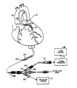
Figure 1 is a ~chematic drawing of the ultrasound
imaging syst-m for incorporating the pre~ent invention
and d-monstrating the use of the device to image a
coronary artery;
FIG 2 i~ an enlarged and partially ~ectioned view
Or a portion Or th- coronary artery Or FIG 1 showing
th- probe a~ bly of an ultra~onic imaging device
located proxi~at- to a balloon;
FIG 3 i~ a ~e~L 1~ - entation Or a corner turner
approach u-ed to generate a set of dynamic image data
for an i~ag~d r gion;
_ FIG ~ i- a block diagra~ ~e~a3entation Or a fir~t
portion o~ ~ color flow ~ or sy-tem in accordance
with a pr-f-rr d embodiment or the ~ qnt invention;
an~
P~G S ia a block diagram ~eyL~-~ntation of a
a con~ portion Or a color rlOw ~ or system in
accord~nc~ with a pref-rred embodiment of the p~ nt
inv ntion;
FIG 6 i~ an illu-trative depiction of a set of 3x3
image point~ that are sa~pled in the course o~ color
mask -p~-~ time filtering in accordance with an aspect
Or th- pre~ent invention;
FIG 7 i~ a flowchart summarizing the step~
performed by an imaging ~ystQm including a color flow
~ or for creating a compo-ite brightness-flow image
CA 02234191 1998-04-07
of an imaged blood vessel in accordance with a preferred
embodiment of the present invention; and
FIG 8 is a flowchart summarizing the space-time
~iltering operations performed by the image processing
sy~t~ of the ~L~nt invention in order to reduce noise
and color rla~ng in a di~played image
n~T~ D~8C~TPTIO~ O~ T~ P~8 DRR~D ~BODT~NT
Turning to the illu~trated e~bodiment and re~erring
to Figs 1-2, a buildup of fatty material or plaque 12
in a coronary artery 14 of a heart 16 may be treatèd in
certain ~ituations by in~erting a balloon 18, in a
deflated ~t te, into the artery via a catheter assembly
20 As illu~trated in Fig 1, th- catheter assembly 20
is a three-part as~embly, having a guide wire 19, a
lS guide catheter 20a for thr~a~n1 through the large
arterie~ such a~ the aorta 22 and a smaller diameter
cathetsr 20b that fit~ in~id- the guid- catheter 20a
Ait-r a .~ n direct~ th- guid- catheter 20a and the
guid- wire 19 through a larg- artery lea~n~ to the
aorta 22, th- ~aller cath-ter 20b i~ in~erted At the-
b~g~n~ng of the coronary artery 14 that is partially
blocked by th- plaqu- 12, th- guide wire 19 is ~irst
extendQd into th- art ry, followed by catheter 20b,
_ which includ-- th- balloon 18 at it~ tip
Onc- th- balloon 18 ha- entered the coronary artery
1~, a- in Fig 2, an ultra~onic imaging devicQ including
a prob a---~bly 2~ hou~ed within the proximal ~leeve 26
of-th~ balloon 18 provid-~ a surg-on with a cross-
~-ctional vi-w o~ th- art-ry on a video display 28 The
probe a~- bly 24 uay eo~pri~- separat- earrler and
t~q ~atarial~ a~ di~elosed in Eberle et al U S
Pat-nt Applieation Serial No 08/712,576 filed on
SeptembQr 13, 1996, whieh i~ expressly incoL~oLated
herain by raf~ r-e Howav-r~ in other e~bodiments, the
transduear array ha~k~ material may also b~ u~ed to
~yGL L th- integrated eireuitry in a flexible eircuit
de~ign of th- type diselo~ed in Eberla et al U S
Patent Applieation Serial No 08/780,437 filed on
January 8, 1997, whieh is e~L~-~ly ineoL~GLated herein
CA 02234191 1998-04-07
by reference The probe assembly 24 comprises an array
Or tranaducers fabricated from highly sen~itive
tranaducer materials Or the type previou~ly disclosed in
th- E~ rle ~t al U S Patent 5,368,037 and the Eberle
s et al patent application serial number 08/712,576 In
the illu~trated e~bodimQnt of th- invention, the
tran~ c-ra e~it 20 MHz ultrasound excitation waveform~
Howa~, oth-r suitable excitation waveform frequencies
would b known to tho~- skilled in the art The
transducers Or the-probe as~embly 24 receive the
rerlected ultra~onic wav-forms and convert the
ultrasound ec~e~ into echo waveforms The amplified
echo waveform~ rrom the probe as~embly 24, indicative of
re~lected ultrasonic wavea, are transrerred along a
microcabl- 25 to a signal ~LG~ssor 30 located outside
th- patient Th- cath-ter 20b enda in a three-part
junction 29 o~ conventional cc _~L~ction that couples
th- catheter to an in~lation source 31, a guide wire
lum-n and th- aignal yL.~ or 30 The inrlation and
guide wir- portJ 29a and 29b, reapectively, are Or
conventional PTCA catheter con~truction The third port
29c provide- a path rOr th- cable 25 to connect with the
signal proc-a-or 30 and v$deo display 28 via an
_ elD_~L~ ic conn-ctor 33
~ It ~hould b not d that th- preaent invention can
be i -~L~Lat~d into a wide variety Or ultrarolnA
i~aging cath t-r assembliea For exa~ple, the present
inv ntion ~ay b ilcoL~o~ated into a probe as~embly
~ount-d upon a ~ o~tic catheter that doea not include
a balloon In addition, the prob~ assembly may also be
mount~d in th- manner taught in Pro~ an et al U S
Patent ~,917,097 and Eberle et al U S Patent
5,167,233, the t-a~h~n~ of which are explicitly
i~o~yoLated~ in all re~pecta, herein by reference
Theae are, however, only ~xamples of varioua probe
aa~bly mounting configurationa Other configurations
would be known to tho~e ~killed in the area of
ultra~o~nA catheter probe design
_g_
CA 02234191 1998-04-07
While a number of techniques and apparatuses will
be known to those skilled in the art for obtaining
ultrasound image data for depicting dynamic ~eatures
within a field of view of an ultrasound imaging probe,
s in a pr~ferr-d embodiment of th~ nt invention, the
color flow sy-tem use~ informat$on from both static
imaging scan~ and dynamic imaging scans to detect and
image moving blood within a ve-~el Such scans are
respectiv-ly r-f-rred to her-in a- B-Scans (Brightness)
and F-Scan~ (Flow) The B-Scans and F-Scans are
constructed from a set of received radio frequen¢y
(hereinafter "RF~) signala known generally as RF A-
Scan~
Turning now to Fig 3, dynamic image data are
initially obtained by mean~ of a multi-port filter bank
approach gen-rally de~cribed in U S Patent 5,453,575,
the content- of which ar- i ~G~.ated herein by
refer-nc- In th- preferred e~bodiment, during F-Scans
eight ad~acent array ele~nt~ act a~ a single
tran~ceiv~r to f$re ultra~onic wave~ into the
vasculatur- Unlike other known imaging t~chniques,
multiple ~ a~urement~, from multipl- firings of the
tran~ducer- at a p~rticular po-ition on the tran~Al~csr
_ as~-~bly, ar- mad- b for- advancing the active aperture
~5 by on el-~-nt For each po~ition, all RF A-Scan echoes
received during F-Scan~ are ~ eA in a known manner
by an 8-port filter bank 40 to obtain eight (8)
dif~~ n~ fr~guency L ~spon~e image signal set~ This
~ign~ filtering t~chn~que, illustratively depicted in
Fig 3, i~ known by tho~e ~killed in the art as a
cla--lc corner turn-r approach
$n Fig 3, each ~x~ Le~L~aents a single digitized,
transduced echo signal rs~Al~g Al~o, each row
L~p~e-~nt~ a ~-t of rea~nq~ at variou~ distan~eq from
3s the sQt of 8 tranc1~ In a preferred emh~l~ent,
2048 such ~s-~lng~ are taken for a single transducer
firing Flnally, each colu~n re~.aaent~ the set of
digitized ~ignal reading~ at a particular depth at a
particular tran~ducer firing po~ition
--10--
CA 02234191 1998-04-07
The eight (8) distinct filters comprising the
f$1ter bank 40 produce eight (8) output RF A-Scans for
~v-ry po~ition within an F-Scan such that each one of
th- eight (8) output signals fro~ the filter bank 40
S repr~sents the signal p~ 6'l by one of the eight (8)
motion filter- tuned to produc- a ~ignal repre~entative
ot a particular spe-d of mater$al within the ~elected
field ot vi-w ot th- ultrasound i~aging device
Ideally, th- pow-r in each Or the eight GU~y~lt. gignalg
fro~ th- e$ght filter port- at any g$v-n depth
repLeFent~ the flow ~ignal for a blood flow speed range
determined by the bandwidth of the motion filter
Turnlng now to Fig 4, the eight filters are
incorporated with$n a Bea~ Former 50 operating at a 34 4
MHz clock rate It i~ noted howev-r, that in other
embodi~ent~ ot th- pres-nt invention the Bea~ Former 50
may co~prise mor- or fewer filter~ Th- y~e-ently
preterr-d ultra ound prob- include 64 ultra~ound array
elen~nt~ Thu~ F-Scan~ ar- p-rfor~ed at 64 aperture
po~ition~, ono for ~ach array el-~ent, to produce 512
dyna~ic i~aging vector~ Th~ 512 dynamic imaging
vector~ consist ot eight (8) tiltered ~ets of image data
at each ot th~ 6~ ap_L~L_ po~itions on the preterred
_ 64--le~ent tran~duc-r array
~5 It i- turth~r noted that nu~ber~ are interposed in
th~ path~ conn~cting th- ~tage~ ot the pre~ently
pr-f rre~ i ag- signal proce~-ing and display ~ystem
Th a~ nu~k-r~ identity the data path width between the
h rdN~r- ~tage~ It i~ noted that such designation~ are
g n-rally d-~ign con~iderations and other path width
configuration~ would bs known to those skilled in the
art in vi~w ot th- de~cription ot the emhc~m~nt
contained h~rein
Th~ B~a~ Form~r 50 i~ contigured to multiplex
b~tween F-Scan (aotion) and B-Scan (~tatic)
mea~urQm nt~, ~uch that ~-parate motion and ~tatic image
data trame~ ar- alternately provided by the Bea~ Former
50 During B-Scàn image data acqui~ition for a frame,
four tiring~ at each aperture position are signal
CA 02234191 1998-04-07
averaged in an Analog to Digital Converter (ADC) board
(not shown), and independent measurement~ are made at
all transmit/receive element pairs over the active
ape.~e The Beam Former 50 is configured for complete
S data s~t, i - , ph~-cA array, reeonstruetion over an
aetiv ap-rtur- of 14 el~ent~ at each of 512 veetors
during B-Sean operation Th- 512 RF A-Sean~ ou~L from
the B-a~ Form-r 50 during 8-Sean operation L ~ e3~nt 512
bea~ unifornly distribut d around a eirele
CO~L~ -pon~n~ to a eylindrieal ultra~ound transducer
array A~ eurrently configur-d for color flow imaging
operation, the pulse repetition intervals during B-Scan
operation ia ~eleeted to produe- a frame period of about
41 6 ~-e (24 frame/~ee)
Th- sa~e B-a~ For~er 50 hardware i~ u~ed during
formation of F-Sean fr~ - Howev-r, a~ previou~ly
mention d abov-, th- hardwar- is cG ~.olled to generatQ
eight (8) s-ts of motion infor~ation at ~aeh one of 64
ap_~Lu~ po-ition- Motion d-teetion i~age data are
obtained by ~horting together ~ight neighboring
tran due-r ~l-- nt~ during eaeh on- of 64 firings at a
single po-ition Eaeh s-t of two firing~ is signal
averag d (-u~ d) in th- ADC, produeing 32 independent
_ signal~ pa~--d to th- B-~n For~ r 50 for ~otion
~5 ~ inq Th- Bea~ Forner 50 i- d-~eribed in
O'~onn-ll ~t al U S Pat-nt 5,453,S75 previou~ly
inc~ a~-d h-r-in by re~e _~ In order to aehieve
2~ fr~-/- ~ operation, the pulse repetition interval
ror notion d-t-etion fra~ - ~hould be about 10 ~ee
Th pri~ry funetion~ of the portion of the flow
i~aglng ~y-t-- illu~trated in Fig~ 4 and 5 are to
sel-ct B-Scan (static i~ag-) and F-Scan (~otion
detection) infor~ation fro~ ~eparately gen-rated "flow"
and ~bri~l.t -~a~ ~can~, and to thereby produc- a
co~poaite fra~e ~G~ -nting a 512 vector, spatial
Y~ tation of blood flow region~ in a field of view
Thi~ i- acco~pli~hed by superi~po~ing portion~ of a ~low
~can i~age meeting a flow i~age ~ignal crit~ria upon an
i~age con~tructed fro~ B-Scan data
-12-
CA 02234191 1998-04-07
Continuing with the description of FIG 4, a 16-bit
Barr-l Shifter S2 receive~ F-Scan or B-Scan data
(d-p~nding on the mode of operation) directly from the
8e~r Former 50 at a 34 4 MHz clock rate As will be
ay~L~_iated by tho~e ~killed in the art, the Barrel
Shifter S2 is selectively ~u L-olled to bit shift input
data in accordance with a ~et of cG LLO1 input~ Barrel
shifted data are ouLy L to a 16 tap Finite Impulse
Respons- (FIR) filter S4
Th- FIR filt-r 54 applie- programrably ~elected
weights to each one of th- 16 tap baDed upon the scan
type For B-Scan frames, coefficients are applied to
the 16 taps to improv- the tran-~ducer impulse response
and reduce axial artifact~ seen on strong reflectors
such as ~tent ~trut~ This is accompli~hed by applying
filt-r coefficients derived by mean~ of well known
filtering method- During F-Scan frame yLoce~sing, th~
weights appli~d to each of th- tap- of the FIR filter S4
are sQlQcted, in a known manner, to narrow the pulse
bandwidth around the carrier fre~uency (nominally 20
Mhz) to both r-duce out-of-band-noise created by the
eight (8) motion detection filter~ and im~Love the
overall electronic signal to noise ratio
Th- o~L~L signal- fror the FIR filter 54, are de-
S multipl~x-d during ~ Sc~n frar ~r~ze~sing to an Image
Vector Dolay RAM 56 ~. e~sing flow images is a multi-
- stag ld ~L~-~dur- In ord-r to im~ove thr~ ghp~t, the
flo~ i~ag - ar~ in a pipeline The pipeline
proc--s-~ up to four flow image~ at a given time The
V ctor D lay RAM 56 compriDeD memory arranged to provide
four-fr~a~ m mory ~torag- that i~ refre~hed (with new
fr~r data) and read in a round-robin manner Thu~, the
Imag- Vector Dolay RAM 56 facilitate~ synchronization of
B-Scan fr~e~ with F-Scan frame~, which a~ mentioned
above ar- obtained fro~ thQ Beam Former 50 on an
alt-rnating ba~i~ Th- ~tored filtered B-Scan data are
ouL~L to an Image and Color O~L~uL Multiplexer (MUX) 58
(d-picted on Fig 5) which ~elect~ ~ignal~ for input to
a digital v-ctor ~ qr~or (not ~hown) which performs
-13-
CA 02234191 1998-04-07
additional processing on the B-Scan data such as
ringdown subtraction and detect~on The B-Scan data
will ultimately be converted into video pixel
coordinate~ and selectively combined with processed F-
Scan image data to generate a composite image displaying
a colorized image of flow region~ imultaneously with
relativ-ly static image f-ature- within a field of view
Tho ou~y~ data of th- FIR filt-r 54, corresponding
to F-Scan frame~ de-multiplexed to an F-Scan signal
~e_a~sing hardwar- chain which i~ separate and distinct
fro~ th- B-Scan image ~ignal processing chain The F-
Scan signal ~L~ ing chain is illustratively depicted
in the remai~ing portion of Fig 4 and creates a set of
color (motion) power data and a ~et of color (motion)
frequency data for each imag- point in an F-Scan frame
Initially, the de-multipl-xed F-Scan data are received
by a Beam 8uffer 60
An azimuthal FIR filt-r 62 r-duce~ artifacts
associated with b a forming in F-Scan frame~ arising
from shorting eight (8~ ad~ac-nt tran~ c~r element~
together which re~ult~ in a les- focused aperture In
the preferr-d e~CA1ment~ th- asimuthal FIR filter 62
includ-- 3 tapa to perfora b a~ smoothing across three
_ (3) ad~acent ap rtur- po~itiona (b am~) for each set of
~5 filter bank data Since ~ight (8) filter bank data set~
are a--ociat d with each apc~k~ position, the Beam
Buff r 60 i~ dim~n~ion-d to ~tor- 8x3 sets of beam data
A ~-t o~ Rang- Varying Co-fficients 64 are applied
to t~ azi~uthal FIR filter 62 during processing of the
8x3=b ~ ~ t~ compQnsat- for rang- (distancQ) dependent
azimuthal boam forming artifact~ in a mann-r known to
thos- skill-d in th- art In th- pr-ferred embodiment
of th~ pre~-nt invention, the valu~ of th- Range
Varying Co-fficient~ 6~ are modifi-d up to 16 times over
the entir- 2048 data point- within a beam It i~ noted
that whil- th- azimuthal filter stage ~ e~-nted ~y the
Beam Buff-r 60, th- azimuthal FIR filter 62 and the
range varying coeffici-nt~ 64 are de~irable, this stage
i~ not pre~-nt in another embodiment of the invention
CA 02234191 1998-04-07
Following azimuthal filtering, the color flow
y~ ing syfitem detect~ all 512 RF A-Scans
con-tituting an F-Scan frame After a rectifier 66
9~ the 512 F-Scan beams rec-ived ~rom the
azimuthal FIR filter 62, the rectified output is
provided to an FIR filter 68 th~t integrate~ the
rec-ived dat~ and pas~e~ th- filt-red data to a
decl~ator 70 The 4 1 decimation r~ c-~ the clock rate
of th- data from 34 4 MHz to 8 6 MHz and the total
length of each be~ from 2048 point~ to 512 points The
8 6 MHz output rate matches the o~L~ rate of a digital
vector prore~or (DVP) de~cribed hereinbelow
- In contra~t to B-Scan data, detected motion ~ignals
usually benefit from additional axial smoothing to
improve th- ~ignal to noise ratio Con~equently, the
ouL~ data from the d-cimator 70 i~ received by an 8
tap FIR filter 72 that provide~ additional filtering
along eacb b ~ Th- ou-~- of the 8 tap FIR filter 72,
i~ received by a compr-s-ion look-up table (LUT) 74
which reduce~ th- bit den~ity from 16 to 8 bits The
for~ of amplitude mapping is ~erely a de~ign choice and
include~ linear mapping and logarithmic mapping a~ two
viabl- alt-rn~tiv-~
_ Th- co~pr-~ed d~ta i~ routed to two distinct flow
_5 imag- proce~ing ~uL ~ which c~lcul~te a signal
power (motion po~-r) and a flo~ sp- d (motion frequency)
for each i~ag point ba~ed upon th- com~.c~ F-Scan
d~a rec-iv d from the compre~ion LUT 74. Because the
~lo~ in~oreation is displayQd as a color image, the
~ign ~ pow r (~otion power) and flow speed (motion
fr-quency) ar- r-fQrred to herein as "color power~ and
"color fr~gu-ncy~ respectiv-ly
The color power ~.~L eaents an estimate of the
signal ~ n~t~ within a motion pixQl In conventional
color Doppler (which in fact may i coL~GLate the ~L~7ent
invention), thi~ signal i8 equivalent to the area under
the curve of the Doppler sp~ To approximate this
power und-r th- pre~ent invention, th- detected outputs
of the filter bank ar- ~umm~d over all eight (8) set~ of
-15-
CA 02234191 1998-04-07
data for a particular aperture position corresponding to
th- eight (8) filter bank bins An eight line buffer ~6
and an accumulator 78 perform the summing operation on
th- compressed F-Scan data Whil- not shown in the
drawing~ accumulator 78 output i8 normalized by bit
~hifting the accumulated output 3 bit~ to render an 8
bit valu- (color power) that i~ r-c-ived by further
color imag- proc-~sing hardwar- illu~tratively depicted
in Fig 5
Not- that th- detail- of th- compres~ion LUT 74
aff~ct the su~ obt~n~~ in th- accumulator If pure
logarithmic com~ 6ion i~ used, then the sum over the 8
filter bank data set~ for a particular aperture is
equivalent to a product over the detected filter
output~ Conv-rsely, if linear com~e3aion i~ used,
th-n all 8 filter bank data s-t~ arQ ~imply ~e~ t~u~
incr-a~ing th- influenc- of noisy filter outputs having
low flow amplitud-- Thu-, an a~o~.iate com~ qion
function mu-t b car-fully ~el-ct d ~o that th- sum over
filt-r bank- approximate~ the area under th- curve of a
Doppl-r ~pectrua
Beforo d-~cribing the circuit~ for generating
"color fr qu-ncy~ valu-~ for F-Scan frames, it i~ noted
_ that, inde d, det-ct d motion F-Scan~ also can be u6ed
to e~timat- th- ~p-~d of flowing blood Thi~ i~, of
cour~o, a ~-ry complicated prob~e~ becau~e flow at the
g~ - ~pe d can ~how up in diff-r-nt filter bank bin~
ba~ d on a nuib~r of factor~, including flow direction
(~n-plan v-r~u~ out-of-plane), local speckle
c~ ract-ri~tic~, filter leakage, and noi~e One method
to dlffer-ntiat- between fast flow and slow flow is to
count th- nu~b r of filt-r bank bin~ above an
e~tabli~h~d noi~ thre~hold
Furthermore, bQcau~- each filter in the bank may
hav ~lightly different characteristic~, especially the
filter clo~e~t to z-ro fr-guency, the noi~s threshold is
independently s-t for each filter bank bin At every
range ~ampl-, each of th- eight (8) filter bank signal~
i~ compared in a Thre~hold On EnvelopQ 80 and a logic 1
-16-
CA 02234191 1998-04-07
is assigned to each of the eight (8) filter bank signals
~x~ ~A~ng the threshold value provided by an adjustable
Env-lop~ Threshold signal input The resulting 8-bit
r-~ult of the thre~hold comparison i8 stored in an 8x512
(slngl-, d~ei~ated beam) buffer 82 Eaeh bit in the 8-
bit word L~PL~ nt~ the ou~p~L of the thre~hold test ~or
a coLLasponding ~ilt-r bin Eaeh of the 8-bit words i5
used to aee-~ an entry in a Color Fr~queney LUT 84 In
an ~~bodlu nt of the pr-~ent invention th- yL~La~med
entri-~ of th- Color Frequeney LUT 8~ nominally estimate
the flow (~otion) fr-queney by counting the number of
bits set in each input word That is, the more bits
set, the higher the flow valoeity Other, more a~vanced
Color Frequency LUTs will likely comprise complicated
lS algorithv~J sine- many different typ-s of patterns can be
expeeted for v-ry si~ilar flow condition~ which are be~t
resolved by elinieal trial- under a wide range Or
eondition~
Fig S sehenatie~lly d-piet~ signal ~ ssing
h~rdwar- ~Lo~u~ably eontroll-d to eombin- eolor
(motion) pow r, eolor (motion) frequeney and B-Sean
fra~o data into a singl- flow estimate In aeeordanee
with on- a~p~et of a pr-ferrQd embodi~Qnt of th- ~ nt
_ invantion, ~ignal stability i- aehi-v-d for both the
_S eolor pow~r ~nd eolor fr quoney valu-~ over timo by
m~an~ of ~ingl- fe~dbaek Infinit- Ilpul~e Ra_~; 7
filt-r/buff r~ 85 and 86 respeetively Eaeh
f~lt-r/buff-r 8S and 8C includes a 64x512x(1 word)
buf~ ~ for ~torin~ th- previou~ly calculated value~ for
~n ntir- F-Scan fra~- Color Pow r Psrsistenc~
Co ffici-nt Xl and it~ a~sociated coefficient M provide
a fir~t p-r~i~tenc- factor Color Freguency Persistence
Co~ffici-nt X2 and its a~ociated coofficient N provide
a s-cond p~r~i~t-nce factor In accordanc- with an
a~pect of the new imaging ~y~t-~, th- fir~t and second
p-r-i~tenc- factor- are ind-~ n~ntly d-signated
It i~ not-d th~t in the above d-scrib d embodiment,
th- fir~t and ~-cond persi~tence factor~ are each
~tabli~h-d by a combination of two variables In other
-17-
CA 02234191 1998-04-07
embodiments of the invention, the first and second
per~i~tencQ factors may consist of single adjustable
variable~ or other combinations of variable~ -- even
variable~ co~mon to both filters An example of such a
sy~t~ is the instance when NM~ and "N~ are the same
value
Th- op-ration of the accumulators within
filt-r/buffer 85 and filt-r/buff-r 86 i~ synchronized
with th- input value- on th- lin-- lab l d ~Color Power"
and ~Color Frequ-ncy~ by ~ an- of co ~ ol lines "PWRRDYn
AND ~FRQRDY~ fro~ th- accu~ulator 78 and th- Color
Frequency LUT, r-spectively Th- structure, functio~
and op-ration of th- filt-r/buff-r- 85 and 86 will be
known to tho~ skilled in the art
A relativ-ly long ter~ per~i~tence value (favoring
little ~- a fro~ a previou~ calculated value) should
b- de~ignated for color pow r ~ignal filtering (via
prop-r de~ignation- of valùe~ for Xl and M) ~ince this
valu- ~hould not fluctuat- nuch over th- cardiac cycle
b cau-- it i- a ~ a-ur- of th- ~cattering co-fficient
fro~ moving blood Color pow r, th~r-fore, ~hould be
averaged ov r long periodc, compared to th~ cardiac
cycle, to ~~ooth noi~- and ~p ckl- fluctuation~ The
color pow~ co ffici-nt Rl and thc valu~ "M~ are
~5 g-n-rally ~-1 ct d to provid- a p r~i~tenc- p-riod of
approxi~at-ly 1 ~econd Th- first p~r~istence factor,
det-ruin-d in tho pr-f-rr~d ~ bodim~nt by the value~ of
Rl and ~, 1- ad~u~tabl- to facilitate ad~u~tment by a
u~r during both te~ting and u~e on a patient
In contr~at, color frequency i~ expected to change
over th~ cardiac cycle because it i~ related to ~low
~p-od Thu-, valu-~ for K2 and N ar- de~ignated to
acco~pliJh th- ti~-baa d ~moothing with a p-rsistence
period ~hort-r than a heartbeat The color frequency
-p-r~i~tenc~ factor d-teroined by the coefficient K2 and
th- valu- ~N~ are g-n rally ~elected to provide a color
fr~qu-ncy per~i~tenc- period of 40-100 ~ill~eco
Th- ~econd per~i~tenc- factor, deter~ined in the
preferred e~bodi~ent by th- value~ X2 and N, i~
-18-
CA 02234191 1998-04-07
adjustable to facilitate ad~ustment by a user during
both testing and use on a patient
Th~ eombined frame averaged 8-bit color power and 7
bit color frequency signal~ are provided by accumulator
output~ fro~ th- filter/buffer~ 85 and 86 respectively
to a Color Value Generation (CVG) LUT 87 The CVG LUT
87 eombine~ motion pow-r and ~otion frequeney
information to produe- a 7 bit eolor signal having both
eolor and brightne~- eoeffieient~ A~ in the ease of
th~ Color Frequeney ~UT 84, th- CVG LUT 87 ean be
y~ ammed in ~any way-, e g , ignore motion frequeney
information and produee only a motion power signal,
ereate a ~ig~oid thre~hold on the power signal to
control luminanee and vary color aeeording to the eolor
frequeney value, ete The de~ignation of partieular
output values for speeified input~ to the CVG LUT are
primarily design eon~ideration- whieh may be re~olved by
elinie~l trial- -
Th- CVG ~UT 87 ou~ a ~ingle eolor ~ignal to an
azi~uthal int-rpolator 88 F-Sean imaging veetor~ are
produeed on C~ bea~; wh-rea~, 8-Sean~ use 512 beams
The azimuthal interpolator 88 ereates 512 beams from the
64 provided b a~ u~ing s~anA~rd linear interpolation
T-; n~~~ int-rpolation i~ adequate bee~ the azimuthal
~5 radiation patt-rn a~oeiated with 8 ~lement~ tied
together ia likely guit- broad, even with azimuthal
filtering of RF data b for- flow ~ ing The
int rpolator 88 tran~it~ the re~ulting interpolated
i~a~ d~ta to a single fram- flow data skew 89 where the
dat~ i~ bu~f-red and ti~e ~k~w~ to produee the 7 bit
eolor valu- ~ignal ap~Lo~iate for o~L~L to a Digital
Veetor ~ or (DVP) and ~ub~equently to a ~ean
eonverter whieh i~ not ~hown but is deseribed in the
O'Donnell et al U S Pat-nt 5,453,575
The top path of FIG 5 aehematieally depiet~ the
imag- ~L~_ ~ssing hardwar- for logieally eomparing F-Sean
eolor power and B-Sean ~ignal level~ to produee a 1 bit
eolor ma~k indieating whether a point on a partieular
bea~ in the final 512x512 ~low frame ~hould be
--19--
CA 02234191 1998-04-07
designated as a color flow image point The single bit
mask de~ignates the source (B-Scan or F-Scan) and manner
of displaying (gray scale or color) a particular image
point on the 512 beams comprising 512 points each The
-sign_l proce~ing schematically depictêd along the top
path of FIG 5 significantly reducQ~ ~flashingn -- that
is, th- ~-Q~ingly random turning on and off of color
pixel~ during th- display of a composite flow/~tatic
tis~ue imaqe
A color power threshold detector 90 compares the
color power ~ignal ou-~- fro~ th- accumulator 78 to a
designated thre~hold level T2 to produce logic signal B
Logic signal 8 is 1 if the color power signal meets or
e~cee'R th~ thre-hold level and 0 if the color power
signal is b~low the threshold level If logic signal B
is 1, then the point i~ identified a~ a potential color
flow image point If logic signal B is 0, then a
particular image point iJ not dynamic (as determined by
the compari~on to T2) and therefor- is designated to
display B-Scan data
In parall-l to th- color power level detection, B-
Scan data (512 beam~) input from a B-Scan data buffer 91
or the DVP ar- compar~d to a thre~hold 1eVQ1 T1 at B-
_ Scan thre~hold detector 92 Th- logic signal produced
by th- B-Sc~n thre~hold detector 92 i~ 1 if the B-Scan
value for an i~ag- point is below a de~ignated threshold
lev~l and 0 i~ it i~ abov- th~ threshold (indicating a
r~gion of ti~ue) If thi~ bit i~ 1, then the B-Scan
1eY ~ i- low and the point is identified as a possible
blood flo~ region A frame identification signal
conn~cted to the "enabl-~ input of a Fr_m~ Mask Buffer
94 8Ql~tCtiV ly pa~ the threshold signal provided by
the B-Scan threshold detector 92 to the Frame Mask
Buffer 94 to ensure that only B-Scan data is involved in
producing logic signal A stored in the Frame Mask Buffer
94
For a given image point within an image, "color
flow~ statu~ i~ design~ted when both signals A and B are
logic 1 That is, an image point must have both
-20-
CA 02234191 1998-04-07
significant motion (color) power and low B-Scan
intensity for it to be identified as blood Logic
signal A, however, is on a 512 beam by 512 image
point/bea~ grid; wherea~, logie ~ignal B i~ on a 64 beam
by 512 image point/bea~ grid Therefore, the 64 input
"B~ b am~ to a Data S~ew and Comparator 95 are expanded
by replieating ~~eh of th- 64 logie ~ignal B beam~ 8
time~ and then p-rforning a logieal AND operation
bcL~en th- logie ~ignal A bea~s and th- skewed logic
signal B be~ The result of th- above deseribed
proee~sing i~ ~tored in a Color Mask Buffer 96
Note that the eolor power threshold decision is
performed b fore the color power signal is frame
averaged by th~ filter/buffer 85 in order to prevent
spatial sm aring of flow information (i e , in a moving
environment, temporal averaging al~o spaee averages)
If tho flow mask infor~ation stor d within the Color
Ma~k Buffer 96 were to b~ derived fro~ temporally ~
averaged eolor power data fro~ the filt-r/buffer 85,
then wall-lu~n interfae-~ would likely blur ~o avoid
this proble~, th- eolor ma~k is eomputed in real-time in
order to a~sur- ~patial alignment between B-Sean and
color infor~ation provided to th~ Data Skew and
Comparator 9S
Th color ~a~k infor~ation stored within th~ Color
Ma~k Buffer 9C ~ay inelud- ~ueh noi~o beeause it is
e~ti~at-d on a fram by fram ba~is Noisy eolor mask
data, in turn, pro~l~r~ eolor flash artifacts To
r~duea th- inei~r-e of eolor fla~h artifaets, eolor
~a ~ data fro~ tha Data Skew and Comparator 95 for the
pr-e~ing eight (8) B-Sean and F-Sean frames is buffered
in tho Color ~a~k Buffer 96 A Sp~e ~ime rank order
filter 97 filt-r~ th- eolor bit mask data over spaee
(c-ntsred around an image point of intere~t) and most
reeent eolor ma~k fra~e~ generated by the syctem
Generally, a linear filter, sueh a~ a low pas~
filt-r, rQdue-s flash artifaet~, but al~o blur~ wall-
lumen edg-~ In cGI~La~t~ a non-linear rank order
filter, ~ueh a~ for example a median filter, reduees the
-21-
CA 02234191 1998-04-07
likelihood of popcorn noise (flashes) in either space or
time while preserving both spatial and temporal edges
Th- Spac--Time rank order filter 97 generates an output
bit ~ask by applying a 3x3x8 spac--frame filter to the
data ~tor-d in the Color Ma~k Buffer 96 A spatial, L=8
co~b i8 u~ed to ~ilt-r over th- samQ ~patial extent as
the original color data ~or 8 fra~e~
Turning briefly to Fig 6, th- 3x3 space referenced
on a ~ingle i~ag- fra~ during th- space-time filtering
is illustratively depict-d As ~hown in the figure, a
first set of three point- are referenced from a first
radial bea~ containing the current i~age point X and two
ad~ac-nt i~age point- Th- re~aining six imaqe points
are referenced (3 each) fro~ two ad~acent beams at
di~t~nce~ ~oLLe_ponding to the three point~ on the first
radial bea~
The value of a particular ma~k bit in the 512 bea~
by 512 point~/bea~ bit mask i~ obtained by su~ming the
value~ of th- bit~ contain-d within the 3x3x8 space-time
~volu~ ~ A ~axi~u~ value for the ~u~ d bita i~
sQventy-two (72) In a particular embodiment the rank
order filter 97 i~ configured as a median filter In
thi~ particular e~bodiment, if th- ~um~ed value of the
_ ~ask bit~ ia ~qual to or greater th n a thre~hold value
~S of thirty-~ix (36),-then the rank order rilter 97,
acting a- a ~ dian filter, o~ a loglc on- (1) value
for the ~a-k bit, thereby indicating that the particular
iu go point i~ to b~ colorized
It i- not-d that while a 3x3 spatial kernel has
b ~n ~el-ct~d, other suitable spatial kernels may be
u~ to provide more robust filtering The choice
of eight (8) frame~ of ma~k data i~ a ~L~7~ntly favored
choice for time filtering, but other values are
cont~plat~d to fall within the ~cop~ of the pL~ s-nt
invention Finally, th- choice of a thre~hold of
thirty-~ix (36) (i e , one-half the maximum -- and thus
a median f ilter) for the rank ~Ld_L filter 97 may be
modlfied in accordance with the needs or preferences of
-22-
CA 02234191 1998-04-07
a particular clinical operation or imaging system
hardware by means of a THRESH signal input
When the threshold is modified to a value other
than the median value, while still a "rank order"
filter, thia filter is generally considered not to be a
"median" filter since th- determining point would not be
the ~iddl- of the range of value~ Of course in
instance~ wh-r- the s-l-cted thr--hold value is
sub-tantially the sa~- a- th- m dian value, one may
refer to such a filter a~ a ~sub-tantial median filter "
For each of the 512 imag- beams and each one of the
512 image points on a single image beam, a Color Vector
Delay RAM 98 combines the ~ingle bit of color mask data
provided by the Space-Time rank order filte~ 97 and the
lS 7-bit color value data fro~ the single frame flow data
skew 89 to generate a color image frame The color
imag- data points of th- color image frame are each
stored a- an 8-bit word Th- ouLyuL of the CVD RAM 98
act~ a~ on~ data input to an Image and Color Ou~yu~
Multiplexer (~UX) 58 Th- oth-r data input to the Image-
and Color O~L~ Mux 58 is the 16 bit B-Scan data from
the FIR filter 54 and image vector delay RAM 56 of FIG
4, i e , th- Vector 8ypa~- ~ignal
_ U-ing a Color Fr~ control aignal to determine
wh~ch data type (B-scan or color flow) to pa~-, the
ou~ of th- MUX 58 i- passed to the DVP If a color
fra~ i~ ~-lect-d, then the DVP passes the proçe~
color i~aga data stored within the Color Vector Delay
RA~ 98 to th- display ~ f~or Alternatively, if a
color fra~ is not selected, then the DVP takes the RF
filtered raw B-Scan data and p~_a~ 7 it (i e ,
deci~ate~ th 2,048 beau sample~ into 512 samples) and
pAqs~ th- B-Scan data to th~ display proce~or for scan
cG v~r-ion, etc The decimat d B-Scan data is stored in
the B-Scan data buff-r 91
Cu~nt DVP- y~oca~ B-Scan data and pass the
resultant to th- di~play processor for scan conversion
and display The DVP according to th- invention,
receive~ both raw B-Scan and processed color flow
-23-
CA 02234191 1998-04-07
fram-s As mentioned above, the DVP processes raw B-
Scan data if the color frame bit is logic 0 by
d-cimating the input A-Scan data into 512 samples per
b- ~ for each of the 512 beams The processed B-Scan
data aro then p~t- ~ on to the display procesqQr If
the Color Fr~me control bit equals logic "1~, then Color
frame data ar- pa~sed directly from the Color Vector
Delay RAM 98 to th- display y~e~ or without processing
by th- DVP
Both fram type- ar- buff-r-d and their location
followed throughout th- ~ntir- ~.e~ ing chain That
is, consecutiv- B-Scan and color frames entering the
display y~e:e~-or should be temporally aligned The
di~play proce~sor separate~ and ind-p-ndently buffers
color flow and B-Scan frames For every ouL~ pixel
after scan conversion (from beam- to pixels), the color
bit specifi~- whether to display the color value
according to a color map or th- B-Scan value according
to a gray scal- map
It should bo not-d that the color bit is not scan
conv-rted u-ing a linear operation such as bi-linear
interpolation For each ~ pixel displayed on a
rectilinear grid, a simpl- logic operation should be
_ perform d on th- color bit- for th- associated nearest
neighbor pix-l- on the polar grld to d-cide if the
ou~ pixel i~ color or gray scale
~a~ing de-cribed system hardwar- and the functions
p~ ~or~ d by the hardwar- in order to generate, for
lat-r di-play, a col-or flow image during ultrasound
intra~a-cular imaging, att-ntion i~ now directed to Fig
7 which ~u~ arize~ the step~ perfor~ed by this
ultrasound imaging syste~ to generate the color flow
- image based upon ultrasound echo information At some
point prior to g-nerating a color mask, a B-Scan (static
image) fram i~ obtained in close temporal proximity to
the F-Scan (motion imag-) frame It is noted that while
the ~tep~ are ordered in a particular manner to
facilitat- a description of the preferred emhq~iment of
th~ ent invention, thos- skilled in the art will
-24-
CA 02234191 1998-04-07
readily appreciate that with respect to a number of the
step- described below, the order in which the steps are
p-rrormed i~ not restricted to the illustrative order
deplcted in th- flowcharts
At step 100, the ultrasound imaging sy~tem acquires
raw digitized RF A-Scan data Sixty-four (64) echo
sample~ (2048 points) are taXen at each of 64 positions
along th- peri~et-r of an ultra~ound tran~ducer
as~embly N-xt, at step 102 these F-Scan data are
filtered by the color flow proce~or hardware
schematically depicted in Fig 4 in order to reduce
noise in the color flow raw data
At ~tep 104, the buffer 76 and accumulator 78
receive color flow data and sum the motion power for
imag- point~ ov-r th- ~ight motion frequency bin~ in
order to obtain a total color power for each image
point During ~tep 106, a thre~hold envelop- i~ app}i~d
to th- ~a~ color flow data ~L._er~-~ during ~tep 104 in
order to ~stimat- th- flow ~peQd from the eight (8) sets
of imag- point data coLLeFronAin~ to the eight (8)
filter bin~ Aft-r thrs~hold y~-e~ing~ the resulting
data are appli-d to a color frequency LUT 84 to render a
color frequ-ncy valu~ for each imag- point
_ In accordanc- with a particular aspQct of the new
~5 imaging ~ tho~, during st-p 108 th- color power data
obtained during ~tep 104 ar- fr~m a~elL~ 1. In other
word-, th- color power data of th- ~rc--~t fram- are
co~bin d with a previous averaged color power data set
to rend-r a n-w averaged color power data ~et The
contsibution Or th- new color power data to the averaged
color pow r data i~ determinQd by a fir~t per~i~tence
factor which i~ d-ternin-d by value~ for the coefficient
Rl and th- valu- of ~ Th- first per~i~tence factor is
preferably ~-l-cted to filt-r ~hort term change~ since
the color power ~hould b- relatively ~table over short
periods Or ti~e
During st-p 110 the color freguency data obtained
during ~tep 106 i~ co~bin-d with ~ previou~ averaged
color frequency data ~et to rend-r a new averaged color
-25-
CA 02234191 1998-04-07
frequency data set. The contribution of the new color
freguency data to the averaged color frequency data is
det-r~ined by a second persi~tence factor which,is
determined by values for the coefficient K2 and the
value of N. The ~conA persistence factor is designated
independently from the ~econA persiatence factor,
thereby enabling a u~er to specify a value for the
-- :n~ persiatence factor which enablea the average
color frequency to pass short term changes thereby
enabling a uaer to observe variations in blood flow
speed during the different stages of a cardiac cycle.
The values for K2 and N, in combination and applied to a
filter as illustrated in Fig. 5, specify a signal
persistence period significantly smaller than the
persistence period specified by the first persistence
factor.
Next, during step 112 the averaged color power and
averaged color frequency data are combined for each
image point over a color flow frame and submitted to the
color value look-up table 87 in order to render a color
image approximation. The color image a~oximation
includes both color and brightness values for each image
point. ~;w~ , since these data are only specified for
_ 64 beama ex~-nA~ng from a circular perimeter
~5 ~G~ rQ~A~ng to an ultrasound tran~Al~cer assembly, at
step 114 the 64 beama are interpolated to 512 beams in
advance of acan CGI~Ve~ aion of the beams into display
coordinates.
At step 116, a color mask for the current frame
generated by the color flow proce~Qr in accordance with
the sub-stepa summarized in Fig. 8 is applied to the
color flow image data computed by the azimuthal
interpolator 88 (during atep 114). Applying the color
mask to the azimuthal interpolated color flow data
rendera a color flow image that specifiea for each
particular image point whether to use the color flow
image data (if the color mask specifies "flow datan) or
alternatively to uae the gray scale static image data
computed from B-Scana (i.e., disregard the computed
-26-
CA 02234191 1998-04-07
color flow data for this particular image point) if the
color mask specifies "static data"
At step 118 the Image and Color Output MUX s8
s-l-ctively pa~sQs either color flow (F-Scan) image
-5 fra~o data or raw, ~tatic (B-Scan) imag- frame data to
the Digital Vector ~L~ _ e--or ba~-d upon the value of-the
Color Fra~ CO~LLO1 signal In th- ca~- of a color flow
image fra~ , th- DVP pa~e~ th- data on to th- display
~._e3~0r for further ouL~uL ~L._e~ing In the case of
B-Scan imag- frame de~ignation, the DVP d-tect~ the RF
A-Scan~ and d-cimate- th- resultant signal to produce a
512 sample beam for each of the 512 beams constituting a
B-Scan fram- Thereafter, the DVP stores the 512 beams
in th- B-Scan frame buffer 91 and r~qe~ th- proc c~c~
B-Scan imag- data on to the display ~L.~ ?r
Next, at ~t-p 120, th- display ~L ~ _ - ~qcr combineg
th- buff-rQd, ~L-~ -A 8-Scan and Color Flow images
receiv d during ~t-p 118 into a co~po~it- imag- Thi~
is achi-ved by u~ing every imag- point fro~ the Color
Flow imag- point having it~ color bit turn-d "on~ to
indicate that th- particular i~age point i~ to b~
di~played in color All the re~aining imag~ points are
fill d in u ing th- cGLL_~ponding i~ag- data from the B-
_ Scan i~ag- fra~ It i- noted that th- B-Scan i~ag- i~
~ynchroniz-d with th- color flow i~ag-, by mean~ of the
I~ag- V-ctor D lay RAM 56, in ord r to ~n~ur- that the
two ~-t~ o~ i~ag- data cGLL~--ponA to raw i~ag- data
- acquir-d within a v-ry short ~a~- timo p-riod (e g ,
ad~ac nt fr~ period~)
Di-play ~L~_ ~sing of th- composite Color Flow/B-
Scan i~ag con~nu-~ during step 120 with th~ conversion
of the co~po-it- imag- point~ fro~ polar coordinate to
display pix-l coordinate~ for ouL~L upon the display
28 For ev-ry ouL~L pix-l aft-r scan conversion, the
color bit ~p-cifi-~ wh-th-r to di~play the color value
according to a color ~ap or the B-Scan value according
to a gray ~ap Th- scan conv-rsion is not a linear
op-ration, ~uch a~ bilinear interpolation Instead, for
each pix-l, th- color bit~ for th- near-~t n-ighbor~ on
-27-
CA 02234191 1998-04-07
the polar grid are observed to determine whether to
designate the output pixel as color or gray scale At
this point, di~play pL~ ing i~ complete and the
compo-it- image is pres-nted upon the display 28 It is
furth-r noted that the ~teps 100 to 120 are performed
with suffici-nt speed to present a substantially real-
tim- i~age of a region, such as an i~aged coronary
vessel.
Turning now to Fig 8, the steps are summarized for
filtering color ~a-k bit~ in both ~pace and time in
order to eli~inate color ~flash ~ During step 130, the
B-Scan threshold detector determine~ for a frame of B-
Scan image point data whether the ~ignal level for each
particular i~age point is too high to L ~ ent blood
flow If the B-Scan signal reaches a specified
threshold (Tl), then the imag- point i~ de~ignated
~color off~ to indicat- that the particular image point
is likely not blood This comparison is p-rformed for
all B-Scan data and stored in a 512 beam by 512
point/b a~ buff-r
At ~tep 132 ~ color mask is constructed for a frame
wherein a maa~ bit is d--ignated "color" for a
particular point if th- B-Scan ~ignal level did not meet-
-~ th- Tl thr-shold and the color power meQts or eYc~
the color pow r tbreshold T2 Otherwi~e the mask bit
for th- particular point i~ ~-t to ~no color ~
Next, at ~t-p 13~ th ~a-k bit~ are stored in a
color ma~k fram buffer Such color mask frames are
buff r-d for a ~LLe~ a~ well a~ a set of previous
fra~ - In th- preferred emh~A~ment of the present
inv ntion, th~ total numk~r of color mask frames
buffQred e~uals eight (8)
In accordance with a particular aspect of the
pre-ent invention, during st-p 136 the buffered color
mas~ data i~ filterQd in space and time to render a
filtered color m-a~k designating whether a particular
imag- point is a color flow i~age point or alternatively
a gray ~cale ~tatic imag- point In a preferred
emhcA~m-nt of tho ~L~s-nt invention, the spatial extent
-28-
CA 02234191 1998-04-07
for a particular image point comprises the point of
intsrest and each one o~ its eight (8) ad~acent
neighbors (in a plane) In addition to the two
din-n~ional plane rendering nine (9) color mask bits,
S the f1lt~ring occurs ov-r time by including the eight
(8) most rocently rend-r-d color ~sk~ stored in the
color ma~k buff-r 96
The pr-f-rred filt-rinq m thod compri~e~ summing
the valu-- represented in th- 3x3x8 ti~e-~pace region
and comparing the re~ult to a m dian value 36 or some
oth-r specified thre~hold in ord-r to determine whether
to designate ~color on~ or ~color off~ for the image
point of intere~t Th- re~ulting ~iltered image point
is provided for further image processing a~ described in
step 116 above
While th- invention ha~ been described in
conn~ction with certain pr-ferr d ~hoA~ments, there i~
no intent to li~it it to tho~ bodiment~ On the
- cv ~ary, th- pr--~nt inv-ntion i~ applicabl- to other
imaging ~ thod- which provid- a co~bination of dynamic
and statlc i~age data Por ~x~ple, the ~ nt
inv-ntion ~ay b advantageously i,~o~o~ated into an
imaging ~y-t~ wherein th- dyna~ic and ~tatic image data
i~ obtain-d via Doppl-r i~aging t-ch~uQ~ The intent
i~ to cov r all alt-rnat~vec, ~odi~ication~, and
equival-nt~ includ-d within th- spirit and scope of the
inv~ntion a- d-fin~d by th- appendQd claim~
-29-