Note: Descriptions are shown in the official language in which they were submitted.
CA 02234787 1998-04-1~
METHOD OF MANUFACTURING
A MEDICATED POROUS METAL PROSTHESIS
BACKGROUND OF THE INVENTION
Field of the Invention:
This invention generally relates to a medicated prosthesis or implant.
More particularly, the invention relates to a medic~te~l intra-vascular prosthesis, such
as a stent, that is radially expan~ble in the vasculature of a patient and delivers a
therapeutic agent to the site of the implantation.
Description of Related Art:
Stents are generally cylindrically-shaped prosthetic implants which
function to hold open and sometimes expand a segment of a blood vessel or other
anatomical lumen. They are particularly suitable for supporting and preventing a torn
or injured arterial lining from occluding a fluid passageway. Intravascular stents
increasingly are useful for treatment of coronary artery stenoses, and for reducing the
likelihood of the development of restenosis or closure after balloon angioplasty.
The success of a stent can be assessed by evaluating a number of factors,
such as thrombosis; neointimal hyperplasia, smooth muscle cell migration and
proliferation following implantation of the stent; injury to the artery wall; overall loss
of luminal patency; stent diameter in~vivo; thickness of the stent; and leukocyte
adhesion to the luminal lining of stented arteries. However, the chief areas of concern
are early subacute thrombosis, and eventual restenosis of the blood vessel due to
intimal hyperplasia.
Therapeutic pharmacological agents have been developed to improve
successful placement of the stent and are delivered to the site of stent implantation.
Stents that are of a common metallic structure were previously unable to deliverloc~ ed therapeutic pharmacological agents to a blood vessel at the location being
treated with the stent. There are polymeric materials that can be loaded with
CA 02234787 1998-04-1~
therapeutic agents including drugs or other pharmacological treatments which agents
then can be released for drug delivery. However, these polymeric materials may not
fulfill the structural and mech~ni~l requirements of a stent, especially when the
polymeric materials are loaded with a drug, since drug loading of a polymeric material
can significantly reduce the structural and mechanical properties of the polymeric
material.
It has been known in the art to coat a metallic stent with a polymeric
material and to load the polymeric material with a drug. Alternatively, stents of
polymeric materials have been reillforced with metal structure. These stent designs
have the strength n~cess~ry to hold open the lumen of the vessel because of the
reinforcement contributed by the metal. Stents made of both polymeric material and
metal have a larger radial profile because the volume occupied by the metal portion of
the stent cannot absorb and retain drugs. Reducing the profile of a stent is desirable
because doing so increases the in vivo diameter of the lumen created by the stent. Thus
it is desirable to configure a metallic stent to deliver drugs to the blood vessel walls
without substantially increasing the profile of the stent. The present invention meets
these needs.
SUMMARY OF THE INVENTION
Briefly and in general terms, the present invention is a method of
m~mlfactl1ring a medicated prosthesis. The method comprises providing a porous
metal material having a plurality of porous cavities or pores, forming the material into
a prosthesis having a plurality of pores, and loading therapeutic agents into the pores of
the prosthesis. In one embodiment, the prosthesis is a stent for implantation into a
blood vessel, biliary duct, esophagus or other body lumen. In one embodiment, the
method comprises sintering metal particles including spherical particles, filaments or
fibers into a wire, a sheet or tube. Then the wire, sheet, or tube further is
m~mlf~ctllred by forming the stent from the same. Sheets or tubes can be formed into
stents by chemical etching or laser cutting the same according to a stent pattern. In
CA 02234787 1998-04-1~
another embodiment, the sheet is formed by weaving metallic fibers and sintering the
metallic fibers into a metal wire or a sheet.
In yet another embodiment, a sheet of stent material is formed in a
plurality of layers. A layer of large ~ m~ter particles are arranged in a first hori_ontal
plane. Two layers of small diameter particles are arranged on both sides of the plane.
The particles are sintered into a sheet of particles that has a large core formed of large
diameter particles that is sandwiched between two layers of small ~ meter particles.
Similarly, a sintered stent wire can be formed by arranging large diameter particles
along a first axis and then arranging small rli~m~ter particles radially outward from and
coaxial to the large diameter particles. Then, the particles are sintered to form a stent
wire that has a substantially porous central cavity and an outer layer that has smaller
pore diameter.
In still another embodiment, the method of forming a stent comprises
arranging a sheet of solid metal between two layers of particles. The particles then are
then sintered to both sides of the sheet. Similarly, the particles can be sintered to one
side of the metal sheet. Alternatively, particles can be oriented radially outward from a
solid metal wire and sintered into a partially porous wire. The partially porous wire
and the stent with a sheet metal core are believed to improve the strength of the overall
stent.
According to one embodiment of the present invention, a therapeutic
agent can be loaded into the pores of the stent by immersing the stent in a liquid
solution cont~ining the therapeutic agent. The stent is immersed for a period of time
sufficient to permit the therapeutic agent to be absorbed into the pores of the stent. The
therapeutic agent may be any number of drugs or chemical agents that treat arterial
diseases and/or treat or tend to minimi7.e or counteract the side effects which sometimes
accompany stent implantation.
In yet another embodiment of the invention the method includes coating
the stent with a polymer. The polymer itself may be loaded with one or more
therapeutic agents or may be applied to delay the release of medicine or otherwise to
control the rate at which the therapeutic agent will diffuse into the body.
CA 02234787 1998-04-1~
These and other features of the present invention will become apparent
from the following more detailed description, when taken in conjunction with theaccompanying drawings which illustrate, by way of example, the principles of thepresent invention.
BRIEF DESCRIPTION OF THE DRAWINGS
FIGURE 1 is a longitll(lin~l sectional view of a blood vessel with stent
m~nllfaçtllred according to one embodiment of the present invention.
FIG. 2 is a porous stent wire or strut in a partially m~gnified, partially
cut-away perspective, m~mlfaçlllred according to one embodiment of the present
invention.
FIG. 3 is a m~gnified, cross-sectional view of un-sintered, packed
particle.
FIG. 4 is a porous stent wire or strut in a partially m~?~nified, partially
cut-away perspective, m~mlfa~tllred according to one embodiment of the present
mvenhon.
FIG. S is a porous stent wire or strut in a partially m~gnified, partially
cut-away perspective, m~mlf~ctured according to one embodiment of the present
mventlon.
FIG. 6 is a cross-sectional view of a stent wire or strut m~mlfactured
according to one embodiment of the present invention.
FIG. 7 is a cross-sectional view of a stent wire or strut manufactured
according to one embodiment of the present invention.
CA 02234787 1998-04-1~
FIG. 8 is a sheet of sintered stent mAnllfactured according to one
embodiment of the present invention.
FIG. 9 is a stent formed from a sheet of sintered metal according to one
embodiment of the present invention.
FIG. 10 is a cross-sectional, partially cut-away view of a sheet of
sintered metal mAmlfactured according to the principles of one embodiment of thepresent invention.
FIG. 11 is a cross-sectional view of a stent wire or strut mAnllfa~tllred
according to the principles of one embodiment of the present invention.
FIG. 12 is a cross-sectional view, partially cut- away of a sheet of
sintered metal mAmlfactured according to the principles of one embodiment of thepresent invention.
DETAILED DESCRIPTION OF THE PREFERRED EMBODIMENTS
Referring to FIGURE 1, the prosthesis of one embodiment is a porous
stent 12 that is radially e~p~n~able a~ainst the walls 14 of a vessel 16. A therapeutic
agent is loaded into the pores 18 (See FIG. 2) of the stent. When placed in the
vasculature, the therapeutic agent is delivered to the tissue that comes into contact with
the stent. The stent of one preferred embodiment is formed of a stent wire that is
porous. An example of a porous stent wire is a sintered metal wire. FIG. 2 illustrates
a partial microscopic view of a sintered wire that is suitable for use in one embodiment
of the present invention. The wire is porous and has several pores 18. The cavities
preferably range in size between 0.01 and 20 microns.
According to one ~lefelled embodiment, the metal is made porous by the
process of sintering metal. Sintering is a process of fabrication where particles are
CA 02234787 1998-04-1~
bonded together to form a coherent mass without entirely melting the particles.
Particles are pressed together or molded into a desired shape. A considerable amount
of pressure first is applied to press the particles together. Then the metal is heated to
tempel~lules slightly below the melting point of the metal. Without entirely melting,
the particles bond to each other. Space remains between the lattice of the particles and
this space defines the pores 18.
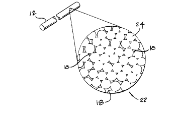
The formation of sintered metal is illustrated with reference to FIG. 3
and contimlecl reference to FIG. 2. FIG. 3 is a microscopic view of a packed lattice 22
of metallic particles 24. Gaps 26 exist between each particle despite the fact that the
particles are ples~uliGed and are in contact with adjacent particles. Particles preferably
are sized between 0.02 microns and 20 microns in diameter. Prior to heating, there are
no chemical bonds formed between the individual particles. When the metal is heated
to slightly below the melting point of the metal, the particles bond with neighboring
particles. The gaps in the packed lattice form pores 18 when the particles are sintered.
Thus in FIG. 2, the metal stent wire formed by the process of sintering has pores 18
extending throughout the entire wire, thereby interconnecting the cavities. The cavities
then can be filled with a therapeutic agent as hereinafter described. The approp~iate
pressure and temperature of sintering a particular metal is specific to that particular
metal. One skilled in the art of metal fabrication would understand how to sinter any
given metal or alloy.
For each of the embodiments~ the metal stent material member can be a
suitable metal such as stainless steel, tantalum, nickel-tit~nium alloy, pl~tinum-iridium
alloy, molybdenum-rhenium alloy, gold, magnesium, combinations thereof, althoughother similar materials also may be suitable. The metal can be modified to exhibit
different hardnesses, and thus varying stiffnesses, by well known ~nn~ling and
m~mlfacturing processes.
One of the most important factors to be considered when m~king a stent
according to one embodiment of the present invention is the porosity of the metal.
Porosity is the total volume of pores in the sintered metal divided by the total volume
of the metal. Porosity determines the amount of a therapeutic agent that can be loaded
CA 02234787 1998-04-1~
into a stent of predetermined dimensions. High porosity means that a stent can deliver
more therapeutic agents or have a narrower profile because it is less dense. High
porosity, according to some embodiments of the present invention, adversely affects the
strength and elasticity of a metal. Consequently, there is an ongoing tradeoff between
stent strength, on the one hand, and stent profile and stent load capacity on the other
hand.
Pore size is a function of the size of the particles which create the gaps
that establish the pores. In one embodiment of the present invention illustrated in FIG.
3, the particles 24 generally are spherical. The size of each pore 18 is proportional to
particle size, particularly with generally spherical particles. When the particles 24 are
not of uniform size, smaller particles tend to fill the gaps between larger particles.
Thus, the porosity of the metal formed with such particles is less predictable than when
more equally-sized particles are used. General uniformity of pore size also is
important to ensure that drugs are dispersed evenly throughout the stent. A generally
uniform distribution of pores insures that the tissue in contact with the stent will receive
an evenly distributed dose of a therapeutic agent.
There are several types of drugs that ~;ulrellLly are a~lmini~tered at the
site that a stent is placed in the vessel. Examples of therapeutic drugs, or agents that
can be combined with the particle layers, include antiplatelets, antifibrin, antithrombin
and antiproliferatives. Examples of anticoagulants, antiplatelets antifibrins and
an~i~ll,o"lbins include but are not limited to sodium heparin, low molecular weight
heparin, hirudin, argatroban, forskolin, vapiprost, prostacyclin and prostacyclin
analogues, dextran, D-phe-pro-arg-chloromethylketone (synthetic antithrombin),
dipyridamole, glycoprotein IIb/IIIa platelet membrane receptor antibody, recombinant
hirudin, thrombin inhibitor (available from Biogen), and an antiplatelet drug sold under
the trademark "7E-3B" by Centorcor, Inc. Examples of cytostatic or antiproliferative
agents include angiopeptin, a somatostatin analogue; angiotensin-converting enzyme
inhibitors, such as those m~mlfactllred under the trademarks "Captopril" (by Squibb
Corp.), "Cilazapril" (by Hoffman-LaRoche, Inc.) and "Lisinopril" (by Merck & Co.,
~ 30 Inc.); calcium channel blockers such as nifedipine; colchicine; fibroblast growth factor
CA 02234787 1998-04-1~
(FGF) antagonists; fish oils, such as omega 3 fatty acids; cholesterol-lowering drugs
such as inhibitors of HMG-CoA recluct~e, one of which is sold under the trademark
"Lovastatin" by Merck & Co., Inc.; methotrexate, monoclonal phosphodiesterase
inhibitors, prost~gl~n-lin inhibitor (available from Glaxo Wellcome, Inc., PDGF
S antagonists such as seramin and triazolopyrimidin~o, serotonin blockers, steroids,
thioprotease inhibitors, and nitric oxide. Other therapeutic drugs which may be
appropriate include alpha-interferon and genetically-engineered epithelial cells, for
example.
The foregoing therapeutic agents have been used to prevent or to treat
restenosis, and each is identified by way of example and not by limitation, as other
therapeutic drugs may be developed which equally are applicable for use with thepresent invention. Using such therapeutic agents to treat vessels or body lumens is
known in the art, as is the calculation of dosages, dosage rates and appropliate duration
of treatment.
The therapeutic agent of one embodiment preferably is in liquid form and
is loaded into a stent by immersing the stent in a medicated solution. The therapeutic
agent may be dissolved in a solvent or suspended in a liquid mixture. If a suspension is
used, it is important that the pore size of the stent is considerably larger than the
suspended particles of the therapeutic agent. An average pore size that is more than ten
(10) times the particle size of a suspended therapeutic agent is suitable. After the stent
is immersed in the medicated solution, the therapeutic agent is absorbed into the pores
of the stent. The loaded stent then can be removed from the solution and implanted
into the vasculature of a patient. Optionally, the loading of the therapeutic agent into
the stent can be facilitated by applying pressure to the fluid in which the agent is
dissolved or suspended. The applied pressure will aid the passage of medicated fluid
into the pores of the stent. This technique might be likened to the physical process of
forcing a fluid through the pores of a filter.
Once loaded into the stent the therapeutic agent remains in place by
reason of the surface tension between the outer surfaces of the particles that form the
pores 18 and the particles of the therapeutic agent. As shown in FIG. 1, the loaded or
CA 02234787 1998-04-1~
medicated porous stent 12 then is deployed to the site of an arterial closure 13 and is
exp~n-le~l. The expanded stent engages the walls 14 of the vessel 16 to m~int~in the
patency of the vessel. Once in the vessel and as is illustrated in FIG. 2, the therapeutic
agent disseminates from the pores 18 and is absorbed into the tissue of the walls of the
vessel that are in contact with the stent.
Chief among the advantages of the stent of the present invention over
prior art medicated stents are its profile and strength. Metal, including sintered metal,
is stronger than synthetic materials, such as polymer blends, which are capable of being
loaded with a therapeutic agent. Thus, in order for a medicated stent to deliver an
approl,liate amount of a therapeutic agent and structurally m~int~in vessel patency, the
radial profile of the stent must be substantially larger than that of metal stents. This is
true whether or not a metal stent is coated with a polymeric material to carry atherapeutic agent, or if the stent is made entirely of a plastic material.
Sintered metal has strength and elasticity that is comparable to non-
sintered metal. Sintered metal further has the added feature of porosity.
Consequently, a sintered metal stent can be m~mlf~ctured with a profile that is
substantially comparable to that of a conventional metal stent, and a therapeutic agent
can be loaded into the pores and delivered to the site of stent implantation without the
aid of medicated polymer coatings.
Additionally, many synthetic materials, including materials that are
bioabsorbable, can cause infl~mm~tion of the tissue. A medicated metal stent having a
therapeutic agent loaded directly into the pores of the stent likely will be less apt to
cause irritation at the site of implantation.
FIG. 4 illustrates an alternative embodiment of a stent wire 30
constructed according to the present invention. The stent is formed of elongatedparticles 32, i.e., filaments and fibers. When generally spherically-shaped particles of
metal are used to compose sintered metal, the resultant porosity typically is in the range
of five to thirty percent. When the particles are elongated, these filaments or fibers can
result in a porosity of greater than thirty percent when sintered. The technique of
fabricating a stent with elongated fil~m~nt~ or fibers is similar to the method described
CA 02234787 1998-04-1
-10-
above for spherical particles or powders. The filaments or fibers are molded andpressurized. Then the fibers are heated to a temperature just below the melting point of
the metal.
A stent made of metal filaments or fibers 32 rather than spherical
particles (such as those illustrated in FIG. 2) exhibits greater porosity because of the
irregular shape of the particles. The particles can be packed less densely than
uniformly-shaped particles but contact between the irregularly-shaped particles
nevertheless can be m~in~in.o~l to allow sintering. Thus, the void space or pores 34 in
the sintered metal tend to be larger than the pores 18 that result from spherical particle
sintering.
The strength of a stent wire 30 using filaments in FIG. 4 is improved
because, upon sintering, the individual strands have a greater surface-area-to-volume
ratio and will contact more neighboring strands than will spherical particles. Thus,
each filament or fiber will have a greater surface area on which to bond with adjacent
filaments or fibers. A matrix of overlapping filaments or fibers thus is formed
exhibiting greater porosity and stronger inter-particle bonding.
In yet another embodiment, wire fibers 36 are woven or twined into a
structure 38 as illustrated in FIG. 5. The individual strands cooperate in a synergistic
manner to reinforce the strength of the wire. Additionally, the wire fibers can be
woven into the form of a sintered metal sheet having improved and reinforced strength
or into a sintered metal tube. Other ~combinations of particle size and shape can be
employed to form a stent wire having dirreLellL characteristics.
In another embodiment illustrated in FIG. 6, the stent wire 42 is formed
of an inner core 44 and an outer layer 46 of sintered particles. The outer layer is
formed from particles having a different diameter than the diameter of the particles that
form the inner core. For example, the core of the metal is formed of particles that
have a diameter in the range of 10-20 microns. Surrounding the core are particles that
have a diameter in the range of 2-4 microns. The larger particles create a core having
larger pores 52. This results in higher porosity and thus a higher load capacity. The
CA 02234787 1998-04-1~
smaller particles on the outer layer form smaller pores 54 which reduce the rate of
diffusion of drugs into the tissue of the vessel.
When a therapeutic agent is loaded into a stent formed of the stent wire
42 illustrated in FIG. 6, a larger volume can be stored in the larger pores 52 at the core
44 of the stent wire. Once the stent is placed into the vessel, the therapeutic agent in
the stent wire is delivered at a rate that is determined by the smaller pores 54 in the
outer layer 46 of the stent wire. Such a structure is expected to be capable of storing a
large amount of therapeutic agent at the core and of delivering the therapeutic agent at a
slower rate than would be accomplished if the pores of the stent wire were of more
uniform size. Consequently, this design is appr~liate when long-term drug therapy at
a low dosage rate is desired.
Alternatively, according to another embodiment of the present invention
shown in FIG. 7, a stent wire 56 is formed from sintered particles 58. The pores 62
formed between the sintered metal particle surrounding the solid core retain thetherapeutic agent. The overall porosity of a stent having a solid core and porous outer
layer is much less than that of a stent wire having similar proportions but which is
composed entirely of sintered particles. However, the solid core reinforces the tensile
strength and elasticity of the metal stent and is considerably stronger than a uniform-
particle sintered stent. Thus, it is desirable to use a sintered stent with a solid core for
applications where maximum tensile strength and elasticity is desirable and only a
relatively small amount of therapeutic agent is needed.
The sintered metal stent of still another embodiment of the present
invention can be made of material formed in the spherically-shaped or filament-like
particles discussed previously. For example, the stent can be formed of a sheet of
sintered metal 64 as shown in FIG. 8 or of a sintered metal tube. By way of example,
metal particles 66 are arranged and pressurized into a sheet. The sheet then is heated
to a temperature below the melting point of the particles as described previously. The
sheet of sintered metal is porous and has a plurality of pores 68.
The same principles that apply to porosity and pore size of a wire apply
equally to a sintered stent that is formed into a sheet or tube. The advantage of
CA 02234787 1998-04-1~
forming the stent from a sheet of metal is that the stent is radially expandable without
placing a great deal of strain on the metal lattice when it is e~r~n-lecl. A sheet or tube
of sintered metal can be cut in the desired shape to form the metal structural member
with a laser, such as a continuous CO2 laser, a pulsed YAG laser, or an excimer laser,
for example, or alternatively, by chemical etching or ~lalllpillg. When cut from a flat
sheet, the stent then is rolled into a cylindrical configuration and is laser welded along
the longitllflin~l edges.
The stent can be formed into a particular pattern known in the art for
stents formed from metal sheets. One such pattern is a rolled locking design and is
illustrated in FIG. 9. The sheet is etched into a stent configuration 70 that has a head
portion 72 that includes one or more slots 74 for receipt of a number of tail sections 76
that correspond to each slot. The tail sections are received into the slots so as to form a
cylindrical loop. Each tail section includes a plurality of teeth 78 that are adapted to
cooperatively engage the slot of the head portion. When the teeth engage the slot, the
tail sections are retained in place, holding the stent configuration in an expanded state.
Additionally, holes 80 are formed throughout the stent to reduce the metal-to-air ratio
of the stent. The less metal that is in contact with the wall 14 of the vessel 16, the
more likely the stent is to be compatible with the blood.
Prior to deployment, the tail sections are coiled into a retracted position
in a "jelly-roll" fashion. Each tail section then is threaded through each corresponding
slot and wound. The stent configuration is e~ran-le~l by a balloon according to
principles that are well known in the art for delivering and implanting stents. As the
stent configuration 70 is expanded by a balloon during deployment, it unwinds and the
teeth 78 lock into the slots 74 at a desired radial diameter to prevent the stent from
returning to its original, retracted state.
A benefit of the coiled stent shown in FIG. 9 is that the stent 70 can be
etched to have a minim~l surface area that comes in contact with the walls of the vessel.
This may be an important feature when it is desired to cover the entire area of the walls
of a blood vessel with a therapeutic agent because the coiled sheet metal stent can be
CA 02234787 1998-04-1~
configured to m~int~in maximum surface area contact with the wall of the blood vessel
in contrast to wire stents.
With reference to FIG. 10, another embodiment of the present invention
is a sheet that has particles that are sintered to both sides 84 and 86 of a metal sheet 82.
S The stent of FIG. 10 is similar in structure to the stent wire of FIG. 7 in that it has a
solid core and particles sintered to the core forming a porous outer layer. The solid
core reinforces the strength of the metal. The metal sheet also provides a barrier
through which a therapeutic agent cannot pass. Thus, a therapeutic agent loaded into
the pores 92 on the top side 84 of the sheet permeates in a first direction 88 outward
from the solid core. A therapeutic agent loaded into the pores 94 on the bottom side 86
of the solid wire permeates only in a second direction 90 which is opposite to the
direction of the therapeutic agent loaded into the pores on the top side.
When a stent as shown in FIG. 10 is looped into a cylindrical formation
and placed into a vessel, only the top side 84, which is directed radially outward,
engages the walls of the vessel. The bottom side 86 faces radially inward and does not
come in contact with the walls of the vessel. Thus, if it is desired, a first therapeutic
agent can be loaded into the top side to treat the tissue in the wall of the vessel. A
second therapeutic agent can be loaded into the bottom side to prevent coagulation of
the blood flowing in the vessel. Additionally, the stent can be formed so that particles
are sintered only to one side of the stent. A therapeutic agent is loaded into the
sintered metal on the porous side of the stent. When a stent is formed with only one
porous side, that side can be oriented radially outward to deliver a therapeutic agent to
the tissue in the wall of the stent.
FIG. 11 illustrates a cross-sectional view of a stent wire according to one
embodiment of the invention. The sheet has a plurality of porous cavities or pores 98.
A therapeutic agent is loaded into the pores of the sintered metal. Then, a coating 100
is applied to the sintered metal. The coating may be used for several purposes as
described hereinafter.
With reference to FIG. 12, another embodiment of the invention is
shown wherein the stent is formed of a sintered sheet 104 of metal having core 106
CA 02234787 l998-04-l~
-14-
formed of larger-diameter particles 108 that form larger pores as a result of sintering.
The core layer 106 is sandwiched between two layers 110 and 112 formed of smaller-
diameter particles 114 or particles that result in the formation of smaller pores. Such a
sheet is formed by orienting the middle or core layer 106 of larger-diameter particles
108 along a plane. A top layer of the smaller-~ meter particles is arranged in a plane
parallel to and above the core layer. A bottom layer of particles are arranged in a
plane parallel to and below the core layer. The three layers then are pressed together
and sintered into a single sheet. The sheet then can be cut or etched into a stent
configuration.
While one of the benefits of the present invention is to provide a stent
that does not require a coating for the purpose of delivering a therapeutic agent to the
blood vessel, the application of a coating after a therapeutic agent has been loaded into
the pores of the sintered metal does not defeat the utility of the present invention. For
example, when a therapeutic agent is loaded into the pores of the stent and also into a
polymeric coating, the profile of the polymeric coating can be recluce~1 When such a
polymeric coating is applied, a larger dosage of a therapeutic agent can be delivered to
the site of stent implantation. Additional benefits can be obtained by loading a stent
with a therapeutic agent in the pores of the metal and by then further applying a
polymeric coating to the stent. Even if a polymeric coating is applied to the stent, the
principles of reducing profile and reinforcing the stent still are realizable because a
greater volume of therapeutic agent can be delivered by a polymeric-coated sintered
stent than by a coated, all-polymeric stent with comparable dimensions.
The polymeric material that coats a sintered metal stent of the invention
preferably comprises a biodegradable, bioabsorbable polymeric film that is capable of
being loaded with and subsequently releasing therapeutic drugs. The polymeric
coatings preferably include, but are not limited to, polycaprolactone (PCL), poly-DL-
lactic acid (DL-PLA) and poly-L-lactic acid (L-PLA) or lactide. Other biodegradable,
bioabsorbable polymers such as polyorthoesters, polyiminocarbonates, aliphatic
polycarbonates, and polyphosphazenes also may be suitable, and other non-degradable
polymers capable of carrying and delivering therapeutic drugs might be apl?ropriate as
CA 02234787 1998-04-1~
~ well. Examples of non-degradable synthetic polymers are polyurethane, polyethylene,
polyethylene teraphth~l~te, ethylene vinyl acetate, silicone and polyethylene oxide
(PEO). The polymeric layer, according to one embodiment, is loaded with a
pharmacologic agent for use in localized drug therapy. As used in this description, the
terms biodegradable, bioabsorbable, reabsorbable, degradable, and absorbable
collectively are meant to encompass materials that are broken down and graduallyabsorbed or elimin~te(l by the body, whether these processes are due to hydrolysis,
metabolic processes, or to bulk or surface erosion. In each of the foregoing
embodiment~, one polymeric layer preferably is about 0.0025 to 0.051 millim~ters(0.0001 to 0.002 inches) thick.
The thin polymeric films used to coat the stent preferably first are
intermixed with the drug or drugs to be delivered, and then typically are l~min~te~ or
~ solvent cast to the surface of the metal structural member. T ~min~tion processing
methods and temperatures can vary widely depending on the polymers used and the
temperature sensitivity of the loaded drugs. Alternatively, the metal structure of the
stent can be encapsulated in the layers of polymeric material by solvent casting, melt
processing, insert molding, and dip coating.
In one embodiment of the present invention, the membrane is
bioabsorbable, but no therapeutic agent is loaded into the polymer. The coating
dissolves after implantation and this delays the time that a therapeutic agent is released
into the vasculature of a patient. The thickness of the coating, as well as the rate at
which the coating is bioabsorbed, determine the length of time that the stent ispositioned in the vascular before a therapeutic agent is delivered from the pores of the
stent. Additionally, a therapeutic agent can be loaded into the bioabsorbable coating.
Thus a therapeutic agent will be delivered to the stent at a rate determined by the
bioabsorbability of the coating. Once the bioabsorbable material has completely
dissolved, the therapeutic agent in the pores can be delivered at a rate dele~ ed by
the pore size and porosity.
In another embodiment, it is preferred that the coating is permeable and
non-absorbable. In such circl-m.ct~nres, the rate at which the drugs permeate into the
CA 02234787 1998-04-1~
-16-
tissue is controlled by the physical properties of the particular coating selected.
Additionally, the coating may be selected to reduce restenosis, thrombosis or other
tissue infl~mm~tion. For example, a heparin coating is known in the art to reduce
blood clotting. Heparin, when coated on a stent, reduces clotting of blood on the
surface of the stent. The heparin coating is affixed to the surface of the stent through
ionic bonding, end-point ~ ching, or by photo-linking the heparin.
In yet another embodiment, a first therapeutic agent is loaded into the
coating and a second therapeutic agent is loaded into the pores of the stent. This may
be the case when a series of drug dosages or concentrations are needed. When such a
stent is placed into the vasculature, the first therapeutic agent is absorbed first by the
stent and a second therapeutic agent is absorbed later by the v~c~ re. This variation
adds a further dimension to drug treatment allowing for sequential drug therapy at the
site of placement of a stent.
It will be apparent from the foregoing that while particular forms of the
invention have been illustrated and described, various modifications can be madewithout departing from the spirit and scope of the invention. Accordingly, it is not
intended that the invention be limit~d, except as by the appended claims.