Language selection

Search

Patent 2235702 Summary

Third-party information liability

Some of the information on this Web page has been provided by external sources. The Government of Canada is not responsible for the accuracy, reliability or currency of the information supplied by external sources. Users wishing to rely upon this information should consult directly with the source of the information. Content provided by external sources is not subject to official languages, privacy and accessibility requirements.

Claims and Abstract availability

Any discrepancies in the text and image of the Claims and Abstract are due to differing posting times. Text of the Claims and Abstract are posted:

  • At the time the application is open to public inspection;
  • At the time of issue of the patent (grant).
(12) Patent Application: (11) CA 2235702
(54) English Title: STEREOTACTIC BREAST BIOPSY COIL AND METHOD FOR USE
(54) French Title: DISQUE STEREOTAXIQUE POUR BIOPSIE MAMMAIRE ET SON MODE D'UTILISATION
Status: Dead
Bibliographic Data
(51) International Patent Classification (IPC):
  • A61B 5/055 (2006.01)
  • A61B 10/00 (2006.01)
(72) Inventors :
  • MEZRICH, REUBEN S. (United States of America)
  • STRENK, LAWRENCE M. (United States of America)
  • STRENK, SUSAN A. (United States of America)
(73) Owners :
  • DIAGNOSTIC INSTRUMENTS, INC. (United States of America)
(71) Applicants :
  • DIAGNOSTIC INSTRUMENTS, INC. (United States of America)
(74) Agent: BORDEN LADNER GERVAIS LLP
(74) Associate agent:
(45) Issued:
(86) PCT Filing Date: 1996-11-20
(87) Open to Public Inspection: 1997-05-29
Availability of licence: N/A
(25) Language of filing: English

Patent Cooperation Treaty (PCT): Yes
(86) PCT Filing Number: PCT/US1996/018592
(87) International Publication Number: WO1997/018751
(85) National Entry: 1998-05-13

(30) Application Priority Data:
Application No. Country/Territory Date
08/562,564 United States of America 1995-11-24

Abstracts

English Abstract


An MRI breast coil (10) is provided with a large transverse access portal (26)
and a stereotactic frame for guiding a biopsy needle (24). The portal is
covered by a thin sheath (30) of plastic to retain the breast but still allows
insertion of the needle to any location. The frame aligns the needle by
azimuth, height, and depth.


French Abstract

L'invention concerne un disque mammaire (10) pour IRM qui comporte une grande fenêtre d'accès transversale (26) et un cadre stéréotaxique servant à guider une aiguille à biopsie (24). La fenêtre est recouverte d'une fine enveloppe (30) en plastique qui retient le sein tout en permettant d'insérer l'aiguille à l'endroit désiré, quel qu'il soit. Le cadre aligne l'aiguille en azimut, hauteur et profondeur.

Claims

Note: Claims are shown in the official language in which they were submitted.


WHAT IS CLAIMED:

1. An MRI coil for providing an image of a protuberance of
tissue, said coil comprising:
a tubular wall having a longitudinal proximal portion
adapted to receive said protuberance and a
longitudinal distal portion, said wall including a
transverse access portal between said distal and
proximal portions, said portal eliminating a
substantial portion of said wall;
a first coil portion about said distal portion; and
a second coil portion about said proximal portion.

2. A coil according to claim 1, further comprising a
fiducial reference providing a correspondence between said image
and the interior of said tubular wall.

3. A coil according to claim 1, further comprising a
puncturable sheath closing said portal, said sheath being readily
replaceable.

4. A coil according to claim 1, further comprising a
bladder inside said wall, said bladder being inflatable to urge
said protuberance toward said portal.

5. A coil according to claim 1, further comprising a member
constrained to move azimuthally about an axis extending
longitudinally within said wall; and a needle guide connected to
said member, said guide including a plurality of guide bores for
locating a needle in a plurality of longitudinal positions
between said proximal and distal portions.

6. A coil according to claim 5, further comprising a
puncturable sheath closing said portal, said sheath being readily
replaceable.

8


7. A coil according to claim 6, further comprising a
bladder inside said wall, said bladder being inflatable to urge
said protuberance toward said portal.

8. A stereotactic frame for orientating a needle, said
frame comprising:
a base;
a member constrained to move azimuthally about an axis
extending transversely from said base;
a needle guide connected to said member, said guide
including a plurality of guide bores for locating said
needle in a plurality of positions spaced away from
said base.

9. A frame according to claim 8, further comprising a
fiducial reference providing a correspondence between an MRI
image and said frame.

10. A frame according to claim 8, wherein guides having
different sizes of guide bores are interchangeably connectable
to said member.

11. A frame according to claim 8, further comprising:
a tubular wall having a longitudinal proximal portion
adapted to receive said protuberance and a
longitudinal distal portion, said wall being attached
to said base and including a transverse access portal
between said distal and proximal portions; and
a puncturable sheath closing said portal, said sheath being
readily replaceable.

12. A frame according to claim 11, further comprising a
bladder inside said wall, said bladder being inflatable to urge
said protuberance toward said portal.

9


13. A frame for immobilizing a protuberance of tissue, said
frame comprising:
at least one wall having a longitudinal proximal portion
closest to said protuberance and a longitudinal distal
portion, said wall including a transverse access
portal between said distal and proximal portions, said
portal eliminating a substantial portion of said wall;
and
a puncturable sheath closing said portal, said sheath being
readily replaceable.

14. A frame for immobilizing a protuberance of tissue, said
frame comprising:
a wall having a longitudinal proximal portion adapted to
receive and engirdle said protuberance and a
longitudinal distal portion, said wall including a
transverse access portal between said distal and
proximal portions, said portal eliminating a
substantial portion of said wall; and
a puncturable sheath closing said portal, said sheath being
readily replaceable.

15. A frame according to claim 14, further comprising a
bladder inside said wall, said bladder being inflatable to urge
said protuberance toward said portal.

16. A frame according to claim 14, further comprising a
fiducial reference providing a correspondence between an MRI
image and the interior of said wall.

17. A frame for immobilizing a protuberance of tissue, said
frame comprising:
a tubular wall having a longitudinal proximal portion
adapted to receive said protuberance and a
longitudinal distal portion, said wall including a


transverse access portal between said distal and
proximal portions; and
a bladder inside said wall, said bladder being inflatable
to urge said protuberance toward said portal.

18. An MRI coil for providing an image of a protuberance
of tissue, said coil comprising:
a tubular wall having a longitudinal proximal portion
adapted to receive said protuberance and a
longitudinal distal portion, said wall including a
transverse access portal between said distal and
proximal portions, said portal eliminating a
substantial portion of said wall;
a first coil portion about said distal portion;
a second coil portion about said proximal portion;
a fiducial reference providing a correspondence between
said image and the interior of said tubular wall;
a puncturable sheath closing said portal, said sheath being
readily replaceable;
a bladder inside said wall, said bladder being inflatable
to urge said protuberance toward said portal;
a member constrained to move azimuthally about an axis
extending longitudinally within said wall; and
a needle guide connected to said member, said guide
including a plurality of guide bores for locating a
needle in a plurality of longitudinal positions
between said proximal and distal portions.

19. A method for maneuvering a needle to a location
indicated in an MRI image of a protuberance of tissue, said
method comprising:
providing an MRI coil for providing said image;
providing a stereotactic frame, said frame including:
a tubular wall having a longitudinal proximal portion

11


adapted to receive said protuberance and a
longitudinal distal portion, said wall including
a transverse access portal between said distal
and proximal portions, said portal eliminating a
substantial portion of said wall;
a fiducial reference providing a correspondence
between said image and the interior of said
tubular wall;
a puncturable sheath closing said portal, said sheath
being readily replaceable;
a member constrained to move azimuthally about an axis
extending longitudinally within said wall; and
a needle guide connected to said member, said guide
including a plurality of guide bores for locating
a needle in a plurality of longitudinal positions
between said proximal and distal portions;
inserting said protuberance into said proximal portion;
imaging said protuberance and said fiducial reference;
determining the correspondence between said location and
the interior of said tubular wall;
moving said needle guide to an azimuth intersecting said
location;
placing said, needle into a guide bore intersecting said
location; and
inserting said needle through said sheath to a depth
intersecting said location.

20. A method according to claim 19, further comprising:
further including in said frame a bladder inside said wall
and;
inflating said bladder to urge said protuberance toward
said portal.

Description

Note: Descriptions are shown in the official language in which they were submitted.


CA 0223~702 1998-0~-13
WO97/18751 PCT~S96/18592


1 STEREOTACTIC BREAST BIOPSY COIL
2 AND METHOD FOR USE

3 BACKGROUND OF THE INVENTION

4 The present invention relates to MRI-guided tissue biopsy
and, in particular, to a stereotaxic breast biopsy coil.
6 Magnetic resonance imaging (MRI) can detect breast
7 malignancies that have previously been sub-clinical (i.e.,
8 neither palpable nor detected by mammography). Unfortunately,
9 a 40 percent false positive rate goes along with this detection.
This results in a large number of extremely invasive open
11 biopsies being performed.
12 It iS desirable to use less invasive methods such as a
13 needle biopsy (core or aspiration), but needle placement re~uires
14 1 mm accuracy in three dimensions.

SUMMARY OF THE INVENTION

16 An MRI coil for providing an image of a protuberance of
17 tissue has a tubular wall having a longitudinal proximal portion
18 adapted to receive the protuberance and a longitudinal distal
19 portion. The wall includes a transverse access portal between
the distal and proximal portions. This portal eliminates a
21 substantial portion of the wall. A first coil portion is located
22 about the distal portion and a second coil portion is located
23 about the proximal portion.

24 BRIEF DESCRIPTION OF THE DRAWINGS

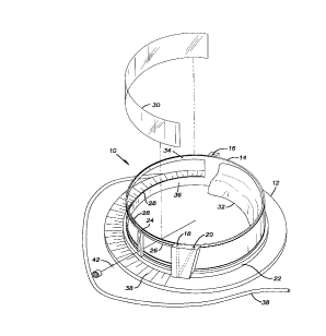
FIG. 1 is a perspective view of a coil according to the
26 invention from above with a portion exploded.
27 FIG. 2 is a front perspective view of a coil according to
28 t~e invention.

CA 0223~702 l998-0~-l3
WO97/18751 PCT~S96/18592


1 FI&. 3 is a top plan view of the a coil according to the
2 invention.
3 FIG. 4 is front elevation view of a needle guide according
4 to the invention.
FIG. 5 is a graphical view of the geometry of an embodiment
6 of the invention.

7 DESCRIPTION OF THE PREFERRED EMBODIMENTS

8 Referring to FIGS. 1, 2 and 3, a breast biopsy coil lO
9 includes a base 12 to which is attached a tubular wall 14 and
three fiducials 16, 18, 20.
11 A ring 22 abuts the external junction of the base 12 and the
12 wall 14. The ring 22 is rotatable about its axis and is slidably
13 retained between the fiducials 16, 18, 20 and base 12 and wall
14 14. A needle guide 24 is removably attached to the ring 22.
These structures can be formed from a suitable non-magnetic, MRI
16 transparent material such as a thermoplastic pol~7mer of methyl
17 methacrylate (e.g., Plexiglas).
18 The wall 14 includes a large aperture or portal 26. The
19 wall 14 contains a slot 28 for receiving a sheath 30. The sheath
30 may be, for example, a thin sheet of plastic of moderate
21 stiffness and being relatively easy to puncture with a sharp
22 instrument (e.g., 15 mil thick polyurethane film such as TEXIN
23 brand polyurethane film from Bayer Polymers Division, Bayer
24 Corporation, or 1/32 inch thick Huntsman ~igh Impact Polystyrene
- Medical Grade).
26 The interior of the wall opposite the portal 26 is provided
27 with an inflatable bladder 32. The bladder 32 may be, for
28 example, formed of latex rubber.
29 The coil 10 may have, for example, an internal diameter of
17.8 cm an~ a height of 7 cm. This provides an internal volume
31 of about 1740 ml for ease of breast placement. When the bladder
32 32 is fully inflated, this volume may be, for example, reduced
33 to 9OO ml.

. .

CA 0223~702 l998-0~-l3

WO 97/18751 PCTIUS96/18592


1 The proximal portion of the wall 14 contains a portion 34
2 of an electrical coil about its circumference. Similarly, the
3 distal portion of the wall 14 contains a portion 36 of an
4 electrical coil about its circumference. The electrical coil
portions 34, 36 may be, for example, single turns. The
6 electrical coil portions are connected by an unshown matching
7 network to a cable 38 and include capacitive splits, a detuning
8 diode, and current traps to protect the coil during transmission
9 by an unshown imager. such features are set forth in U.S. Patent
Application Serial Number 08/530,576, filed September 19, 1995,
11 and incorporated herein by reference. The coil may operate, for
12 example, at 63.89 MHz. The distal and proximal coil portions
13 allow for the portal to be, for example, 5.7 cm high and to
14 extend around nearly 180 degrees of the wall 14.
The base 12 can be provided with a graduated scale 38 to
16 provide an azimuthal positioning of the needle guide 24 as the
17 ring 22 is rotated.
18 Referring to FIG. 4, the needle guide 24 has a series of
19 guide bores 40. The bores 40 are in two staggered rows to
provide increased spatial resolution. For use with an 18 gauge
21 needle (nominally 1 mm), the bores 40 are bored 0.07 mm over
22 needle size. They are on 2 mm centers with the staggering
23 providing plus or minus 0.5 mm resolution in the vertical
24 direction. A different guide 24 is provided for each size needle
to be used. The guide 24 can be conveniently made to insert into
26 a receptacle in the ring 22. The number of staggered rows can
27 be varied to insure the desired resolution for different
28 needle/bore sizes.
29 In general, a needle 42 inserted into the guide 24 will
follow a radial path with respect to the ring 22. The
31 graduations scale 38 may be, for example, spaced to provide 1.5
32 mm arc len~th at the interior of the wall 14. This then provides
33 a resolution of plus or minus 0.77 mm at the wall 14 and as the
34 needle tip continues to the center the resolution approaches
zero.

CA 0223~702 1998-0~-13
WO97/187S1 PCT~S96/18592


1 The depth of insertion of the needle 42 can be conveniently
2 determined, for example, by either a graduated scale on the
3 needle or a moveable stop placed on the needle to prevent further
4 insertion through a guide bore 40.
The combination of adjusting the ring 22 in azimuth,
6 choosing the proper guide bore 40 and inserting the needle 42 a
7 desired depth into the guide bore can easily provide better than
8 1 mm resolution for positioning the needle tip in three
9 dimensions within the coil lO.
The vertical fiducials 16, 18 consist of 5 mm bores parallel
11 to the axis the coil lO. The fiducials 16, 18 are located on a
12 diameter of the ring 22. The fiducial 20 consists o~ a 5 mm bore
13 at an angle to the fiducial 18. The bores of the fiducials 18,
14 20 intersect to ~orm the zero height reference of the coil lO.
The angle between the fiducials 18, 20 may be, for example, 45
16 degrees. The bores are filled with a material that will provide
17 an MRI image, for example, 0.04M CuSo4-5H2O.
18 In operation, a breast or other protuberance of tissue is
19 inserted into the proximal portion 34 of the wall 14 and extends
toward the distal portion 36. In the preferred embodiment, the
21 patient lies in a prone position upon the coil lO (with suitable
22 surrounding support3.
23 The bladder 32 is inflated by unshown means to compress the
24 breast against the portal 26. The sheath 30 keeps the breast
from "leaking" in a transverse direction out of the portal 26.
26 The sheath 30 provides support of the lateral portion of breast.
27 Despite the large portal 24, there is no compromise of
28 immobilization. The sheath 30 is readily replaceable between
29 patients or procedures.
The breast is then imaged using the coil lO with an unshown
31 MRI imager. Referring to FIG. 5, in a coronal image, the
32 fiducials ~8, 20 produce dots separated by a distance d. The
33 height h above the base of the coil is given by: h = d-tan(45~).
34 I~ the coordinates of the fiducials 16, 18 are ~x1~ Y1) and
tXz~ Y2), respectively, then the midpoint between them (xm/ Ym) is

CA 02235702 1998-0~-l3

WO 97/18751 PCT/US96/18592


A l given by: (xm, Ym) = ((x1 + x2)/2, (Y1 ~ y2)/2). The distance
2 between the fiducials 16, 18 is D given by: D = (~x1 - x2)Z + (Y
3 - y2)2)(1/Z)
4 The distance p from the midpoint to the imaged lesion or
target (Xt, Yt) is given by: p = ((xt - Xm) 2 + (Yt - y~) 2~ ~1/2), The
6 distance c from the fiducial 16 to the target is given by c =
7 ((xt ~ x2)2 ~ (Yt + Y2)Z)(1/2) The azimuthal angle ~ is given by:
8 ~ = arCcos((c2 _ p2 -(~/2)2)/( 2.p.(D/2))
9 The needle insertion length N i5 given by: N = R - p where
R is the length of the needle. Thus the image can be used to
ll provide the anteroposterior distance, the needle insertion
12 length, and the angular position necessary to locate the target
13 in three dimensions and insert the tip the needle 42 to that
14 location.
Based on the data from the image, the needle guide 24 is
l~ rotated to the correct azimuth and the needle inserted into the
17 correct guide bore 40. The needle 42 is then inserted through
18 the sheath 30 into the breast (the needle and sheath are sterile)
l9 to the desired depth. For particularly large bore needles, the
breast skin (and the sheath) can be nicked to assist in inserting
2l the needle. The tip of the needle is then located within less
22 than l mm of the desired location.
23 A biopsy of the lesion can be performed by using either a
24 coring biopsy needle or an aspiration biopsy needle. In
addition, because of MRI detects tumor borders more accurately
26 than other modalities, the needle can be used to localize the
27 lesion.
28 The large po!rtal 24 provides ready access to any point
29 within the breast and makes such procedures as skin nicks easy
to accomplish. This large portal 24 is made possible by the
3l sheath 30.
32 The s~eath 30 provides support and immobilization for the
33 tissue, while still permitting access to any location of the
34 tissue with needles or other puncturing or cutting instruments.
This combination of portal and sheath can be used in other

CA 0223~702 1998-0~-13
WO97/18751 PCT~S96/18592


l coil/frame structures, for example, a tubular wall having an oval
2 or rectangular cross-section, or a frustoconical chamber can be
3 conveniently provided with a large portal that can be covered
4 with a sheath.
In the preferred embodiment, the necessary position settings
6 for the needle 42 are automatically calculated by either software
7 in the imager or in an auxiliary computer such as a laptop
8 computer.
9 The coil lO as described provides needle access to within
about l cm of the chest wall. If access to the blocked area is
ll desired, the needle guide 24 can be provided with guide bores at
12 a polar angle to the plane o~ the coil. With minor modifications
13 to the calculations, the needle can then be accurately inserted
14 into this area.
Other configurations of the invention are also possible, for
16 example, the electrical coil portions 34, 36 can be provided in
17 the form of a separate electrical coil or coils that are placed
18 about the coil lO during operation. The coil lO is then more
l9 accurately described as a frame even though its structure remains
otherwise unchanged. This structure provides a stereotactic
21 frame for orienting a needle. It also may be advantageous to use
22 the coil lO in combination with additional electrical coil
23 geometries (e.g.,. Helmoltz, saddle, or surface coil), either
24 integral or external.
It should be evident that this disclosure is by way of
26 example and that various changes may be made by adding, modifying
27 or eliminating details without departing from the fair scope of
28 the teaching contained in this disclosure. The invention is
29 therefore not limited to particular details of this disclosure
except to the extent that the following claims are necessarily
3l so limited.
.
Representative Drawing
A single figure which represents the drawing illustrating the invention.
Administrative Status

For a clearer understanding of the status of the application/patent presented on this page, the site Disclaimer , as well as the definitions for Patent , Administrative Status , Maintenance Fee  and Payment History  should be consulted.

Administrative Status

Title Date
Forecasted Issue Date Unavailable
(86) PCT Filing Date 1996-11-20
(87) PCT Publication Date 1997-05-29
(85) National Entry 1998-05-13
Dead Application 2002-11-20

Abandonment History

Abandonment Date Reason Reinstatement Date
2001-11-20 FAILURE TO PAY APPLICATION MAINTENANCE FEE
2001-11-20 FAILURE TO REQUEST EXAMINATION

Payment History

Fee Type Anniversary Year Due Date Amount Paid Paid Date
Registration of a document - section 124 $100.00 1998-05-13
Application Fee $300.00 1998-05-13
Maintenance Fee - Application - New Act 2 1998-11-20 $100.00 1998-10-06
Maintenance Fee - Application - New Act 3 1999-11-22 $100.00 1999-09-17
Maintenance Fee - Application - New Act 4 2000-11-20 $100.00 2000-11-03
Owners on Record

Note: Records showing the ownership history in alphabetical order.

Current Owners on Record
DIAGNOSTIC INSTRUMENTS, INC.
Past Owners on Record
MEZRICH, REUBEN S.
STRENK, LAWRENCE M.
STRENK, SUSAN A.
Past Owners that do not appear in the "Owners on Record" listing will appear in other documentation within the application.
Documents

To view selected files, please enter reCAPTCHA code :


To view images, click a link in the Document Description column. To download the documents, select one or more checkboxes in the first column and then click the "Download Selected in PDF format (Zip Archive)" or the "Download Selected as Single PDF" button.

List of published and non-published patent-specific documents on the CPD .

If you have any difficulty accessing content, you can call the Client Service Centre at 1-866-997-1936 or send them an e-mail at CIPO Client Service Centre.


Document
Description 
Date
(yyyy-mm-dd) 
Number of pages   Size of Image (KB) 
Representative Drawing 1998-08-05 1 11
Abstract 1998-05-13 1 48
Description 1998-05-13 6 299
Cover Page 1998-08-05 1 40
Drawings 1998-05-13 3 68
Claims 1998-05-13 5 202
Assignment 1998-05-13 5 227
PCT 1998-05-13 6 214